A Girl with Cutaneous Lesions, Polyarthritis - downloads - Hindawi
4
International Scholarly Research Network ISRN Dermatology Volume 2011, Article ID 657673, 3 pages doi:10.5402/2011/657673 Case Report A Girl with Cutaneous Lesions, Polyarthritis, and Antinuclear Antibodies Positivity Jorge L. Musuruana and Javier A. Cavallasca Section of Rheumatology and Autoimmune Diseases, Hospital JB Iturraspe, Bv. Pellegrini 3551, 3000 Santa Fe, Argentina Correspondence should be addressed to Jorge L. Musuruana, [email protected] Received 24 January 2011; Accepted 8 March 2011 Academic Editors: A. Belloni-Fortina, G. Kirtschig, and Y. Tuzun Copyright © 2011 J. L. Musuruana and J. A. Cavallasca. This is an open access article distributed under the Creative Commons Attribution License, which permits unrestricted use, distribution, and reproduction in any medium, provided the original work is properly cited. On October 1996, a 14-year-old girl was admitted to the hospital because cutaneous lesions, asthenia, and arthralgias. On examination, there was nonscarring hair thinning with a widened part over the frontal hairline, polymorphic papulosquamous rash on her face, neck, arms, and trunk, and livedo reticularis in her legs. Multiple aphtous ulcers were present on the buccal and nasal mucosa. There was polyarthritis involving the wrist, metacarpophalangeal joints, proximal interphalangeal joints, and metatarsophalangeal joints of both hands and feet. Skin biopsy of the face was compatible with subacute cutaneous lupus erythematosus. She started on prednisone 60 mg/d without improvement, and later hdroxhchloroquine (HCQ) 6 mg/kg/d was added for one year. Cutaneous lesions were almost healed, with just a hypopigmented macules left. Over the last 14 years, she has not shown any cutaneous or systemic manifestations. 1. Case Report On October 1996, a 14-year-old girl was admitted to the hospital because of cutaneous lesions, asthenia, and arthral- gias. She had been in good health until four months earlier, when she began to experience asthenia, decreased appetite, photosensitivity, hair loss, erythematous papules on her face, and diffuse arthralgias. On examination, the patient’s blood pressure was 110/60 mm Hg, her temperature was 36,8 ◦ C, her pulse rate was 68 beats/min, and her respiration rate was 16 breaths/min. There was nonscarring hair thinning with a widened part over the frontal hairline, polymorphic papulosquamous rash on her face, neck, arms, and trunk, (Figures 1 and 2), and livedo reticularis in her legs. Multiple aphtous ulcers were present on the buccal and nasal mucosa. There was pol- yarthritis involving the wrist, metacarpophalangeal joints, proximal interphalangeal joints, and metatarsophalangeal joints of both hands and feet. The rest of the physical examination findings were normal. A complete blood count with differential analysis, liver function tests, chemistry panel, muscle enzymes, and urinal- ysis was normal. A test for antinuclear antibodies (ANA) was positive at a titer of 1 : 160 with a homogeneous-speckled pattern, but the remainder of the antinuclear-antibody screening (anti-double-stranded DNA, anti-Ro/SSA, anti- La/SSB, and anti ENA) was negative. An antineutrophil cytoplasmic antibody (ANCA) test, rheumatoid factor, anti- cardiolipin antibodies Ig G and Ig M, VDRL, and HIV were all negative. Complement levels (C3, C4, and CH50) were within normal limits. A chest X-ray, an electrocardiogram, an transthoracic echocardiography, and an abdominal US were normal. Skin biopsy of the face was compatible with subacute cutaneous lupus erythematosus (Figure 3). She started with prednisone 60 mg/d without improve- ment; after two months this medication was discontinued. Later hydroxychloroquine (HCQ) 6 mg/kg/d was added for one year, then, the dose was tapered to 3 mg/kg/d. The patient had an extraordinary response to HCQ. Cutaneous lesions were almost healed, with just a hypopigmented macules left. Over the last 14 years, she has not shown any cutaneous or systemic manifestations. 2. Discussion The skin lesions seen in patients with lupus can be classified into those that are lupus-specifichistologically and those
Transcript of A Girl with Cutaneous Lesions, Polyarthritis - downloads - Hindawi
International Scholarly Research NetworkISRN DermatologyVolume 2011, Article ID 657673, 3 pagesdoi:10.5402/2011/657673
Case Report
A Girl with Cutaneous Lesions, Polyarthritis, andAntinuclear Antibodies Positivity
Jorge L. Musuruana and Javier A. Cavallasca
Section of Rheumatology and Autoimmune Diseases, Hospital JB Iturraspe, Bv. Pellegrini 3551, 3000 Santa Fe, Argentina
Correspondence should be addressed to Jorge L. Musuruana, [email protected]
Received 24 January 2011; Accepted 8 March 2011
Academic Editors: A. Belloni-Fortina, G. Kirtschig, and Y. Tuzun
Copyright © 2011 J. L. Musuruana and J. A. Cavallasca. This is an open access article distributed under the Creative CommonsAttribution License, which permits unrestricted use, distribution, and reproduction in any medium, provided the original work isproperly cited.
On October 1996, a 14-year-old girl was admitted to the hospital because cutaneous lesions, asthenia, and arthralgias. Onexamination, there was nonscarring hair thinning with a widened part over the frontal hairline, polymorphic papulosquamousrash on her face, neck, arms, and trunk, and livedo reticularis in her legs. Multiple aphtous ulcers were present on the buccaland nasal mucosa. There was polyarthritis involving the wrist, metacarpophalangeal joints, proximal interphalangeal joints,and metatarsophalangeal joints of both hands and feet. Skin biopsy of the face was compatible with subacute cutaneous lupuserythematosus. She started on prednisone 60 mg/d without improvement, and later hdroxhchloroquine (HCQ) 6 mg/kg/d wasadded for one year. Cutaneous lesions were almost healed, with just a hypopigmented macules left. Over the last 14 years, she hasnot shown any cutaneous or systemic manifestations.
1. Case Report
On October 1996, a 14-year-old girl was admitted to thehospital because of cutaneous lesions, asthenia, and arthral-gias. She had been in good health until four months earlier,when she began to experience asthenia, decreased appetite,photosensitivity, hair loss, erythematous papules on her face,and diffuse arthralgias. On examination, the patient’s bloodpressure was 110/60 mm Hg, her temperature was 36,8◦C,her pulse rate was 68 beats/min, and her respiration rate was16 breaths/min.
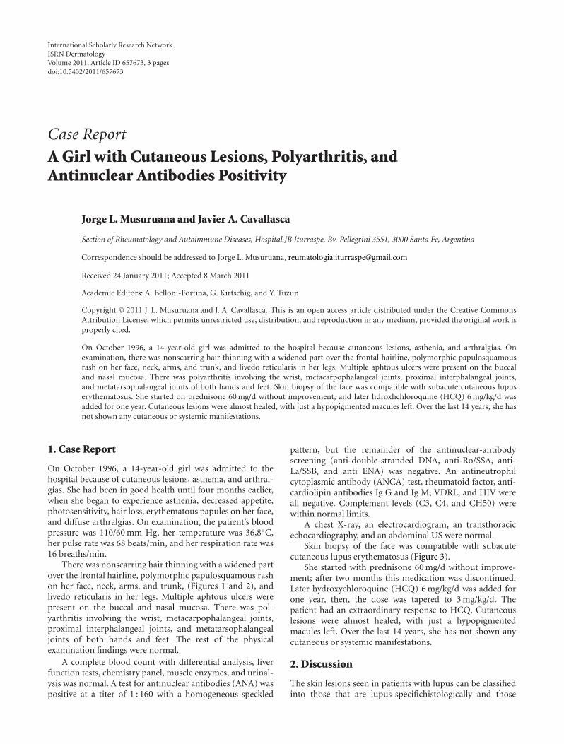
There was nonscarring hair thinning with a widened partover the frontal hairline, polymorphic papulosquamous rashon her face, neck, arms, and trunk, (Figures 1 and 2), andlivedo reticularis in her legs. Multiple aphtous ulcers werepresent on the buccal and nasal mucosa. There was pol-yarthritis involving the wrist, metacarpophalangeal joints,proximal interphalangeal joints, and metatarsophalangealjoints of both hands and feet. The rest of the physicalexamination findings were normal.
A complete blood count with differential analysis, liverfunction tests, chemistry panel, muscle enzymes, and urinal-ysis was normal. A test for antinuclear antibodies (ANA) waspositive at a titer of 1 : 160 with a homogeneous-speckled
pattern, but the remainder of the antinuclear-antibodyscreening (anti-double-stranded DNA, anti-Ro/SSA, anti-La/SSB, and anti ENA) was negative. An antineutrophilcytoplasmic antibody (ANCA) test, rheumatoid factor, anti-cardiolipin antibodies Ig G and Ig M, VDRL, and HIV wereall negative. Complement levels (C3, C4, and CH50) werewithin normal limits.
A chest X-ray, an electrocardiogram, an transthoracicechocardiography, and an abdominal US were normal.
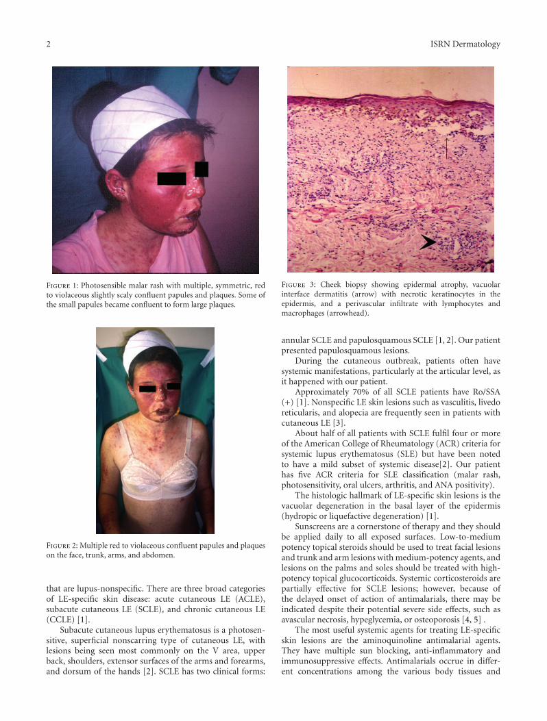
Skin biopsy of the face was compatible with subacutecutaneous lupus erythematosus (Figure 3).
She started with prednisone 60 mg/d without improve-ment; after two months this medication was discontinued.Later hydroxychloroquine (HCQ) 6 mg/kg/d was added forone year, then, the dose was tapered to 3 mg/kg/d. Thepatient had an extraordinary response to HCQ. Cutaneouslesions were almost healed, with just a hypopigmentedmacules left. Over the last 14 years, she has not shown anycutaneous or systemic manifestations.
2. Discussion
The skin lesions seen in patients with lupus can be classifiedinto those that are lupus-specifichistologically and those
2 ISRN Dermatology
Figure 1: Photosensible malar rash with multiple, symmetric, redto violaceous slightly scaly confluent papules and plaques. Some ofthe small papules became confluent to form large plaques.
Figure 2: Multiple red to violaceous confluent papules and plaqueson the face, trunk, arms, and abdomen.
that are lupus-nonspecific. There are three broad categoriesof LE-specific skin disease: acute cutaneous LE (ACLE),subacute cutaneous LE (SCLE), and chronic cutaneous LE(CCLE) [1].
Subacute cutaneous lupus erythematosus is a photosen-sitive, superficial nonscarring type of cutaneous LE, withlesions being seen most commonly on the V area, upperback, shoulders, extensor surfaces of the arms and forearms,and dorsum of the hands [2]. SCLE has two clinical forms:
Figure 3: Cheek biopsy showing epidermal atrophy, vacuolarinterface dermatitis (arrow) with necrotic keratinocytes in theepidermis, and a perivascular infiltrate with lymphocytes andmacrophages (arrowhead).
annular SCLE and papulosquamous SCLE [1, 2]. Our patientpresented papulosquamous lesions.
During the cutaneous outbreak, patients often havesystemic manifestations, particularly at the articular level, asit happened with our patient.
Approximately 70% of all SCLE patients have Ro/SSA(+) [1]. Nonspecific LE skin lesions such as vasculitis, livedoreticularis, and alopecia are frequently seen in patients withcutaneous LE [3].
About half of all patients with SCLE fulfil four or moreof the American College of Rheumatology (ACR) criteria forsystemic lupus erythematosus (SLE) but have been notedto have a mild subset of systemic disease[2]. Our patienthas five ACR criteria for SLE classification (malar rash,photosensitivity, oral ulcers, arthritis, and ANA positivity).
The histologic hallmark of LE-specific skin lesions is thevacuolar degeneration in the basal layer of the epidermis(hydropic or liquefactive degeneration) [1].
Sunscreens are a cornerstone of therapy and they shouldbe applied daily to all exposed surfaces. Low-to-mediumpotency topical steroids should be used to treat facial lesionsand trunk and arm lesions with medium-potency agents, andlesions on the palms and soles should be treated with high-potency topical glucocorticoids. Systemic corticosteroids arepartially effective for SCLE lesions; however, because ofthe delayed onset of action of antimalarials, there may beindicated despite their potential severe side effects, such asavascular necrosis, hypeglycemia, or osteoporosis [4, 5] .
The most useful systemic agents for treating LE-specificskin lesions are the aminoquinoline antimalarial agents.They have multiple sun blocking, anti-inflammatory andimmunosuppressive effects. Antimalarials occrue in differ-ent concentrations among the various body tissues and

ISRN Dermatology 3
organs. The highest of all concentrations occurs in melanin-containing cells such as found in the retina and skin. Hydrox-ychloroquine is the initial treatment of choice. Its onset ofaction is approximately 1 month, even though its full benefitmight not be seen for several months. It is typically dosed at200 or 400 mg per day and should not exceed 6.5 mg/kg/d.At these doses, it is usually well tolerated. Chloroquine ismore toxic and it is used in a 4 mg/kg/d dose. Quinacrinedoes not cause retinal toxicity, and thus can be used incombination with either hydroxychloroquine or chloroquine[6]. Approximately 75% of SCLE patients improved withantimalarials agents, smokers are less likely to do so [5].
Other treatments include thalidomide, topical tacroli-mus, dapsone, gold salts, oral retinoids, and oral fenitoin.Cytotoxic immunosuppressive agents, such as azathioprine,mycophenolate mofetil, and methotrexate are reserved forpatients with more severe disease who have failed the lesstoxic forms of therapy [4].
References
[1] N. Rothfield, R. D. Sontheimer, and M. Bernstein, “Lupuserythematosus: systemic and cutaneous manifestations,” Clinicsin Dermatology, vol. 24, no. 5, pp. 348–362, 2006.
[2] R. D. Sontheimer, “Subacute cutaneous lupus erythematosus:25-Year evolution of a prototypic subset (subphenotype)of lupus erythematosus defined by characteristic cutaneous,pathological, immunological, and genetic findings,” Autoim-munity Reviews, vol. 4, no. 5, pp. 253–263, 2005.
[3] B. Tebbe, “Clinical course and prognosis of cutaneous lupuserythematosus,” Clinics in Dermatology, vol. 22, no. 2, pp. 121–124, 2004.
[4] J. P. Callen, “Update on the management of cutaneous lupuserythematosus,” British Journal of Dermatology, vol. 151, no. 4,pp. 731–736, 2004.
[5] A. Kuhn, F. Ochsendorf, and G. Bonsmann, “Treatment ofcutaneous lupus erythematosus,” Lupus, vol. 19, pp. 1125–1136,2010.
[6] A. Wozniacka, A. Carter, and D. P. McCauliffe, “Antimalarialsin cutaneous lupus erythematosus: mechanisms of therapeuticbenefit,” Lupus, vol. 11, no. 2, pp. 71–81, 2002.

Submit your manuscripts athttp://www.hindawi.com
Stem CellsInternational
Hindawi Publishing Corporationhttp://www.hindawi.com Volume 2014
Hindawi Publishing Corporationhttp://www.hindawi.com Volume 2014
MEDIATORSINFLAMMATION
of
Hindawi Publishing Corporationhttp://www.hindawi.com Volume 2014
Behavioural Neurology
International Journal of
EndocrinologyHindawi Publishing Corporationhttp://www.hindawi.com
Volume 2014
Hindawi Publishing Corporationhttp://www.hindawi.com Volume 2014
Disease Markers
BioMed Research International
Hindawi Publishing Corporationhttp://www.hindawi.com Volume 2014
OncologyJournal of
Hindawi Publishing Corporationhttp://www.hindawi.com Volume 2014
Hindawi Publishing Corporationhttp://www.hindawi.com Volume 2014
Oxidative Medicine and Cellular Longevity
PPARRe sea rch
Hindawi Publishing Corporationhttp://www.hindawi.com Volume 2014
The Scientific World JournalHindawi Publishing Corporation http://www.hindawi.com Volume 2014
Immunology ResearchHindawi Publishing Corporationhttp://www.hindawi.com Volume 2014
Journal of
ObesityJournal of
Hindawi Publishing Corporationhttp://www.hindawi.com Volume 2014
Hindawi Publishing Corporationhttp://www.hindawi.com Volume 2014
Computational and Mathematical Methods in Medicine
OphthalmologyJournal of
Hindawi Publishing Corporationhttp://www.hindawi.com Volume 2014
Diabetes ResearchJournal of
Hindawi Publishing Corporationhttp://www.hindawi.com Volume 2014
Hindawi Publishing Corporationhttp://www.hindawi.com Volume 2014
Research and TreatmentAIDS
Hindawi Publishing Corporationhttp://www.hindawi.com Volume 2014
Gastroenterology Research and Practice
Parkinson’s DiseaseHindawi Publishing Corporationhttp://www.hindawi.com Volume 2014
Evidence-Based Complementary and Alternative Medicine
Volume 2014Hindawi Publishing Corporationhttp://www.hindawi.com