A genetic screen for new Polycomb Group genes in...
97
Universidad Autónoma de Madrid Facultad de Ciencias Departamento de Biología Molecular A genetic screen for new Polycomb Group genes in Drosophila melanogaster Andrés Gaytán de Ayala Alonso Experimental work performed at the European Molecular Biology Laboratory under the supervision of Dr Jürg Müller
Transcript of A genetic screen for new Polycomb Group genes in...

Universidad Autónoma de Madrid Facultad de Ciencias Departamento de Biología Molecular
A genetic screen for new Polycomb Group genes in
Drosophila melanogaster
Andrés Gaytán de Ayala Alonso
Experimental work performed at the European Molecular Biology Laboratory underthe supervision of Dr Jürg Müller

2

3
Acknowledgements
I would like to thank Jürg Müller for giving me the opportunity to perform this project, and
for his continuous support; he provided me with an excellent supervision, balanced with a
great deal of independency. Besides, he started the genetic screen for new PcG genes,
together with Cornelia Fritsch and Dirk Beuchle; most of the screen on the third chromosome
was performed by them. Cornelia Fritsch was also a superb teacher for "fly pushing".
Parallel to our screen, Gary Struhl carried out another genetic screen using the same crosses
scheme and fly lines; there was a fruitful exchange of mutants, from which several of our
mutants on the third chromosome come.
Bernadett Papp was always an excellent labmate, providing useful ideas and help in a most
kind way. She made a key contribution to the mapping of penelope/skd, and provided one
anti-Pc antibody.
All members of the lab were always willing to give a helping hand with whatever issue, and
created a great working atmosphere.
Sonsoles Campuzano, Eileen Furlong and Pernille Rørth, members of my Thesis Advisory
Committee (TAC), proved to be excellent evaluators and did not hesitate to provide me with
any time I needed. Sonsoles Campuzano was also my tutor at the Universidad Autónoma de
Madrid; I thank her kind replies to all my questions about procedures, and her dealings with
bureaucracy on my behalf.
This work would have not been possible without the availability of Flybase
(www.flybase.org) and the public stock centres of Bloomington (USA) and Szeged
(Hungary). I also thank other colleagues for reagents and fly lines: Mátyás Végh and Konrad
Basler for providing us with the fly lines we used in the screen; Hsin-Ho Sung and Pernille
Rørth for the FRT42D ovoD1 and khc8 lines; Peter Verrijzer for anti-USP7 antibodies and
helpful discussions; Bernhard Mechler for capsuleen alleles; Jessica Treisman for kto and skd
alleles; and the Tübingen stock centre for brh, cra and thi mutants.
Ann-Mari Voie made an excellent work injecting constructs in flies for the establishment of
transgenic lines. Likewise, I received perfect assistance from the EMBL Gene Core facility
and Laboratory Animal Resources.
All the genetic screen project, including my PhD fellowship, was funded by the Deutsche
Forschungsgemeinschaft (DFG; Schwerpunktprogramm 1129: Epigenetics).
I dedicate this PhD thesis to my family and friends.

4

5
TABLE OF CONTENTS
Resumen en castellano ......................................................................................... 7
Summary ............................................................................................................. 19
1. Introduction ..................................................................................................... 19
1.1. PcG genes are required to maintain the HOX gene pattern of
expression ......................................................................................... 20
1.2. PcG genes ......................................................................................... 23
1.3. Regulation of PcG function .............................................................. 26
1.4. The trithorax Group .......................................................................... 32
2. Objectives ........................................................................................................ 35
3. Materials and methods ..................................................................................... 37
4. Results .............................................................................................................. 47
4.1. A genetic screen for new PcG genes on the second chromosome .... 47
4.2. calypso, a new PcG gene on the second chromosome ...................... 54
4.3. A genetic screen for new PcG genes on the third chromosome ........ 62
4.4. penelope and siren1, new PcG genes on the third chromosome ....... 65
4.5. Enhancers of Pc recovered from the screen on the third
chromosome ...................................................................................... 68
4.6. Mutants not recovered from the screen ............................................. 69
5. Discussion ......................................................................................................... 73
5.1. A deubiquitinating activity required for PcG-mediated silencing ..... 73
5.2. Role of Mediator in PcG-mediated silencing ..................................... 77
5.3. A genetic screen for new PcG genes in Drosophila .......................... 78

6
6. Conclusions ....................................................................................................... 83
6.1. The screen methodology allows to recover PcG mutants .................. 83
6.2. New PcG genes in Drosophila melanogaster were recovered
from the screen ................................................................................... 83
6.3. calypso encodes a deubiquitinating enzyme required for
PcG-mediated repression .................................................................... 83
6.4. The Med12 and Med13 Mediator components are involved
in HOX gene silencing ........................................................................ 84
References .............................................................................................................. 85

7
Un "screen" genético dirigido a encontrar nuevos genes del Grupo
Polycomb en Drosophila melanogaster
Los genes del grupo Polycomb (PcG) codifican represores transcripcionales
necesarios para el mantenimiento de estados de silenciamiento génico de genes diana
de generación en generación celular. En metazoos el silenciamiento génico mediado
por el PcG mantiene el patrón de expresión de los genes HOX durante todo el
desarrollo. En esta memoria describo los resultados de un "screen" genético dirigido a
encontrar nuevos genes del PcG en Drosophila melanogaster. Aparte de nuevos
alelos para genes ya descritos del PcG, se encontraron mutantes para nuevos loci del
PcG, todos necesarios para el mantenimiento del silenciamiento de genes HOX. Tres
han sido caracterizados y mapeados. calypso codifica una enzima deubiquitinasa, y
muestra fenotipos característicos clásicos del PcG en estadio embrionario y larvario;
muestro además evidencia experimental que indica que la actividad deubiquitinasa de
Calypso es estrictamente necesaria para el silenciamiento de genes HOX. Los otros
dos loci que se identificaron corresponden a kohtalo y skuld, cuyos productos son
componentes de un submódulo del complejo Mediador con función represora de la
transcripción. Mutantes para kohtalo y skuld muestran fenotipos típicos del PcG en
clones en discos imaginales, sugiriendo que formas represoras del complejo Mediador
están implicadas en el silenciamiento mediado por el PcG.
Introducción
Los genes del grupo Polycomb y del grupo trithorax como memoria celularDurante el desarrollo embrionario las células del embrión van recibiendo una serie de
señales que va demarcando su destino de diferenciación. Sin embargo, muchas veces
sucede que, una vez iniciado un programa de desarrollo, las señales que lo iniciaron
dejan de estar presentes, y la célula, así como sus descendientes, continúa el programa
de desarrollo en su ausencia, "recordando" lo que esas señales indicaron. Cuando esto
ocurre, se dice que la célula ha sido especificada a ese programa de desarrollo, lo que

8
se comprueba explantando la célula del embrión y cultivándola in vitro. Si la célula
estaba realmente especificada, ella y sus descendientes continuarán su programa
normal de desarrollo en cultivo. Se deduce de esto que existe una "memoria celular"
que mantiene el patrón de expresión génica necesario para la progresión del programa
de desarrollo, de generación en generación celular.
Los genes del grupo Polycomb y del grupo trithorax (PcG y trxG, respectivamente)
constituyen un sistema de "memoria celular". Los genes del grupo Polycomb
mantienen estados de represión génica; si las señales que iniciaron la especificación
reprimieron un gen diana del PcG, éste mantendrá dicha represión de generación en
generación celular. Los genes del trxG ejercen una función antagónica, de
mantenimiento del estado de activación génica; son fundamentales para que aquellos
genes activados por la señales iniciales se mantengan activos de generación en
generación celular. Tanto el PcG como el trxG parecen controlar la expresión de sus
genes diana a nivel de la transcripción.
Función del PcG y del trxG sobre los genes HOXEn animales segmentados de simetría bilateral, como los artrópodos, los nemátodos o
los vertebrados, la identidad de distintas regiones del cuerpo viene definida por el
patrón de expresión de los genes homeóticos selectores del complejo genético HOX, o
de los complejos Antennapedia (ANT-C) y Bithorax (BX-C) en D. melanogaster
(llamados "genes HOX", en general). Mutaciones que causan la pérdida de función, o
la ganancia de función, en los genes HOX dan lugar a fenotipos homeóticos: regiones
del cuerpo adquieren identidades inapropiadas, y se transforman, parcial o totalmente,
según la identidad errónea (Lewis, 1978). El ejemplo típico es el gen Ultrabithorax
(Ubx): mutaciones en D. melanogaster que eliminan la actividad de Ubx dan lugar a
transformaciones en el embrión, de tal modo que el tercer segmento del tórax y el
primer segmento del abdomen, cuya identidad normalmente viene especificada por
Ubx, se transforman hacia una identidad de segundo segmento torácico (revisado por
Lewis, 1978). Es por tanto evidente que un patrón correcto de expresión de los genes
HOX es fundamental para un desarrollo embrionario normal. Este patrón de expresión
es establecido relativamente pronto en el desarrollo embrionario; en el caso de
Drosophila se encargan de ello los factores de transcripción codificados por los genes
de tipo "pair-rule" y "gap". Pero estos factores de transcripción dejan de expresarse

9
poco después de establecer el patrón de expresión de los HOX; los genes del PcG y
del trxG pasan entonces a mantener dicho patrón, y lo hacen durante el resto del
desarrollo. Las proteínas del PcG mantienen la represión transcripcional de los HOX
en aquellos segmentos donde previamente se había establecido dicha represión; las
proteínas del trxG son esenciales para mantener la actividad transcripcional de los
HOX en aquellos segmentos donde previamente se había establecido dicha actividad.
Todavía se desconoce cómo se produce el relevo de los factores iniciales a las
proteínas del PcG y del trxG.
Puesto que los genes del PcG mantienen el estado de represión de los genes HOX,
mutaciones en genes del PcG dan lugar a desrepresión, o expresión ectópica, de genes
HOX; esto da lugar a transformaciones homeóticas parecidas a las causadas por
mutaciones de ganancia de función en genes HOX. Así, mutaciones severas en el gen
Polycomb producen, en heterozigosis, transformaciones parciales de antena a pata
(desrepresión de Antennapedia en este apéndice), de ala a halterio (desrepresión de
Ubx), etc. La más famosa de estas transformaciones es la transformación parcial de
patas medias y traseras en patas delanteras, por desrepresión de Scr, que normalmente
se expresa sólo en patas delanteras. En moscas macho esto causa la aparición en patas
medias y traseras de peines sexuales, estructuras normalmente presentes sólo en patas
delanteras; esta aparición de peines sexuales extra da nombre a la mutación Polycomb
(Pc). Lewis (1978) fue quien primero describió este fenotipo como indicativo de
desrepresión de genes HOX.
Con el paso del tiempo se han ido describiendo nuevos mutantes con fenotipos
parecidos al de Pc , indicando que estos genes ejercen la misma función de
mantenimiento de la represión de los genes HOX. Como Pc, la mayoría de estos
genes son fundamentales para la supervivencia; mutantes en homozigosis suelen
morir pronto en el desarrollo embrionario, a veces mostrando un fenotipo homeótico
en su cutícula. Algunos de ellos, al igual que Pc, muestran transformaciones
homeóticas parciales en heterozigosis. Además, animales que son mutantes para
varios de estos loci frecuentemente muestran fenotipos mucho más severos que
mutantes para sólo uno de estos loci; este exacerbamiento del fenotipo se consideró
indicativo de que estos genes ejercen cooperativamente la misma función, con lo que
se los agrupó en el grupo Polycomb (Jürgens, 1985). La tabla 1 indica todos los genes
del PcG conocidos en la mosca de la fruta, el brazo cromosómico donde se

10
encuentran, así como el complejo multiproteico al que pertenece su producto, si se
conoce.
Todos los genes del PcG y del trxG de Drosophila tienen homólogos en vertebrados,
donde también llevan a cabo su función de mantenimiento del patrón de expresión de
los genes HOX (van der Lugt et al., 1994; Müller et al., 1995; Schumacher et al.,
1996; Coré et al., 1997). En plantas y nemátodos se encuentran homólogos para
algunos genes del PcG y del trxG; en nemátodos conservan su función de
mantenimiento del patrón de expresión de los HOX (Ross y Zarkower, 2003),
mientras que en plantas regulan la expresión de otros genes (Grossniklaus et al., 1998;
Luo et al., 1999; Ohad et al., 1999; Gendall et al., 2001; Köhler et al., 2003).
Un detalle importante: la mayoría de las proteínas del PcG y del trxG se expresan
ubicuamente: en todo momento y en todas las células del organismo (Paro y Hogness,
1991; Bornemann et al., 1998; Buchenau et al., 1998; Simon, 1995). Ello implica que,
en cada célula, las proteínas del PcG y del trxG no deben actuar sobre todos sus genes
diana, sino sólo sobre aquéllos que previamente hubieran sido reprimidos o activados,
respectivamente. Por otro lado, esto también implica que los genes del PcG suelen
tener un fuerte componente materno, el cual alivia en gran medida el fenotipo de
embriones mutantes para el PcG (Breen y Duncan, 1988; Simon et al., 1992; Soto et
al., 1995).
Las proteínas del PcG y trxG actúan sobre la cromatinaHoy día se conocen varios complejos del grupo Polycomb y del grupo trithorax.
Todos ellos ejercen funciones en la cromatina para mantener el patrón de expresión de
sus dianas.
Así, el Polycomb Repressive Complex 2 (PRC2) metila la lisina 27 de la histona 3
hasta el estado de trimetilación (H3-K27me3) (Cao et al., 2002; Kuzmichev et al.,
2002; Müller et al., 2002). Esto sirve como punto de anclaje para el PRC1 (Cao et al.,
2002; Czermin et al., 2002), un complejo que inhibe la remodelación de la cromatina
por parte de remodeladores dependientes de ATP (Shao et al., 1999); precisamente,
un complejo del trxG pertenece a este tipo de remodeladores (Tamkun et al., 1992;
Simon and Tamkun, 2002). Dos complejos del trxG también metilan histonas, aunque
con una especificidad distinta de la del PRC2 (Smith et al., 2004; Beisel et al., 2002);

11
un trabajo reciente indica que estos dos complejos del trxG no son necesarios para la
activación de la transcripción per se, sino que realmente protegen a sus dianas de la
represión ejercida por el PcG (Klymenko y Müller, 2004).
Cuál es el mecanismo por el cual los complejos del PcG son reclutados sobre sus
genes diana, es actualmente motivo de controversia (Brown et al., 2003; Wang et al.,
2004b; Klymenko et al., 2006).
Un "screen" genético en busca de nuevos genes del PcGHasta la fecha no ha habido "screens" sistemáticos que alcanzaran saturación y
buscaran desrepresión de genes HOX para encontrar genes nuevos del PcG
(Landecker et al., 1994; Fauvarque et al., 2001; Mollaaghababa et al., 2001).
Nosotros hemos decidido realizar tal "screen". A continuación expondré la
metodología del "screen", así como tres mutantes identificados mediante el mismo.
Uno de estos mutantes muestra una fuerte desrepresión de genes HOX, y codifica una
enzima deubiquitinasa. Los otros dos codifican proteínas que pertenecen a un mismo
complejo multiproteico, un submódulo con función represora dentro del Mediador, un
coactivador transcripcional.
Resultados
"Screen" genético para nuevos genes del PcG en el segundo cromosoma
Se mutagenizaron machos de D. melanogaster mediante etilmetanosulfonato (EMS),
un agente mutagénico, a una concentración de 25 mM en una solución de sacarosa.
Los machos mutagenizados, así como las hembras con las que se cruzaron después,
portaban transgenes con secuencias FRT flanqueando el centrómero del segundo
cromosoma (FRT40 y FRT42D). Las hembras portaban además transgenes que
expresaban la recombinasa FLP únicamente en discos imaginales de ala y halterio
(esta expresión es dirigida por el "enhancer" del gen vestigial para el límite dorso-
ventral, vg D/V Boundary Enhancer, vgBE. Líneas descritas por Vegh y Basler,
2003). En la descendencia de los machos mutagenizados, heterozigótica para aquellas
mutaciones causadas por el EMS, la presencia de FRTs y la expresión de la Flp
generaron clones de células homozigóticas para mutaciones en el segundo
cromosoma, tanto en disco de ala como de halterio. Cuando un gen del PcG había

12
sido lesionado por el EMS, estos clones homozigóticos para la mutación daban lugar
a un fenotipo característico en las alas y el tórax, al que nosotros llamamos "síndrome
del PcG" (la figura 1 muestra cómo funciona el sistema Flp-FRT; la figura 2 muestra
ejemplos de alas con síndrome del PcG, causado por mutaciones en el tercer
cromosoma). Se buscaron moscas con síndrome del PcG entre estas moscas mutantes;
aquéllos que se encontraron fueron a su vez retrocruzados con la línea madre, para ver
si el fenotipo era heredable. Una vez superaron este "retest", se establecieron "stocks"
con los mutantes encontrados. Cada uno de estos "stocks" era, por tanto, portador de
una mutación para un posible gen del PcG en el segundo cromosoma (la figura 3
muestra un esquema de cruces).
Una definición estricta del PcG exige desrepresión de genes HOX en mutantes del
PcG. Para cada uno de los mutantes obtenidos, todos inviables en homozigosis, se
originaron otra vez clones homozigóticos mutantes en el disco de ala, se disectaron
las larvas, y se utilizó inmunohistoquímica para detectar la proteína Ubx. Ubx, un gen
HOX, no se expresa normalmente en el disco del ala, pues el PcG lo mantiene
silenciado en este tejido; aquéllos de nuestros mutantes que expresaron Ubx en los
clones mutantes en el disco del ala fueron considerados verdaderos mutantes del PcG
(figura 4).
Tests de complementación con mutantes ya conocidos del PcG permitieron distinguir
mutaciones en genes ya conocidos de mutaciones en genes nuevos. A su vez, se
realizaron tests de complementación entre estas mutaciones en genes nuevos, para
agruparlos en distintos grupos de complementación. Cada uno de estos grupos
representa, en principio, un nuevo locus del PcG.
Todo el proceso, incluyendo mutagénesis, "screen", análisis de expresión de HOX y
tests de complementación, se repitió siete veces; las dos primeras por Cornelia
Fritsch, Dirk Beuchle, y Jürg Müller; las otras cinco restantes por mí. Los resultados
pueden verse en la figura 5. Se obtuvieron varios allelos para genes del PcG ya
conocidos en el segundo cromosoma, lo que indica que la metodología del "screen"
realmente es capaz de encontrar mutaciones en el PcG; esto es además indicativo de
que se alcanzó saturación en el "screen". También se obtuvieron mutantes para dos
nuevos loci del PcG, siren5 y calypso.

13
siren5
Se obtuvieron varios alelos de siren5; todos salvo uno muestran una débil expresión
de Ubx en clones en el disco de ala (figura 4). He empezado a mapear el locus, en el
brazo derecho del segundo cromosoma, mediante tests de complementación con
deficiencias. Hasta el momento he conseguido estrechar su posible localización en
algunas regiones delecionadas en la deficiencia Df(2R)CX1 (ver Materials and
Methods para una descripción detallada).
calypso
calypso muestra una fuerte expresión de Ubx en clones del disco de ala, indicando un
papel fundamental en la represión de genes HOX (figura 8A). También muestra
desrepresión de Scr en discos de segunda y tercera pata (figura 8B). Los individuos
homozigóticos para calypso mueren como larvas de primer o segundo ínstar, y los
embriones no muestran transformaciones homeóticas; sin embargo, embriones
mutantes carentes de componente materno muestran transformaciones homeóticas
típicas del PcG, transformando los segmentos abdominales 5 al 7 hacia la identidad
del octavo segmento abdominal (figura 9A). Estos embriones desreprimen el gen
HOX Abd-B en la epidermis aunque, curiosamente, también muestran una reducción
de su expresión en el sistema nervioso central (figura 9B).
Originalmente obtuve un único alelo de calypso, y posteriormente hice un "screen" en
busca de alelos nuevos (figura 7), obteniendo calypso2. Paralelamente mapeé el locus
calypso, también en el brazo derecho de segundo cromosoma, mediante tests de
complementación entre calypso1 y deficiencias; estos tests de complementación, en
caso de resultar negativos, fueron confirmados con cruces con calypso2. Tras reducir
la posible localización del gen calypso a un corto segmento del segundo cromosoma,
y utilizando las anotaciones sobre los genes residentes en Flybase (www.flybase.org),
identifiqué unos "genes candidatos". El primero fue capsuleen, una metiltransferasa
de argininas; mutaciones en calypso complementan mutaciones en capsuleen. La
secuenciación de CG8445, sin embargo, reveló una mutación en ambos alelos de
calypso : un desoxinucleótido de Citosina (C) ha sido sustituido por un
desoxinucleótido de Timina (T), cambiando el codón 41, que codifica Gln, a un codón

14
de STOP (figura 10A). Sorprendentemente, esta mutación se encuentra en ambos
alelos de calypso, aunque proceden de mutagénesis independientes. Dicha mutación,
sin embargo, no se halla presente en el cromosoma isogénico sobre el que se realizó la
mutagénesis, ni en otros dos mutantes procedentes de una de dichas mutagénesis.
Como prueba última, he llevado a cabo un experimento de rescate: un CG8445
transgénico expresado bajo choque térmico es capaz de rescatar la represión de Ubx
en clones mutantes para calypso en el disco de ala (figura 11); y un transgén
expresando una proteína de fusión TAP-CG8445 bajo el promotor de la Tubulina alfa
es capaz de rescatar la viabilidad de moscas hemizigóticas para calypso2 (resultados
no mostrados). CG8445 codifica una deubiquitinasa de la familia UCH (Ubiquitin C-
terminal Hydrolase); una proteína CG8446 transgénica carente de un residuo esencial
para la actividad catalítica es incapaz de rescatar la represión de Ubx en clones
mutantes para calypso en el disco de ala, sugiriendo que la actividad deubiquitinasa
de CG8445/Calypso es fundamental para la represión mediada por el PcG.
Se ha descrito el exacerbamiento genético de mutantes de Pc por mutantes para otra
deubiquitinasa, USP7 (van der Knaap et al., 2005). Sin embargo, no he observado
desrepresión de Ubx en clones mutantes para USP7 en disco de ala, indicando que
USP7 no fenocopia calypso (figura 12).
"Screen" genético para nuevos mutantes del PcG en el tercer cromosoma
Este "screen" en el tercer cromosoma fue llevado a cabo, casi por completo, por
Cornelia Fritsch, Dirk Beuchle y Jürg Müller. La metodología fue igual a la utilizada
para el segundo cromosoma (ver figura 13). Otra vez, se obtuvieron múltiples alelos
de genes ya conocidos del PcG, así como de nuevos loci (figura 14). Sin embargo,
todos los mutantes para nuevos loci muestran niveles débiles de desrepresión en
clones en el disco de ala (figura 15). Mi labor fue analizar estos mutantes en mayor
profundidad.
La reducida desrepresión de Ubx en clones mutantes para los nuevos loci podría
deberse a deficiencias en el crecimiento de dichos clones. Esta posibilidad fue
descartada cuando generé clones marcados por la ausencia de GFP y observé que
crecían normalmente (resultados no mostrados). Ninguno de los mutantes de nuevos
loci muestra un fenotipo embrionario obvio (resultados no mostrados).

15
Por otro lado, mutantes para los nuevos loci siren1 y penelope muestran un
incremento en su desrepresión de Ubx en clones en discos de ala cuando se
encuentran en un contexto heterozigótico para una mutación de Pcl, un gen del PcG
(figura 16). Esta interacción genética fue una evidencia más de que estos dos loci
pertenecen al PcG.
siren1 es kohtalo, y penelope es skuld
Dada la interacción genética entre Pcl y siren1 y penelope, decidimos mapear estos
dos loci; Bernadett Papp, una estudiante del laboratorio, mapeó siren1; yo mapeé
penelope. Estando ambos loci en el brazo izquierdo del tercer cromosoma, el proceso
de mapeo fue llevado a cabo en paralelo; primero mediante recombinación meiótica
con marcadores visibles en el tercer cromosoma, luego mediante tests de
complementación con deficiencias, y finalmente mediante tests de complementación
con mutantes. siren1 resultó ser kohtalo (kto), y penelope resultó ser skuld (skd), loci
que previamente habían sido incluidos en el trxG debido a que mutaciones en los
mismos presentan, en heterozigosis, supresión del fenotipo de mutantes Pc (Kennison
y Tamkun, 1988). Analicé clones mutantes en disco de ala para alelos de kto y skd
obtenidos en un "screen" distinto al nuestro (Janody et al., 2004), y también observé
desrepresión de Ubx (figura 17), indicando que dicha desrepresión no es un fenotipo
específico de los alelos obtenidos en nuestro "screen". Por otro lado, comprobé que
ninguno de los alelos de penelope/skd obtenidos en nuestro "screen" exacerba la
aparición de peines sexuales extra en animales heterozigóticos para Pc, y de hecho
uno de ellos suprime este fenotipo (tabla 2), tal como los alelos de skd originalmente
descritos por Kennison y Tamkun (1988).
skd y kto parecen por tanto necesarios para la correcta represión de genes HOX, si
bien no podemos descartar que su papel sea indirecto.
kto codifica Med12, y skd codifica Med13, componentes de un submódulo con
función represora de la transcripción dentro del complejo Mediador (Akoulitchev et
al., 2000; Borggrefe et al., 2002; Taatjes et al., 2002; Lewis y Reinberg, 2003.
Bourbon et al., 2004 describe la nueva nomenclatura para los componentes de
Mediador).

16
Interacción genética entre nuevos PcG loci y PcLa tabla 3 muestra que la mayoría de loci obtenidos en el "screen" en el tercer
cromosoma exacerban el número de peines sexuales extra de animales heterozigóticos
para Pc; tal interacción es otro fenotipo clásico de mutantes del PcG.
Loci no obtenidos en el "screen"He realizado tests de complementación entre mutantes obtenidos en nuestro "screen"
y mutantes para loci que podrían haber haber sido aislados según distintos estudios.
Nuestro "screen" no ha obtenido mutantes de: eff (descrito como PcG en Fauvarque et
al., 2001); Ino80 (cuyo producto interacciona físicamente con la proteína Pho, del
PcG; Klymenko et al., 2006); dSfmbt (cuyo producto interacciona físicamente con la
proteína Pho, del PcG, y además es necesario para el silenciamiento mediado por el
PcG; Klymenko et al., 2006); pr-set7 (metiltransferasa que podría crear puntos de
anclaje para la proteína dSfmbt; Karachentsev et al., 2005; Klymenko et al., 2006);
cdk8 y cycC (codifican componentes del mismo submódulo de Mediador con función
represora al que pertenecen los productos de kto y skd; Akoulitchev et al., 2000;
Borggrefe et al., 2002; Taatjes et al., 2002; Lewis y Reinberg, 2003).
Discusión
Una actividad deubiquitinasa en el PcG
Calypso es una proteína del PcG cuya actividad deubiquitinasa es esencial para el
mantenimiento de la represión de genes HOX. Sería por tanto interesante encontrar
sus sustratos in vivo, y determinar el papel de su deubiquitinación en el
mantenimiento de la represión de genes HOX. Poco se conoce sobre la especificidad
de sustrato en enzimas deubiquitinasas; Calypso pertenece a la familia de las
Ubiquitin C-terminal Hydrolases (UCH), al principio consideradas incapaces de
deubiquitinar sustratos mayores que oligopéptidos (Larsen et al., 1998), aunque
posteriores estudios han revelado que esto no es cierto para todas las UCHs (Misaghi
et al., 2005). Un posible sustrato serían histonas, cuya ubiquitinación parece relevante
en el control de la expresión génica (Zhang, 2003; Henry et al., 2003; Pavri et al.,
2006). Otra posibilidad sería la deubiquitinación de proteínas del PcG, evitando así su

17
degradación por el proteasoma; se asume que muchas de las proteínas del PcG se
encuentran en concentración limitante en la célula, dado que mutaciones en los loci
correspondientes dan lugar a fenotipos mutantes ya en heterozigosis, luego esta
regulación de su estabilidad podría ser relevante. Sce/Ring es una proteína del PcG
con actividad ubiquitin ligasa (E3); estudios recientes (Buchwald et al., 2006) han
mostrado que Sce/Ring puede autoubiquitinarse in vitro; de darse tal situación in vivo
una función de Calypso podría ser la deubiquitinación de Sce/Ring, previniendo por
tanto su degradación por el proteasoma. Varias E3 han sido descritas en asociación
con deubiquitinasas (Nijman et al., 2005), posiblemente por esta función
estabilizadora de la deubiquitinasa asociada (Wu et al., 2004); en este sentido hay que
destacar que el ortólogo en mamíferos de Calypso es la BRCA1-Associated Protein 1
(BAP1), que interacciona físicamente con el dominio RING de BRCA1 (Jensen et al.,
1998). No hay homólogos de BRCA1 en Drosophila, pero Calypso podría
interaccionar con y estabilizar otras proteínas con dominio RING, como Sce/Ring.
He establecido líneas transgénicas que expresan fusiones TAP-Calypso y Calypso-
TAP para llevar a cabo una Tandem Affinity Purification (TAP) de Calypso y sus
proteínas asociadas. Identificar el complejo multiproteico en el que Calypso se
encuentre podría ser útil para determinar sus sustratos in vivo.
kto y skd como loci del PcG
kto y skd fueron descritos como miembros del grupo trithorax, y sin embargo
muestran un fenotipo de desrepresión de genes HOX. Cabe la posibilidad de que este
fenotipo sea una consecuencia indirecta de, por ejemplo, unos niveles menores de
expresión de genes del PcG, o de genes necesarios para la función del PcG. Aunque
los ortólogos de kto y skd también han sido definidos como represores de la
transcripción por otros grupos y en otros sistemas, también en estos casos puede
deberse a consecuencias indirectas (Carlson, 1997; Treisman, 2001; Yoda et al.,
2005).
Sería interesante comprobar si mutantes para los otros componentes de Mediador con
función represora, Cdk8 y CycC, muestran el mismo fenotipo que kto y skd; y
entonces comparar el fenotipo con el de mutantes para componentes de Mediador con
función activadora de la transcripción. A esto hay que añadir que ningún mutante para
cdk8 o cycC ha sido obtenido en nuestro "screen" en el tercer cromosoma.

18
El método de nuestro "screen" es válido para obtener mutantes del PcG
Hemos obtenido de nuestro "screen" varios mutantes para genes ya conocidos del
PcG, lo que demuestra su utilidad en este sentido. Hemos obtenido además genes
nuevos (como calypso); junto con el hecho de que Klymenko et al. (2006)
identificaron también un gen nuevo del PcG, esto indica que hay genes del PcG
todavía por encontrar en Drosophila, y la mayoría pueden encontrarse con nuestro
método.
No obstante, nuestro "screen" tiene limitaciones: no puede identificar genes
necesarios para la viabilidad celular, ni cuyos mutantes carezcan de fenotipo en
adultos (como phol), o localizados entre el centrómero y las secuencias FRT (tal es el
caso de sxc).
Conclusiones
1. El método del "screen" permite recuperar mutantes en el PcG
2. Hemos obtenido del "screen" mutantes para nuevos loci del PcG
3. calypso codifica una deubiquitinasa necesaria para la represión mediada por el
PcG
4. Los componentes del complejo Mediador Med12 y Med13 están implicados en
el silenciamiento de los genes HOX

19
Summary
Polycomb Group (PcG) genes encode transcriptional repressors that are
required for the heritable long-term silencing of target genes in animals and
plants. In animals, PcG-mediated silencing ensures that HOX gene expression
patterns are maintained through time and cell division during development.
Here I report the results of a genetic screen that was aimed at identifying novel
PcG genes in Drosophila melanogaster. In addition to isolating novel alleles in
most of the known PcG loci, I identified and characterized three novel PcG genes
that are needed for HOX gene silencing. ca lypso, which encodes a
deubiquitinating enzyme, shows classic PcG phenotypes in embryos and in
larvae; I provide evidence that the deubiquitinase activity of Calypso is critically
required for HOX gene silencing. The two other identified loci are kohtalo and
skuld, which encode components of a repressor submodule within the Mediator
complex. kohtalo and skuld mutants show PcG phenotypes in clones in imaginal
discs, suggesting that repressive forms of Mediator participate in PcG-mediated
gene silencing.
1. Introduction
Cell specification is a fundamental process in developmental biology by which cells
get committed to a particular fate. This commitment is independent from
environmental clues, so if the specified cell is explanted and cultured in vitro, it and
its daughter cells will still pursue the fate it was specified to (Wolpert et al., 1998).
Specification involves acquisition of the appropriate gene expression pattern, and
requires a cell memory system that keeps such pattern of expression from one cell
generation to the next, even in the absence of environmental clues and of those signals
that first established the specified state. Knowledge about the components of this

20
cellular memory and how they work is important for understanding cell specification
during development, during regeneration and many cases of cell differentiation, e.g.
hematopoiesis. Knowledge of the mechanism of cell memory may also have an
impact on oncology, since cancer represents a failure in the maintenance of the
differentiated state; it may as well be relevant for cell therapy and stem cell research,
where it could provide means to revert terminally differentiated cells back, maybe
even to the point of totipotency.
The Polycomb and trithorax groups of genes (abbreviated PcG and trxG, respectively)
are part of this cellular memory system. PcG genes have been shown to be involved in
maintenance of the silent state of genes from one cell generation to the next; trxG
genes are required to keep the active state.
1.1. PcG genes are required to maintain the HOX gene pattern of expression
1.1.1. HOX selector genes specify segmental identities in Drosophila
The best known targets of the PcG and the trxG system are the homeotic segment
selector genes in the Antennapedia and Bithorax complexes (ANT-C and BX-C,
respectively) in Drosophila melanogaster, collectively known as HOX. HOX genes
are so-called "master regulator" genes that specify the identity of the different
segments in the embryo, larva and adult. Mutations in HOX genes cause homeotic
phenotypes, that is, segments in one part of the body are transformed and develop like
segments present in a different region of the body. An example of such a HOX gene is
Ultrabithorax (Ubx), whose null mutations in homozygosis give place to embryos
displaying transformation of the third thoracic and first abdominal segments towards
the identity of the second thoracic segment (reviewed by Lewis, 1978).
1.1.2. Establishment and maintenance of HOX gene expression pattern
The pattern of expression of the HOX genes is established already in the blastoderm
stage of the embryonic development of the fruit fly. The pattern of expression of each
HOX gene is established by the products of the segmentation genes, which are DNA-
binding transcription regulators. For instance, transcription of the Ubx HOX gene is
activated by the products of the even skipped (eve) and fushi tarazu (ftz) pair-rule
genes (Ingham and Martinez-Arias, 1986; Qian et al., 1991; Müller and Bienz, 1992;

21
Qian et al., 1993), and it is repressed by the products of the gap genes hunchback (hb)
and tailless (tll). Hb represses U b x from the anterior end of the embryo in
parasegments (PS) 1 - 4; Tll represses Ubx from parasegment 13 to the posterior end
of the embryo (White and Lehmann, 1986; Qian et al., 1991; Müller and Bienz, 1992;
Qian et al., 1993). This way, already at the blastoderm stage, Ubx expression is
established from parasegment 5 to parasegment 12 (with some weak expression in
parasegment 13).
But both pair-rule and gap genes are only transiently expressed and no longer present
after 4 hrs of development. A “cellular memory” system then maintains the pattern of
HOX gene expression for the rest of development. This cellular memory system relies
on the products of the PcG and trxG genes. PcG and trxG genes are ubiquitously
expressed throughout development but, in each segment, PcG proteins silence only
those HOX genes that had been previously repressed by the gap proteins in that
segment, whereas trxG proteins keep HOX genes active in those segments where they
must stay active. This maintenance of the HOX gene pattern of expression became
first known with the discovery of the first Polycomb (Pc) mutant. The phenotype of
Pc mutant animals suggested that multiple HOX genes became active in segments
where they are normally not functioning. In particular, Pc heterozygous adults show
partial transformation of wings into halteres (as we now know, due to de-repression of
Ubx in the wing disc), of antennae into legs (due to de-repression of Antp), of the
fourth abdominal segment into the fifth (so, A4 to A5 transformation, as seen by
characteristic pigmentation in males. This shows the de-repression of Abd-B in more
anterior abdominal segments); and partial transformation of mid and posterior legs
into anterior legs, which in the case of males involves the appearance of sex combs,
normally present only in the anterior legs, in the mid and posterior legs as well (this
phenotype is the reason for the name "Polycomb"). This last transformation follows
the de-repression of Scr in second and third leg discs. Pc homozygous embryos show
almost complete transformation of the thoracic and first seven abdominal segments
into copies of the eighth abdominal segment (A8) (Lewis, 1978).
It was Lewis who first appreciated that these phenotypes were the consequence of
HOX gene gain of function; he provided proof for this by showing that deficiencies
deleting HOX genes suppressed the phenotype of Pc, while additional copies of the
HOX Bithorax Complex (BX-C) enhanced it (Lewis, 1978). Mutations in a number of

22
loci have since been shown to cause similar phenotypes (Struhl, 1981; Duncan, 1982;
Jürgens, 1985). Moreover, Jürgens (1985) also found that several of these genes
interact genetically: double mutants for these loci usually showed a much more severe
phenotype than the corresponding single mutants. This similarity in phenotype and
the mutual enhancement they displayed was the basis to group these loci in the
Polycomb Group (PcG) (Jürgens, 1985).
1.1.3. PcG genes keep HOX gene pattern of expression by repressing HOX genes
out from their expression domains
In the beginning the role of PcG genes in repressing HOX genes was deduced by
means of homeotic transformations in adults and in embryos (Lewis, 1978; Struhl,
1981). Nucleic acid probes and antibodies for HOX gene products subsequently
allowed to directly analyze HOX gene expression in PcG mutants and it was found
that most HOX genes indeed became activated in most body segments (Akam, 1983;
White and Wilcox, 1984; Beachy et al., 1985; Wedeen et al., 1986; Simon et al.,
1992; Soto et al., 1995). Using PcG mutant embryos lacking maternal component,
and following HOX gene expression in a time-course basis on those embryos, it was
shown that the HOX gene pattern of expression is at first indistinguishable in these
embryos from the wild-type situation, but as development proceeds the pattern of
expression is lost because HOX genes gradually become de-repressed all over the
embryo (Struhl and Akam, 1985; Simon et al., 1992). This way it was established that
PcG genes are in charge of maintaining, but not initially defining, the HOX gene
pattern of expression; PcG proteins maintain the repressed state of HOX genes,
previously established by gap proteins.
1.1.4. Other PcG target genes
Although initially described as repressors of HOX genes, PcG genes were
subsequently found to also participate in the repression of other genes. Among the
best-studied of these are engrailed and hedgehog (Busturia and Morata, 1988;
Maschat et al., 1998; Randsholt et al., 2000; Maurange and Paro, 2002). Cell
proliferation or cell cycle progression genes may also be controlled by PcG genes,
(Beuchle et al., 2001; Martinez et al., 2006). Reports on PcG protein binding on
approximately 100 sites on polytene chromosomes in Drosophila (Franke et al., 1992;
23
Rastelli et al., 1993) suggest that the PcG may exert its repression on many targets
apart from the HOX genes; in fact, PcG proteins appear to bind some of the PcG loci
themselves, and may regulate their expression (Rastelli et al., 1993; Fauvarque et al.,
1995). Recent studies identified additional PcG target genes by analyzing the binding
of PcG proteins on a genome-wide level, using chromatin-immunoprecipitation or
DamID assays (Nègre et al., 2006; Schwartz et al., 2006; Tolhuis et al., 2006).
However, for most of these putative targets, it remains to be demonstrated whether
they are indeed regulated by the PcG system.
1.2. PcG genes
1.2.1. PcG genes in Drosophila melanogaster
PcG genes were originally identified as repressors of HOX genes. In this thesis, I will
only define genes as PcG members if mutations in these genes cause de-repression of
HOX genes. However, there are some exceptions to this; in particular, for some PcG
genes, two loci encoding highly related proteins exist in the genome and only
mutations in one of these loci causes a PcG phenotype whereas mutations in the other
locus do not cause such a phenotype or are even viable. As described below, double
mutants in those loci typically show much more severe PcG phenotypes, uncovering
redundancy among the two genes and showing that the second gene is in fact also
required for PcG-mediated repression. In such cases, I refer to the “second” locus also
as a PcG gene. Table 1 lists the known PcG genes that have been cloned to date. In
addition there is one locus that has not been cloned yet: super sex combs (sxc)
(Ingham, 1984).
1.2.2. Several PcG genes are duplicated in the Drosophila genome
Mutants for all the PcG loci listed in table 1 show de-repression of HOX genes, with a
few exceptions:
Genetic studies originally suggested that extra sex combs (esc) is primarily required
during the first four hours of embryonic development and that esc function is to a
large extent dispensable during later developmental stages (Struhl, 1981; Struhl and
Brower, 1982). In particular, maternally deposited esc+ product suffices for this
requirement, meaning that esc homozygous animals show HOX gene de-repression

24
only if the maternal component is removed (Struhl, 1981; Struhl and Akam, 1985).
However, the recent identification and characterization of esc-like, a gene that
encodes a protein that is very closely related to Esc, suggests that Esc-like may
substitute for Esc at later developmental stages (Wang et al., 2006).
Gene name Abbreviation Chromosome arm Encodes component of
Additional sex combs Asx 2R Unknown
cramped crm X Unknown
Enhancer of zeste E(z) 3L PRC2
extra sex combs esc 2L PRC2
extra sex combs-like escl 2L Putative PRC2-like
multi sex combs mxc X Unknown
pleiohomeotic pho 4 Pho-INO80, PhoRC
pleiohomeotic-like phol 3L PhoRC-like
Polycomb Pc 3L PRC1
Polycomb-like Pcl 2R PRC2
(substoichiometrically)
Polyhomeotic-distal ph-d X PRC1
Polyhomeotic-proximal ph-p X PRC1
Posterior sex combs Psc 2R PRC1
Scm-like with four MBTs dSfmbt 2L PhoRC
Sex combs extra Sce 3R PRC1
Sex comb on midleg Scm 3R PRC1
(substoichiometrically)
Suppressor of zeste 2 Su(z)2 2R Unknown
Suppressor of zeste 12 Su(z)12 3L PRC2
Table 1. Cloned PcG genes in Drosophila melanogaster. For "chromosome arm", "L"means "left arm" and "R" "right arm".
There are two copies of polyhomeotic (ph) arranged in tandem in the Drosophila
genome, polyhomeotic-proximal (ph-p) and polyhomeotic-distal (ph-d), which are
functionally redundant; disruption of both transcription units is required for a
complete ph loss of function (Dura et al., 1987).
Functional redundancy is also observed between the closely related neighboring genes
Posterior sex combs (Psc) and Suppressor of zeste 2 (Su(z)2) loci. Su(z)2 mutants do

25
not show any HOX gene misexpression; Psc mutant clones either do not show any
HOX gene misexpression (Beuchle et al., 2001), or only very subtle misexpression
(this work), probably depending on the alleles examined. However, Psc-Su(z)2 double
mutants show strong HOX gene de-repression, both in embryos and in somatic clones
(Soto et al., 1995; Beuchle et al., 2001).
Pleiohomeotic (pho) and pleiohomeotic-like (phol) are highly similar (80% identity)
in their DNA-binding domain, and the two proteins function in a partially redundant
manner (Brown et al., 2003). pho mutants die as pharate adults and show only mild
misexpression of HOX genes in imaginal discs, whereas Phol homozygotes are viable
and do not show any obvious homeotic transformations, although phol mutant
females are sterile. Redundancy between the two genes is again demonstrated by the
fact that phol; pho double homozygotes show extensive misexpression of HOX genes
in imaginal discs (Brown et al., 2003; Klymenko et al., 2006).
1.2.3. Conservation of PcG genes in animals and plants
Vertebrates possess homologues for all the fly PcG genes listed in Table 1, with
usually several copies present in the genome. PcG mutant mice show axial patterning
defects indicative of HOX gene misexpression (van der Lugt et al., 1994; Schumacher
et al., 1996; Coré et al., 1997). Functional conservation of the PcG system is further
demonstrated by experiments that showed that the mouse Pc homologue M33 is able
to rescue Drosophila Pc mutants (Müller et al., 1995). This indicates that the PcG has
been maintained in evolution as a system of transcriptional repressor in charge of
maintaining HOX gene expression patterns. The C. elegans genome encodes
homologues only for some of the fly and vertebrate PcG genes. In particular, C.
elegans has only esc and E(z) homologues, which have been shown to be required for
the maintenance of HOX gene repression in development (Ross and Zarkower, 2003).
Interestingly, several PcG genes are also present in plants. Molecular and genetic
studies in Arabidopsis thaliana have shown that homologues of E(z), Su(z)12 and esc
act as repressors in processes like vernalization and seed development (Grossniklaus
et al., 1998; Luo et al., 1999; Ohad et al., 1999; Gendall et al., 2001; Köhler et al.,
2003).

26
1.3. Regulation of PcG function
1.3.1. PcG proteins are ubiquitously expressed and required for HOX gene
repression
Most PcG genes are expressed ubiquitously at all developmental stages of Drosophila
melanogaster (Paro and Hogness, 1991; Bornemann et al., 1998; Buchenau et al.,
1998; Simon, 1995), consistent with them being continuously required to maintain
silencing of HOX genes throughout development (Busturia and Morata, 1988;
Beuchle et al., 2001). Moreover, all PcG genes are expressed in the germ line and
maternally deposited PcG gene products are present in the early embryo; these
maternally-deposited PcG products typically rescue homozygous mutant embryos to a
certain extent: In the case of some PcG genes, PcG homozygous embryos derived
from heterozygous mothers show only mild transformations, and only removal of the
maternal component reveals the complete lack of function phenotype (Struhl, 1981;
Breen and Duncan, 1988; Simon et al., 1992; Soto et al., 1995).
1.3.2. PcG-mediated repression is not ubiquitous for each target gene
Since PcG proteins are present in all cells, this raises the question as to how they
acquire the specificity for repressing HOX genes in segment-specific patterns.
Different mechanisms seem to contribute to generating such segment-specific PcG-
dependent “OFF“ states. On one hand, there appear to be mechanisms that relay the
repression mediated by the early repressors (i.e. the gap proteins) to the PcG proteins.
One such link appears to be the dMi-2 protein (Kehle et al., 1998). dMi-2 has been
shown to physically and genetically interact with Hunchback, and a mutation on dMi-
2 enhances HOX de-repression in PcG mutants and somatic clones (Kehle et al.,
1998). On the other hand, several studies suggest that PcG proteins may by default
silence HOX genes in all segments, but that this default silencing does not occur on
HOX genes that are actively transcribed; this allows the previously established
expression pattern to be maintained. This view is supported by the fact that PcG
proteins by default repress reporters to which they are artificially tethered (Müller,
1995; Roseman et al., 2001), unless such reporters had been activated early in
development by the same elements that initially activate HOX gene expression (Poux

27
et al., 2001). At least some trxG proteins appear to be involved in this prevention of
PcG-mediated silencing, as detailed below (section 1.4.2).
1.3.3. Polycomb-Group Response Elements (PREs)
HOX genes appear to be regulated by a complex system of DNA elements in cis
which are bound by different transcriptional activators and repressors. Different
elements and the factors that bind them direct the expression of HOX genes in
different segments and stages of development. Genetic data from mutants on DNA
control elements (Lewis, 1978) already gave hints on the location and function of
such control elements, and higher resolution knowledge of the system was gained by
studying transgenic lines in which a reporter was placed under different DNA
fragments of the ANT-C and the BX-C; characterizing the pattern of expression of the
reporter in different lines it was possible to identify the different DNA control
elements and their associated pattern of expression. This way two basic types of
control elements were identified: some of the elements appeared to direct the early
activation or repression of HOX genes in specific segments; other elements, however,
instead of directing early expression patterns, appeared involved in the maintenance
of the expression pattern directed by the other, earlier-acting elements. The first
elements, or "initiating elements", are the binding sites for the early activators and
repressors that specify the HOX gene pattern of expression. The second type of
elements confer "memory" to the pattern of expression, keeping repression of the
associated HOX (or reporter) gene where it had previously been repressed. These
elements are dependent on PcG gene function, losing their repressive capability in
PcG mutants; that is why they were called Polycomb-Group Response Elements
(PREs) (Simon et al., 1993; Chan et al., 1994).
PREs can be introduced in transgenes, where they appear to repress the associated
reporter by default, independently from the promoter and enhancer sequences that are
included in the transgene and acting on the reporter (Sengupta et al., 2004), unless
such promoter and enhancer sequences activate the reporter's expression early during
embryonic development; in this case the included PRE does not repress the reporter
where it was originally expressed (Fritsch et al., 1999). It is thus possible to induce a
pattern of expression on the reporter through an initiation element that acts early in
development, and then include an enhancer that induces the reporter later in

28
development; the included PRE will repress any activation from the late enhancer that
happens out from the original pattern of expression, but will be permissive to
activation happening inside the early pattern of expression (Fritsch et al., 1999). The
continuous presence of the PRE in the transgene is required to maintain silencing of
the reporter (Busturia et al., 1997; Sengupta et al., 2004). Recent studies have shown
that transcription through a transgenic PRE can prevent silencing of the associated
reporter, and have suggested that transcription through endogenous PREs also
happens in vivo, correlating with expression of the gene associated to the PRE
(Schmitt et al., 2005).
Chromatin immunoprecipitation (ChIP) studies have shown that PcG proteins are
directly bound to PREs (Orlando and Paro, 1993). PREs are currently seen as
"docking sites" for PcG proteins, which after being anchored to the chromosome
through the PREs can exert their repressive function upon their targets (which can be
tens of kilobases away from the associated PRE). In fact, all known PREs, which have
a length of several hundred base-pairs, include several copies of the sequence
recognized by Pho and Phol, the only known PcG proteins with sequence-specific
DNA-binding activity. Furthermore, Pho has been shown to co-localize with several
PcG proteins on polytene chromosomes, supposedly on PREs (Brown et al., 2003).
Assays on transgenic PREs that recapitulate PRE function have demonstrated that
they are bound by Pho; these PREs lose their repressive properties when their Pho-
(and Phol-) binding sites are mutated, precluding Pho (and Phol) binding (Fritsch et
al., 1999; Klymenko et al., 2006).
1.3.4. PcG complexes and chromatin
The fact that PcG genes are involved in the same function and enhance each other's
phenotype had led to the suggestion that these proteins might act as multiprotein
complexes. Early co-immunoprecipitation and co-localization studies supported this
view (Franke et al., 1992), but it took several years until PcG protein complexes were
eventually biochemically purified.
1.3.4.1. Polycomb Repressive Complex 1 (PRC1) was first isolated as a 2 MDa-
sized complex. The PcG proteins Polycomb (Pc), Pleiohomeotic (Ph), Posterior sex
combs (Psc), Ring (encoded by the Sex combs extra (Sce) gene; Fritsch and Müller,

29
2003; Gorfinkiel, 2004) and, in substoichiometric amounts, Sex comb on midleg
(Scm) are components of this complex, together with zeste, TATA-Binding Protein
(TBP), TBP-Associated Factors (TAFs), chaperones and several other polypeptides
(Shao et al., 1999; Saurin et al., 2001). PRC1 has been shown to inhibit chromatin
remodeling by ATP-dependent remodelers of the SWI/SNF type (Shao et al., 1999).
Baculovirus-expressed Pc, Ph, Psc and Ring/Sce could be reconstituted into a
recombinant PRC1 core complex that recapitulated the SWI/SNF-inhibiting activity
of PRC1 (Francis et al., 2001). Psc appears to be a key component in this complex,
since, on its own, it is still able to inhibit SWI/SNF activity to quite a considerable
extent (Francis et al., 2001). Recent analysis by electron microscopy suggest that
core PRC1, and also Psc protein alone, are able to compact nucleosomal arrays in an
in vitro assay, indicating that this may be the mechanism by which PRC1 inhibits
chromatin remodeling (Francis et al., 2004).
However, Ring/Sce protein, and its mammalian homologue Ring1B, have been shown
to ubiquitinate histone H2A, in the context of HOX gene silencing in the fruit fly, and
in X-chromosome inactivation in mammals, respectively (Wang et al, 2004a; de
Napoles et al., 2004). Ring1B protein performs this activity more efficiently if in
complex with Bmi-1 (a mammalian Psc homologue) and Ring1A (another
mammalian Ring/Sce homologue) (Wang et al, 2004a; Cao et al., 2005; Buchwald et
al., 2006).
The Pc protein, a core component of PRC1, contains a chromo domain. This domain
was first found in the Heterochromatin Protein 1(HP1) a protein that was shown to
bind to methylated lysine 9 in the N-terminal tail of histone H3 (H3-K9me) (Bannister
et al., 2001; Lachner et al., 2001). Studies on Drosophila Pc showed that the chromo
domain of Pc binds trimethylated lysine 27 of histone H3 (H3-K27me3), in a similar
way to the binding of HP1 to H3-K9me3 (Cao et al., 2002; Czermin et al., 2002;
Fischle et al., 2003). I will discuss the relevance of this binding below.
1.3.4.2. Polycomb Repressive Complex 2 (PRC2) has a molecular mass of 600 kDa,
and has been shown to include the PcG proteins Enhancer of zeste (E(z)), Extra sex
combs (Esc) and Suppressor of zeste 12 (Su(z)12), together with the histone-binding
protein Caf1/p55/Nurf-55. A core complex with these four components shows
Histone MethylTransferase (HMTase) activity towards H3-K27; an activity that is

30
critical for HOX gene silencing in vivo (Müller et al., 2002). A mammalian complex
containing the homologues of the four core components was isolated and shown to
have the same HMTase activity towards H3-K27 (Cao et al., 2002; Kuzmichev et al.,
2002).
E(z) is the enzyme responsible for the HMTase activity of PRC2, although it shows a
very low activity when it is not in complex with other components of PRC2; Su(z)12,
Esc and Caf1 boost the HMTase activity of E(z) to the levels displayed by the
reconstituted complex (Nekrasov et al., 2005). E(z) activity is required for mono-, di-
and trimethylation of H3-K27 in Drosophila (Ebert et al., 2004). H3-K27me3 is
bound by the chromo domain of Pc (Cao et al., 2002; Czermin et al., 2002; Fischle et
al., 2003), so it has been proposed that methylation of HOX gene chromatin by PRC2
marks this target gene chromatin for binding by PRC1 (Cao et al., 2002; Czermin et
al., 2002; Müller et al., 2002).
Moreover, the fact that PcG proteins dissociate from chromosomes during mitosis
(Buchenau et al., 1998) creates the necessity for an epigenetic mark on those genes
that must be silenced, so that PcG complexes can reassemble on them after each cell
division. H3-K27me3 could be such an epigenetic mark.
1.3.4.3. Two distinct Pleiohomeotic (Pho)-protein complexes were recently
purified. One of them is the Pho-INO80 complex, which contains the SWI/SNF type
ATP-dependent chromatin remodeling complex dINO80 (Klymenko et al., 2006).
The other Pho-containing complex is the so-called Pho-Repressive Complex
(PhoRC), which apart from Pho contains the Drosophila Scm-like protein with four
MBT domains (dSfmbt). A targeted knock-out of the dSfmbt gene showed that dSfmbt
is a PcG gene that is strictly required to maintain HOX gene silencing (Klymenko et
al., 2006).
1.3.5. Mechanisms of transcriptional repression by PcG-proteins
It is nowadays firmly established that PcG proteins repress HOX and other target
genes at the level of transcription. Although it was previously thought that PcG
proteins create a "closed" chromatin conformation that prevented the basal
transcriptional machinery from accessing repressed genes, there are several lines of
evidence that argue against such a model. In particular, PcG complexes permit the

31
recruitment of transcriptional activators and the basal transcription machinery to
repressed reporter genes (Dellino et al., 2004) as well as to endogenous repressed
HOX promoters (Papp and Müller, 2006). The available evidence suggests that PcG
repression blocks transcription initiation at a late step, prior to the transition to
elongation (Dellino et al., 2004; Papp and Müller, 2006).
On polytene chromosomes, E(z) function is required for PRC1 components to be
recruited to target genes (Rastelli et al.,1993; Czermin et al., 2002; Wang et al.,
2004). Furthermore, E(z), in the context of PRC2, creates the histone methyl mark
recognized by Pc (and so, supposedly, by PRC1) (Cao et al., 2002; Czermin et al.,
2002; Kuzmichev et al., 2002). This implies that PRC2 is “upstream” of PRC1 in
PcG-mediated silencing. But how is PRC2 first recruited to target genes? Since PRC2
binding is tightly localized at PREs (Cao et al., 2002; Wang et al., 2004; Papp and
Müller, 2006), and since Pho and Phol are the only PcG proteins known to bind PRE
DNA (Brown et al., 1998; Fritsch et al., 1999; Brown et al., 2003), it was
hypothesized that Pho directly binds PRC2 and recruits it to PREs. In support of this
hypothesis, tests for direct physical interactions between Pho and Phol and the PRC2
components in in vitro binding assays suggest that E(z) and Esc directly bind to Pho
and Phol. Moreover, it has been reported that in pho, phol double mutant imaginal
discs, PcG proteins are absent from the chromatin of the BX-C (Wang et al., 2004b).
However, biochemical purification of Pho-containing complexes by Klymenko and
co-workers (2006) failed to provide evidence that PRC1 or PRC2 components would
be stably associated with Pho. Furthermore, analysis of PcG protein binding on
polytene chromosomes in pho, phol double mutants has shown that PcG proteins are
still recruited to most of their targets, including the ANT-C and BX-C, in the absence
of Pho and Phol, even though such animals show widespread and severe HOX gene
misexpression; this suggests that Pho and Phol are required for transcription
repression per se, but not for recruitment of PcG complexes to PREs (Brown et al.,
2003). The discrepancy between the results from Wang et al. (2004b) and Brown et
al. (2003) in pho, phol double mutants may be due to tissue differences: Wang et al.
checked for PcG complex binding in wing discs, while Brown et al. performed
polytene squashes.
Apart from the Pho/Phol cognate sequences, PRE function requires other DNA
sequences (Mohd-Sarip et al., 2005; Busturia et al., 2001; Mishra et al., 2001;
32
Hodgson et al., 1999; Americo et al., 2002; Dejardin et al., 2005; Brown et al., 2005).
Thus, there may be other DNA-binding factors involved in the recognition of PREs
and the recruitment of PcG complexes.
1.4. The trithorax Group (trxG)
1.4.1. Definition of the trxG
If the PcG is defined as "genes required to maintain gene silencing from one cell
generation to the next", the trxG is defined as those genes required to maintain the
active state of gene expression from one cell generation to the next. As with the PcG,
the trxG was first defined with respect to the maintenance of the HOX gene
expression patterns. However, the trxG is much more heterogeneous than the PcG,
because many genes required for transcription in a more general way are also required
for the continuous expression of HOX genes. Many trxG genes were first identified in
screens for dominant suppressors of the weak homeotic transformation phenotype
that is observed in Pc heterozygotes (Kennison and Tamkun, 1988) whereas others
were identified as positive regulators of HOX genes, e.g. trx was originally identified
as Regulator of bithorax (Rg(bx)) (Capdevila and García-Bellido, 1981).
trxG mutations also show genetic interactions among each other and, in addition to
the property to suppress the phenotype of PcG mutants (Shearn, 1989), this has been
another reason why they have been grouped together. It should also be pointed out
that some loci have been included in the trxG solely on the basis of this genetic
interaction, rather than by directly assaying whether mutants really show loss of HOX
gene expression.
There are many described trxG genes, but the function of most of them is poorly
characterized (Ringrose and Paro, 2004). I will only briefly discuss those relevant for
this introduction or for the PhD work itself.
1.4.2. trxG proteins with antirepressor function
The trithorax (trx), and absent, small and homeotic discs 1 and 2 (ash1 and ash2) loci
are classic trxG genes that show homeotic transformations consistent with loss of
activity of multiple HOX genes (Ingham and Whittle, 1980; Capdevila and García-
Bellido, 1981; LaJeunesse and Shearn, 1995). Indeed, trx, ash1 and ash2 mutants

33
have all been reported to show complete loss of HOX gene expression (LaJeunesse
and Shearn, 1995; Beltran et al., 2003; Klymenko and Müller, 2004). Biochemical
studies suggest that Ash1 and Trx are part of different multiprotein complexes
(Papoulas et al., 1998; Petruk et al., 2001; Smith et al., 2004). Trx and Ash1 proteins
have been shown to act as HMTases; more specifically, Ash1 has been reported to
methylate H3-K4, H3-K9 and H4-K20 (Beisel et al., 2002), while Trx has been
shown to methylate H3-K4 (Smith et al., 2004). Although Ash1 and Trx have often
been considered to act as transcriptional co-activators, a recent report suggests that trx
and ash1 are not directly required for HOX gene expression, but rather to prevent PcG
complexes from repressing HOX genes in cells where HOX genes must be active
(Klymenko and Müller, 2004).
1.4.3. The Brahma complex
brahma (brm), osa and moira (mor) mutations were isolated as dominant suppressors
of Pc and were thus classified as trxG members (Kennison and Tamkun, 1988;
Tamkun et al., 1992). The Brahma protein has been shown to be the ATPase subunit
of the Drosophila SWI/SNF chromatin remodeling complex, (also called Brahma
complex); whereas osa and moira turned out to be two other subunits of the same
complex (reviewed in Simon and Tamkun, 2002). The BRM complex has been shown
to be present at many sites on polytene chromosomes and it appears to play a general
role in transcription at many loci in addition to the HOX genes (Armstrong et al.,
2002).
In nucleosome remodeling assays in vitro, Polycomb Repressive Complex 1 inhibits
chromatin remodeling by mammalian SWI/SNF complex (Shao et al., 1999; Francis
et al., 2001), providing a molecular explanation for the genetic interaction between
brm and Pc.
1.4.4. kohtalo and skuld encode Mediator components
The same screen for Pc suppressor mutations that led to the isolation of mutations in
brm, osa and moira also identified mutations in kohtalo (kto) and skuld (skd) as Pc
suppressors (Kennison and Tamkun, 1988). kto and skd have been shown to encode
Med12 (formerly known as TRAP230) and Med13 (TRAP240), respectively. Med12
and Med13 are subunits of the Mediator complex, a multiprotein complex with

34
transcription co-activator function that bridges the interaction between transcription
regulator factors and the basal transcription machinery (Lewis and Reinberg, 2003;
for an up-to-date nomenclature on Mediator subunits, check Bourbon et al., 2004).
However, Med12 and Med13, together with Cdk8 and CycC, define a biochemically
separable submodule of Mediator complex that appears to possess repressor function.
Mediator preparations without this submodule activate transcription in vitro, but
preparations that include the submodule do not (Borggrefe et al., 2002; Akoulitchev
et al., 2000; Taatjes et al., 2002). Yeast mutants for Med12 and Med13 also show de-
repression phenotypes (Carlson 1997).

35
2. Objectives
2.1. A genetic screen for new Polycomb Group genes in Drosophila melanogaster
The known PcG genes were either identified because the mutants showed homeotic
phenotypes in adults (Lewis, 1978; Struhl, 1981) or in pharate adults (Gehring, 1970;
Ingham, 1984), or because homozygotes are embryonic lethal and show Pc-like
homeotic transformations (Jürgens, 1985). Other PcG genes were found because of
similarity to a known PcG gene for which some genetic redundancy was expected
(Brown et al., 2003), or because the gene product was biochemically identified as a
component of a PcG protein complex (Klymenko et al., 2006). Several genetic
screens aimed at identifying novel PcG genes have been performed, but most of these
screens were actually looking for dominant modifiers of PcG mutant phenotypes,
rather than directly checking for HOX gene misexpression phenotypes. Moreover
most screens have been performed either using chromosomal deficiencies (Landecker
et al., 1994) or P-element insertions (Fauvarque et al., 2001) that did not cover the
whole genome. One screen was performed on 1-ethyl-1-nitrosourea-induced mutants,
although it did not reach saturation and, again, looked for enhancers of PcG mutants
(Mollaaghababa, et al., 2001). So far there have been no systematic genetic screens
for new PcG mutants that covered the whole genome, reached saturation, and used
HOX gene de-repression as the read-out of PcG loss of function. We decided to
perform such a screen in the hope to identify important unknown components of the
PcG system. For instance, as discussed above, Pho and Pho-like are probably not the
only PRE-binding factors and it is likely that more DNA-binding proteins with a role
in PcG repression exist. In addition, genetic interaction tests between PcG mutations
and chromosomal deficiencies had suggested that there may be as many as 40 PcG
genes in the Drosophila genome (Jürgens, 1985).
In this thesis I report the results from a systematic genetic screen for novel PcG
mutants on the second chromosome and the characterization of mutants isolated in
previous screens for mutations on the third chromosome. The basic approach of the
screen was to make use of the Flp-FRT technology to screen for homeotic

36
transformations in homozygous mutant cell clones in the wing of adult flies that are
heterozygous for mutagenized chromosomes. Such an F1 screen has allowed me to
perform a saturation mutagenesis and, as reported below, I indeed identified
mutations in almost all known PcG genes; in addition, I also isolated novel genes that
play an important role in PcG-mediated repression. The cloning of one of these genes
is reported.
37
3. Materials and methods
Fly husbandry
Flies were grown on standard apple medium without antibiotics. Crosses and theiroffspring were kept at 25ºC unless indicated otherwise.
EMS mutagenesis
Drosophila males were starved for 4-6 hours without water or food. Then they werefed over-night (ON) with 25 mM ethyl methanesulfonate (EMS) in a 5 % saccharosesolution. Mutagenized males were then changed to bottles with fresh food to recoverfor 4 hours, and were crossed to tester female virgins afterwards; this cross wasflipped for several days, and offspring developed at 18ºC. Larvae from this cross weredistributed into new bottles if that was necessary to prevent overcrowding.
Scheme of crosses for screen on the 3rd chromosome and establishment of stocks
y w; FRT2A FRT82B males (isogenized for the 3rd chromosome) were mutagenizedand crossed to y w; vg-gal4 UAS-flp; FRT2A FRT82B y+ females (tester line). Themutant offspring was grown at 18ºC, and screened for PcG syndrome; picked animalswere backcrossed to the tester line to check that they transmitted the PcG syndrome totheir offspring, which was again grown at 18ºC. Males that showed PcG syndrome inthe next generation were then crossed to y w; Dr/TM6C virgins to establish stocks.Three rounds of mutagenesis and screen were carried out as indicated by DirkBeuchle and Jürg Müller."vg" represents the D/V-boundary enhancer of the vestigial gene; in this context itdrives Gal4 expression (and thus, indirectly, FLP expression) all over the wing disc,as shown by Vegh and Basler (2003).
Scheme of crosses for screen on the 2nd chromosome, establishment of stocks andlethality tests
Males of the genotype y w hsp70-flp; FRT40 FRT42D y+ (isogenized for the 2nd
chromosome) were fed with EMS and crossed to the FLP-expressing (tester) line y whsp70-flp; vg-gal4 UAS-flp FRT40 FRT42D (again, FLP expression is driven by theD/V boundary enhancer of vestigial). The balancer-bearing line was y w; wgIG22 cn bwsp/SM6b (eve-LacZ). Otherwise, the procedure was as for the screen on the 3rd
chromosome.Seven rounds of mutagenesis and screen were carried out as indicated; the first tworounds were performed by Dirk Beuchle and Jürg Müller.Lethality tests were carried out for several rounds of mutagenesis by randomlypicking 20 males during the screen, establishing balanced stocks bearing theirmutagenized chromosome, and then checking for the presence of flies withoutbalancer in these stocks. This way I found that approximately half of the males carriedno lethal hits on the second chromosome; this allows me to conclude that with the

38
EMS concentration used (25 mM) the appearance of more than one lethal hit perchromosome is rare.
Complementation tests
Mutants from the screen were crossed to known PcG mutants on the correspondingchromosome: Pc, E(z), Su(z)12, Scm and Sce for the third chromosome (by CorneliaFritsch); PclD5, Asx and Su(z)21.b8 (deficiency that uncovers both Psc and Su(z)2) forthe second chromosome. Those mutants that did not complement Su(z)21.b8 were thenchecked for complementation with Psch27 and Su(z)21.b7 single mutants. Afterwards,complementation tests were carried out between those mutants that misexpress Ubx inwing-disc clones and complement all the known PcG genes on the correspondingchromosome, to define complementation groups.Complementation tests for Pho-like (on the 3rd chromosome) and esc (on the 2nd
chromosome) were not performed because mutants for these loci were not expected tobe recovered in the screen. sxc mutants could not be recovered in the screen either,since locus sits between the centromere and FRT42D and thus no sxc mutant clonescan be generated.For those complementation tests where the result was no complementation more than100 flies were checked. This applies to all the complementation tests performed in thecourse of this PhD work.
Larvae dissection and disc staining
3rd-instar larvae were dissected in PBS by splitting them in half and turning theanterior part inside-out. Fatty bodies, salivary glands and gut were eliminated, and thecarcass with the discs was fixed for 20 min in 4% formaldehyde in PBT (0,1 %Tween in PBS); carcasses were then blocked by several washes with BBT (1%Bovine Serum Albumin (BSA) and 0,1% Triton X-100 in PBS). Staining wasperformed at 4ºC ON with the corresponding primary antibodies diluted in BBT.Primary antibody was washed with BBT (changed 5-6 times; 30-60 min in total).Carcasses were then stained at 4ºC ON with fluorescently-labeled secondary antibodydiluted in BBT. Afterwards there were two washes with BBT, and 3 to 4 washes withPBT for several hours. Discs were then ripped from the carcass and mounted inFluoromount-G. When Ubx expression was assayed, haltere and 3rd leg discs werealso mounted as positive control. When Scr expression was assayed, 1st leg discs andlarval brains were mounted as positive controls.
Anti-Ubx antibody (mouse monoclonal FP.3.38. Described in White and Wilcox,1984).Anti-Scr antibody (mouse monoclonal 6H4.1. Described in Glicksman and Brower,1988).Anti-Abd-B antibody (mouse monoclonal 1A2E9. Described in Celniker et al., 1990).Anti-Pc antibody (polyclonal and raised in rabbit against peptide spanning aminoacids15 to 78 of Pc, by Bernadett Papp).Affinity-purified anti-Scm antibody was kindly provided by Jeffrey Simon (describedin Bornemann et al., 1998).Affinity-purified anti-USP7 antibody was kindly provided by Peter Verrijzer (directedagainst amino acids 1-343, as described by van der Knaap et al., 2005).

39
Clones were generated by crosses with the same lines used for the screen. Whenmarked clones were needed, the following lines were used:hs-flp122; FRT80 Ubi-GFP (for ktoT241, skdT606, and ktoT241 skdT606 double mutants,described in Janody et al., 2003).y w 122(hs-flp); vg-gal4 UAS-flp; GN20(hs-GFP) FRT2A (for mutants on 3L)y w 122(hs-flp); FRT82B GN8(hs-GFP) (for mutants on 3R)y w 122(hs-flp); FRT42D GNI13(hs-GFP) (for mutants on 2R)y w hs-flp GN20(hs-GFP) FRT101 (for USP7 mutant, on the X chromosome)
FLP expression was induced by 1-hour heat-shock at 37ºC, 24-48 h after egg laying at25ºC (except when FLP-expression was driven by the D/V boundary enhancer ofvestigial); dissection was performed 96-120 hours after heat-shock. GFP expressionwas induced by 1-hour heat-shock at 37 ºC, followed by 1 hour at 25ºC beforedissection. To enhance the GFP signal, a rabbit anti-GFP polyclonal antibody fromTorrey Pines Biolabs, Inc. was used.
Embryonic cuticle preparation
Flies were allowed to mate and lay eggs on fly-food plates at 25ºC. Eggs werecollected 24-48 hours after laying, and decorionated with bleach for 1 min. Eggs werethen rinsed with water, embedded in Hoyer’s oil with lactic acid on a slide, and gentlypressed with the coverslip to breach the vitelline membrane (or before embedding inHoyer's oil were devitellanized with a fine capillary glass). Slides were then heated-up at 65 ºC for several hours.
HOX-gene staining in embryos
12 to 18 hour-old embryos were dechorionated with bleach and fixed for 30 min ingentle agitation in the following mixture: 1800 μl PEM (100 mM pipes pH 7.0, 2 mMMgSO4, 1 mM EGTA pH 8.0) 220 μl formaldehyde 37 % and 6 ml Heptane (fixation,and posterior devitellanization were performed in 15-ml little bottles; other vesselswould require different volumes to ensure complete immersion of the embryos).Fixing solution was then removed and washed with 6 ml heptane. Devitellanizationwas performed in 3 ml heptane plus approx. 8 ml methanol; embryos were stronglyagitated in this liquid, being those that sank into the methanol phase the properlydevitellanized ones. Liquid was removed and embryos on the bottom of the bottlewere washed several times with methanol.Embryos were stained like larval carcasses, that is, blocking and staining ON withprimary and secondary antibody in BBT, being washes for secondary antibody firstwith BBT and then with PBT for several hours. Primary antibodies were the same asfor larval discs; secondary antibodies were biotinylated. A 10μl:10μl mixture ofreagents A and B from Vectastain "Elite" ABC kit (Vector Laboratories, Inc) wasmade in 1 ml PBS and left rotating in wheel for 30 min (time for each Avidinmolecule to bind three biotin-Horseradish Peroxidase H molecules as an average,leaving free a binding site for the biotinylated secondary antibody); afterwardsembryos were left rotating in wheel in this mixture for 30 min. This was followed byat least 8 washes with PBT for 30 min. Embryos were taken to a glass well andimmersed in 1 ml PBT with 12 μl 1% NiCl2, CoCl2 and 25 μl 20mg/ml 3,3'-diaminobenzidine (DAB). Most of liquid was removed and substituted by 1 ml PBTwith 12 μl 1% NiCl2, CoCl2, 25 μl DAB and 2 μl 3% H2O2. DAB oxidization reaction

40
was left to ensue until appearance of background; reaction mixture was removed andembryos washed several times with PBT, then sequential washes with increasingethanol concentration (30%, 50%, 70% and then several with 100% ethanol), onewash with 1:1 ethanol:methylsalicylate, and then left in methylsalicylate. Embryos atappropriate stage were then immersed in Durcupan on slides; 1.5 standard (0.16-0.19mm)-thick coverslips were positioned flanking Durcupan, so that coverslip on top didnot crush embryos, which could then be rotated by sliding top coverslip. Preparationswere conserved at -20ºC to prevent Durcupan from becoming solid.
Checking for genetic interaction with PclD5
Virgin females from each of the candidate new PcG genes on the 3rd chromosomewere crossed to w; FRT42D PclD5/CyO; TM2/TM6B males. Descendant males of thegenotype y w; FRT42D PclD5/+; mut FRT2A FRT82B/TM6B (“mut” representscandidate new PcG mutation on 3rd chromosome) were crossed to y w; vg-gal4 UAS-flp; FRT2A FRT82B virgin females, and the offspring was dissected at the 3rd-instarlarval stage and stained for Ubx misexpression as described above.Also, w; FRT42D PclD5/+; mut FRT2A FRT82B/TM6B animals were crossed to eachother and embryo cuticles prepared from their offspring to check for enhancement ofPclD5 homeotic transformation.
Mapping penelope
w; penelope32A24 FRT2A(w+) FRT82B/TM2 males were crossed to w; ru h th st cu sr eca (ru cu ca, to abbreviate) virgins. Those virgin females from this offspring whichwere w; penelope32A24 FRT2A(w+) FRT82B/ ru cu ca were then crossed to w; ru cu camales. In the offspring there were some individuals with orange eyes (eye colorcomes from the mini-white included in the FRT2A) and with the phenotype of someof the markers of the ru cu ca chromosome, which are all recessive. These animalshad thus inherited recombinant chromosomes; to see which of them kept the penelopemutation in the recombinant chromosome, we took the males among them andcrossed them to y w; vg-gal4 UAS-flp; FRT2A FRT82B virgins. For those animalsthat kept the penelope mutation in the recombinant chromosome, some offspringwould show the PcG-syndrome.I could observe that all the recombinant chromosomes of the sort ru h th FRT2A(w+)kept the penelope mutation (eight of these recombinant males gave offspring withPcG-syndrome. There were more recombinant males like these, but they all diedwithout offspring). This suggested that penelope is between th (which is onchromosome band 72D1) and FRT2A(w+) (on chromosome band 79D/F).
I carried out complementation tests with the following deficiencies on 3L: W10, Cat,kto2, XS572, ri-79c, ri-XT1, Pc-101, Pc-2q, ED219, ED220, ED4606, ED223,ED224, ED225, ED4782, ED4786, ED4789, ED4799, ED228, ED229, ED4858,ED230, and ED231. penelope complements all of them. There were some gapsbetween the deficiencies, gaps in which penelope should be located; one such gap isuncovered by Df(3L)ME107, although I had to change its balancer to a more suitableone for our complementation tests. However at that time, as explained in the Resultssection, we thought that penelope may be a skd allele; a complementation test wasperformed between each of the three penelope alleles and two skd alleles: skd2 and

41
skd3. No trans-heterozygous flies hatched, with a minimum number of 100 offspringscored in each test.
Checking for modification of extra sex comb phenotype of PcG mutants
w; Pc15 FRT2A(w+)/TM6B virgin females were crossed to y w; penelopeX FRT2AFRT82B/TM6C males (where penelopeX represents any of the three penelope alleles).The first twenty males to hatch with genotype w; Pc15 FRT2A(w+)/TM6C and the firsttwenty males of genotype w; Pc15 FRT2A(w+)/ penelopeX FRT2A(w-) FRT82B werepicked, and their sex comb teeth on mid and posterior legs counted. The sameprocedure was followed for other mutants recovered from the screen on the thirdchromosome (indicated in table 3) and skd2.As a positive control for enhancement of the extra sex comb teeth phenotype, wecrossed w; Pc15 FRT2A(w+)/TM6B virgins to w; FRT42D PclD5/CyO males, and sexcomb teeth were counted from the first twenty males that hatched of the genotypes w;+; Pc15 FRT2A(w+)/+ and w; FRT42D PclD5/+; PcXT109 FRT2A(w+)/+.Genetic interaction between Pcl and penelope mutants was also checked by crossingw; F42 PclD5/CyO virgins with y w; penelopeX FRT2A FRT82B/TM6C males, andthen counting extra sex comb teeth in the first twenty PclD5and PclD5; penelopex
mutant males to hatch.
Genetic screen for new calypso alleles
y w hsp70-flp; FRT40 FRT42D y+ males (same isogenized line for the secondchromosome as parental line of calypso1) were mutagenized with 30 mM EMS andcrossed to y w; wgIG22 cn bw sp/SM6b (eve-LacZ) virgin females. More than 2500male offspring of the kind y w hsp70-flp; FRT40 FRT42D (mut) y+/ SM6b wereindividually crossed to (y) w (hsp70-flp) ; UAS-flp F40 F42 y+ calypso1/ CyO virginfemales, and checked for viability of the mutagenized chromosome on the calypso1-bearing chromosome. Three mutants were found to be sub-viable in trans-heterozygosis with calypso1, and SM6b-balanced stocks were established; out of thosethree mutants, only one turned out to be really lethal in trans-heterozygosis withcalypso1, showed Ubx misexpression in clones in the wing disc, and Scrmisexpression in clones in the second and third leg discs. This mutant was namedcalypso2.
Generation of calypso germ-line clones
(y) w; FRT40 FRT42D y+ calypso/SM6b virgin females were crossed to hs-flp slbo-LacZ; FRT42D ovoD1/ CyO males (kindly provided by Hsin-Ho Sung and PernilleRørth), and their offspring heat-shocked at 3rd-instar or pupal stage once or twice at37ºC for between 60 and 90 minutes. hs-flp slbo-LacZ / (y) w; FRT40 FRT42D y+
calypso/ virgin females from this heat-shocked offspring were then crossed to (y) w;FRT40 FRT42D y+ calypso/ CyO ubi-GFP (GFP is driven by an ubiquitiouslyexpressed promoter) males, laying eggs on fly-food plates. The same procedure wasused for both calypso alleles. Embryonic cuticle preparations and embryo HOX-genestaining were performed from calypso2 embryos.

42
Mapping calypso and siren5
Ubx misexpression and PcG syndrome in the adult wing appeared only when siren5and calypso clones were generated after crossing to y w 122(hs-flp); FRT42DGNI13(hs-GFP), and not with y w 122(hs-flp); GN9(hs-GFP) FRT40, indicating thatsiren5 and calypso loci are on the right arm of the second chromosome. siren5 andcalypso were checked for complementation with crack (cra), thickhead (thi) andbrown head (brh) mutants, which are dominant enhancers of Scm on 2R (Jürg Müller,unpublished results); siren521D2 and calypso1 complemented thiIIN43, brhIID6, brhIB andcraIJ23 (kindly sent from Tübingen). calypso1 was also checked for complementationwith Df(2R)59AB and Df(2R)59AD, dominant enhancers of Scm (Heins, 1998);calypso1 complemented both deficiencies.Both loci were further mapped by complementation tests with the followingdeficiencies on 2R from the Bloomington Stock center: min, H3C1, H3E1, X58-12,CX1, stan1, vg-C, en30, PC4, bw[VDe2L]Px[KR], or-BR6, X3, B5, Pu-D17, ES1,AA21, Jp6, Jp7, Np5, nap14, CB21, Kr10, Egfr5, BSC3, 42490, BSC11, BSC22,XTE-11, 14H10W-35, BSC26, BSC29, BSC39, BSC40, 3-70, BSC45, BSC49 and50C-38. calypso1 was also checked for complementation with the followingmolecularly mapped deficiencies from the Drosdel project (Ryder et al., 2004),obtained from the Szeged stock center: ED1552, ED1612, ED1618, ED1715,ED1725, ED2219, ED3683, ED3923, 3952, ED1 and ED2436. siren521D2 was crossedto ED1618, ED1725, ED3683 and ED3952.siren521D2 did not complement deficiencies B5, CB21, CX1, 50C-38 nor Kr10, whichare not overlapping. Two other siren5 alleles, siren52 2 G E 3 7 and siren522N10,complemented these deficiencies except for CX1. The siren522GE37 and siren524B2
alleles were then crossed to the deficiencies vg-D and vg135; siren522GE37 was alsocrossed to the following molecularly mapped Exelixis deficiencies (Parks et al.,2004): Exel6062, Exel7121, Exel7123, Exel7124 and Exel8057. siren524B2 wascrossed to Exel7128, Exel7129, Exel7130 and Exel8056. siren5 mutantscomplemented all these deficiencies, leaving three gaps inside the CX1 deletion aspossible siren5-containing regions.calypso1 did not complement Df(2R)Jp6 nor Df(2R)Jp7. I then checked forcomplementation with the set of overlapping deficiencies Jp1, Jp4, Jp5 and Jp8 (fromBloomington). calypso1 complements Jp1, but not Jp4, Jp5 nor Jp8. The Exelixisdeficiencies Df(2R)Exel7137, 7138, 7139, 9060, 6063, 7142, 7144, 6064, 7145 and6065 map in the same region, so they were checked for complementation withcalypso1; only Df(2R)Exel6063 did not complement. The calypso2 did notcomplement Jp4 nor Exel6063 either, confirming that the calypso locus is in region ofoverlap between these two deficiencies. I checked that Df(2R)Jp4 complements Khc8
(kindly provided by Pernille Rørth), mapping calypso between the centromere-proximal end of Df(2R)Exel6063 (52F7 in cytogenetic map) and the Khc locus(53A3-4 in cytogenetic map). capsuleen (csul) is included in that region, and encodesthe only Arg-methyltransferase that gives place to asymmetric methylation in theDrosophila genome; calypso1 and calypso2 complement both csulRM (a viable butfemale sterile P-element insertion) and csulRL2 (a deficiency that deletes csul, Khc andfidipidine). csul alleles kindly provided by Bernhard Mechler.

43
Genomic DNA preparation from adult flies
Young adult flies were collected and frozen in liquid nitrogen. 50 were taken to aneppendorf tube, added 300 μl solution A (0.1M Tris-HCl pH 9.0, 0.1 M EDTA pH8.0, 1% SDS, fresh DEPC to 0.1%), and then crushed with plastic pestle, which wasthen rinsed with 200 μl solution A, which were added to homogenate. Mixture wasput at 70ºC for 30 min, at room temperature (RT) for 10 min, 70 μl 8 M potassiumacetate were added, and then mixture was put on ice for 30 min. Mixture wascentrifuged at maximum speed in a microfuge (more than 16000g) for 15 min at 4ºC,and supernatant was taken to a new tube with half a volume of isopropanol. This tubewas centrifuged at RT at 16100g for 5 min, supernatant was discarded and pellet waswashed with 100 μl 70% ethanol. After a new centrifugation at RT at 16100g for 5min supernatant was discarded and pellet resuspended in 100 μl Tris-HCl pH 8.0.
Sequencing CG8445 in calypso mutants
Genomic DNA was prepared from (y) w; FRT40 FRT42D y+ calypso1/SM6b and (y)w; FRT40 FRT42D y+ calypso2/SM6b adult flies. Different segments of CG8445 wereamplified by PCR, purified from an 0.8% agarose gel, and sequenced. Pairs ofprimers used were: Pair 1: CAGAAGATAACCGGCAGC and CCAAAATCCTTGAGAAGC. Pair 2: CACCACCATGCCTATGG and CCGTCCTTTCTAGACG. Pair 3: AACACTCCGGAGCTGG and GTCAGCCAGATGCTGC. Pair4: CCACATTATTGGAGCCGAGC and GGGTTAATGGTGCTTCTTGG. CG8445sequence in calypso mutants shows some polymorphisms with respect to publishedsequence that do not give place to amino acid changes, except for a G to Tsubstitution in codon 17, leading to a change of Ala to Ser, and a non-sense mutationin codon 41: CAA is changed to TAA, changing a Gln for a STOP. This last mutationappears in both calypso alleles, and was confirmed with a new DNA preparation fromadult c a l y p s o mutants and using a new pair of primers:GTGTATATCCTTCTCTGTGC and GTCGCTCTTCGATCCAGC. Furthermore,this mutation was confirmed by cloning the PCR product from this last pair of primersinto TOPO pCR2.1 vector (Invitrogen) according to the manufacturer's instructions,and then sequencing bacterial clones. Several polymorphisms in region amplified bythis PCR allowed to distinguish sequence from balancer chromosome from that fromcalypso mutant chromosome. The Q41STOP non-sense mutation was present only insequences from calypso mutant chromosomes; it was absent from the parental FRT40FRT42D y+ chromosome, and from two non-calypso mutants recovered in screen fornew calypso alleles (these are mutants that are sublethal over calypso, and were thusrecovered in allele screen). The A17S mutation was present in both calypso alleles, onthe parental chromosome, and in the two other mutants from the screen for calypsoalleles; it was not present on the SM6b balancer.
calypso transgenes
The CG8445-RA cDNA, cloned into pOT2 vector, belongs to the GH library by LingHong, and was kindly provided by Vladimir Benes. Site-directed mutagenesis wasperformed to make a C131S CG8445 mutant, which would also create a BlpI siteusable to identify the C131S-bearing plasmids after site-directed mutagenesis. Thismutagenesis was performed by PCR reaction amplifying the whole pOT2-CG8445-RA plasmid with 1 pmol of each of the mutation-bearing primers

44
GTCCCCAATAGCTCAGCCACACACGCG and GCGGTGTGTGGCTGAGCTATTGGGGAC. Procedure was as indicated by Stratagene for QuickChange Site-Directed Mutagenesis Kit, but PCR consisted of 18 cycles, its product wastransformed into TG1 bacteria after DpnI digestion, and a control PCR withoutprimers was used to estimate number of background colonies transformed withtemplate wild-type plasmid. Posterior sequencing showed that only the desiredmutation had been introduced by the PCR.To make CaSpeR-hs-CG8445 vectors (where the CG8445-RA cDNA is under thehsp70 promoter), CG8445-RA cDNA (both wt and C131S) was excised from pOT2vector as an EcoRI-XhoI (blunted XhoI end) fragment and cloned into StuI-EcoRI-digested CaSpeR-hs vector following standard molecular biology methods.To make CaSpeR-tub-NTAP-calypso and CaSpeR-tub-calypso-CTAP vectors (where"tub" stands for alpha-tubulin promoter), primers were designed to amplify calypsoORF by PCR; for N-terminal TAP fusion (NTAP), only ORF including STOP codonwas amplified; for C-terminal TAP fusion (CTAP), the 5'UTR of calypso wasamplified together with its ORF, and no STOP codon was included. Primers weredesigned to include an EcoRI site upstrem of calypso sequence, and a SmaI sitedownstream. PCR product was cloned into TOPO pCR2.1 vector from Invitrogenfollowing manufacturer's instructions. After sequencing to check that PCR had notintroduced any mutations, calypso fragment was excised by digestion with EcoRI andSmaI, and was then cloned into CaSpeR-NTAP or CaSpeR-CTAP after digestion ofvector with NotI, blunting of ends, and then digestion with EcoRI.Primers for PCR of calypso ORF with STOP codon, for NTAP fusion:CGATGAATTCATGAACGCTGCTGGAGGAGG and GCTACCCGGGCTACTTCCGTTTTCTGCACTTG.Primers for PCR of calypso 5'UTR-ORF (without STOP codon), for CTAP fusion:CTGGGAATTCGGCACGAGGCTAAGG and GCTACCCGGGCTTCCGTTTTCTGCACTTGTTGC.CaSpeR constructs were injected together with 2-3 helper plasmid into w embryos.Isolation of transgenic lines and identification of chromosome of insertion was bycrossing injected flies with w; If/CyO, followed by crossing transformants (identifiedby orange or red eye color) with y w; Dr/TM6C, and then selecting insertions on thethird chromosome for crosses with (y) w; FRT40 FRT42D y+ calypso2/SM6b virgins,obtaining (y) w; FRT40 FRT42D y+ calypso2/CyO ; transgene after additional cross toselect for transgene-homozygous lines.
calypso rescue assay
Males of the genotype (y) w; FRT40 FRT42D y+ calypso2/CyO; (hs-calypso or hs-calypsoC131S) were crossed to virgin females of the line y w 122(hs-flp); FRT42DGNI13(hs-GFP), clones were induced in offspring by heat-shock (1 hour at 37ºC)around 28 hours after egg laying, and then calypso transgene was induced by heat-shock every 12 hours. Rest of the time development was kept at 25ºC. 3rd instar larvaewere dissected and their discs stained as indicated above, 96 or 120 hours after firstheat-shock. At least three wild-type transgene bearing lines showed complete rescueof Ubx silencing; four C131S mutant transgene-bearing lines were assayed, all ofthem being incapable of rescuing Ubx silencing.tub-NTAP-calypso and tub-calypso-CTAP transgenes were checked for rescue of Ubxsilencing in calypso clones in the wing disc. Procedure was as for hs-calypsotransgenes, but there was only one 1h-long heat-shock approx. 28 hours after

45
removing parents, and then one more 2 hours before dissection to induce GFPexpression.tub-NTAP-calypso and tub-calypso-CTAP transgenes were not capable of rescuingviability in calypso2 homozygotes, but were capable of rescuing viability in calypso2 /Df(2R)Exel6063 flies.
Anti-Calypso antibody production
calypso full-length ORF was cloned into the expression vector pETMCN, whichexpresses a His-tag N-terminal fusion with a TEV cleavage site between the His-tagand the beginning of cloned protein (described in Fribourg et al., 2001; vector used isa modified version with the Ampicillin-resistance gene).The following primers were designed to amplify the calypso ORF with a XhoI siteupstream and a BamHI site downstream: CGGCCTCGAGATGAACGCTGCTGGAGGAGGTAGT and CGGCGGATCCCTACTTCCGTTTTCTGCACTTGTTG. PCRproduct was digested with XhoI and BamHI and ligated into pETMCN digested withthe same enzymes. Several clones were sequenced to check that PCR had notintroduced any mutations.pETMCN-calypso was transformed into BL-21 (DE3) pLysS bacteria fromStratagene. Preculture was grown overnight at 37ºC agitating at 180 rpm in 1 l Luria-Bertani medium with 0.1 μg/ml Ampicilline (LB+Amp). Optical Density at 600 nm(OD600) was measured and culture was diluted to OD600= 0.1 in 3 l LB+Amp and leftat 37ºC 180 rpm until OD600=0.5. Protein expression was then induced by addingIPTG to 1 mM and left growing at 37ºC 180 rpm for 3 to 4 hours. Bacteria were thencentrifuged at 4000g for 20 min, resuspended in 5 ml urea buffer per g of pellet (ureabuffer: 8 M urea, 100 mM NaH2PO4, 10 mM Tris, pH=6), and sonicated. ß-mercaptoethanol was added to a concentration of 20 mM. Homogenate wascentrifuged at 17200g 30 min, and supernatant was added imidazole to 20 mM, triton-X to 2% and 1 ml 50% Ni-NTA from QIAGEN. Mixture was left rocking at RT formore than 30 min, and poured into chromatography column. Beads were thenrecovered and left rocking with 50 ml urea buffer at RT more than 20 min. Beadswere sedimented by centrifugation at 4000g 5 min and resuspended in fresh ureabuffer; like this, another wash in urea buffer. Two washes were by rocking in ureabuffer with 20 mM ß-mercaptoethanol, 0.5 M NaCl, 1% triton-X, 30 mM imidazolepH6.5. Beads were then taken to a 2-ml tube; several elutions were performedovernight (ON) at RT in same buffer as last wash but with 330 mM imidazole.Several intramuscular injections with approx. 100 μg Calypso protein plus Freund'sadjuvant each were performed according to standard protocols. Sera were obtainedand checked by Western blot.
Recombination of USP7KG06814 on FRT101
The USP7KG06814 mutant stock was obtained from the Bloomington Stock Center(USP7 appears as CG1490 in Flybase and the Bloomington database). yCG1490KG06814/FM7c virgins were crossed to y w v FRT101 males. y CG1490KG06814/yw v FRT101 offspring females were crossed to FM7c males. From the offspring ofthis cross, FM7c-bearing females with the strongest intensity of eye color werecrossed individually to FM7c males to establish stocks. These stocks were checkedfirst for non-FM7c male lethality, and then virgins were crossed to y w hs-flpGN20(hs-GFP) FRT101 to check for clone appearance after heat-shock, dissection

46
and staining, as explained above. One line showed appearance of GFP-negativeclones, which were negative for USP7 immunostaining and for Ubx misexpression inwing disc.
Pair-wise and multiple alignments
Pair-wise alignments between Calypso and human BAP1 were performed on-lineusing the EMBOSS:water program (for whole sequence alignment), and theEMBOSS:needle program (when a l igning speci f ic regions)(http://www.ebi.ac.uk/emboss/align/index.html?). Multiple alignment betweenCalypso and other UCHs (figure 10) was performed on-line using MAFFT 5.8ß(Katoh et al., 2002) (http://timpani.genome.ad.jp/~mafft/server/).

47
4. Results
4.1. A genetic screen for new PcG genes on the second chromosome
I performed a genetic screen for new PcG genes on the second chromosome.
Mutations were generated in the male germ-line by a chemical agent, and the
heterozygous mutant offspring was screened for the presence of a characteristic
phenotype (the "PcG-syndrome") in clones of homozygous mutant cells in wing and
thorax; mutant stocks were established from animals showing such phenotype. Bona
fide PcG mutants were identified by assaying for HOX gene misexpression in
homozygous mutant clones in imaginal discs, and subsequent complementation tests
allowed to distinguish between mutants for known PcG genes and mutants for new
PcG loci. Next, I detail each step of the screen:
4.1.1. Mutagenesis and screen
EMS is a chemical agent that is known to alkylate DNA nitrogen bases, usually
giving place to single-base replacements, although it can give place to little deletions
as well (Mathews et al., 2000; Sullivan et al., 2000). Being a small molecule, it is able
to reach the whole genome, in principle behaving as an unbiased mutagen. I
mutagenized Drosophila males by feeding them 25 mM EMS diluted in a saccharose
solution. This is a standard EMS concentration frequently used for mutagenesis in
Drosophila: it is high enough to give place to numerous mutations in the germ line,
but not so high as to induce multiple lesions in each single chromosome (which would
make the subsequent genetic analysis more difficult). Nevertheless, in parallel to the
process of screen and stock establishment, I checked for the number of lethal hits
generated with this concentration of EMS (see Materials and Methods); I found that
around half of the mutant chromosomes do not bear lethal mutations, indicating that
multiple lesions are rare.
The offspring from these mutagenized males was heterozygous for those mutations
produced by EMS in their germ line. In principle a mutant phenotype is not expected
from heterozygous animals, or at least not an obvious or easy to screen one. But in the
wing disc of these heterozygous mutant animals homozygous mutant cells were
generated using the Flp-FRT technique. This technique takes advantage of a
recombinase, Flp, that recognizes one specific DNA sequence: the Flp Recombination

48
Target (FRT). When a pair of homologous chromosomes have an FRT in the same
chromosomal site, the Flp recombinase can recognize both FRTs and catalyze the
exchange of material between both chromosomes; if the cell is in S or G2 phase this
recombination event may take place between one chromatid of one chromosome and
one chromatid of the homologous chromosome. If there is a mutation in one of the
chromosomes, after the recombination event one chromatid of both chromosomes
may bear the mutation. During chromatid segregation in mitosis, depending on the
arrangement of the chromatids in the division plane, it may happen that one of the
daughter cells inherits both mutant chromatids, thus becoming homozygous mutant
(figure 1) (Golic, 1991). A variant exists that allows marking clones: a reporter
transgene (usually expressing GFP) is located on the homologous arm of that bearing
the mutation of interest; after FRT recombination, cells homozygous for the mutation
will no longer bear the reporter transgene, being thus distinguishable from the
surrounding tissue.
Figure 1. FLP-FRT system to generate homozygous mutant clones. Figure represents apair of homologous chromosomes in a heterozygous mutant cell after DNA replication(mutation represented by a cross). Flp is expressed and exchanges chromatid material distalto the FRT (triangle). Depending on how chromosomes are arranged at metaphase, they maysegregate mutant chromatids together, giving place to a homozygous mutant daughter cell.
Mutagenized males bore FRT transgenes flanking the centromere of the second
chromosome (these are FRT40, at the beginning of the left arm, and FRT42D, at the
beginning of the right arm of the second chromosome) (Vegh and Basler, 2003). They
were crossed to virgin females ("tester line") bearing the same FRT insertions and a
transgene encoding the Flp recombinase, whose expression was driven by the dorsal-
ventral boundary enhancer of the vestigial gene (vgBE). This combination of
FRT
mut
FLP expression
FRT
mut
FRT
mut
cell divisionplane
FRTFRT mutmut

49
transgenes generates mutant clones all over the wing and haltere discs, as published
by Vegh and Basler (2003).
Thus, the offspring of the mutagenized males that bore mutations in a PcG gene on
the second chromosome generated clones homozygous for such mutation in the wing
disc. These clones no longer kept silencing of genes repressed by the PcG, and this
gave place to the phenotype we dubbed "PcG syndrome": wings displayed a reduced
size and were misshapen, usually bearing wrinkles and blisters (we interpret this as a
partial transformation towards haltere due to the loss of silencing of Ubx); sometimes
we observed gaps in the anterior row of bristles (a partial posteriorization of the wing
due to the loss of silencing of engrailed); there were several areas of necrotic tissue,
often sorted out from the wing blade (revealing lack of recognition between the clone
and the surrounding tissue) (see figure 2 for an example of clone-bearing wings from
mutants on the third chromosome). Since the wing disc also contributes to the cuticle
of the thorax, malformations in the wing were usually accompanied by defects in the
thorax, such as disappearance, shortening or bending of thoracic bristles, and
occurrence of dark areas in the cuticle (or underlying it), possibly necrotic tissue. I
screened for animals displaying this "PcG syndrome" among the offspring of the
wild-type E(z) 32A40 Pc33A3
penelope32A24 siren132E16
Figure 2. PcG-syndrome due to the presence of PcG mutant clones in the wing disc.Clones homozygous for the indicated mutants were generated in the wing disc by vgBE-driven Flp expression; a wild-type wing is included for comparison. The four mutants wererecovered from the screen on the third chromosome, and later identified as alleles of Pc, E(z)and the candidate new PcG genes penelope and siren1.

50
y w; * FRT40 FRT42D y+ X y w; vg-G4 UAS-Flp FRT40 FRT42D (mutagenized male) (virgin female from tester line)
y w; vg-G4 UAS-Flp FRT40 FRT42D/ * FRT40 FRT42D y+ X y w; vg-G4 UAS--Flp FRT40 FRT42D(PcG-syndrome) (tester line)
y w; vg-G4 UAS-Flp FRT40 FRT42D/ * FRT40 FRT42D y+ X y w; wg cn bw sp/ SM6b eve-LacZ(male with PcG-syndrome) (balancer-bearing virgin female)
y w; * FRT40 FRT42D y+ / SM6b eve-LacZ (balanced stock)
Figure 3. Scheme of crosses for mutagenesis and screen on the second chromosome.Generations are succeeded from top to bottom. Mutagenized males were crossed to virginsbearing a Flp transgene (expressed in wing and haltere disc, as driven by vg-G4), FRT40 andFRT42D. Offspring that showed PcG-syndrome was picked and checked for transmission ofPcG-syndrome to the next generation. Balanced stocks were established for those mutantsthat passed this retest. An asterisk represents the EMS-induced mutation.
mutagenized males, since they were candidate PcG mutants on the second
chromosome. To make sure the observed defects were due to a heritable mutation,
these animals were back-crossed to the line bearing the FRTs and driving Flp
expression in the wing disc ("tester line"); as expected, in most of the cases the PcG
syndrome was transmitted to the offspring. Balanced stocks were established from
these animals.
The whole series of crosses for the screen on the second chromosome is summarized
in figure 3.
4.1.2. Checking for Ubx misexpression
The landmark of PcG loss of function is the misexpression (here understood as loss of
silencing) of HOX genes, the target genes for which PcG-mediated repression is best
known. I carried out the screen relying on a morphological phenotype that we
consider indicative of misexpression of Ubx; but after establishing stocks it was
necessary to carry out a more stringent test to identify bona fide PcG mutants: I
crossed males from the different mutant lines to the tester line, and dissected larvae
from the offspring to check for Ubx expression in clones in the wing disc by
immunostaining with an anti-Ubx antibody. Those mutants that showed such
misexpression were considered bona fide PcG mutants (figure 4).

51
wild-type calypso1 siren522GE37
Pcl22M21 Asx22P4 Psc22K4
Ubx
Figure 4. Ubx misexpression in clones in the wing disc from mutants recovered fromscreen on second chromosome. Mutant clones for candidate PcG mutants obtained fromthe screen on the second chromosome were generated in the wing disc; Ubx expression (asshown by anti-Ubx immunostaining, in red) identifies bona fide PcG mutants. Pcl, Asx andcalypso mutants showed high levels of Ubx misexpression. Psc and siren5 mutants showedlow levels of Ubx misexpression (pointed by white arrowheads; Ubx expression in the tracheais normal and must not be considered Ubx misexpression). All discs are oriented anterior tothe left.
4.1.3. Identification of complementation groups
At the same time I checked for Ubx misexpression, I did complementation tests
between the new mutants and mutants that affect the known PcG genes on the second
chromosome: Pcl, Asx, Psc and Su(z)2. There are two other PcG genes on the second
chromosome, esc and sxc, for which no complementation tests were performed: esc is
known to be sufficiently contributed by the maternal component in the oocyte for
homozygotes to reach adulthood showing only subtle defects (Struhl, 1981), so it
could not be recovered in our screen, where we select for heterozygous animals with
an obvious adult phenotype coming from clones generated in late embryonic and

52
larval stages. sxc has not been mapped, but is known to be located between the
centromere and FRT42D, so with our system it is not possible to generate
homozygous mutant clones for sxc; lacking a dominant phenotype, those animals
heterozygous for sxc could not be recovered from our screen.
I also did complementation tests between those mutants that showed Ubx
misexpression in the wing disc and complemented Pcl, Asx, Psc and Su(z)2, to group
them into different complementation groups. Each of these complementation groups
in principle corresponds to a new PcG locus.
4.1.4. Results from the screen on the second chromosome
In total, seven rounds of mutagenesis, screen, assay for Ubx misexpression and
complementation tests were carried out for the second chromosome. The first two
were carried out by Dirk Beuchle, Cornelia Fritsch and Jürg Müller; I carried out the
last five rounds. Results are summarized in figure 5.
Figure 5. Summary of results from screen on second chromosome
Over 200 000 chromosomes were screened in total. Balanced stocks were established
for 97 of them; Ubx misexpression and complementation tests with known PcG genes
on the second chromosome were performed as indicated for all of them. 20 mutants
showed Ubx misexpression in clones in the wing disc; in 10 of them this Ubx
misexpression was strong. After complementation tests they turned out to be 6 alleles
of Asx, 3 alleles of Pcl, and one allele of a new PcG locus, which we named calypso.
The other 10 mutants showed weak Ubx misexpression: 3 of them are new alleles of
Asx 6Pcl 3calypso1 1Psc 3siren5 6single hits 2
# alleles
2 R
After complementation tests:
• Seven rounds of mutagenesis on the 2nd chromosome (Over 200 000 chromosomes screened)• 97 mutants assayed for misexpression of HOX genes in imaginal discs. Of these, 20 mutants show misexpression of Ubx
Strong Ubxmisexpression
Weak Ubxmisexpression

53
Psc. 6 mutants fall into a new complementation group, siren5; all of them showed
weak Ubx misexpression, except for one that did not show any misexpression at all
(in last 5 rounds of the screen I did complementation tests with a siren5 mutant as
well, being able to identify them independently from the results of the Ubx
misexpression assay). 2 mutants show weak Ubx misexpression, but they complement
all the known PcG genes on the second chromosome and all the other mutants we
recovered from the screen (labeled "single hits" in figure 5).
4.1.5. Checking for clone generation efficiency for FRT40 and FRT42D
All our Ubx-misexpressing mutants (but, perhaps, the single hits) are on the right arm
of the second chromosome. I decided to check whether our system had any kind of
bias towards generating clones for the right arm of the second chromosome rather
than for the left arm; I crossed the tester line with lines bearing either FRT40 or
FRT42D, and checked for clone formation; figure 6 shows that GFP-marked clones
are generated equally well for FRT40 and FRT42D.
Figure 6. Generation of clones by Flp-mediated recombination on FRT40- or FRT42D-bearing chromosomes. Clones (marked by the absence of GFP signal) are equally wellgenerated when Flp-mediated recombination takes place on FRT40 (top row) or on FRT42(bottom row). Two representative wing dics are shown for each FRT. All discs are orientedanterior to the left.
FRT40
FRT42D
GFP

54
I decided to characterize and map calypso (below). Besides, I have mapped siren5 to
some regions uncovered by Df(2R)CX1 (detailed in Materials and Methods section).
4.2. calypso, a new PcG gene on the second chromosome
4.2.1. A genetic screen for new calypso alleles
calypso homozygous clones in the wing disc show Ubx misexpression at levels
comparable to those of Asx or Scm mutants, indicating that calypso is a new PcG
gene. I had isolated a single calypso allele in the screen. However, relying on a single
allele can be problematic when mapping by lethality over deficiencies, or when
identifying the mutant locus by sequencing candidate open reading frames, so I
decided to perform an allele screen to isolate additional calypso alleles.
Figure 7. Scheme of crosses to screen for new calypso alleles. Males from the sameisogenic line as the original calypso allele were mutagenized with 30 mM EMS and crossed toan SM6b balancer line. "MUT" represents an EMS-induced mutation. Mutant chromosomeswere then individually checked for complementation with the original calypso allele; when amutant did not complement, offspring was used to establish a balanced stock (after crossingto y w; wg/SM6b line).
y w; wg/SM6b(balancer line)
y w; FRT40 FRT42D y+ MUT(mutagenized male)
y w; FRT40 FRT42D y+ MUT/ SM6b(candidate mutant male)
w; FRT40 FRT42D y+ calypso1/ CyO(calypso1 female)
(y) w; FRT40 FRT42 y+ MUT/ FRT40 FRT42D y+ calypso1
(likely dead if MUT is calypso allele)(y) w; FRT40 FRT42D y+ calypso1/
(y) w; FRT40 FRT42D y+ MUT/ CyO X y w; wg/SM6b(establishment of stock if MUT is calypso allele)
X
X

55
A higher concentration of EMS (30 mM) was used to mutagenize males from the
same isogenic line as the original calypso mutant (called calypso1 from now on).
Mutagenized males were crossed to a balancer line, and 2500 balancer-bearing
offspring males were individually crossed to calypso1 virgin females to check for
viability of transheterozygotes in offspring (crosses scheme in figure 7). 3 mutants
showed an apparent lack of complementation; a subsequent complementation test
showed that two of them were sublethal over calypso1, but actually complemented it;
these two mutants did not show any Ubx misexpression upon clone induction in the
wing disc. The other mutant is 100% lethal in transheterozygosis with the original
calypso allele, and shows comparable levels of Ubx misexpression in clones in the
wing disc (figure 8A). This is thus another calypso allele, calypso2.
calypso1 calypso2
Figure 8. HOX gene misexpression in calypso clones. Both calypso1 (left) and calypso
2
(right) homozygous mutant clones (marked by absence of GFP signal, in green) show HOXgene misexpression in imaginal discs. (A) Ubx misexpression (in red) in calypso mutantclones in wing disc. (B) Scr misexpression (in red) in calypso mutant clones in 2nd-leg disc.All discs are oriented anterior to the left.
B. 2nd-leg discs
ScrGFP
A. Wing discs
UbxGFP

56
4.2.2. calypso mutant clones show Scr misexpression in the second and third leg
discs
I next checked whether calypso mutant clones show misexpression of other HOX
genes. Clone induction all over the organism by heat-shock-driven Flp expression
gave place to sex comb teeth in mid legs, as I had seen in previous experiments; this
suggested that the HOX gene Scr, which is normally expressed only in the forelegs,
and whose misexpression in the 2nd- and 3rd-leg discs in PcG mutants gives place to
their characteristic ectopic sex combs (Pattatucci and Kaufman, 1991), was
misexpressed in calypso mutant clones. This turned out to be the case, as seen by Scr
immunostaining of calypso1 and calypso2 mutant clones in leg discs (figure 8B).
Many PcG mutants show Abd-B misexpression in clones in the wing disc (Beuchle et
al., 2001). However, neither of the two calypso mutations showed any Abd-B
expression in the wing disc after clone induction (at least when Abd-B expression was
examined, i.e. 96 hours after clone induction; results not shown).
4.2.3. calypso maternal component rescues viability and HOX silencing in calypso
mutant embryos
calypso homozygous animals survive to first or second instar larval stage, as seen in
stocks where calypso is over a GFP-bearing balancer, and calypso homozygous
embryos do not display any homeotic transformations or other obvious phenotype
(results not shown). I next generated calypso homozygous mutant embryos derived
from calypso mutant germ cells using the FLP-Dominant Female Sterile technique
(Chou and Perrimon, 1996). In short, I induced calypso germ-line clones using an
FRT42D ovoD1 strain. ovoD1 is a dominant female-sterile mutation, preventing germ-
cell development from ovoD1-bearing female germ stem-cells. Clones of calypso-
(ovoD1)- germ cells were obtained by heat-shocking hs-flp; FRT40 FRT42D calypso
y+/ FRT42D ovoD1 females during their 3rd-instar larval or pupal stage. Embryos
lacking maternal and zygotic wild-type calypso product were obtained by crossing
females with calypso mutant germ cells to calypso/CyO ubi-GFP males. Paternally
rescued calypso/CyO ubi-GFP animals (i.e. from eggs fertilized by CyO ubi-GFP
sperm) developed into adults without obvious defects, indicating that zygotic calypso+
expression is sufficient for viability and for providing calypso+ activity needed for
PcG silencing. However, calypso2 homozygous embryos derived from calypso2

57
mutant germ cells died as embryos with mild homeotic transformations in the
embryonic cuticle (figure 9A). In particular, embryos lacking maternal and zygotic
Calypso+ product (calypso m- z-) show normal segmentation, but segments A5 to A7
are transformed towards an A8 identity (figure 9A). These homeotic transformations
suggest that Abd-B is being ectopically expressed. To test this, I stained calypso2
homozygous embryos derived from germ-line clones with antibodies directed against
Abd-B, and I indeed observed misexpression of Abd-B in segments anterior to PS10,
the anterior boundary of Abd-B expression in wild-type embryos. Misexpression of
Abd-B is observed in a subset of cells in the epidermis of each abdominal segment
and in a few scattered cells in thoracic and head segments. Misexpression of Abd-B
in calypso mutants is thus not as widespread as in e.g. Pc mutants. Interestingly, I did
not observe any misexpression of Abd-B in the Central Nervous System (CNS);
rather, I found that Abd-B expression within ps 10-14 is reduced compared to wt
embryos (figure 9B).
I also generated calypso mutant embryos lacking maternal product using the calypso1
allele, but I found that these embryos show additional phenotypes in addition to the
homeotic transformations: they are smaller and show segmentation defects (data not
A. Embryo cuticles B. Abd-B expression
Figure 9. Homeotic transformations and homeotic gene misexpression in calypso2
embryos derived from germ-line clones. (A) Cuticles from a wild-type embryo (top), and acalypso
2 mutant embryo without maternal product (m
- z
-; bottom). calypso
2 mutant embryos
derived from germ-line clones show homeotic transformations in the abdominal cuticular beltsA5 to A7 directed towards an A8 identity (indicated by arrowheads). (B) Anti-Abd-B stainingshows a partial loss of expression in the CNS of calypso
2 (m
- z
-) mutant embryos, and ectopic
expression in the epidermis. All embryos are lying on the dorsal side; oriented anterior to theleft.
wild-type
calypso2
(m- z-)

58
shown). As described below, there is good reason to believe that these additional
phenotypes are not due to lack of calypso function but are likely due a second-site
mutation on the calypso1 mutant chromosome.
4.2.4. calypso encodes a deubiquitinating enzyme
I then mapped the calypso mutations by performing complementation tests between
calypso1 and a series of deficiencies that uncover regions on 2R. I thus found that
calypso1 failed to complement several deficiencies in the region 52F; 53A1--5. In
particular, calypso1 does not complement Df(2R)Jp6 nor Df(2R)Jp7. I performed
further complementation tests with the partly overlapping deficiencies Df(2R)Jp1,
Df(2R)Jp4, Df(2R)Jp5, Df(2R)Jp8, and with Df(2R)Exel7137, Df(2R)Exel7138,
Df(2R)Exel7139, Df(2R)Exel6063, Df(2R)Exel7142, Df(2R)Exel7144,
Df(2R)Exel6064, Df(2R)Exel7145, Df(2R)Exel6065, deficiencies generated by
Exelixis (Parks et al., 2004), with better defined limits and known complementation
behaviour with mutations in known genes in the region. This allowed me to narrow
down the location of the calypso gene to a short interval between CG8445 and the
Khc locus. Complementation tests with calypso2 confirmed these results (more details
can be found in the Materials and Methods section).
I then took a candidate gene approach: capsuleen was the only candidate locus for
which I could perform complementation tests with calypso; both calypso alleles
complement capsuleen mutants. Using PCR, I then amplified the open reading frame
of CG8445 from genomic DNA isolated from calypso1/ SM6b and from calypso2/
SM6b heterozygous animals and then sequenced the PCR fragments. I found a single
nucleotide exchange in both calypso alleles that was not present on the parental
FRT40 FRT42D chromosome. This result was confirmed sequencing new PCR
products that were cloned.
CG8445 encodes a Ubiquitin C-terminal Hydrolase (UCH), a member of the UCH
family of deubiquitinating enzymes (DUB). In both calypso alleles, I found a point
mutation that results in a premature stop codon in the beginning of the protein,
upstream from the UCH domain. Strikingly, the same mutation is present in both
calypso alleles, even though they were generated in two independent mutagenesis
experiments. A single C has been replaced by a T, leading codon 41, encoding Gln, to
become a premature STOP codon (figure 10A). However, since this point mutation is

59
absent on the parental FRT40 FRT42D chromosome, and it is also not present in
either of the two other mutants that were isolated together with calypso2 in the allele
screen, the molecular lesion in CG8445 in the calypso1 and calypso2 alleles strongly
suggests that calypso corresponds to CG8445.
To confirm whether CG8445 indeed corresponds to the calypso locus in an
independent way, I next performed a rescue assay using a CG8445-expressing
transgene. I constructed a transgene that expresses CG8445 under the control of the
hsp70 promoter, (hs-CG8445). This transgene was introduced into the calypso mutant
background and I found that upon heat-shock induction the CG8445 product is able to
rescue silencing of Ubx in calypso mutant clones in the wing disc (figure 11).
Together with the molecular lesions in CG8445, this establishes that calypso
corresponds to the CG8445 locus.
The human orthologue of CG8445/calypso encodes the BRCA1-Associated Protein 1
(BAP1) (Jensen et al., 1998). Calypso protein and BAP1 show an overall 29%
identity and 38% similarity; their UCH domains are 58.7% identical, and 73.5%
similar (figure 10B). There are two other regions of extensive similarity: near the C-
terminal end, and the C-terminus itself, with 67.8% and 58.8% similarity, respectively
(figure 10C). BAP1 has been reported to physically interact with the BRCA1 protein
in vivo, and to be deleted in some cancer lines, suggesting a tumor suppressor role in
mammals (Jensen et al., 1998).
DUBs of the UCH family are cysteine proteases. It was reported by Larsen et al.
(1996) that the substitution of this catalytic cysteine residue by a serine residue gives
place to a properly folded but inactive protein. A CG8445 transgene bearing such a
mutation (C131S) is incapable of rescuing Ubx silencing in calypso mutant clones
(figure 11), which suggests that the deubiquitinating activity of calypso is strictly
required for repression of HOX genes. Anti-Calypso staining of mutant clones shows
that both wild-type and C131S-mutant transgenes are well expressed (figure 11),
ruling out that the inability to silence Ubx by the C131S mutant transgene is due to
inefficient expression.

60
A. CG8445/Calypso protein and location of molecular lesion
B. Alignment of UCH domains
C. Alignment of C-terminus of Calypso/BAP1 homologues
Figure 10. calypso encodes a deubiquitinating enzyme from the UCH family. (A)Diagram of the CG8445/Calypso protein. The molecular lesion found in calypso
1 and calypso
2
mutant lines is indicated: A point mutation changes the Gln-41 codon to a premature Stopcodon, giving place to a truncated protein that lacks the UCH domain (indicated as a graybox). (B) MAFFT 5.8ß alignment (Katoh et al., 2002) of part of the UCH domains of flyCalypso, human and murine BAP1 orthologs (HsBAP1 and MmBAP1, respectively), humanUCH-L1 (a different UCH) and the only UCH in Saccharomyces cerevisiae, Yuh1. Threeresidues required for deubiquitinating activity (C131, H113 and D228 in Calypso; Larsen etal., 1996) are conserved in all five proteins (arrows). (C) MAFFT 5.8ß alignment of C-terminalend conserved in Calypso, HsBAP1 and MmBAP1. Other UCHs lack this C-terminalextension. Nuclear location signals 1 and 2 (NLS-1 and -2) predicted in HsBAP1 by Jensen etal. (1998) are indicated.
UCH
CAA > TAAGln-41 > STOP
calypso1
calypso2
NLS-1
NLS-2

61
No transgene hs-calypso hs-calypsoC131S
Figure 11. calypso rescue assay. calypso2 mutant clones in the wing disc show Ubx
misexpression (in red, top left panel), unless rescued by a heat-shock-driven transgeneexpressing wild-type CG8445/Calypso (top middle panel). Mutation of the catalytical cysteineresidue (C131S) in the CG8445/calypso transgene (hs-calypso
C131S) precludes rescue of Ubx
silencing in calypso clones (top right panel), even though both wild-type and C131S calypsotransgenes are equally well expressed, as seen by anti-Calypso staining (in red in bottompanels) in calypso
2 clones (marked by absence of GFP in panels in middle row). All discs are
oriented anterior to the left.
4.2.5. USP7 does not phenocopy calypso
A recent study (van der Knaap et al., 2005) reported that mutants for USP7, a DUB of
the Ubiquitin-Specific Proteases (USP) family, are dominant enhancers of Pc. I
decided to check whether USP7 mutant clones misexpress HOX genes. I recombined
the USP7KG06814 allele, a P-element insertion that gives place to a null mutant and
which was used in the study of van der Knaap and co-workers, onto FRT101, and
stained clones in the wing disc for Ubx expression. USP7KG06814 mutant clones do not
UbxGFP
GFP
Calypso

62
show any Ubx misexpression in the wing disc (figure 12); furthermore, clone-bearing
adult flies do not show any PcG syndrome in the wing or other body parts after
hatching. I conclude that the Ubx misexpression phenotype I see in calypso mutant
clones is not a general phenotype of deubiquitinating enzymes.
GFP Ubx GFP USP7
Figure 12. USP7 mutant clones in wing disc do not show Ubx misexpression.USP7
KG06814 mutant clones (marked by the absence of GFP signal, in green) were stained for
Ubx misexpression in the wing disc (in red; disc on the left). USP7KG06814
is a protein-nullmutant, as seen by anti-USP7 immunostaining (in red, disc on the right; compare USP7staining with GFP signal in the same disc, shown in the middle). All discs are oriented anteriorto the left.
4.3. A genetic screen for new PcG genes on the third chromosome
4.3.1. Results from the screen on the third chromosome
A genetic screen for new PcG genes on the third chromosome, following the same
methodology as for the second chromosome (see figure 13), had already been almost
completely performed by Dirk Beuchle, Cornelia Fritsch and Jürg Müller by the time
I started my PhD. There had been three rounds of mutagenesis, screen, assay for Ubx
misexpression in the mutants with the most severe phenotypes, and subsequent
identification of complementation groups. Results are shown in figure 14. Having
found several alleles for most of the known PcG genes on the third chromosome, we
considered that the screen on this chromosome was already close to saturation. Again,
there were mutants that showed strong Ubx misexpression, and mutants that showed
weak Ubx misexpression (figure 15). All those that showed strong Ubx misexpression
turned out to be alleles of already known PcG genes on the third chromosome; none
of the new complementation groups showed strong Ubx misexpression. I decided to
characterize them further.

63
y w; * FRT2A FRT82B X y w; vg-G4 UAS-Flp; FRT2A FRT82B y+ (mutagenized male) (virgin female. Tester line)
y w; vg-G4 UAS-Flp/+; * FRT2A FRT82B/FRT2A FRT82B y+ X y w; vg-G4 UAS-Flp; FRT2A FRT82B y+(PcG-syndrome) (Tester line)
y w; vg-G4 UAS-Flp/(+); * FRT2A FRT82B/FRT2A FRT82B y+ X y w; Dr/TM6C(male with PcG-syndrome) (balancer-bearing virgin female)
y w; (vg-G4 UAS-Flp); * FRT2A FRT82B/TM6C (balanced stock)
Figure 13. Scheme of crosses for mutagenesis and screen on the third chromosome.Generations are succeeded from top to bottom. Mutagenized males are crossed to virginsbearing Flp transgene and FRTs. Offspring that shows PcG-syndrome is picked and checkedfor transmission of PcG-syndrome to the next generation. Balanced stocks are established forthose mutants that pass this retest. An asterisk represents the EMS-induced mutation.
Figure 14. Results from screen on third chromosome
4.3.2. Clones homozygous mutant for new PcG loci grow normally
When Ubx misexpression was checked for candidate PcG mutants recovered from the
screen, mutant clones were not marked. This did not allow distinguishing whether
weak Ubx misexpression was actually due to reduced clone growth. Using a variant
of the Flp-FRT system that allows identification of mutant clones by GFP expression
in surrounding cells, I observed that mutant clones for all the new PcG loci on the
third chromosome grow in the wing disc normally, ruling out that reduced clone
growth was the reason for the weak Ubx de-repression observed (results not shown).
E(z) 13Pc 5Su(z)12 6penelope 3siren1 6siren2 2siren7 2siren8 2single hits 5
Scm 1Sce 1Ubxreg 1siren3 4siren4 5siren6 2telemachos 1single hits 5
# alleles
3 L 3 R
After complementation tests:
• Three rounds of EMS mutagenesis on the 3rd chromosome (approx. 120 000 3rd chromosomes screened)• 243 mutants with PcG syndrome recovered• 124 mutants assayed for misexpression of HOX genes in imaginal discs of these, 64 mutants show misexpression of Ubx
# alleles
Strong Ubxmisexpression
Weak Ubxmisexpression
Strong Ubxmisexpression
Weak Ubxmisexpression

64
wild-type E(z)32A40 Pc33A3
penelope32A24 siren132E16
Figure 15. Ubx misexpression in clones in the wing disc from mutants recovered fromscreen on third chromosome. Mutant clones for candidate PcG mutants obtained from thescreen on the third chromosome were generated in the wing disc; Ubx expression (as shownby anti-Ubx immunostaining, in red) identifies bona fide PcG mutants. Mutants for known PcGgenes, like E(z) or Pc, show strong Ubx misexpression. Mutants for new PcG genes, likepenelope or siren1 show weak Ubx misexpression (pointed by arrowheads to distinguish fromwild-type Ubx expression in trachea). All discs are oriented anterior to the left.
4.3.3. Mutants on new PcG genes on the third chromosome do not show
embryonic homeotic transformations
I prepared embryonic cuticles from mutants representing all the new complementation
groups, including single hits. No obvious homeotic transformations were observed
(results not shown).
4.3.4. Checking for genetic interaction with Pcl
PcG genes are known to interact genetically with each other, usually enhancing their
phenotype, so that double mutants show a much more severe phenotype than either
single mutant alone. I decided to check for genetic interaction with Pcl with regard to
the Ubx misexpression in clones.
Ubx

65
Pcl heterozygous larvae show weak levels of Ubx misexpression in the wing disc. I
generated mutant clones for all the new complementation groups (including single
hits) on the third chromosome in Pcl heterozygous larvae. Only penelope and siren1
mutants displayed enhanced Ubx misexpression in this sensitized genetic background
(figure 16).
wild-type Su(z)122 penelope32A24 siren132E16
Ubx
Figure 16. Ubx misexpression in penelope and siren1 mutant clones is enhanced in aPcl-mutant background. Mutant clones for novel PcG loci identified in screen on the thirdchromosome were generated in wing discs that were either Pcl-wild-type (top row), orheterozygous for Pcl
D5, and stained for Ubx expression (in red). Only penelope and siren1
mutant clones showed increased Ubx misexpression in this sensitized background, behavingin this respect as Su(z)12
2, a known PcG mutant. Arrowheads point Ubx misexpression when
wild-type tracheal Ubx expresssion is also observed. All discs are oriented anterior to the left.
4.4. penelope and siren1, new PcG genes on the third chromosome
4.4.1. Mapping penelope
Since penelope and siren1 are the only mutants that show genetic interactions with
Pcl (at least with respect to the Ubx misexpression in wing disc clones), we decided
to map them. Bernadett Papp, a PhD student in the lab, mapped siren1, whereas I
mapped penelope. The mapping of both genes was largely carried out in parallel. It
was known that penelope and siren1 were on the left arm of the third chromosome by

66
checking for PcG syndrome in clones after crosses with single FRT-bearing lines
(performed by Cornelia Fritsch).
Mapping was first by meiotic recombination with the so-called ru cu ca chromosome,
which bears multiple visible recessive markers on both arms of the third chromosome.
Meiotic recombination took place in w; penelope32A24 FRT2A(w+) FRT82B/ ru cu ca
females, which were crossed to ru cu ca males. Males from this offspring were
screened for red eyes (indicating presence of FRT2A(w+)) and the presence of one or
more of the recessive markers on the left of the ru cu ca chromosome, that is, males
bearing a recombinant chromosome. These males were then crossed to the same tester
line as in the screen on the third chromosome, to check for PcG syndrome in their
offspring after clone generation in the wing disc; this way it was possible to check for
the presence of the penelope mutation in the recombinant chromosome.
Males bearing ru, ru h, or ru h th recombinant chromosomes (with FRT2A(w+))
always gave place to offspring with PcG syndrome, that is, recombination events
taking place between the FRT2A and the ru, h or th markers had also taken place
between those markers and the penelope mutation. This allowed me to conclude that
most likely penelope was between the th marker and FRT2A.
I then performed complementation tests with a series of deficiencies that uncovered
most of the chromosomal segment between th and FRT2A. Unfortunately, it was
difficult to reach conclusive results, since some of the deficiencies were not on
suitable balancers. However, by this time, Bernadett Papp had successfully mapped
siren1 to the kohtalo locus. Given the reported phenotypic similarity between kohtalo
(kto) and skuld (skd) (Treisman, 2001), and the fact that siren1 and penelope also
show similar phenotypes, I performed complementation tests between penelope and
skd and found that penelope alleles do not complement mutations in skd.
kto and skd had been described as trxG genes on the basis of their dominant
suppression of Pc (Kennison and Tamkun, 1988). kto and skd encode Med12 and
Med13, respectively, components of a repressor submodule in the Mediator complex
(see introduction).
4.4.2. skd/penelope alleles do not enhance dominant phenotypes of Pc or Pcl
skd had been described as a dominant suppressor of the extra-sex-comb-teeth (ESCT)
phenotype of Pc heterozygous males, that is, the appearance of ectopic sex comb teeth

67
on the second and third legs of Pc-/+ males. We decided to check whether penelope
mutants behave in this regard the same as the other skd alleles; we checked for
interaction with Pc15, a null allele (Müller et al., 1995). For some penelope alleles, we
also checked for interaction with a Pcl mutant. As shown in table 2, the penelope18.55
allele suppresses Pc15 to the same extent as described for a skd mutant (Kennison and
Tamkun, 1988); the penelope10.52 allele does not appear to modify P c15. The
penelope32A24 allele appeared to be a mild enhancer of both Pc15 and PclD5, but when
the assay was performed with ru h th penelope32A24, that is, the penelope32A24 allele on
the "cleaned chromosome" after recombination with ru cu ca chromosome,
enhancement of Pc15 was no longer seen, suggesting that the Pc15 enhancement was
due to a second hit on the chromosome.
Thus, only one of the three penelope alleles, penelope18.55, behaves in its interaction
with Pc like described skd alleles. The other two penelope alleles do not show
dominant suppression of Pc, although they do not enhance it either. All three
penelope alleles show Ubx misexpression in clones in the wing disc. I conclude that
HOX gene misexpression is not a phenotype derived from a radically different kind of
mutation on the skd locus from the ones that were previously recovered.
Extra Sex Comb Teeth (ESCT) per male
Pc15 /Bal Pc15 /mut Ratio PclD5/+;+/Bal PclD5/+;+/mut Ratio
penelope32A24 1.65 3.89 2.36 0.9 2.41 2.67
ru h th penelope32A24 1.05 0.9 0.86 not determined
penelope18.55 2.1 1.3 0.62 1.71 1.85 1.08
penelope10.52 1.15 1.25 1.18 not determined
skd2 1.95 1.25 0.64 1.35 1.6 1.18
Table 2. Extra sex comb teeth in PcG, skd mutants
4.4.3. Ubx misexpression in other skd and kto mutants
To check whether the Ubx misexpression shown by the penelope and siren1 mutants
is a peculiarity due to the bias of our screen, or whether it is a phenotype observed in
other skd and kto mutants as well, I generated mutant clones in the wing disc of skdT606
and ktoT241 animals, recovered in a screen for mutants affecting the pattern of

68
photoreceptor differentiation (Janody et al., 2004). I could see weak levels of Ubx
misexpression in these mutant clones, similar to the ones observed in penelope and
siren1 clones (figure 17). This suggests that HOX gene de-repression is not a
phenotype specific for the kto and skd alleles recovered in our screen, but a general
phenotype of kto and skd mutants. kto and skd could then be considered bona fide
PcG genes, unless this misexpression phenotype is indirect (see Discussion, point
5.3).
Janody et al. (2003) reported that kto and skd single mutants' phenotype was
indistinguishable from the phenotype of the double mutant; I checked whether this
was true for Ubx misexpression in clones in the wing disc. ktoT241, skdT606 double
mutant clones in the wing disc do not show higher levels of Ubx misexpression than
either single mutant (figure 17), suggesting that the products of kto and skd do not
function independently but require each other for their function; apparently there is no
functional redundancy between them.
ktoT241 skdT606 ktoT241,skdT606
Figure 17. kto and skd mutant alleles recovered from another screen show Ubxmisexpression in mutant clones. kto
T241, skd
T606 single and double mutant clones were
generated in assayed for Ubx expression (in red) in the wing disc. Mutant clones are markedby absence of GFP signal (in green). Note that the level of Ubx misexpression (in red) in kto,skd double mutant clones is the same as for single mutant clones. All discs are orientedanterior to the left.
4.5. Enhancers of Pc recovered from the screen on the third chromosome
Mutants for different PcG genes are known to interact with each other, usually
enhancing their phenotype, i.e., the phenotype of the double mutant is much more
severe than the phenotype of each of the single mutants. I checked whether different
mutants recovered in our screen on the third chromosome interact with Pc; more
specifically, whether they modify the extra sex comb phenotype of Pc15 heterozygous
UbxGFP

69
males, being Pc15 a null allele (Müller et al., 1995). As can be seen in table 3, most of
the mutants recovered from the screen on the 3rd chromosome show dominant
enhancement of the Pc15 extra sex comb teeth phenotype.
Table 3. Enhancement of Pc15
extra sex comb teeth phenotype by mutants recoveredfrom the screen on the 3
rd chromosome. Pc
15 enhancement by Pcl
D5 was determined as a
positive control in the two bottom rows: Last row indicates number of extra sex comb teethper male of the genotype indicated in row above; ratio in last row indicates fold-enhancementof number of extra sex comb teeth per male between Pcl; Pc double heterozygous and theirPc single heterozygous brothers.
4.6. Mutants not recovered from the screen
Mutants for several genes that have not been characterized as belonging to the PcG
but which may have a function in keeping silencing of HOX genes may have been
recovered from our screen on the third chromosome. When it was possible, I
performed complementation tests between mutants from our screen and available
mutants for the corresponding loci.
extra sex comb teeth per male
Pc15/TM6C Pc15/mut Ratio
siren231H4 2 4.2 2.1
siren232B15 1.85 7.65 4.14
siren333C8 0.9 1.8 2
siren433E6 0.75 7.5 10
siren433M19 1.55 4.7 3
siren633U1 1.57 5.55 3.53
siren731P7 1.7 20.75 12.2
siren833U17 3.5 14.9 4.26
PclD5/+;+/TM6B +/CyO;Pc15/+ PclD5/+; Pc15/+ Ratio
1.5 0.76 27.3 35.9

70
4.6.1. No effete mutants were recovered from the screen
effete (eff), a locus on the right arm of the 3rd chromosome encoding an Ubiquitin
conjugating enzyme, or E2, has been defined as a new PcG gene on the basis of its
enhancement of PcG mutants, suppression of Df(2R)Dll-MP (regarded as a trxG
mutant by Fauvarque et al., 2001), the occasional appearance of sex comb teeth in
mid legs of effmer4 heterozygous males, and partial de-repression of the Scr HOX gene
in effmer4 homozygous embryos (Fauvarque et al., 2001). I prepared effmer4 embryonic
cuticles, and I could not see any homeotic transformations (results not shown). I set
up complementation tests between effmer4 and mutants representing all the
complementation groups and single hits recovered from our screen on the right arm of
the 3rd chromosome; all of them complement effmer4.
4.6.2. No complementation group from the screen corresponds to Ino80, dSfmbt
or pr-set7
As indicated in the introduction, two complexes containing the PcG protein Pho were
recently isolated: one of them is the homologue of the INO80 complex, and the other
one includes the dSfmbt protein (Klymenko et al., 2006).
Although no involvement of Ino80 in PcG-mediated silencing has been shown, I
decided to check mutants from our screen on 3R for complementation with Df(3R)DI-
BX12, a deficiency uncovering the Ino80 locus (for which there are no single-gene
mutants available). None of the complementation groups or single hits from our
screen on 3R fail to complement Df(3R)DI-BX12.
A targeted knock-out of dSfmbt shows HOX gene de-repression, indicating that
dSfmbt is in fact a PcG gene (Klymenko et al., 2006). The dSfmbt locus sits on the left
arm of the second chromosome, so in principle it could have been recovered from our
screen on this chromosome. However, none of the single hits recovered from our
screen, which may be located on 2L, fails to complement Df(2L)BSC30, a deficiency
that deletes dSfmbt. I will discuss possible reasons why we did not recover dSfmbt
mutants from our screen in the Discussion section.
The MBT repeats of dSfmbt have been reported to recognize mono- and dimethylated
lysine 20 of histone 4 (H4-K20me1 and H4-K20me2) (Klymenko et al., 2006), which

71
suggests that such histone modification may have a role in PcG gene silencing. PR-
Set7 has been recently shown to be a histone methyltransferase (HMT) that is
required for H4-K20 mono-, di- and trimethylation to reach wild-type level in
Drosophila, indicating that it is involved in creating the methylhistone mark
recognized by dSfmbt (Karachentsev et al., 2005). PR-Set7 could then be required for
PcG-mediated repression.
I carried out complementation tests between the mutant allele pr-set720 and
telemachos33R11, siren332E5, siren433E6 and siren633U1, mutants from our screen on 3R.
All of them complement pr-set720. This was not completely unexpected, since PR-
Set7 has been shown to be required for imaginal disc cells to undergo mitosis
(Karachentsev et al., 2005), and accordingly I observed that pr-set720 mutant clones in
imaginal discs do not grow (data not shown), suggesting that it is not possible to
isolate pr-set7 null mutants in our screen.
4.6.3. No cdk8 nor cycC mutants were recovered from the screen
We recovered kto and skd mutants from our screen. The kto and skd products are
components of a submodule of the Mediator complex, together with Cyclin-dependent
kinase 8 (Cdk8) and Cyclin C (CycC). The four proteins define the Cdk8-CycC
submodule, and their mutant phenotypes in S. cerevisiae are very similar to each
other, at least with respect to their expression profile (van de Peppel et al., 2005). This
submodule is biochemically separable from the Mediator complex, and appears to
have transcription repression function in vitro (Akoulitchev et al., 2000; Borggrefe et
al., 2002; Taatjes et al., 2002; Lewis and Reinberg, 2003). kto and skd show PcG
phenotype, so cdk8 and cycC mutants may show it as well, and we could have
recovered such mutants in our screen. I performed complementation tests between
siren2, siren7 and siren8 mutants and Df(3L)AC1, which uncovers the cdk8 locus; I
also checked for complementation between siren3, siren4, siren6 and telemachos
mutants and Df(3R)Exel6275, which uncovers the cycC locus. All mutants checked
complemented the corresponding deficiency.

72

73
5. Discussion
A new PcG locus, calypso, which encodes a deubiquitinating enzyme, was identified
in a genetic screen for new PcG genes in Drosophila melanogaster. calypso mutants
show HOX gene de-repression phenotypes characteristic of PcG mutants. The
deubiquitinating activity of calypso appears to be required for PcG-mediated
repression; I will discuss this interesting aspect of calypso below.
Apart from calypso two other new PcG genes were identified in the screen: siren1/kto
and penelope/skd, which had been previously included in the trxG due to their
dominant suppression of Pc mutant phenotype. kto and skd encode components of a
submodule of the Mediator complex; this suggests an involvement of the basal
transcriptional machinery in PcG-mediated repression.
This screen has allowed us to recover mutants for most of the known PcG genes, apart
from the isolation of mutants for new PcG loci. This indicates that the screen
methodology is valid to isolate mutations in PcG genes. Furthermore, saturation has
been reached on the second and third chromosomes, indicating that probably all the
PcG loci on these chromosomes with a straight-forward phenotype of the sort of most
known PcG mutants have already been found.
5.1. A deubiquitinating activity required for PcG-mediated silencing
calypso is a new PcG gene identified from our screen. calypso mutant clones show
strong HOX gene de-repression, and mutant embryos without maternal component
show homeotic transformations and Abd-B misexpression. As different from most
other PcG mutants, which show generalized Abd-B expression all over the embryo
(Celniker et al., 1990; Simon et al., 1992), calypso embryos show Abd-B de-
repression in stripes in the epidermis, but loss of expression in the Central Nervous
System.
5.1.1 Deubiquitinating enzymes
Ubiquitin is a highly conserved, 76 amino acid-long protein present in all eukaryotes
that can be covalently bound via its C-terminus to amino groups, forming amide
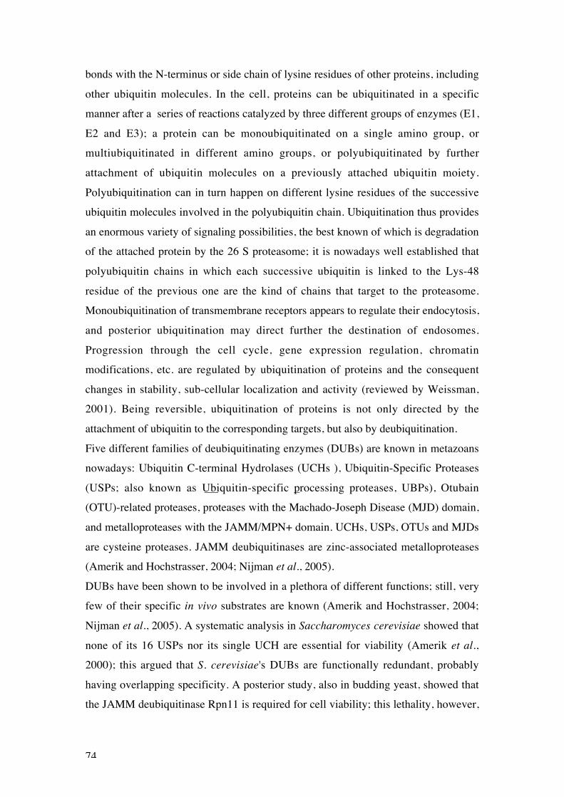
74
bonds with the N-terminus or side chain of lysine residues of other proteins, including
other ubiquitin molecules. In the cell, proteins can be ubiquitinated in a specific
manner after a series of reactions catalyzed by three different groups of enzymes (E1,
E2 and E3); a protein can be monoubiquitinated on a single amino group, or
multiubiquitinated in different amino groups, or polyubiquitinated by further
attachment of ubiquitin molecules on a previously attached ubiquitin moiety.
Polyubiquitination can in turn happen on different lysine residues of the successive
ubiquitin molecules involved in the polyubiquitin chain. Ubiquitination thus provides
an enormous variety of signaling possibilities, the best known of which is degradation
of the attached protein by the 26 S proteasome; it is nowadays well established that
polyubiquitin chains in which each successive ubiquitin is linked to the Lys-48
residue of the previous one are the kind of chains that target to the proteasome.
Monoubiquitination of transmembrane receptors appears to regulate their endocytosis,
and posterior ubiquitination may direct further the destination of endosomes.
Progression through the cell cycle, gene expression regulation, chromatin
modifications, etc. are regulated by ubiquitination of proteins and the consequent
changes in stability, sub-cellular localization and activity (reviewed by Weissman,
2001). Being reversible, ubiquitination of proteins is not only directed by the
attachment of ubiquitin to the corresponding targets, but also by deubiquitination.
Five different families of deubiquitinating enzymes (DUBs) are known in metazoans
nowadays: Ubiquitin C-terminal Hydrolases (UCHs ), Ubiquitin-Specific Proteases
(USPs; also known as Ubiquitin-specific processing proteases, UBPs), Otubain
(OTU)-related proteases, proteases with the Machado-Joseph Disease (MJD) domain,
and metalloproteases with the JAMM/MPN+ domain. UCHs, USPs, OTUs and MJDs
are cysteine proteases. JAMM deubiquitinases are zinc-associated metalloproteases
(Amerik and Hochstrasser, 2004; Nijman et al., 2005).
DUBs have been shown to be involved in a plethora of different functions; still, very
few of their specific in vivo substrates are known (Amerik and Hochstrasser, 2004;
Nijman et al., 2005). A systematic analysis in Saccharomyces cerevisiae showed that
none of its 16 USPs nor its single UCH are essential for viability (Amerik et al.,
2000); this argued that S. cerevisiae's DUBs are functionally redundant, probably
having overlapping specificity. A posterior study, also in budding yeast, showed that
the JAMM deubiquitinase Rpn11 is required for cell viability; this lethality, however,

75
is probably not due to the defect of deubiquitination of a specific substrate, but rather
because Rpn11, a component of the proteasome, is required for the deubiquitination
of proteins targeted for degradation, and this deubiquitination is required for ubiquitin
recycling and to prevent the proteasome from being blocked with substrates
conjugated with large polyubiquitin chains (Verma et al., 2002).
In flies, however, there appears to be some degree of substrate specificity. The Fat
facets (Faf) USP, for instance, has been shown to deubiquitinate and stabilize the
Liquid facets (Lqf) protein in the eye disc. Stabilization of Lqf by Faf is required for
intercellular signaling in the eye disc and the subsequent determination of the wild-
type number of photoreceptors in the adult eye (Chen et al., 2002). Does also Calypso
have specificity of substrate? The fact that USP7 mutants do not display the same
phenotype as calypso mutants argues for this (see Results, section 4.2.5).
5.1.2. Possible Calypso substrates
UCHs were first described as being able to cleave ubiquitin only from small adducts,
like single amino acids or short peptides (Larsen et al., 1998). Posterior biochemical
and structural studies, however, indicated that at least some UCHs may be able to
cleave ubiquitin from larger adducts (Misaghi et al., 2005). Calypso may then be
involved in deubiquitinating large proteins.
Ubiquitin is never synthesized as a monomer in vivo, but as fusion proteins of several
head-tail-linked ubiquitin units, or fused to ribosomal proteins. Several DUBs have
been shown to cleave such peptides, releasing ubiquitin monomers. Other DUBs are
associated to the proteasome, and are in charge of deubiquitinating substrates that
must not be degraded, or, quite opposite, of releasing the ubiquitin chain so that it is
recycled and the substrate degraded. Finally, some DUBs are in charge of releasing
ubiquitin monomers from such deconjugated polyubiquitin chains, or from little
naturally occurring adducts of ubiquitin with glutation or other metabolites (Amerik
and Hochstrasser, 2004; Nijman et al., 2005). I do not expect Calypso to perform this
kind of basic ubiquitin metabolism functions, at least not in the context of PcG-
mediated silencing.
One possible Calypso target could be histones. Histones are known to be
ubiquitinated in vivo, and this ubiquitination appears to have consequences in
transcription, chromatin structure and DNA repair (Zhang, 2003; Amerik and
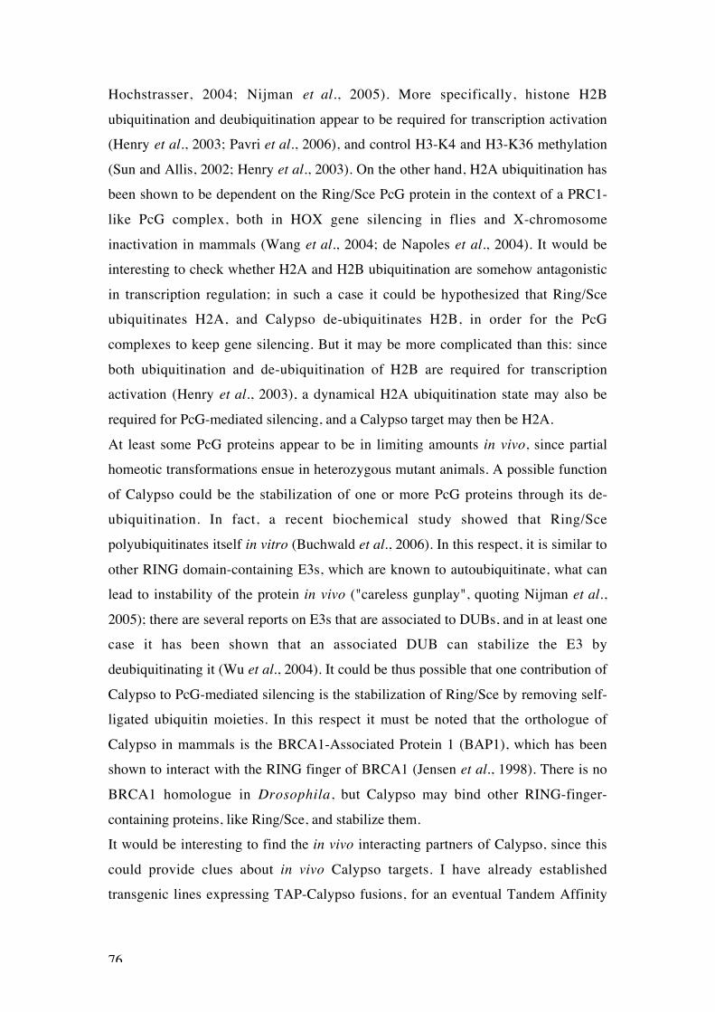
76
Hochstrasser, 2004; Nijman et al., 2005). More specifically, histone H2B
ubiquitination and deubiquitination appear to be required for transcription activation
(Henry et al., 2003; Pavri et al., 2006), and control H3-K4 and H3-K36 methylation
(Sun and Allis, 2002; Henry et al., 2003). On the other hand, H2A ubiquitination has
been shown to be dependent on the Ring/Sce PcG protein in the context of a PRC1-
like PcG complex, both in HOX gene silencing in flies and X-chromosome
inactivation in mammals (Wang et al., 2004; de Napoles et al., 2004). It would be
interesting to check whether H2A and H2B ubiquitination are somehow antagonistic
in transcription regulation; in such a case it could be hypothesized that Ring/Sce
ubiquitinates H2A, and Calypso de-ubiquitinates H2B, in order for the PcG
complexes to keep gene silencing. But it may be more complicated than this: since
both ubiquitination and de-ubiquitination of H2B are required for transcription
activation (Henry et al., 2003), a dynamical H2A ubiquitination state may also be
required for PcG-mediated silencing, and a Calypso target may then be H2A.
At least some PcG proteins appear to be in limiting amounts in vivo, since partial
homeotic transformations ensue in heterozygous mutant animals. A possible function
of Calypso could be the stabilization of one or more PcG proteins through its de-
ubiquitination. In fact, a recent biochemical study showed that Ring/Sce
polyubiquitinates itself in vitro (Buchwald et al., 2006). In this respect, it is similar to
other RING domain-containing E3s, which are known to autoubiquitinate, what can
lead to instability of the protein in vivo ("careless gunplay", quoting Nijman et al.,
2005); there are several reports on E3s that are associated to DUBs, and in at least one
case it has been shown that an associated DUB can stabilize the E3 by
deubiquitinating it (Wu et al., 2004). It could be thus possible that one contribution of
Calypso to PcG-mediated silencing is the stabilization of Ring/Sce by removing self-
ligated ubiquitin moieties. In this respect it must be noted that the orthologue of
Calypso in mammals is the BRCA1-Associated Protein 1 (BAP1), which has been
shown to interact with the RING finger of BRCA1 (Jensen et al., 1998). There is no
BRCA1 homologue in Drosophila, but Calypso may bind other RING-finger-
containing proteins, like Ring/Sce, and stabilize them.
It would be interesting to find the in vivo interacting partners of Calypso, since this
could provide clues about in vivo Calypso targets. I have already established
transgenic lines expressing TAP-Calypso fusions, for an eventual Tandem Affinity

77
Purification. Both N-terminal TAP-Calypso and C-terminal Calypso-TAP fusions are
able to rescue Ubx silencing in calypso mutant clones in the wing disc, and viability
in calypso2 hemizygotes (data not shown).
5.2. Role of Mediator in PcG-mediated silencing
Two mutants recovered in our screen, siren1/kto and penelope/skd, correspond to
genes encoding components of the Mediator complex. siren1/kto and penelope/skd
mutant clones in the wing disc show de-repression of Ubx, which is enhanced in a Pcl
heterozygous background. kto and skd alleles recovered in a different screen (Janody
et al., 2004) show the same HOX gene de-repression phenotype, indicating that this is
a common phenotype of kto and skd mutants, and not specific of mutants recovered
from our screen.
5.2.1. Med12 and Med13 belong to a submodule in Mediator with repressive
function
kto encodes Med12, and skd encodes Med13, components of a biochemically
separable submodule of the Mediator complex with repressive function. Preparations
of Mediator that include the CRSP70 protein and without the submodule that includes
Cdk8, CycC, Med12 and Med13 (the Cdk8-CycC submodule) are capable of
activating transcription in vitro. However, Mediator complexes that include the Cdk8-
CycC submodule, which lack CRSP70, do not activate transcription in vitro
(Borggrefe et al., 2002; Taatjes et al., 2002; Lewis and Reinberg, 2003. Check
Bourbon et al., 2004, for an update on Mediator component nomenclature).
Furthermore, a recent study reported that the expression profiles of S. cerevisiae
mutants for components of the Cdk8-CycC submodule are very similar to each other,
mostly showing de-repression of different genes; these expression profiles are very
different, actually opposite, from the expression profiles of mutants for other
components of the Mediator, which mainly show gene expression down-regulation.
These results support the view that the Cdk8-CycC submodule has repressive function
inside the otherwise transcription-activating Mediator complex (van de Peppel et al.,
2005).

78
Med12 and Med13 yeast, fly and nematode mutants show de-repression of several
different genes, further suggesting that Med12 and Med13 have repression function
(Carlson, 1997; Treisman, 2001; Yoda et al., 2005). However, these reported de-
repression phenotypes may be an indirect consequence of Med12 and Med13
mutations, and our siren1/kto and penelope/skd mutants present the same problem: are
kto and skd really PcG genes, or are they somehow indirectly required for PcG
function? Being Mediator components, Med12 and Med13 may have transcription
activation function in vivo, at least in some circumstances; they may be required for
optimal expression of, for instance, PcG genes. In such a case, kto and skd mutants
may show a PcG phenotype without being repressors or PcG genes themselves.
A way to address this question would be checking whether the phenotype of
Drosophila cdk8 and cycC mutants is similar to the phenotype of kto and skd, and
whether this phenotype is different from the one of mutants for other components of
the Mediator complex. If all Mediator mutants showed the same phenotype as kto and
skd, it would strongly argue in favor of an indirect effect of Med12 and Med13 on
PcG function, probably by inducing the expression of proteins involved in PcG-
mediated silencing, maybe PcG proteins themselves. However, if kto, skd, cdk8 and
cycC mutants show a PcG phenotype, and other components of the Mediator do not, it
would support a more direct involvement of the Cdk8-CycC submodule in HOX gene
repression. Still, we did not recover any cdk8 nor cycC mutants from our screen,
indicating that perhaps they behave in a different way from kto and skd.
5.3. A genetic screen for new PcG genes in Drosophila
5.3.1. EMS mutagenesis: advantages and disadvantages
The use of EMS in our screen provides the advantage that it is unbiased in its
mutagenizing activity, reaching the whole genome. In this respect it is different from
transposon insertional mutagenesis, which displays some tendencies depending on the
transposon; some genes appear not to be ever disrupted by transposon insertions,
while other genes appear highly likely to be disrupted. Furthermore, use of P-element
"jumping" is not possible in our screen, since it would also mobilize FRTs, P-
elements themselves; Piggy-bac transposons would then be the only available choice.
It could be argued that EMS mutagenesis is not completely unbiased, in the sense that

79
some genomic regions are probably more easily repaired than others by the DNA
repair systems (in fact, the striking finding that both calypso alleles, generated in
independent EMS mutagenesis, show exactly the same base-pair substitution, would
argue for some kind of bias in this genomic region). Still, a saturation mutagenesis
will likely end up producing lesions all over the genome.
The disadvantage of chemical mutagenesis is the posterior mapping of mutations,
which must be carried out by traditional genetic methods. Fortunately, the abundance
nowadays of different markers (like those on the ru cu ca chromosome), transposon
insertions and deficiencies (many of which are now being "custom-made" and
molecularly mapped; Parks et al., 2004; Ryder et al., 2004) makes mapping much
easier. Once the interval of a mutant's locus has been narrowed down, the availability
of the D. melanogaster genome permits taking the candidate-gene approach.
Additional hits on mutagenized chromosomes may complicate mapping, but this
problem can be circumvented by performing mutagenesis on isogenized
chromosomes, thus avoiding lethal lesions in the genetic background. Using EMS at a
25mM concentration made multiple lethal hits rare, as seen by lethality tests in which
roughly half F1-males carried a lethal mutation. Having several alleles obtained in
different mutagenesis is also very useful to confirm results for mapping, and it is
anyway an aim to be fulfilled as an indication that saturation was reached.
5.3.2. Usefulness of the Flp-FRT system
The Flp-FRT system has proven very useful as a way to obtain phenotypes in adult
flies in individuals heterozygous for a lethal mutation. It has made possible to screen
through the F1 generation from mutagenized flies, so that interesting mutants could be
identified and the rest of the offspring discarded, without need for further crosses to
score embryonic phenotypes in the screening stage. Since clones were generated only
in wing and haltere discs, PcG-syndrome-bearing flies were viable and fertile and
could be used for a retest and then to establish stocks. However, it must be said that
severe cases of PcG-syndrome still had survival problems: sometimes, for unknown
reason, those flies with strong PcG-syndrome displayed bent legs with mobility
problems. Maybe there is some leaky expression of the Flp driver (the vestigial
Boundary Enhancer) or of the UAS-Flp transgene itself in the leg discs; this leaky
expression appears not to perturb leg development in mutants with a weak PcG-

80
syndrome, but can be quite deleterious in other cases. It was difficult to obtain Pcl
alleles, since many such mutants usually got stuck to the food and died. While
screening, I had to avoid leaving flies in their "natal" bottles for long to be able to
isolate weak mutants into clean vials.
Temperature also played an important role: Cornelia Fritsch, Dirk Beuchle and Jürg
Müller noted that letting the offspring of mutagenized males develop at 25ºC gave
place to very few severe mutants, being their survival seriously compromised. That is
why the F1 generation has to develop at 18ºC. Overcrowding can also pose a problem
for individuals with a strong phenotype, so it must be avoided.
One important advantage of the Flp-FRT system is the possibility of screening one
chromosome at a time; it also allows mapping mutations to the right or left arm,
facilitating mapping considerably. Having interesting mutations on an FRT
chromosome can be very useful afterwards, as it was the case to obtain calypso germ-
line mutant clones, for instance; or more importantly, to check for HOX gene de-
repression in clones, as discussed below.
5.3.3. Assay for Ubx misexpression in clones in wing disc
A strict definition of PcG genes demands HOX gene de-repression in bona fide PcG
mutants. It is desirable to assay such misexpression directly, rather than inferring it
from homeotic phenotypes, when HOX gene inter-regulation can give place to
misleading results (Kennison, 2004). Since mutants from our screen were generated
on FRTs it was possible to assay for HOX gene misexpression in clones; wing discs
are easy to dissect, vestigial Boundary Enhancer-driven Flp expression works well in
this tissue, and Ubx misexpression appears shortly after PcG-mutant clone generation
(Beuchle et al., 2001). We considered Ubx misexpression in clones in wing disc
would then be a good read-out for PcG loss of function.
Other screens have assayed for PcG phenotype enhancement to find new PcG mutants
(Landecker, 1994; Fauvarque et al., 2001; Mollaaghababa et al., 2001). In my
experience, however, genetic interaction is very dependent on the genetic background
and environmental conditions. This was particularly disturbing in the case of calypso,
which enhanced Pc15 phenotype very variably (results not shown). In fact, the
absolute number of extra sex comb teeth in Pc15 heterozygous embryos was also
highly variable; this makes it always necessary to compare sibling animals that

81
developed in the same conditions when performing this kind of experiment. On the
other hand, not every PcG mutant genetically enhances the phenotype of other PcG
mutants (Landecker et al., 1994); and again HOX gene inter-regulation can
sometimes make genetic interactions between PcG and trxG mutants difficult to
interpret (Kennison, 2004). Genetic interaction assessments may be useful, for
example to see whether our penelope alleles behave like already described skd alleles.
Still, we favor direct HOX gene misexpression assessment to include mutants into the
PcG.
5.3.4. Most mutants for new PcG loci generally display low levels of HOX gene
misexpression
With the exception of calypso, all mutants for new PcG loci recovered from our
screen show low levels of Ubx de-repression in clones in the wing disc. This is not
completely unexpected: those PcG loci with strong misexpression phenotypes are
more likely to give place to partial homeotic transformations in heterozygosis, or to
strong homeotic transformations in homozygous mutant embryos, that is, are more
likely to have already been isolated in screens for homeotic mutations. Those loci we
have recovered from our screen may not play such a central role in HOX gene
silencing as already known PcG genes, giving thus place to weak HOX gene de-
repression when mutated. May be higher de-repression levels would be observed if
clones were allowed to develop further, since different PcG mutants show different
time points of onset of HOX gene misexpression in clones (Beuchle et al., 2001). In
fact, in the beginning of my PhD I tried dissecting pupal wings instead of wing
imaginal discs to assay for Ubx misexpression in clones (results not shown); this way
I hoped to obtain stronger Ubx de-repression in mutants for the new PcG loci we
isolated. This approach turned out to be too time-consuming, and by that time I could
start to assess for enhancement of the misexpression in a PclD5/+ background, so I did
not pursue it any further.
Another possibility is functional redundancy: if several of the new PcG loci are
present in multiple copies in the genome, mutants for only one of such copies are not
expected to show strong HOX gene de-repression; such is in fact the case for Psc and
Su(z)2 (Beuchle et al., 2001), ph-p and ph-d (Dura et al., 1987) and pho and phol

82
(Brown et al., 2003). We plan to map these new PcG loci eventually; we may then
realize that some of them are capable of compensating for one another.
5.3.5. Limitations of the screen
We already knew before-hand that our screen would have limitations and would not
allow recovery of some types of mutants. To start with, those PcG genes with a strong
compensating redundancy, like phol, could not be recovered from our screen. The
Flp-FRT system allows formation of mutant clones only for loci distal from the FRT;
mutations on sxc, being between the centromere and FRT42D, on the right arm of the
second chromosome, could not be recovered either; the same would happen with any
non-described PcG locus on a similar site. Of course, cell-lethal mutations cannot be
recovered from our screen, since homozygous cells cannot give place to clones and
the surrounding non-mutant tissue takes over.
Recently Tetyana Klymenko purified the Pho-Repressive Complex (PhoRC), which
contains Pho and dSfmbt. dSfmbt targeted knock-out mutants show HOX gene
misexpression (Klymenko et al., 2006). The dSfmbt locus is on the left arm of the
second chromosome, but we did not recover any dSfmbt mutants in our screen. To see
whether dSfmbt mutants, subjected to the same procedure as in the screen, would
show PcG syndrome, I induced dSfmbt mutant clones in the wing disc using the same
Flp driver as in the screen, and let flies develop at 18ºC. Surprisingly, almost all
dSfmbt clone-bearing flies that hatched got stuck in the pupal case by one of their
wings; eventually they all died almost completely emerged from the pupal case, but
one fly, which died stuck to the food. However, the wing phenotype was not strong; it
was comparable to the one of calypso or Asx, which are in turn much weaker than the
phenotype of Pcl mutants. So it seems it would be difficult to recover dSfmbt mutant
flies from our screen. Weaker mutants would perhaps survive, but then wing
phenotype would probably be negligible (take note that the dSfmbt allele I generated
clones for is a targeted knock-out predicted to be null; Klymenko et al., 2006).

83
6. Conclusions
6.1. Screen methodology allows to recover PcG mutants
We have designed a genetic screen for new PcG genes. Mutagenesis is performed
with a chemical mutagen, ethyl methanesulfonate (EMS), producing mutations in an
unbiased way. The Flp-FRT system allows to screen the offspring of mutagenized
males directly for candidate PcG mutants; it also determines for which chromosome
mutations may show a phenotype, so that the screen can be focused on specific
chromosomes. Candidate PcG mutants are then assayed for HOX gene de-repression,
a more stringent test to identify bona fide PcG mutants. Complementation tests have
shown that many of the HOX-misexpressing mutants recovered in the screen actually
correspond to already known PcG loci, demonstrating that the screening methodology
is valid to find PcG mutants. Several alleles have been recovered for most known and
new PcG genes from the screen on the second and third chromosomes, indicating that
saturation has been reached.
6.2. New PcG genes in Drosophila melanogaster were recovered from the screen
Our screen has allowed the isolation of several mutants with HOX gene de-repression
phenotypes that complement mutants for already known PcG genes, and which fall
into several different complementation groups. Thus, each of these complementation
groups highly likely uncovers a new PcG gene that can be mapped and characterized
to the molecular level. calypso, siren1/kto and penelope/skd represent such new PcG
loci.
6.3. calypso encodes a deubiquitinating enzyme required for PcG-mediated
repression
Our screen allowed the identification a new PcG gene, calypso, which encodes a
deubiquitinating enzyme. I have shown that Calypso is required throughout

84
development for maintenance of the silenced state of HOX genes. A rescue assay has
confirmed the identity of the calypso locus, and has indicated that its deubiquitinating
activity is required for HOX gene silencing. USP7, another deubiquitinating enzyme
whose mutants had been reported to enhance Pc mutant phenotype, is not required for
HOX gene silencing.
The calypso locus is conserved in vertebrates, having been described as a tumor
suppressor associated to the BRCA1 protein. We are currently pursuing biochemical
methods to find Calypso interacting partners in Drosophila melanogaster.
6.4. The Med12 and Med13 Mediator components are involved in HOX gene
silencing
Two loci identified in our screen, kto and skd, encode Med12 and Med13,
respectively, components of a submodule with transcription repressive function in the
Mediator complex. This suggests that Med12 and Med13 may be PcG proteins that
repress HOX genes in the context of the Mediator complex; however, we cannot rule
out the possibility that the role of Med12 and Med13 in HOX gene silencing is
indirect.

85
References
Akam, M. E. (1983). The location of Ultrabithorax transcripts in Drosophila tissuesections. EMBO J. 2, 2075-2084.
Akoulitchev, S., Chuikov, S. and Reinberg, D. (2000). TFIIH is negativelyregulated by cdk8-containing mediator complexes. Nature 407, 102-106.
Americo, J., Whiteley, M., Brown, J. L., Fujioka, M., Jaynes, J. B. and Kassis, J.A. (2002). A complex array of DNA-binding proteins required for pairing-sensitivesilencing by a Polycomb group response element from the Drosophila engrailed gene.Genetics 160, 1561-1571.
Amerik, A. Y. and Hochstrasser, M. (2004). Mechanism and function ofdeubiquitinating enzymes. Biochim. Biophys. Acta 1695, 189– 207.
Amerik, A. Y., Li, S.-J. and Hochstrasser, M. (2000). Analysis of thedeubiquitinating enzymes of the yeast Saccharomyces cerevisiae. Biol. Chem. 381,981-992.
Armstrong, J. A., Papoulas, O., Daubresse, G., Sperling, A. S., Lis, J. T., Scott,M. P. and Tamkun, J. W. (2002). The Drosophila BRM complex facilitates globaltranscription by RNA polymerase II. EMBO J. 21, 5245-5254.
Bannister, A., Zegerman, P., Partridge, J. F., Miska, E. A., Thomas, J. O.,Allshire, R. C. and Kouzarides, T. (2001). Selective recognition of methylatedlysine 9 on histone H3 by the HP1 chromo domain. Nature 410, 120-124.
Beachy, P. A., Helfand, S. L. and Hogness, D. S. (1985). Segmental distribution ofbithorax complex proteins during Drosophila development. Nature 313, 545-551.
Beisel, C., Imhof, A., Greene, J., Kremmer, E. and Sauer, F. (2002). Histonemethylation by the Drosophila epigenetic transcriptional regulator Ash1. Nature 419,857-862.
Beltran, S., Blanco, E., Serras, F., Pérez-Villamil, B., Guigó , R., Artavanis-Tsakonas, S. and Corominas, M. (2003). Transcriptional network controlled by thetrithorax-group gene ash2 in Drosophila melanogaster. Proc. Natl. Acad. Sci. USA100, 3293–3298.
Beuchle, D., Struhl, G. and Müller, J. (2001). Polycomb group proteins andheritable silencing of Drosophila Hox genes. Development 128, 993-1004.
Birve, A., Sengupta, A. K., Beuchle, D., Larsson, J., Kennison, J. A., Rasmuson-Lestander, Å. and Müller, J. (2001). Su(z)12, a novel Drosophila Polycomb groupgene that is conserved in vertebrates and plants. Development 128, 3371-3379.

86
Borggrefe, T., Davis, R., Erdjument-Bromage, H., Tempst, P. and Kornberg, R.D. (2002). A complex of the Srb8, -9, -10, and -11 transcriptional regulatory proteinsfrom yeast. J. Biol. Chem. 277, 44202–44207.
Bornemann, D., Miller, E. and Simon, J. (1998). Expression and properties of wild-type and mutant forms of the Drosophila Sex Comb on Midleg (SCM) repressorprotein. Genetics 150: 675–686.
Bourbon, H.-M., Aguilera, A., Ansari, A. Z., Asturias, F. J., Berk, A. J.,Bjorklund, S., Blackwell, T. K., Borggrefe, T., Carey, M., Carlson, M., Conaway,J. W., Conaway, R. C., Emmons, S. W., Fondell, J. D., Freedman, L. P.,Fukasawa, T., Gustafsson, C. M., Han, M., He, X., Herman, P. K., Hinnebusch,A. G., Holmberg, S., Holstege, F. C., Jaehning, J. A., Kim, Y.-J., Kuras, L.,Leutz, A., Lis, J. T., Meisterernest, M., Naar, A. M., Nasmyth, K., Parvin, J. D.,Ptashne, M., Reinberg, D., Ronne, H., Sadowski, I., Sakurai, H., Sipiczki, M.,Sternberg, P. W., Stillman, D. J., Strich, R., Struhl, K., Svejstrup, J. Q., Tuck, S.,Winston, F., Roeder, R. G. and Kornberg, R. D. (2004). A unified nomenclaturefor protein subunits of Mediator complexes linking transcriptional regulators to RNAPolymerase II. Molecular Cell 14, 553–557.
Breen, T. R. and Duncan, I. M. (1986). Maternal expression of genes that regulatethe Bithorax Complex of Drosophila melanogaster. Dev. Biol. 118, 442-456.
Brown, J. L., Fritsch, C., Mueller, J. and Kassis, J. A. (2003). The Drosophilapho-like gene encodes a YY1-related DNA binding protein that is redundant withpleiohomeotic in homeotic gene silencing. Development 130, 285-294.
Brown, J. L., Grau, D. J., DeVido, S. K. and Kassis, J. A. (2005). An Sp1/KLFbinding site is important for the activity of a Polycomb group response element fromthe Drosophila engrailed gene. Nucl. Acids Res. 33, 5181-5189.
Brown, J. L., Mucci, D., Whiteley, M., Dirksen, M.-L. and Kassis, J. A. (1998).The Drosophila Polycomb Group gene pleiohomeotic encodes a DNA binding proteinwith homology to the transcription factor YY1. Mol. Cell 1, 1057–1064.
Buchenau, P., Hodgson, J., Strutt, H. and Arndt-Jovin, D. J. (1998). Thedistribution of Polycomb-Group proteins during cell division and development inDrosophila embryos: Impact on models for silencing. J. Cell Biol. 141, 469–481.
Buchwald, G., van der Stoop, P., Weichenrieder, O., Perrakis, A., van Lohuizen,M. and Sixma, T. K. (2006). Structure and E3-ligase activity of the Ring-Ringcomplex of Polycomb proteins Bmi1 and Ring1b. EMBO J. 25, 2465-2474.
Busturia, A., Lloyd, A., Bejarano, F., Zavortink, M., Xin, H. and Sakonju, S.(2001). The MCP silencer of the Drosophila Abd-B gene requires both Pleiohomeoticand GAGA factor for the maintenance of repression. Development 128, 2163-2173.
Busturia, A. and Morata, G. (1988). Ectopic expression of homeotic genes causedby the elimination of the Polycomb gene in Drosophila imaginal epidermis.Development 104, 713-720.

87
Busturia, A., Wightman, C. D. and Sakonju, S. (1997). A silencer is required formaintenance of transcriptional repression throughout Drosophila development.Development 124, 4343-4350.
Cao, R., Tsukada, Y. and Zhang, Y. (2005). Role of Bmi-1 and Ring1A in H2Aubiquitylation and Hox gene silencing. Mol. Cell 20, 845–854.
Cao, R., Wang, L., Wang, H., Xia, L., Erdjument-Bromage, H., Tempst, P.,Jones, R. S. and Zhang, Y. (2002). Role of histone H3 lysine 27 methylation inPolycomb-Group silencing. Science 298, 1039-1043.
Capdevila, M. P. and García-Bellido, A. (1981). Genes involved in the activation ofthe Bithorax Complex of Drosophila. Wilhelm Roux's Arch. 179, 125-143.
Carlson, M. (1997). Genetics of transcriptional regulation in yeast: Connections tothe RNA Polymerase II CTD. Annu. Rev. Cell Dev. Biol. 13:1–23.
Celniker, S. E., Sharma, S., Keelan, D. J. and Lewis, E. B. (1990). The moleculargenetics of the bithorax complex of Drosophila: cis-regulation in the Abdominal-Bdomain. EMBO J. 9, 4277-4286.
Chan, C.-S., Rastelli, L. and Pirrotta, V. (1994). A Polycomb response element inthe Ubx gene that determines an epigenetically inherited state of repression. EMBO J.13, 2553-2564.
Chen, X., Zhang, B. and Fischer, J. A. (2002). A specific protein substrate for adeubiquitinating enzyme: Liquid facets is the substrate of Fat facets. Genes Dev. 16,289–294.
Chenna, R., Sugawara, H., Koike, T., Lopez, R., Gibson, T. J., Higgins, D. G. andThompson, J. D. (2003). Multiple sequence alignment with the Clustal series ofprograms. Nucleic Acids Res. 31, 3497-3500.
Chou, T. and Perrimon, N. (1996). The autosomal FLP-DFS technique forgenerating germline mosaics in Drosophila melanogaster. Genetics 144, 1673-1679.
Coré, N., Bel, S., Gaunt, S. J., Aurrand-Lions, M., Pearce, J., Fisher, A. andDjabali, M. (1997). Altered cellular proliferation and mesoderm patterning inPolycomb-M33-deficient mice. Development 124, 721-729.
Czermin, B., Melfi, R., McCabe, D., Seitz, V., Imhof, A. and Pirrota, V. (2002).Drosophila Enhancer of zeste/ESC complexes have a histone H3 methyltransferaseactivity that marks chromosomal Polycomb sites. Cell 111, 185-196.
Dejardin, J., Rappailles, A., Cuvier, O., Grimaud, C., Decouville, M., Locker, D.and Cavalli, G. (2005). Recruitment of Drosophila Polycomb group proteins tochromatin by DSP1. Nature 434, 533-538.

88
Dellino, G. I., Schwartz, Y. B., Farkas, G., McCabe, D., Elgin, S. C. R. andPirrota, V. (2004). Polycomb silencing blocks transcription initiation. Mol. Cell 13,887-893.
de Napoles, M., Mermoud, J. E., Wakao, R., Tang, Y. A., Endoh, M., Appanah,R., Nesterova, T. B., Silva, J., Otte, A. P., Vidal, M., Koseki, H. and Brockdorff,N. (2004). Polycomb Group proteins Ring1A/B link ubiquitylation of histone H2A toheritable gene silencing and X inactivation. Dev. Cell 7, 663–676,
Duncan, I. M. (1982). Polycomblike: A gene that appears to be required for thenormal expression of the Bithorax and Antennapedia gene complexes of Drosophilamelanogaster. Genetics 102, 49-70.
Dura, J.-M., Randsholt, N. B., Deatrick, J., Erk, I., Santamaria, P., Freeman, J.D., Freeman, S. J., Weddell, D. and Brock, H. W. (1987). A complex genetic locus,polyhomeotic, is required for segmental specification and epidermal development inD. melanogaster. Cell 51, 829-839.
Ebert, A., Schotta, G., Lein, S., Kubicek, S., Krauss, V., Jenuwein, T. andReuter, G. (2004). Su(var) genes regulate the balance between euchromatin andheterochromatin in Drosophila. Genes Dev. 18, 2973–2983.
Fauvarque, M.-O., Zuber, V. and Dura, J.-M. (1995). Regulation of polyhomeotictranscription may involve local changes in chromatin activity in Drosophila. Mech.Dev. 52, 343-355.
Fauvarque, M.-O., Laurenti, P., Boivin, A., Bloyer, S., Griffin-Shea, R.,Bourbon, H. M., and Dura, J.-M. (2001). Dominant modifiers of the polyhomeoticextra-sex-combs phenotype induced by marked P element insertional mutagenesis inDrosophila. Genet. Res., Camb. 78, 137-148.
Fischle, W., Wang, Y., Jacobs, S. A., Kim, Y., Allis, C. D. and Khorasanizadeh,S. (2003). Molecular basis for the discrimination of repressive methyl-lysine marks inhistone H3 by Polycomb and HP1 chromodomains. Genes Dev. 17, 1870–1881.
Francis, N. J., Saurin, A. J., Shao, Z. and Kingston, R. E. (2001). Reconstitution ofa Functional Core Polycomb Repressive Complex. Mol. Cell 8, 545–556.
Francis, N. J., Kingston, R. E. and Woodcock, C. L. (2004). Chromatin compactionby a Polycomb Group protein complex. Science 306, 1574-1577.
Franke, A., DeCamillis, M., Zink, D., Cheng, N., Brock, H. W. and Paro, R.(1992). Polycomb and polyhomeotic are constituents of a multimeric protein complexin chromatin of Drosophila melanogaster. EMBO J. 11, 2941-2950.
Fribourg, S., Romier, C., Werten, S., Gangloff, Y. G., Poterszman, A. andMoras, D. (2001). Dissecting the interaction network of multiprotein complexes bypairwise coexpression of subunits in E. coli. J. Mol. Biol 306, 363-373.

89
Fritsch, C., Brown, J. L., Kassis, J. A. and Müller, J. (1999). The DNA-bindingPolycomb group protein Pleiohomeotic mediates silencing of a Drosophila homeoticgene. Development 126, 3905-3913.
Fritsch, C., Beuchle, D., and Müller, J. (2003). Molecular and genetic analysis ofthe Polycomb group gene Sex combs extra/Ring in Drosophila. Mech Dev. 120, 949-954.
Gehring, W. J. (1970). A recessive lethal (l(4)29) with a homeotic effect in D.melanogaster. Dros. Inform. Serv. 45, 103.
Gendall, A. R., Levy, Y. Y., Wilson, A. and Caroline Dean. (2001). TheVERNALIZATION 2 gene mediates the epigenetic regulation of vernalization inArabidopsis. Cell 107, 525–535.
Glicksman, M. A. and Brower, D. L. (1988). Expression of the Sex combs reducedprotein in Drosophila larvae. Dev. Biol. 127, 113-118.
Golic, K. G. (1991). Site-specific recombination between homologous chromosomesin Drosophila. Science 252, 958-961.
Gorfinkiel, N., Fanti, L., Melgar, T., García, E., Pimpinelli, S., Guerrero, I. andVidal, M. (2004). The Drosophila Polycomb group gene Sex combs extra encodes theortholog of mammalian Ring1 proteins. Mech. Dev. 121, 449–462.
Grossniklaus, U., Vielle-Calzada, J.-P., Hoeppner, M. A. and Gagliano, W. B.(1998). Maternal control of embryogenesis by MEDEA, a Polycomb Group gene inArabidopsis. Science 280, 446-450.
Heins, N. (1998). Interaktionen von Polycomb-Gruppen Genen des drittenChromosoms mit Genen des zweiten Chromosoms bei Drosophila melanogaster.Diploarbeit, Eberhard-Karls University, Tübingen, Germany.
Henry, K. W., Wyce, A., Lo, W.-S., Duggan, L. J., Emre, N. C. T., Kao, C-F.,Pillus, L., Shilatifard, A., Osley, M. A. and Berger, S. L. (2003). Transcriptionalactivation via sequential histone H2B ubiquitylation and deubiquitylation, mediatedby SAGA-associated Ubp8. Genes Dev. 17, 2648–2663.
Hodgson, J. W., Argiropoulos, B. and Brock, H. W. (2001). Site-specificrecognition of a 70-base-pair element containing d(GA)n repeats mediates bithoraxoidPolycomb Group response element-dependent silencing. Mol. Cell. Biol. 21, 4528-4543.
Ingham, P. W. (1984). A gene that regulates the Bithorax Complex differentially inlarval and adult cells of Drosophila. Cell 37, 815-823.
Ingham, P. and Whittle, R. (1980). Trithorax: A new homoeotic mutation ofDrosophila melanogaster causing transformations of abdominal and thoracic imaginalsegments. Mol. Gen. Genet. 179, 607-614.

90
Ingham, P. W. and Martinez-Arias, A. (1986). The correct activation ofAntennapedia and bithorax complex genes requires the fushi tarazu gene. Nature 324,592-597.
Janody, F., Martirosyan, Z., Benlali, A. and Treisman, J. E. (2003). Two subunitsof the Drosophila mediator complex act together to control cell affinity. Development130, 3691-3701.
Janody, F., Lee, J. D., Jahren, N., Hazelett, D. J., Benlali, A., Miura, G. I.,Draskovic, I. and Treisman, J. E. (2004). A mosaic genetic screen reveals distinctroles for trithorax and Polycomb Group genes in Drosophila eye development.Genetics 166, 187–200.
Jensen, D. E., Proctor, M., Marquis, S. T., Gardner, H. P., Ha, S. I., Chodosh, L.A., Ishov, A. M., Tommerup, N., Vissing, H., Sekido, Y., Minna, J., Borodovsky,A., Schultz, D. C., Wilkinson, K. D., Maul, G. G., Barlev, N., Berger, S. L.,Prendergast, G. C. and Rauscher, F. J., 3rd. (1998). BAP1: a novel ubiquitinhydrolase which binds to the BRCA1 RING finger and enhances BRCA1-mediatedcell growth suppression. Oncogene 16, 1097-1112.
Jürgens, G. (1985). A group of genes controlling the spatial expression of thebithorax complex in Drosophila. Nature 316, 153, 155.
Karachentsev, D., Sarma, K., Reinberg, D. and Steward, R.(2005). PR-Set7-dependent methylation of histone H4 Lys 20 functions in repression of geneexpression and is essential for mitosis. Genes Dev. 19, 431–435.
Katoh, K., Misawa, K., Kuma, K. and Miyata, T. (2002). MAFFT version 5.25:multiple sequence alignment program. Nucleic Acids Res. 30, 3059-3066.
Kehle, J., Beuchle, D., Treuheit, S., Christen, B., Kennison, J. A., Bienz, M. andMüller, J. (1998). dMi-2, a Hunchback-interacting protein that functions in Polycombrepression. Science 282, 1897-1900.
Kennison, J. A. and Tamkun, J. W. (1988). Dosage-dependent modifiers ofPolycomb and Antennapedia mutations in Drosophila. Proc. Natl. Acad. Sci. USA 85,8136-8140.
Kennison, J. A. (2004). Introduction to Trx-G and Pc-G genes. Meth. Enzymol. 377,61-70.
Klymenko, T. and Müller, J. (2004). The histone methyltransferases Trithorax andAsh1 prevent transcriptional silencing by Polycomb group proteins. EMBO Rep. 5,373-377.
Klymenko, T., Papp, B., Fischle, W., Köcher, T., Schelder, M., Fritsch, C., Wild,B., Wilm, M. and Müller, J. (2006). A Polycomb group protein complex withsequence-specific DNA-binding and selective methyl-lysine-binding activities. GenesDev. 20, 1110–1122.

91
Köhler, C., Hennig, L., Bouveret, R., Gheyselinck, J., Grossniklaus, U. andGruissem, W. (2003). Arabidopsis MSI1 is a component of the MEA/FIE Polycombgroup complex and required for seed development. EMBO J. 22, 4804-4814.
Kuzmichev, A., Nishioka, K., Erdjument-Bromage, H., Tempst, P. and Reinberg,D. (2002). Histone methyltransferase activity associated with a human multiproteincomplex containing the Enhancer of Zeste protein. Genes Dev. 16, 2893–2905.
Lachner, M., O'Carroll, D., Rea, S., Mechtler, K. and Jenuwein, T. (2001).Methylation of histone H3 lysine 9 creates a binding site for HP1 proteins. Nature410, 116-120.
LaJeunesse, D. and Shearn, A. (1995). Trans-regulation of thoracic homeoticselector genes of the Antennapedia and bithorax complexes by the trithorax groupgenes: absent, small and homeotic discs 1 and 2. Mech. Dev. 53, 123-139.
Landecker, H. L., Sinclair, D. A. R. and Brock, H. W. (1994). Screen forenhancers of Polycomb and Polycomblike in Drosophila melanogaster. Dev. Genet.15, 425-434.
Larsen, C. N., Price, J. S. and Wilkinson, K. D. (1996). Substrate binding andcatalysis by Ubiquitin C-Terminal Hydrolases: Identification of two active siteresidues. Biochemistry 35, 6735-6744.
Larsen, C. N., Krantz, B. A. and Wilkinson, K. D. (1998). Substrate specificity ofdeubiquitinating enzymes: Ubiquitin C-Terminal Hydrolases. Biochemistry 37, 3358-3368.
Lewis, E. B. (1978). A gene complex controlling segmentation in Drosophila. Nature276, 565-570.
Lewis, B. A. and Reinberg, D. (2003). The mediator coactivator complex: functionaland physical roles in transcriptional regulation. J. Cell Sci. 116, 3667-3675.
Lindsley, D. L. and Zimm, G. G. (1992) The genome of Drosophila melanogaster.Academic Press, Elsevier group.
Luo, M., Bilodeau, P., Koltunow, A., Dennis, E. S., Peacock, W. J. andChaudhury, A. M. (1999). Genes controlling fertilization-independent seeddevelopment in Arabidopsis thaliana. Proc. Natl. Acad. Sci. USA 96, 296-301.
Martinez, A.-M., Colomb, S., Déjardin, J., Bantignies, F. and Cavalli, G. (2006).Polycomb group-dependent Cyclin A repression in Drosophila. Genes Dev. 20,501–513.
Maschat, F., Serrano, N., Randsholt, N. B. and Géraud, G. (1998). engrailed andpolyhomeotic interactions are required to maintain the A/P boundary of theDrosophila developing wing. Development 125, 2771-2780.

92
Mathews, C. K., van Holde, K. E. and Ahern, K. G. (2000). Biochemistry, 3rd
edition. Benjamin Cummings.
Maurange, C. and Paro, R. (2002). A cellular memory module conveys epigeneticinheritance of hedgehog expression during Drosophila wing imaginal discdevelopment. Genes Dev. 16, 2672–2683.
Misaghi, S., Galardy, P. J., Meester, W. J. N., Ovaa, H., Ploegh, H. L. andGaudet, R. (2005). Structure of the Ubiquitin Hydrolase UCH-L3 complexed with asuicide substrate. J. Biol. Chem. 280, 1512–1520.
Mishra, R. K., Mihaly, J., Barges, S., Spierer, A., Karch, F., Hagstrom, K.,Schweinsberg, S. E. and Schedl, P. (2001). The iab-7 Polycomb response elementmaps to a nucleosome-free region of chromatin and requires both GAGA andPleiohomeotic for silencing activity. Mol. Cell. Biol. 21, 1311-1318.
Mohd-Sarip, A., Cleard, F., Mishra, R. K., Karch, F. and Verrijzer, C. P. (2005).Synergistic recognition of an epigenetic DNA element by Pleiohomeotic and aPolycomb core complex. Genes Dev 19, 1755-1760.
Mollaaghababa, R., Sipos, L., Tiong, S. Y. K., Papoulas, O., Armstrong, J. A.,Tamkun, J. W. and Bender, W. (2001). Mutations in Drosophila heat shock cognate4 are enhancers of Polycomb. Proc. Natl. Acad. Sci. USA 98, 3958-3963.
Müller, J. and Bienz, M. (1991). Long range repression conferring boundaries ofUltrabithorax expression in the Drosophila embryo. EMBO J. 10, 3147-3155.
Müller, J. and Bienz, M. (1992). Sharp anterior boundary of homeotic geneexpression conferred by the fushi tarazu protein. EMBO J. 11, 3653 - 3661.
Müller, J., Gaunt, S., and Lawrence, P. A. (1995). Function of the Polycombprotein is conserved in mice and flies. Development 121, 2847-2852.
Müller, J. (1995). Transcriptional silencing by the Polycomb protein in Drosophilaembryos. EMBO J. 14, 1209-1220.
Müller, J., Hart, C. M., Francis, N. J., Vargas, M. L., Sengupta, A., Wild, B.,Miller, E. L., O’Connor, M. B., Kingston, R. E. and Simon, J. A. (2002). HistoneMethyltransferase Activity of a Drosophila Polycomb Group Repressor Complex.Cell 111, 197–208.
Nègre, N., Hennetin, J., Sun, L. V., Lavrov, S., Bellis, M., White, K. P. andCavalli, G. (2006). Chromosomal distribution of PcG proteins during Drosophiladevelopment. PLoS Biol. 4, e170. DOI: 10.1371/journal.pbio.0040170.
Nekrasov, M., Wild, B. and Müller, J. (2005). Nucleosome binding and histonemethyltransferase activity of Drosophila PRC2. EMBO Rep. 6, 348-353.
Nijman, S. M. B., Luna-Vargas, M. P. A., Velds, A., Brummelkamp, T. R.,,Dirac, A. M. G., Sixma, T. K. and Bernards, R. (2005). A genomic and functional

93
inventory of deubiquitinating enzymes. Cell 123, 773-786.
Ohad, N., Yadegari, R., Margossian, L., Hannon, M., Michaeli, D., Harada, J. J.,Goldberg, R. B. and Fischer, R. L. (1999). Mutations in FIE, a WD PolycombGroup gene, allow endosperm development without fertilization. Plant Cell 11, 407-415.
Orlando, V. and Paro, R. (1993). Mapping Polycomb-repressed domains in theBithorax Complex using in vivo formaldehyde cross-linked chromatin. Cell 75, 1187-1198.
Papoulas, O., Beek, S. J., Moseley, S. L., McCallum, C. M., Sarte, M., Shearn, A.and Tamkun, J. W. (1998). The Drosophila trithorax group proteins BRM, ASH1and ASH2 are subunits of distinct protein complexes. Development 125, 3955-3966.
Parks, A. L., Cook, K. R., Belvin, M., Dompe, N. A., Fawcett, R., Huppert, K.,Tan, L. R., Winter, C. G., Bogart, K. P., Deal, J. E., Deal-Herr, M. E., Grant, D.,Marcinko, M., Miyazaki, W. Y., Robertson, S., Shaw, K. J., Tabios, M.,Vysotskaia, V., Zhao, L., Andrade, R. S., Edgar, K. A., Howie, E., Killpack, K.,Milash, B., Norton, A., Thao, D., Whittaker, K., Winner, M. A., Friedman, L.,Margolis, J., Singer, M. A., Kopczynski, C., Curtis D., Kaufman, T. C.,Plowman, G. D., Duyk, G. and Francis-Lang, H. L. (2004). Systematic generationof high-resolution deletion coverage of the Drosophila melanogaster genome. Nat.Genet. 36, 288-92.
Paro, R. and Hogness, D. S. (1991). The Polycomb protein shares a homologousdomain with a heterochromatin-associated protein of Drosophila. Proc. Natl. Acad.Sci. USA 88, 263-267.
Pattatucci, A. M. and Kaufmann, T. C. (1991). The homeotic gene Sex combsreduced of Drosophila melanogaster is differentially regulated in the embryonic andimaginal stages of development. Genetics 129, 443-461.
Pavri, R., Zhu, B., Li, G., Trojer, P., Mandal, S., Shilatifard, A. and Reinberg, D.(2006). Histone H2B monoubiquitination functions cooperatively with FACT toregulate elongation by RNA Polymerase II. Cell 125, 703–717.
Petruk, S., Sedkov, Y., Smith, S., Tillib, S., Kraevski, V., Nakamura, T.,Canaani, E., Croce, C. M. and Mazo, A. (2001). Trithorax and dCBP acting in acomplex to maintain expression of a homeotic gene. Science 294, 1331-1334.
Qian, S., Capovilla, M. and Pirrota, V. (1991). The bx region enhancer, a distantcis-control element of the Drosophila Ubx gene and its regulation by hunchback andother segmentation genes. EMBO J. 10, 1415-1425.
Qian, S., Capovilla, M. and Pirrota, V. (1993). Molecular mechanisms of patternformation by the BRE enhancer of the Ubx gene. EMBO J. 12, 3865-3877.
Randsholt, N. B., Maschat, F. and Santamaria, P. (2000). polyhomeotic controlsengrailed expression and the hedgehog signaling pathway in imaginal discs. Mech.

94
Dev. 95, 89-99.
Rastelli, L., Chan, C. S. and Pirrota, V. (1993). Related chromosome binding sitesfor zeste, suppressors of zeste and Polycomb group proteins in Drosophila and theirdependence on Enhancer of zeste function. EMBO J. 12, 1513-1522.
Ringrose, L. and Paro R. (2004). Epigenetic regulation of cellular memory by thePolycomb and trithorax group proteins. Annu. Rev. Genet. 38, 413–443.
Roseman, R. R., Morgan, K., Mallin, D. R., Roberson, R., Parnell, T. J.,Bornemann, D. J., Simon, J. A. and Geyer, P. K. (2001). Long-range repression bymultiple Polycomb Group (PcG) proteins targeted by fusion to a defined DNA-binding domain in Drosophila. Genetics 158, 291-307.
Ross, J. M. and Zarkower, D. (2003). Polycomb Group regulation of Hox geneexpression in C. elegans. Dev. Cell 4, 891–901.
Ryder, E., Blows, F., Ashburner, M., Bautista-Llacer, R., Coulson, D.,Drummond, J., Webster, J., Gubb, D., Gunton, N., Johnson, G., O’Kane, C. J.,Huen, D., Sharma, P., Asztalos, Z., Baisch, H., Schulze, J., Kube, M., Kittlaus,K., Reuter, G., Maroy, P., Szidonya, J., Rasmuson-Lestander, Å., Ekström, K.,Dickson, B., Hugentobler, C., Stocker, H., Hafen, E., Lepesant, J. A., Pflugfelder,G., Heisenberg, M., Mechler, B., Serras, F., Corominas, M., Schneuwly, S., Preat,T., Roote, J. and Russell, S. (2004). The DrosDel Collection: A set of P-elementinsertions for generating custom chromosomal aberrations in Drosophilamelanogaster. Genetics 167, 797-813.
Saurin, A. J., Shao, Z., Erdjument-Bromage, H., Tempst, P. and Kingston, R. E.(2001). A Drosophila Polycomb group complex includes Zeste and dTAFII proteins.Nature 412, 655-660.
Schmitt, S., Prestel, M. and Paro, R. (2005). Intergenic transcription through aPolycomb group response element counteracts silencing. Genes Dev. 19, 697–708.
Schumacher, A., Faust, C. and Magnuson, T. (1996). Positional cloning of a globalregulator of anterior-posterior patterning in mice. Nature 383, 250-253.
Schwartz, Y. B., Kahn, T. G., Nix, D. A., Li, X.-Y., Bourgon, R., Biggin, M. andPirrotta, V. (2006). Genome-wide analysis of Polycomb targets in Drosophilamelanogaster. Nat. Genet. 6, 700-705.
Sengupta, A. K., Kuhrs, A. and Müller, J. (2004). General transcriptional silencingby a Polycomb response element in Drosophila. Development 131, 1959-1965.
Shao, Z., Raible, F., Mollaaghababa, R., Guyon, J. R., Wu, C.-T., Bender, W.and Kingston, R. E. (1999). Stabilization of Chromatin Structure by PRC1, aPolycomb Complex. Cell 98, 37–46.
Shearn, A. (1989). The ash-1, ash-2 and trithorax genes of Drosophila melanogasterare functionally related. Genetics 121, 517-525.
95
Simon, J. (1995). Locking in stable states of gene expression: Transcriptional controlduring Drosophila development. Curr. Opin. Cell Biol. 7, 376-385.
Simon, J., Chiang, A. and Bender, W. (1992). Ten different Polycomb group genesare required for spatial control of the abdA and AbdB homeotic products.Development 114, 493-505.
Simon, J., Chiang, A., Bender, W., Shimell, M. J. and O'Connor, M. (1993).Elements of the Drosophila Bithorax Complex that mediate repression by Polycombgroup products. Dev. Biol. 158, 131-144.
Simon, J. A. and Tamkun, J. W. (2002). Programming off and on states inchromatin: mechanisms of Polycomb and trithorax group complexes. Curr. Opin.Genet. Dev. 12, 210-218.
Smith, S. T., Petruk, S., Sedkov, Y., Cho, E., Tillib, S., Canaani, E. and Mazo, A.(2004). Modulation of heat shock gene expression by the TAC1 chromatin-modifyingcomplex. Nat. Cell Biol. 6, 162-167.
Soto, M. C., Chou, T.-B. and Bender W. (1995). Comparison of germline mosaicsof genes in the Polycomb group of Drosophila melanogaster. Genetics 140, 231-243.
Struhl, G. (1981). A gene product required for correct initiation of segmentaldetermination in Drosophila. Nature 293, 36-41.
Struhl, G. and Akam, M. (1985). Altered distributions of Ultrabithorax transcriptsin extra sex combs mutant embryos of Drosophila. EMBO J. 4, 3259-3264.
Struhl, G. and Brower, D. (1982). Early role of the esc+ gene product in thedetermination of segments in Drosophila. Cell 31, 285-292.
Struhl, G. and White, R. A. H. (1985). Regulation of the Ultrabithorax gene ofDrosophila by other Bithorax Complex genes. Cell 43, 507-519.
Sullivan, W., Ashburner, M. and Hawley, R. S. (2000). Drosophila protocols. ColdSpring Harbor Laboratory Press.
Sun, Z.-W. and Allis, C. D. (2002). Ubiquitination of histone H2B regulates H3methylation and gene silencing in yeast. Nature 418, 104-108.
Taatjes, D. J., Naar, A. M., Andel, F., 3rd, Nogales, E. and Tjian, R. (2002).Structure, function, and activator-induced conformations of the CRSP coactivator.Science 295, 1058-1062.
Tamkun, J. W., Deuring, R., Scott, M. P., Kissinger, M., Pattatucci, A. M.,Kaufman, T. C. and Kennison, J. A. (1992). brahma: A regulator of Drosophilahomeotic genes structurally related to the yeast transcriptional activator SNF2/SWI2.Cell 68, 561-572.

96
Tolhuis, B., Miujrers, I., de Wit, E., Teunissen, H., Talhout, W., van Steensel, B.and van Lohuizen, M. (2006). Genome-wide profiling of PRC1 and PRC2 Polycombchromatin binding in Drosophila melanogaster. Nat. Genet., in press.
Treisman, J. E. (2001). Drosophila homologues of the transcriptional coactivationcomplex subunits TRAP240 and TRAP230 are required for identical processes in eyeantennal disc development. Development 128, 603-615.
van de Peppel, J., Kettelarij, N., van Bakel, H., Kockelkorn, T. T., van Leenen,D. and Holstege, F. C. (2005). Mediator expression profiling epistasis reveals asignal transduction pathway with antagonistic submodules and highly specificdownstream targets. Mol. Cell 19, 511-522.
van der Knaap, J. A., Kumar, B. R. P., Moshkin, Y. M., Langenberg, K.,Krijgsveld, J., Heck, A. J. R., Karch, F. and Verrijzer, C. P. (2005). GMPSynthetase stimulates histone H2B deubiquitylation by the epigenetic silencer USP7.Mol. Cell 17, 695-707.
van der Lugt, N. M. T., Domen, J., Linders, K., van Roon, M., Robanus-Maandag, E., te Riele, H., van der Valk, M., Deschamps, J., Sofroniew, M., vanLohuizen, M. and Berns, A. (1994). Posterior transformation, neurologicalabnormalities, and severe hematopoietic defects in mice with a targeted deletion ofthe bmi-1 proto-oncogene. Genes Dev. 8, 757-769.
Vegh, M. and Basler, K. (2003). A genetic screen for Hedgehog targets involved inthe maintenance of the Drosophila Anteroposterior Compartment Boundary. Genetics163, 1427–1438.
Verma, R., Aravind, L., Oania, R., McDonald, W. H., Yates, 3rd, J. R., Koonin,E. V. and Deshaies, R. J. (2002). Role of Rpn11 metalloprotease in deubiquitinationand degradation by the 26S proteasome. Science 298, 611-615.
Wang, H., Wang, L., Erdjument-Bromage, H., Vidal, M., Tempst, P., Jones, R. S.and Zhang, Y. (2004a). Role of histone H2A ubiquitination in Polycomb silencing.Nature 431, 873-878.
Wang, L., Brown, J. L., Cao, R., Zhang, Y., Kassis, J. A. and Jones, R. S.(2004b). Hierarchical recruitment of Polycomb Group silencing complexes. Mol. Cell14, 637–646.
Wang, L., Jahren, N., Vargas, M. L., Andersen, E. F., Benes, J., Zhang, J.,Miller, E. L., Jones, R. S. and Simon, J. A. (2006). Alternative ESC and ESC-Likesubunits of a Polycomb Group histone methyltransferase complex are differentiallydeployed during Drosophila development. Mol. Cell. Biol. 26, 2637–2647.
Wedeen, C., Harding, K. and Levine, M. (1986). Spatial regulation ofAntennapedia and Bithorax gene expression by the Polycomb locus in Drosophila.Cell 44, 739-748.

97
Weissman, A. M. (2001). Themes and variations in ubiquitylation. Nat. Rev. Mol.Cell Biol. 2, 169-178.
White, R. A. H. and Lehmann, R. (1986). A gap gene, hunchback, regulates thespatial expression of Ultrabithorax. Cell 47, 311-321.
White, R. A. H. and Wilcox, M. (1984). Protein products of the Bithorax Complexin Drosophila. Cell 39, 163-171.
Wolpert, L., Beddington, R., Brockes, J., Jessel, T., Lawrence, P. andMeyerowitz, E. (1998). Principles of development. Current biology Ltd. OxfordUniversity Press.
Wu, X., Yen, L., Irwin, L., Sweeney, C. and Carraway, K. L., 3rd. (2004).Stabilization of the E3 ubiquitin ligase Nrdp1 by the deubiquitinating enzyme USP8.Mol. Cell. Biol. 24, 7748-7757.
Wysocka J., Allis, C. D. and Coonrod, S. (2006). Histone arginine methylation andits dynamic regulation. Front. Biosci. 11, 344-355.
Yoda, A., Kouike, H., Okano, H. and Sawa, H. (2005). Components of thetranscriptional Mediator complex are required for asymmetric cell division in C.elegans. Development 132, 1885-1893.
Zhang, Yi. (2003). Transcriptional regulation by histone ubiquitination anddeubiquitination. Genes Dev. 17, 2733-2740.