Mitochondrial diseases: expanding the diagnosis in the era ...
1
A Diagnosis of “Possible” Mitochondrial Disease: An Existential Crisis
Sumit Parikh*, MD, Mitochondrial Medicine Center, Neurologic Institute, Cleveland Clinic,
Cleveland, OH
Amel Karaa*, MD, Mitochondrial Disease Program, Genetics Unit, Massachusetts General
Hospital, Boston, MA.
Amy Goldstein, MD, Mitochondrial Medicine Frontier Program, Division of Human Genetics,
Department of Pediatrics, Children’s Hospital of Philadelphia, Philadelphia, PA 19104, USA;
University of Pennsylvania Perelman School of Medicine, Philadelphia, PA 19104, USA
Enrico Bertini, Unit of Neuromuscular and Neurodegenerative Disorders, Bambino Gesu’
Children’s Research Hospital, Rome, Italy
Patrick F. Chinnery, MRC Mitochondrial Biology Unit & Department of Clinical Neurosciences,
University of Cambridge, Cambridge Biomedical Campus, Cambridge, CB2 0QQ, UK
John Christodoulou, Neurodevelopmental Genomics Research Group, Murdoch Children’s
Research Institute, Melbourne and Department of Paediatrics, Melbourne Medical School,
University of Melbourne, Melbourne, Victoria, Australia
Bruce H. Cohen, Department of Pediatrics and Rebecca D. Considine Research Institute, Akron
Children’s Hospital, Akron, OH, USA and Northeast Ohio Medical University, Rootstown, OH,
USA
Ryan L. Davis, University of Sydney, Faculty of Medicine and Health, Northern Clinical School,
Sydney, New South Wales, Australia; Department of Neurogenetics, Kolling Institute, University
of Sydney and Royal North Shore Hospital, Sydney, New South Wales, Australia
2
Marni J. Falk, Mitochondrial Medicine Frontier Program, Division of Human Genetics,
Department of Pediatrics, Children’s Hospital of Philadelphia, Philadelphia, PA 19104, USA;
University of Pennsylvania Perelman School of Medicine, Philadelphia, PA 19104, USA
Carl Fratter, NHS Specialized Services for Rare Mitochondrial Disorders of Adults and Children
UK, Oxford, UK and Oxford Medical Genetics Laboratories, Oxford University Hospitals, Oxford,
UK
Rita Horvath, Wellcome Centre for Mitochondrial Research, Institute of Genetic Medicine,
Newcastle University, Central Parkway, Newcastle upon Tyne, NE1 3BZ UK; Department of
Clinical Neurosciences at the University of Cambridge Box 165, Addenbrooke’s Hospital,
Cambridge Biomedical Campus, Cambridge CB2 0QQ
Mary Kay Koenig, Department of Pediatrics, Mitochondrial Center, University of Texas
McGovern Medical School, Houston, Texas, USA
Michelangelo Mancuso, Department of Experimental and Clinical Medicine, Neurological
Institute, University of Pisa, Italy
Shana McCormack, Mitochondrial Medicine Frontier Program, Division of Human Genetics,
Department of Pediatrics, Children’s Hospital of Philadelphia, Philadelphia, PA 19104, USA;
University of Pennsylvania Perelman School of Medicine, Philadelphia, PA 19104, USA
Elizabeth M. McCormick, Mitochondrial Medicine Frontier Program, Division of Human
Genetics, Department of Pediatrics, Children’s Hospital of Philadelphia, Philadelphia, PA 19104,
USA
3
Robert McFarland, Wellcome Centre for Mitochondrial Research, Institute of Neuroscience,
Newcastle University, Newcastle upon Tyne, NE 2 4HH UK
Victoria Nesbitt, NHS Highly Specialised Services for Rare Mitochondrial Disorders, Oxford
University Hospitals, Oxford, OX3 9DU, UK; Wellcome Centre for Mitochondrial Research,
Institute of Neuroscience, Newcastle University, Newcastle upon Tyne, NE2 4HH, UK
Manuel Schiff, Reference Center for Inborn Errors of Metabolism, Robert-Debré University
Hospital, APHP, UMR1141, PROTECT, INSERM, Université Paris-Diderot, Sorbonne Paris Cité,
Paris, France
Hannah Steele, Department of Neurology, Sunderland Royal Hospital, Sunderland, SR4 7TP, UK.
Previously at Wellcome Centre for Mitochondrial Research, Institute of Genetic Medicine,
Newcastle University, Central Parkway, Newcastle upon Tyne, NE1 3BZ UK
Sylvia Stockler, Department of Pediatrics, Division of Biochemical Diseases, University of British
Columbia
Carolyn Sue, University of Sydney, Faculty of Medicine and Health, Northern Clinical School,
Sydney, New South Wales, Australia; Department of Neurogenetics, Kolling Institute, University
of Sydney and Royal North Shore Hospital, Sydney, New South Wales, Australia; Department of
Neurology, Royal North Shore Hospital, Northern Sydney Local Health District, Sydney, New
South Wales, Australia
Mark Tarnopolsky, Department of Pediatrics, Neuromuscular and Neurometabolic Clinic,
McMaster University, Hamilton, Canada
David Thorburn, Murdoch Children’s Research Institute, Royal Children’s Hospital, Melbourne,
Victoria, Australia; Victorian Clinical Genetics Services, Royal Children’s Hospital, Melbourne,
4
Victoria, Australia; Department of Paediatrics, University of Melbourne, Melbourne, Victoria,
Australia
Jerry Vockley, Department of Pediatrics, University of Pittsburgh School of Medicine; Center for
Rare Disease Therapy, Children’s Hospital of Pittsburgh, Pittsburgh, PA 15224
Shamima Rahman, Mitochondrial Research Group, UCL Great Ormond Street Institute of Child
Health, London, UK; Metabolic Unit, Great Ormond Street Hospital NHS Foundation Trust,
London, UK
*These authors contributed equally to this manuscript.

5
Corresponding Author
Sumit Parikh, MD
Cleveland Clinic, 9500 Euclid Ave, S60, Cleveland, OH 44195.
Word Count (text) 2997
Number of Figures: 0
Number of Tables: 5
6
Abstract
Primary genetic mitochondrial diseases are often difficult to diagnose, and the term “possible”
mitochondrial disease is used frequently by clinicians when such a diagnosis is suspected. There
are now many known phenocopies of mitochondrial disease. Advances in genomic testing have
shown that some patients with a clinical phenotype and biochemical abnormalities suggesting
mitochondrial disease may have other genetic disorders. In instances when a genetic diagnosis
cannot be confirmed, a diagnosis of “possible” mitochondrial disease may result in harm to
patients and their families, creating anxiety, delaying appropriate diagnosis and leading to
inappropriate management or care. A categorization of “diagnosis uncertain”, together with a
specific description of the metabolic or genetic abnormalities identified, is preferred when a
mitochondrial disease cannot be genetically confirmed.
Author contributions
SP, AK, AG conceived the presented idea and prepared a document outline including needed
supplementary material.
SP, AK, AG, SR developed and supervised the manuscript.
SP, AK, AG, EB, PFC, JC, BHC, RLD, MJF, CF, RH, MKK, MM, SM, EMM, RM, VN, MS, HS, SS, CS,
MT, DT, JV, SR commented on, approved and helped expand on the presented idea, drafted and
revised portions of the manuscript including supplementary materials and commented on and
approved the final draft.
Details of funding: This project did not receive direct funding.
Patient consent and IRB: IRB involvement was not required for this project
Key Words:
Mitochondrial Disease Diagnosis, Genetic Disorders, Patient Harm

7
8
Introduction
Primary mitochondrial disorders (PMD) are genetic metabolic disorders that directly impair
normal mitochondrial structure or function including electron-transport chain (ETC) activity.[1]
They are due to mutations in either maternally-inherited mitochondrial DNA (mtDNA) or one of
hundreds of nuclear DNA (nDNA) genes that encode components involved in mitochondrial
structure and function. PMDs can present at any age and be multisystemic or selectively involve
only a single organ. They can present as a well-defined canonical syndrome or a constellation of
phenotypes, although typically at least one “red-flag” symptom is usually present at disease
onset.[2]
With advances in next-generation sequencing (NGS) and the discovery of a multitude of new
disease genes, the ability to diagnose PMDs has improved enormously compared to just a few
years ago. The diagnosis still remains challenging due to heterogeneous manifestations
combined with limitations of currently available biochemical and genetic testing methods.
Despite recent advances, many individuals with suspected mitochondrial disease may remain
without a confirmed genetic diagnosis, presenting a challenge to the clinician in not only
establishing a mitochondrial disease diagnosis but also in knowing how to categorize, counsel
and manage the patient with a suspected PMD where a genetic diagnosis is not yet possible.
[3–7]
These limitations contribute to continued variation in diagnostic categorization of patients
depending on the opinion of the treating provider.[8] Diagnostic terms such as “unlikely,”
“possible” or “probable” mitochondrial disease, originally proposed as part of research
diagnostic criteria[9–11] were developed prior to genetic advances and may end up being
inaccurate and misleading to patients and care providers, impacting or limiting proper
counseling and the pursuit of further diagnostic testing. The complex and variable clinical
presentation of mitochondrial diseases means that many unexplained disorders could
conceivably have a mitochondrial etiology, so if a concrete alternative diagnosis cannot be

9
made using conventional investigations, there is a tendency to use the label ‘possible’
mitochondrial as a working diagnosis until an alternative emerges. Patients and families may
inadvertently be burdened by the fear of the progressive nature of PMDs, the potential
complications and early demise, as PMDs have no known cure. Thus, a diagnosis of “possible”
mitochondrial disease may do more harm than good and consequently a new categorization for
these patients is necessary.
Mitochondrial Disease can no longer be diagnosed on the basis of phenotypic features alone
A high index of suspicion for the possibility of a mitochondrial disease is appropriate when
there is multi-system involvement or the presence of so-called “red-flags” such as stroke-like
episodes in a non-vascular distribution (seen in Mitochondrial Encephalomyopathy, Lactic
Acidosis and Stroke [MELAS] syndrome), bilateral symmetrical T2-weighted hyperintense MRI
lesions in the basal ganglia and/or brainstem (Leigh syndrome), or chronic progressive external
ophthalmoplegia (CPEO) and myopathy. Some of these “red-flag” symptoms were the subject
of a previous review.[2] However, the growing list of genetically confirmed mitochondrial
diseases has also led to an expanding list of variable phenotypes that should be suspected in
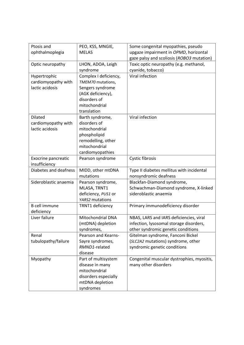
the differential diagnosis of PMD, some of which are outlined in Table 1. [12–14]
Table 1 Differential diagnosis of selected phenotypes commonly associated withmitochondrial disease
Phenotype Mitochondrial cause Limited differential diagnosis
Dystonia Leigh syndrome,deafness-dystoniasyndrome, othermitochondrialencephalomyopathies
Biotinidase deficiency, thiaminetransporter deficiency 2, ADAR mutations(Aicardi- Goutières syndrome 6), organicacidaemias (especially glutaric aciduriatype I), NBIA, acute (viral) necrotisingencephalopathy, mutations in NUP62,RANBP2 and PDE8B, primary geneticdystonias
Epilepticencephalopathy
Alpers-Huttenlochersyndrome, many other
Many genetic epileptic encephalopathies,including Dravet syndrome and KCNQ2mutations, Pyridoxine dependent
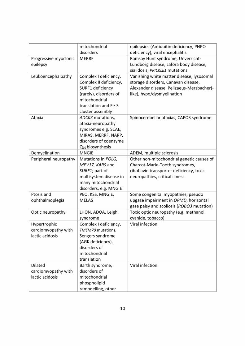
10
mitochondrialdisorders
epilepsies (Antiquitin deficiency, PNPOdeficiency), viral encephalitis
Progressive myoclonicepilepsy
MERRF Ramsay Hunt syndrome, Unverricht-Lundborg disease, Lafora body disease,sialidosis, PRICKLE1 mutations
Leukoencephalpathy Complex I deficiency,Complex II deficiency,SURF1 deficiency(rarely), disorders ofmitochondrialtranslation and Fe-Scluster assembly
Vanishing white matter disease, lysosomalstorage disorders, Canavan disease,Alexander disease, Pelizaeus-Merzbacher(-like), hypo/dysmyelination
Ataxia ADCK3 mutations,ataxia-neuropathysyndromes e.g. SCAE,MIRAS, MERRF, NARP,disorders of coenzymeQ10 biosynthesis
Spinocerebellar ataxias, CAPOS syndrome
Demyelination MNGIE ADEM, multiple sclerosis
Peripheral neuropathy Mutations in POLG,MPV17, KARS andSURF1; part ofmultisystem disease inmany mitochondrialdisorders, e.g. MNGIE
Other non-mitochondrial genetic causes ofCharcot-Marie-Tooth syndromes,riboflavin transporter deficiency, toxicneuropathies, critical illness
Ptosis andophthalmoplegia
PEO, KSS, MNGIE,MELAS
Some congenital myopathies, pseudoupgaze impairment in OPMD, horizontalgaze palsy and scoliosis (ROBO3 mutation)
Optic neuropathy LHON, ADOA, Leighsyndrome
Toxic optic neuropathy (e.g. methanol,cyanide, tobacco)
Hypertrophiccardiomyopathy withlactic acidosis
Complex I deficiency,TMEM70 mutations,Sengers syndrome(AGK deficiency),disorders ofmitochondrialtranslation
Viral infection
Dilatedcardiomyopathy withlactic acidosis
Barth syndrome,disorders ofmitochondrialphospholipidremodelling, other
Viral infection
11
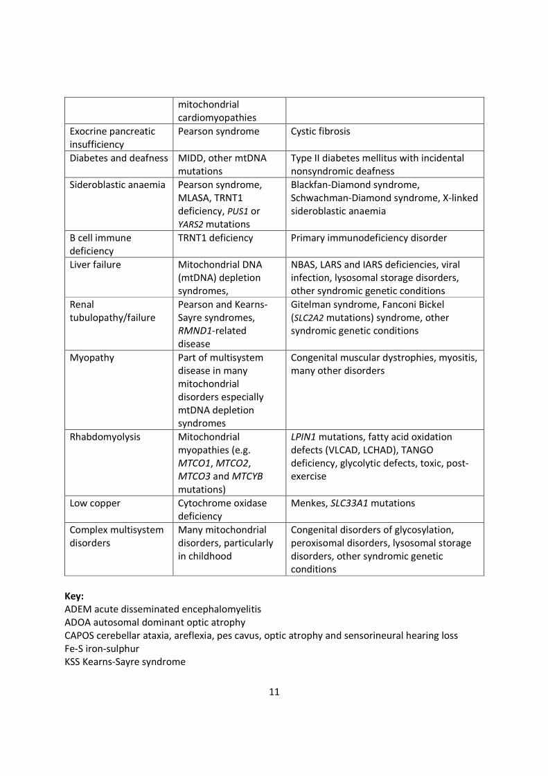
mitochondrialcardiomyopathies
Exocrine pancreaticinsufficiency
Pearson syndrome Cystic fibrosis
Diabetes and deafness MIDD, other mtDNAmutations
Type II diabetes mellitus with incidentalnonsyndromic deafness
Sideroblastic anaemia Pearson syndrome,MLASA, TRNT1deficiency, PUS1 orYARS2 mutations
Blackfan-Diamond syndrome,Schwachman-Diamond syndrome, X-linkedsideroblastic anaemia
B cell immunedeficiency
TRNT1 deficiency Primary immunodeficiency disorder
Liver failure Mitochondrial DNA(mtDNA) depletionsyndromes,
NBAS, LARS and IARS deficiencies, viralinfection, lysosomal storage disorders,other syndromic genetic conditions
Renaltubulopathy/failure
Pearson and Kearns-Sayre syndromes,RMND1-relateddisease
Gitelman syndrome, Fanconi Bickel(SLC2A2 mutations) syndrome, othersyndromic genetic conditions
Myopathy Part of multisystemdisease in manymitochondrialdisorders especiallymtDNA depletionsyndromes
Congenital muscular dystrophies, myositis,many other disorders
Rhabdomyolysis Mitochondrialmyopathies (e.g.MTCO1, MTCO2,MTCO3 and MTCYBmutations)
LPIN1 mutations, fatty acid oxidationdefects (VLCAD, LCHAD), TANGOdeficiency, glycolytic defects, toxic, post-exercise
Low copper Cytochrome oxidasedeficiency
Menkes, SLC33A1 mutations
Complex multisystemdisorders
Many mitochondrialdisorders, particularlyin childhood
Congenital disorders of glycosylation,peroxisomal disorders, lysosomal storagedisorders, other syndromic geneticconditions
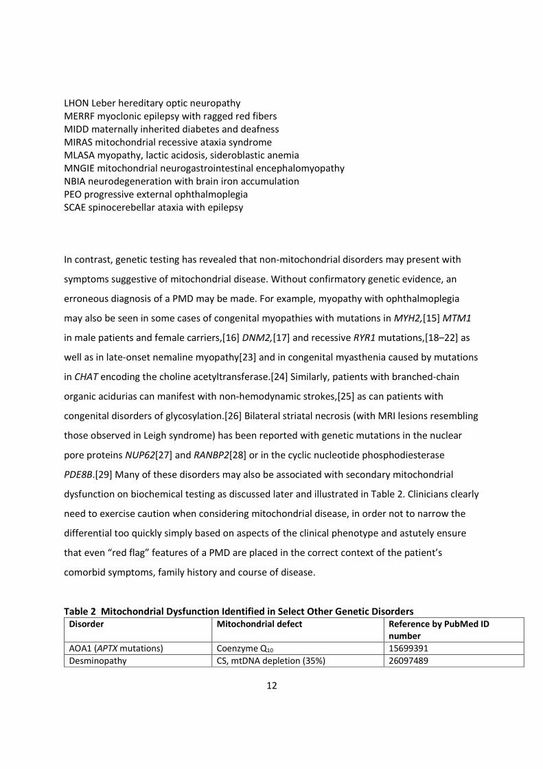
Key:ADEM acute disseminated encephalomyelitisADOA autosomal dominant optic atrophyCAPOS cerebellar ataxia, areflexia, pes cavus, optic atrophy and sensorineural hearing lossFe-S iron-sulphurKSS Kearns-Sayre syndrome
12
LHON Leber hereditary optic neuropathyMERRF myoclonic epilepsy with ragged red fibersMIDD maternally inherited diabetes and deafnessMIRAS mitochondrial recessive ataxia syndromeMLASA myopathy, lactic acidosis, sideroblastic anemiaMNGIE mitochondrial neurogastrointestinal encephalomyopathyNBIA neurodegeneration with brain iron accumulationPEO progressive external ophthalmoplegiaSCAE spinocerebellar ataxia with epilepsy
In contrast, genetic testing has revealed that non-mitochondrial disorders may present with
symptoms suggestive of mitochondrial disease. Without confirmatory genetic evidence, an
erroneous diagnosis of a PMD may be made. For example, myopathy with ophthalmoplegia
may also be seen in some cases of congenital myopathies with mutations in MYH2,[15] MTM1
in male patients and female carriers,[16] DNM2,[17] and recessive RYR1 mutations,[18–22] as
well as in late-onset nemaline myopathy[23] and in congenital myasthenia caused by mutations
in CHAT encoding the choline acetyltransferase.[24] Similarly, patients with branched-chain
organic acidurias can manifest with non-hemodynamic strokes,[25] as can patients with
congenital disorders of glycosylation.[26] Bilateral striatal necrosis (with MRI lesions resembling
those observed in Leigh syndrome) has been reported with genetic mutations in the nuclear
pore proteins NUP62[27] and RANBP2[28] or in the cyclic nucleotide phosphodiesterase
PDE8B.[29] Many of these disorders may also be associated with secondary mitochondrial
dysfunction on biochemical testing as discussed later and illustrated in Table 2. Clinicians clearly
need to exercise caution when considering mitochondrial disease, in order not to narrow the
differential too quickly simply based on aspects of the clinical phenotype and astutely ensure
that even “red flag” features of a PMD are placed in the correct context of the patient’s
comorbid symptoms, family history and course of disease.
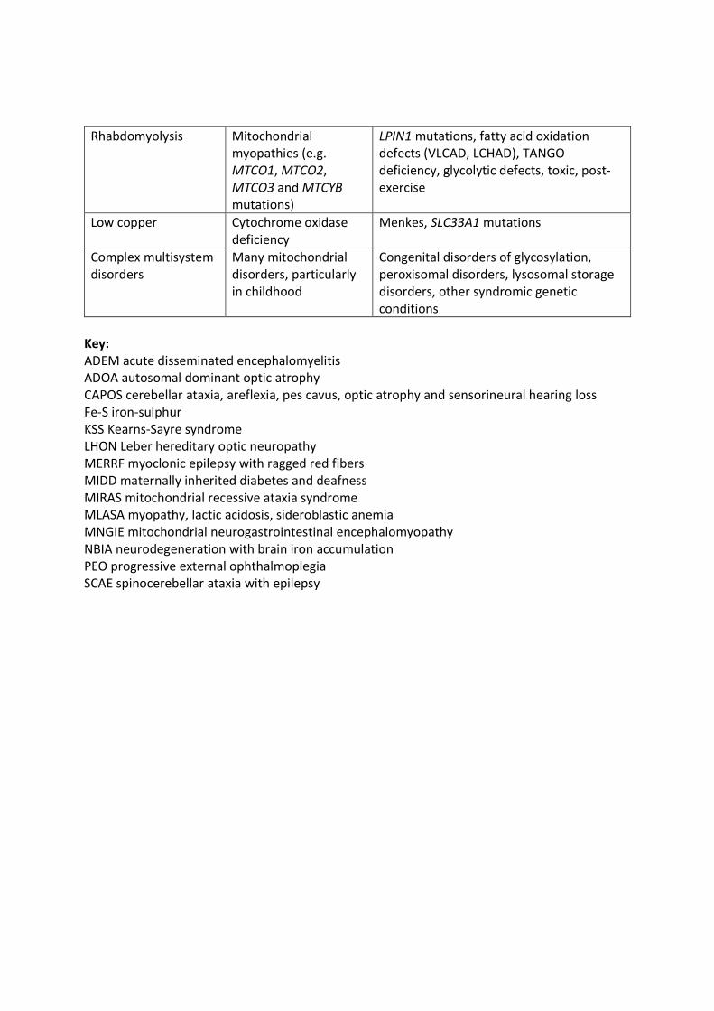
Table 2 Mitochondrial Dysfunction Identified in Select Other Genetic DisordersDisorder Mitochondrial defect Reference by PubMed ID
number
AOA1 (APTX mutations) Coenzyme Q10 15699391
Desminopathy CS, mtDNA depletion (35%) 26097489

13
Dravet syndrome (SCN1Amutations)
Variable OXPHOS deficiencies 20392657; 21906962
EXOSC3 and EXOSC8 relateddiseases
Low Complex I and pyruvatedehydrogenase activities, low mtDNAcopy number, increased expression ofmitochondrial genes
28687512; 24989451
GLUT1 deficiency Complex I 22156785
GM3 synthase deficiency Respiratory chain dysfunction infibroblasts and liver
22990144
LCHADD Complex III, COX 16417669
Limb immobilization COX and CS 19654872
Lysosomal diseases: GM1-gangliosidosis,mucopolysaccharidosis IIIC,multiple sulfatase deficiency,Krabbe disease, Gaucher disease,Niemann Pick disease type C
Multiple OXPHOS deficiencies attributedto excessive production of mitochondrialreactive oxygen species anddysregulated calcium homeostasis withmitochondria-induced apoptosis andneurodegeneration
28132808
MADD (ETFDH, ETFA or ETFBmutations)
Complex I and II deficiencies;Riboflavin and Coenzyme Q10
responsive
17412732
Molybdenum co-factor deficiency COX 16417669
MTHFR mutations Complex I deficiency 21131308
Multiple carboxylase deficiency Complex III 16417669
NBIA (PKAN) Complex III 16417669
Neonatal haemochromatosis Complex III (liver) 16417669
Neuroferritinopathy (FTL1) Complex I or multiple Complexdeficiency
17142829
NPHS3 (PLCE1 deficiency) COX 21365190
Neuronal Ceroid Lipofuscinosis(CLN2 and CLN3-related)
Partial deficiency in fatty acid oxidationenzymes and the storage of subunit c ofmitochondrial ATP synthase infibroblasts
8971698
ORAI1 related disease Impaired lipid metabolism and fatty acidoxidation in skeletal muscle, heart andliver due to abnormal store-operatedCa2+ entry
28132808
Organic acidemias Coenzyme Q10, multiple OXPHOSdeficiencies and free radical inducedoxidative damage
21329767; 28753922; 28753922
Ras/MAPK pathway mutations Variable OXPHOS deficiencies 26097489
Rett syndrome (MECP2 mutations) Variable OXPHOS deficiencies 26741492
SCAR10 Coenzyme Q10 25182700
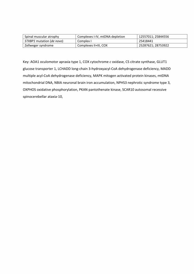
Spinal muscular atrophy Complexes I-IV, mtDNA depletion 12557011; 25844556
STXBP1 mutation (de novo) Complex I 25418441
Zellweger syndrome Complexes II+III, COX 25287621; 28753922

14
Biochemical diagnostic tests remain imperfect
Consensus criteria to help standardize the evaluation of patients with potential PMD, outlining
a streamlined approach and reviewing the strengths and limitations of many of the current
testing modalities were suggested in 2015 by the Mitochondrial Medicine Society (MMS), an
international group of clinicians specializing in mitochondrial disease.[30] This exercise aimed
to decrease the variability that exists in approaches used by clinicians to diagnose PMDs.[8]
When a mitochondrial disorder is suspected, biochemical screening in blood, urine and
cerebrospinal fluid (CSF) remain the initial tests of choice quickly followed by next-generation
sequencing (NGS) of mtDNA and nDNA from white blood cells, with additional genetic studies in
muscle when needed, particularly in adult-onset cases. Whole exome sequencing (WES) is
useful, and along with whole genome sequencing (WGS) is quickly becoming the first or second
line genetic test in patients with suspected mitochondrial disease.[1, 5]
Histopathological, biochemical and genetic analysis of tissue including muscle remain important
tools to further delineate the phenotype and ascertain the relevance of any genetic variants
identified in blood, but should no longer be considered first or second line tests when suspicion
of a PMD is high and appropriate genetic testing is available.[30] Select disorders, such as CPEO,
may warrant the need for further diagnostic testing in muscle. Additional considerations
regarding these tests have been reviewed previously[30] and are summarized below and in
Table 3 and discussed in detail in the supplementary material (Supplement – Testing).
Table 3 Limitations of Testing
Current Limitations of Biochemical TestingImperfect sensitivity and specificitySecondary mitochondrial dysfunction leading to abnormal results
15
Inter-lab variability of methods and reference rangesChallenges with tissue processing
Current Limitations of Genetic TestingIncomplete understanding of the role of the entire genome in mitochondrial functionNovel genes still being identifiedInterpretation of nuclear and mtDNA variants of uncertain significanceLack of understanding of tissue-specificity of mtDNA mutationsUnclear relevance of low heteroplasmy levels of pathogenic mtDNA mutationsmtDNA deletions and depletion may be observed in non-mitochondrial disease
Challenges with biochemical testing
Biochemical studies in blood and urine such as lactate, amino acids, organic acids, and including
the recently identified biomarkers growth differentiation factor 15 (GDF15) and fibroblast
growth factor 21 (FGF21), along with functional assays in various tissues such as ETC enzyme
analysis, all have less than optimal sensitivity and specificity, especially when interpreted in
isolation from the clinical context.[30–35]
Abnormalities on ETC enzyme analysis may occur for a multitude of reasons outside of PMD
including secondary mitochondrial dysfunction from other causes such as other genetic
diseases, limb immobilization,[36] and in liver failure from non-mitochondrial causes.[37, 38]
The list of other genetic disorders where some degree of secondary mitochondrial dysfunction
in various tissues is seen seems ever-growing (Table 2) and includes spinal muscular atrophy
(SMA),[39] X-linked adrenoleukodystrophy (ALD),[40] Phelan-McDermid syndrome, Down
syndrome, Zellweger syndrome, the “rasopathies” (disorders caused by mutations in the Ras-
MAPK pathway) and a variety of other conditions.[41–46] Causes of this secondary dysfunction
have been discerned for very few of these disorders and the extent of mitochondrial
dysfunction is variable and may not meet the diagnostic criteria threshold for ‘definite’
mitochondrial disease.[47] Therefore, evidence of biochemical dysfunction on functional
testing alone, especially when mild or moderate, should not lead to a conclusive diagnosis of
PMD.[42, 45, 48][49][47] When used with rigor, mitochondrial disease criteria may help the

16
clinician selectively better stratify truly high-risk patients.[50] However, mitochondrial disease
diagnostic criteria were all developed at a time prior to the advent of NGS, when only limited
genetic testing was available, and strongly emphasized the importance of abnormal
biochemical findings in tissue.[10, 50, 51] This inevitably led to many patients being diagnosed
with ‘possible’ mitochondrial disease.
Challenges with genetic testing
The advent of rapid, relatively low cost, NGS technologies has allowed for a genetic diagnosis to
be made in many more patients with PMD. A growing number of nuclear genes has been
associated with mitochondrial function (1500 to-date) [52, 53] although only around 350 or so
have firmly been linked to causing human mitochondrial disease.[1, 54, 55] With more routine
use of WES, new nuclear genes impacting mitochondrial function continue to be discovered. In
some patients with a prior suspected but unconfirmed mitochondrial disease diagnosis, WES
has also identified non-mitochondrial diseases.[56] In other cases, variant and milder
phenotypes of PMD have been identified.[57] The ability to detect clearly pathogenic mutations
in suspected PMD via genetic studies remains imperfect, with a reported diagnostic yield
ranging from 25-75%.[3–7] The lack of understanding of the entire genome beyond the exome
and increasing findings of variants of unknown significance (VUS) add to the diagnostic
complexity.
MtDNA can now be accurately sequenced in its entirety for a relatively low cost and it is
possible to detect levels of heteroplasmy of less than 5% in tissue, including blood. Genetic
testing of mtDNA continues to be impacted by aspects of tissue specificity of mutations in
mtDNA and varying degrees of heteroplasmy in easily attainable tissue. With newer testing
methods able to detect low levels of heteroplasmy, common pathogenic mtDNA mutations
(such as m.3243A>G) at low mutation load may mistakenly be attributed to cause a patient’s
phenotype.[58] These issues and others are discussed in further detail in the supplementary
material (Supplement – Testing) but lead to the clear concern that simply testing the
mitochondrial genome in leukocytes is not always adequate, and that mtDNA testing including
17
quantification and deletion analysis in other tissues (skeletal muscle, liver, buccal, urine
sediment) may be needed. Furthermore, even though many defects in mtDNA maintenance
may be diagnosed by WES, there remains a significant number in which the causative genes
remain unknown. Muscle or liver biopsy (depending on the phenotype), along with reliable
assessment of mtDNA copy number compared to age specific control ranges and/or long PCR
for multiple deletions, are needed to diagnose these patients.
Despite the current limitations of genetic testing, the need for genetic confirmation of a PMD
diagnosis is becoming a necessity. The number of phenocopies identified together with the less
than perfect specificity of biochemical studies raises the concern of a mistaken diagnosis and
the potential of missing a separate treatable disease. Accurate genetic diagnosis of a PMD
allows care providers and affected families to better understand the condition, for the provision
of appropriate genetic counseling, and for the development of targeted therapies. For some
PMDs where the natural history is better known, clinicians and families can more accurately
predict the disease course and provide appropriate clinical management and preventative
care.[59] The need for a genetic diagnosis in PMD is now essential for eligibility in clinical trials.
Pre-implantation genetic diagnosis for nuclear and mtDNA disorders and mitochondrial
donation techniques also requires a prior confirmed genetic diagnosis.
Ending a “Possible” Diagnosis of Mitochondrial Disease
Previously established diagnostic criteria,[9–11] developed prior to advances in genetic testing,
relied heavily on biochemical functional tests. They were intended to serve as research
categorization tools in the era of only a basic understanding of mtDNA as it relates to
mitochondrial illness and prior to our knowledge of any but a handful of the hundreds of
nuclear genes that are now known to cause mitochondrial disease. In addition, they were often
not adhered to in the strictest fashion by clinicians. These diagnostic categorizations
subsequently infiltrated the clinic and many more patients began to be labelled as having
18
“possible” mitochondrial disease. Others have received the diagnosis of “mitochondrial
myopathy” because of abnormalities seen in muscle histology or microscopy alone, even
though this finding may exist due to other genetic, metabolic or neurodegenerative diseases.
While genetic testing has improved, it is not currently possible to confirm the diagnosis at a
genomic level in every case. Some patients may have a coincidentally identified pathogenic
mtDNA mutation with low levels of heteroplasmy or a VUS in a nuclear gene bioinformatically
predicted to impact mitochondrial function that may make a clinician consider a “possible”
mitochondrial disease diagnosis.
Given that patients with symptoms suggestive of mitochondrial disease may or may not
ultimately have a PMD, it is increasingly important to establish better diagnostic criteria, or at
least a unified approach to categorizing these patients, to avoid significant variability in
diagnostic labelling, genetic counseling and management. With the growing number of clinical,
biochemical and genetic phenocopies of PMD being identified, it has become prudent that a
definitive diagnosis of mitochondrial disease should only be provided when a confirmed
pathogenic genetic defect has been identified. Utmost caution must be used when providing a
diagnosis based on biochemical abnormalities in tissue alone and the strictest application of
biochemical diagnostic criteria is needed. Patients with strong biochemical and clinical
evidence for a PMD should be periodically re-evaluated as diagnostic testing advances.
There is a clear concern that a diagnosis of “possible” mitochondrial disease may result in harm.
First and foremost, some patients who receive a diagnosis of a “possible” or “suspected”
mitochondrial disease may not recognize the impermanence of such a diagnosis and remain
carrying this label for many years without having their symptoms periodically reassessed and a
more specific diagnosis investigated as knowledge and diagnostic tools improve. Over time, the
categorization of “possible” is often dropped by some providers and non-mitochondrial
specialists providing routine care for the patient. Some families may cling to the diagnosis even
after having had a different genetic disease confirmed, as it is the diagnosis they have become
19
most familiar with over time. Testing for another disorder may be delayed from the clinician’s
side if they are not aware of this diagnostic uncertainty. Other treatable disorders may not be
diagnosed, or diagnosis may be delayed.
A diagnosis of “possible” mitochondrial disease may also create an unfounded fear of
worsening morbidity and mortality. Certain families of patients given a diagnosis of “possible”
mitochondrial disease often overlook the uncertainty of the diagnosis and become overly
concerned that they or a family member may manifest all of the symptoms a patient with a
PMD may develop, including neurodegeneration or early death, even in instances where their
symptoms are relatively mild.
Lastly, patients with a diagnosis of “possible” mitochondrial disease may receive inappropriate
care or be over-medicalized. Counseling of disease expectations and management may vary
based on how patients are categorized.[60] Unnecessary medical interventions may be offered
to some during times of catabolic stress. Some medications may not be used due to a concern
of potential mitochondrial toxicity. New symptoms that a patient may manifest may
inappropriately be explained away by the underlying diagnostic label rather than looking for
other potentially treatable causes. These and other concerns are summarized in Table 4.
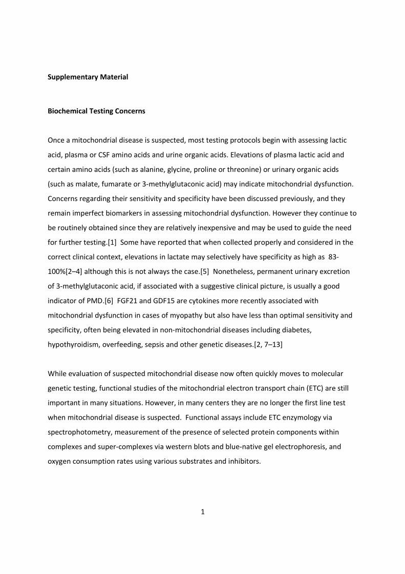
Table 4 Potential Harms Arising from a Diagnosis of “Possible” Mitochondrial Disease
Ending diagnostic odyssey prematurelyMissing potentially treatable disordersPsychological burden of mitochondrial disease diagnosis: parent/patient fear of progressive ordegenerative disorderInaccurate recurrence risk counselingInappropriate preventative careUnnecessary medical interventions at times of catabolic stressAvoidance of needed medications owing to fear of mitochondrial toxicityInappropriate reproductive decisions taken

20
Some of these very issues and challenges are outlined in example cases provided in the
supplementary material (Supplement – Cases). In addition to the disorders outlined in Table 2,
the supplementary cases illustrate instances where a patient may have symptoms suggesting
the possibility of mitochondrial disease, often with biochemical abnormalities suggesting
mitochondrial dysfunction, but the final diagnosis is not a PMD. Diagnosis is often delayed due
to the mistaken diagnosis. Examples include a manganese transporter disorder with bilateral
basal ganglia hyperintensities and elevated FGF21 levels (Case 1), oculopharyngeal muscular
dystrophy with ragged red and cytochrome c oxidase (COX)-negative fibers (Case 2), Lesch-
Nyhan syndrome with putaminal and thalamic abnormalities, lactic acidosis and reduced
Complex I enzymatic activity in muscle (Case 3) and Niemann-Pick Type C with Complex I
deficiency leading to a delay in being prescribed Miglustat (Case 4). In some of these instances,
mitochondrial functional testing was notably abnormal, meeting biochemical diagnostic criteria
for a mitochondrial disease. In contrast, select other cases (Cases 5-8) illustrate a delayed PMD
diagnosis due to limitations of genetic testing in blood, findings of low levels of heteroplasmy or
findings of a VUS. Case 5 illustrates an instance of a female with MELAS-like symptoms. Other
cases (Cases 6-8) show the challenges in interpreting nuclear and mtDNA VUS.
Recommendations
In patients without a confirmed genetic diagnosis, there is a need for clinicians and the
mitochondrial disease community to utilize diagnostic labels that clearly state that the
diagnosis is uncertain even when mitochondrial dysfunction has been identified. A category of
“genetic diagnosis uncertain; mitochondrial biochemical dysfunction or mitochondrial genetic
variant of unknown significance identified” is preferable to a diagnosis of “possible” or
“probable” or “suspected” mitochondrial disease. Other terminology that should be avoided is
listed in Table 5. Depending on the clinical situation, patients may be further stratified into a
“high risk” for a PMD to guide management.
Table 5 Terminology to avoid when a mitochondrial diagnosis is uncertain“Possible,” “Probable” or “Suspected” Mitochondrial DiseaseMitochondrial Myopathy

21
Mitochondrial CytopathyMitochondrial Metabolism DisorderDefect of Mitochondrial Metabolism
Our proposal to utilize a diagnostic label of “genetic diagnosis uncertain” for all such cases
would allow clinicians and patients to remain actively engaged in the diagnostic odyssey, review
the prior data periodically and take advantage of technological advances in genetic testing and
new disease descriptions. Conducting relevant screening of other systems and monitoring for
other organ involvement would allow better definition of the phenotype and not overlook
disease progression. The clarity of the diagnostic label may prevent inappropriate or
unnecessary care and allay fears of a progressive or degenerative disease.
Further categorization of selected patients as possible “high risk” for a PMD would allow for
closer monitoring for mitochondrial disease related systemic comorbidities or extra cautions
during times they are at risk of metabolic decompensations. If the phenotype is especially
suggestive of a PMD, it may be appropriate to manage such a patient as if they have a
genetically confirmed PMD for the time being – especially if they have previously experienced
metabolic decompensation during times of illness or medical stress. Unexpected, acute changes
in clinical status warrant thorough medical evaluation including laboratory testing to investigate
potential mitochondrial dysfunction. However, the “diagnosis uncertain” designation would
prevent any misunderstanding among medical teams. If the phenotype is not as suggestive of a
PMD, it may be prudent to avoid over-medicalization of the patient and simply continue more
routine monitoring.
As diagnostic standards for mitochondrial disease continue to evolve, these patients should
remain under the care of a clinician who can assist in providing up-to-date recommendations
regarding further testing. The MMS has such recommendations available online at
www.mitosoc.org.
22
Conclusion
Despite advances in diagnostic techniques and molecular genetics, a subset of patients with
suspected mitochondrial disease remains without a confirmed genetic diagnosis. The path
these patients take to receiving a diagnosis is arduous and, at times, circuitous. Newer NGS
based genetic studies offer the ability to streamline the approach to diagnosis for some
patients. Others remain with a constellation of symptoms, findings of mitochondrial
dysfunction on functional testing, and no clear pathogenic genetic mutation. Patients
diagnosed with a “possible” mitochondrial disease might be found to have a non-mitochondrial
genetic disorder once new testing modalities are utilized. A mistaken diagnosis of mitochondrial
disease may prematurely end their diagnostic journey, over-medicalize their care, and
potentially limit access to appropriate treatments for the actual underlying condition. To
alleviate this dilemma, such patients would be better served by clinicians avoiding the
diagnostic term “possible” mitochondrial disease.
23
Acknowledgements
RH is a Wellcome Trust Investigator (109915/Z/15/Z), who receives support from the Wellcome
Centre for Mitochondrial Research (203105/Z/16/Z), Medical Research Council (UK)
(MR/N025431/1), the European Research Council (309548), the Wellcome Trust Pathfinder
Scheme (201064/Z/16/Z) and the Newton Fund (UK/Turkey, MR/N027302/1). PFC is a
Wellcome Trust Senior Fellow in Clinical Science (101876/Z/13/Z) and a UK NIHR Senior
Investigator, who receives support from the Medical Research Council Mitochondrial Biology
Unit (MC_UP_1501/2), the Medical Research Council (UK) Centre for Translational Muscle
Disease (G0601943), EU FP7 TIRCON, and the National Institute for Health Research (NIHR)
Biomedical Research Centre based at Cambridge University Hospitals NHS Foundation Trust and
the University of Cambridge. SR is supported by a Great Ormond Street Hospital Children's Charity
Research Leadership Award (V1260) and by research grant funding from the NIHR Great Ormond Street
Hospital Biomedical Research Centre and the Lily Foundation.
EB, SR and MR are members of the European Reference Network for Rare Hereditary Metabolic
Disorders (METABERN) - Project ID No 739543. MetabERN is partly co-funded by the European
Union in the framework of the Third Health Programme “ERN-2016 - Framework Partnership
Agreement 2017-2021.
MM is supported by research grants from Telethon and MITOCON Italian patients’ association
(grants GSP09004 and GSP16001)
MT would like to thank Dan Wright and family for a kind donation for research in mitochondrial
disease and to Kathy Corkins and Warren Lammert and family for a kind donation to establish
the Corkins-Lammert Mitochondrial Disease Center at McMaster University.
24
REFERENCES
1 Rahman J, Rahman S. Mitochondrial medicine in the omics era. Lancet (London, England)
2018;391:2560–74.
2 Haas RH, Parikh S, Falk MJ, Saneto RP, Wolf NI, Darin N, Cohen BH. Mitochondrial
Disease: A Practical Approach for Primary Care Physicians. Pediatrics 2007;120:1326–33.
3 Calvo SE, Compton AG, Hershman SG, Lim SC, Lieber DS, Tucker EJ, Laskowski A, Garone
C, Liu S, Jaffe DB, Christodoulou J, Fletcher JM, Bruno DL, Goldblatt J, Dimauro S,
Thorburn DR, Mootha VK. Molecular diagnosis of infantile mitochondrial disease with
targeted next-generation sequencing. Sci Transl Med 2012;4:118ra10.
4 Wortmann SB, Koolen DA, Smeitink JA, van den Heuvel L, Rodenburg RJ. Whole exome
sequencing of suspected mitochondrial patients in clinical practice. J Inherit Metab Dis
2015;38:437–43.
5 Taylor RW, Pyle A, Griffin H, Blakely EL, Duff J, He L, Smertenko T, Alston CL, Neeve VC,
Best A, Yarham JW, Kirschner J, Schara U, Talim B, Topaloglu H, Baric I, Holinski-Feder E,
Abicht A, Czermin B, Kleinle S, Morris AAM, Vassallo G, Gorman GS, Ramesh V, Turnbull
DM, Santibanez-Koref M, McFarland R, Horvath R, Chinnery PF. Use of whole-exome
sequencing to determine the genetic basis of multiple mitochondrial respiratory chain
complex deficiencies. Jama 2014;312:68–77.
6 Lieber DS, Calvo SE, Shanahan K, Slate NG, Liu S, Hershman SG, Gold NB, Chapman BA,
Thorburn DR, Berry GT, Schmahmann JD, Borowsky ML, Mueller DM, Sims KB, Mootha
VK. Targeted exome sequencing of suspected mitochondrial disorders. Neurology
2013;80:1762–70.
7 Kohda M, Tokuzawa Y, Kishita Y, Nyuzuki H, Moriyama Y, Mizuno Y, Hirata T, Yatsuka Y,
Yamashita-Sugahara Y, Nakachi Y, Kato H, Okuda A, Tamaru S, Borna NN, Banshoya K,
Aigaki T, Sato-Miyata Y, Ohnuma K, Suzuki T, Nagao A, Maehata H, Matsuda F, Higasa K,
Nagasaki M, Yasuda J, Yamamoto M, Fushimi T, Shimura M, Kaiho-Ichimoto K, Harashima
H, Yamazaki T, Mori M, Murayama K, Ohtake A, Okazaki Y. A Comprehensive Genomic
Analysis Reveals the Genetic Landscape of Mitochondrial Respiratory Chain Complex
Deficiencies. PLoS Genet 2016;12. doi:10.1371/journal.pgen.1005679
25
8 Parikh S, Goldstein A, Koenig MK, Scaglia F, Enns GM, Saneto R, Anselm I, Collins A,
Cohen BH, DeBrosse SD, Dimmock D, Falk MJ, Ganesh J, Greene C, Gropman AL, Haas R,
Kahler SG, Kamholz J, Kendall F, Korson MS, Mattman A, Milone M, Niyazov D, Pearl PL,
Reimschisel T, Salvarinova-Zivkovic R, Sims K, Tarnopolsky M, Tsao CY, van Hove J, Walsh
L, Wolfe LA. Practice patterns of mitochondrial disease physicians in North America. Part
1: Diagnostic and clinical challenges. Mitochondrion 2014;14:26–33.
9 Morava E, van den Heuvel L, Hol F, de Vries MC, Hogeveen M, Rodenburg RJ, Smeitink JA.
Mitochondrial disease criteria: diagnostic applications in children . Neurology
2006;67:1823–6.
10 Bernier FP, Boneh A, Dennett X, Chow CW, Cleary MA, Thorburn DR. Diagnostic criteria
for respiratory chain disorders in adults and children. Neurology 2002;59:1406–11.
11 Thorburn DR, Sugiana C, Salemi R, Kirby DM, Worgan L, Ohtake A, Ryan MT. Biochemical
and molecular diagnosis of mitochondrial respiratory chain disorders. Biochim Biophys
Acta 2004;1659:121–8.
12 Martikainen MH, Chinnery PF. Mitochondrial disease: mimics and chameleons. Pract
Neurol 2015;15:424–35.
13 Needham M, Duley J, Hammond S, Herkes GK, Hirano M, Sue CM. Mitochondrial disease
mimicking Charcot-Marie tooth disease. BMJ Case Rep Published Online First: 2009.
doi:10.1136/bcr.06.2009.2001
14 Gardeitchik T, Wyckmans J, Morava E. Complex Phenotypes in Inborn Errors of
Metabolism: Overlapping Presentations in Congenital Disorders of Glycosylation and
Mitochondrial Disorders. Pediatr. Clin. North Am. 2018;65:375–88.
15 D’Amico A, Fattori F, Bellacchio E, Catteruccia M, Servidei S, Bertini E. A new de novo
missense mutation in MYH2 expands clinical and genetic findings in hereditary myosin
myopathies. Neuromuscul Disord 2013;23:437–40.
16 Biancalana V, Scheidecker S, Miguet M, Laquerrière A, Romero NB, Stojkovic T, Abath
Neto O, Mercier S, Voermans N, Tanner L, Rogers C, Ollagnon-Roman E, Roper H, Boutte
C, Ben-Shachar S, Lornage X, Vasli N, Schaefer E, Laforet P, Pouget J, Moerman A,
Pasquier L, Marcorelle P, Magot A, Küsters B, Streichenberger N, Tranchant C, Dondaine
26
N, Schneider R, Gasnier C, Calmels N, Kremer V, Nguyen K, Perrier J, Kamsteeg EJ, Carlier
P, Carlier RY, Thompson J, Boland A, Deleuze JF, Fardeau M, Zanoteli E, Eymard B,
Laporte J. Affected female carriers of MTM1 mutations display a wide spectrum of
clinical and pathological involvement: delineating diagnostic clues. Acta Neuropathol
2017;134:889–904.
17 Catteruccia M, Fattori F, Codemo V, Ruggiero L, Maggi L, Tasca G, Fiorillo C, Pane M,
Berardinelli A, Verardo M, Bragato C, Mora M, Morandi L, Bruno C, Santoro L, Pegoraro E,
Mercuri E, Bertini E, D’Amico A. Centronuclear myopathy related to dynamin 2
mutations: Clinical, morphological, muscle imaging and genetic features of an Italian
cohort. Neuromuscul Disord 2013;23:229–38.
18 Bevilacqua JA, Monnier N, Bitoun M, Eymard B, Ferreiro A, Monges S, Lubieniecki F,
Taratuto AL, Laquerrière A, Claeys KG, Marty I, Fardeau M, Guicheney P, Lunardi J,
Romero NB. Recessive RYR1 mutations cause unusual congenital myopathy with
prominent nuclear internalization and large areas of myofibrillar disorganization.
Neuropathol Appl Neurobiol 2011;37:271–84.
19 Wilmshurst JM, Lillis S, Zhou H, Pillay K, Henderson H, Kress W, Müller CR, Ndondo A,
Cloke V, Cullup T, Bertini E, Boennemann C, Straub V, Quinlivan R, Dowling JJ, Al-Sarraj S,
Treves S, Abbs S, Manzur AY, Sewry CA, Muntoni F, Jungbluth H. RYR1 mutations are a
common cause of congenital myopathies with central nuclei. Ann Neurol 2010;68:717–
26.
20 Clarke NF, Waddell LB, Cooper ST, Perry M, Smith RLL, Kornberg AJ, Muntoni F, Lillis S,
Straub V, Bushby K, Guglieri M, King MD, Farrell MA, Marty I, Lunardi J, Monnier N, North
KN. Recessive mutations in RYR1 are a common cause of congenital fiber type
disproportion. Hum Mutat 2010;31. doi:10.1002/humu.21278
21 Kondo E, Nishimura T, Kosho T, Inaba Y, Mitsuhashi S, Ishida T, Baba A, Koike K, Nishino I,
Nonaka I, Furukawa T, Saito K. Recessive RYR1 mutations in a patient with severe
congenital nemaline myopathy with ophthalomoplegia identified through massively
parallel sequencing. Am J Med Genet A 2012;158A:772–8.
22 Das S, Dowling J, Pierson CR. X-Linked Centronuclear Myopathy. 1993.
27
http://www.ncbi.nlm.nih.gov/pubmed/20301605 (accessed 27 Apr2018).
23 Wengert O, Meisel A, Kress W, Dekomien G, Angstwurm K, Heppner FL, Goebel HH,
Stenzel W. Progressive external ophthalmoplegia as initial manifestation of sporadic late-
onset nemaline myopathy. J. Neurol. 2011;258:915–7.
24 Mullaney P, Vajsar J, Smith R, Buncic JR. The natural history and ophthalmic involvement
in childhood myasthenia gravis at the hospital for sick children. Ophthalmology
2000;107:504–10.
25 Testai FD, Gorelick PB. Inherited metabolic disorders and stroke part 2: Homocystinuria,
organic acidurias, and urea cycle disorders. Arch. Neurol. 2010;67:148–53.
26 Briones P, Vilaseca MA, García-Silva MT, Pineda M, Colomer J, Ferrer I, Artigas J, Jaeken J,
Chabás A. Congenital disorders of glycosylation (CDG) may be underdiagnosed when
mimicking mitochondrial disease. Eur J Paediatr Neurol 2001;5:127–31.
27 Basel-Vanagaite L, Muncher L, Straussberg R, Pasmanik-Chor M, Yahav M, Rainshtein L,
Walsh CA, Magal N, Taub E, Drasinover V, Shalev H, Attia R, Rechavi G, Simon AJ, Shohat
M. Mutated nup62 causes autosomal recessive infantile bilateral striatal necrosis. Ann
Neurol 2006;60:214–22.
28 Neilson DE, Adams MD, Orr CMD, Schelling DK, Eiben RM, Kerr DS, Anderson J, Bassuk
AG, Bye AM, Childs A-MM, Clarke A, Crow YJ, Di Rocco M, Dohna-Schwake C, Dueckers G,
Fasano AE, Gika AD, Gionnis D, Gorman MP, Grattan-Smith PJ, Hackenberg A, Kuster A,
Lentschig MG, Lopez-Laso E, Marco EJ, Mastroyianni S, Perrier J, Schmitt-Mechelke T,
Servidei S, Skardoutsou A, Uldall P, van der Knaap MS, Goglin KC, Tefft DL, Aubin C, de
Jager P, Hafler D, Warman ML. Infection-triggered familial or recurrent cases of acute
necrotizing encephalopathy caused by mutations in a component of the nuclear pore,
RANBP2. Am J Hum Genet 2009;84:44–51.
29 Appenzeller S, Schirmacher A, Halfter H, Bäumer S, Pendziwiat M, Timmerman V, De
Jonghe P, Fekete K, Stögbauer F, Lüdemann P, Hund M, Quabius ES, Ringelstein EB,
Kuhlenbäumer G. Autosomal-dominant striatal degeneration is caused by a mutation in
the phosphodiesterase 8B gene. Am J Hum Genet 2010;86:83–7.
30 Parikh S, Goldstein A, Koenig MK, Scaglia F, Enns GM, Saneto R, Anselm I, Cohen BH, Falk
28
MJ, Greene C, Gropman AL, Haas R, Hirano M, Morgan P, Sims K, Tarnopolsky M, Van
Hove JLK, Wolfe L, DiMauro S. Diagnosis and management of mitochondrial disease: a
consensus statement from the Mitochondrial Medicine Society. Genet Med
2015;17:689–701.
31 Lehtonen JM, Forsström S, Bottani E, Viscomi C, Baris OR, Isoniemi H, Höckerstedt K,
Österlund P, Hurme M, Jylhävä J, Leppä S, Markkula R, Heliö T, Mombelli G, Uusimaa J,
Laaksonen R, Laaksovirta H, Auranen M, Zeviani M, Smeitink J, Wiesner RJ, Nakada K,
Isohanni P, Suomalainen A. FGF21 is a biomarker for mitochondrial translation and
mtDNA maintenance disorders. Neurology 2016;87:2290–9.
32 Montero R, Yubero D, Villarroya J, Henares D, Jou C, Rodríguez MA, Ramos F, Nascimento
A, Ortez CI, Campistol J, Perez-Dueñas B, O’Callaghan M, Pineda M, Garcia-Cazorla A,
Oferil JC, Montoya J, Ruiz-Pesini E, Emperador S, Meznaric M, Campderros L, Kalko SG,
Villarroya F, Artuch R, Jimenez-Mallebrera C. GDF-15 is elevated in children with
mitochondrial diseases and is induced by mitochondrial dysfunction. PLoS One
2016;11:1–15.
33 Yatsuga S, Fujita Y, Ishii A, Fukumoto Y, Arahata H, Kakuma T, Kojima T, Ito M, Tanaka M,
Saiki R, Koga Y. Growth differentiation factor 15 as a useful biomarker for mitochondrial
disorders. Ann Neurol 2015;78:814–23.
34 Davis RL, Liang C, Edema-Hildebrand F, Riley C, Needham M, Sue CM. Fibroblast growth
factor 21 is a sensitive biomarker of mitochondrial disease. Neurology Published Online
First: 18 October 2013. doi:10.1212/01.wnl.0000436068.43384.ef
35 Davis RL, Liang C, Sue CM. A comparison of current serum biomarkers as diagnostic
indicators of mitochondrial diseases. Neurology 2016;86:2010–5.
36 Abadi A, Glover EI, Isfort RJ, Raha S, Safdar A, Yasuda N, Kaczor JJ, Melov S, Hubbard A,
Qu X, Phillips SM, Tarnopolsky M. Limb immobilization induces a coordinate down-
regulation of mitochondrial and other metabolic pathways in men and women. PLoS One
2009;4:e6518.
37 McKiernan P, Ball S, Santra S, Foster K, Fratter C, Poulton J, Craig K, McFarland R, Rahman
S, Hargreaves I, Gupte G, Sharif K, Taylor RW. Incidence of Primary Mitochondrial Disease
29
in Children Younger Than 2 Years Presenting With Acute Liver Failure. J Pediatr
Gastroenterol Nutr 2016;63:592–7.
38 Lane M, Boczonadi V, Bachtari S, Gomez-Duran A, Langer T, Griffiths A, Kleinle S, Dineiger
C, Abicht A, Holinski-Feder E, Schara U, Gerner P, Horvath R. Mitochondrial dysfunction
in liver failure requiring transplantation. J Inherit Metab Dis 2016;39:427–36.
39 Berger A, Mayr JA, Meierhofer D, Fötschl U, Bittner R, Budka H, Grethen C, Huemer M,
Kofler B, Sperl W. Severe depletion of mitochondrial DNA in spinal muscular atrophy.
Acta Neuropathol 2003;105:245–51.
40 Fourcade S, Ferrer I, Pujol A. Oxidative stress, mitochondrial and proteostasis
malfunction in adrenoleukodystrophy: A paradigm for axonal degeneration. Free Radic.
Biol. Med. 2015;88:18–29.
41 Frye RE, Cox D, Slattery J, Tippett M, Kahler S, Granpeesheh D, Damle S, Legido A,
Goldenthal MJ. Mitochondrial Dysfunction may explain symptom variation in Phelan-
McDermid Syndrome. Sci Rep 2016;6. doi:10.1038/srep19544
42 Hui J, Kirby DM, Thorburn DR, Boneh A. Decreased activities of mitochondrial respiratory
chain complexes in non-mitochondrial respiratory chain diseases. Dev Med Child Neurol
2006;48:132–6.
43 Niyazov DM, Kahler SG, Frye RE. Primary Mitochondrial Disease and Secondary
Mitochondrial Dysfunction: Importance of Distinction for Diagnosis and Treatment. Mol.
Syndromol. 2016;7:122–37.
44 Garcia-Cazorla A, Serrano M, Perez-Duenas B, Gonzalez V, Ormazabal A, Pineda M,
Fernandez-Alvarez E, Campistol JM, Artuch RM. Secondary abnormalities of
neurotransmitters in infants with neurological disorders. Dev Med Child Neurol
2007;49:740–4.
45 Prince J, Jia S, Båve U, Annerén G, Oreland L. Mitochondrial enzyme deficiencies in
Down’s syndrome. J Neural Transm - Park Dis Dement Sect 1994;8:171–81.
46 Kleefstra T, Wortmann SB, Rodenburg RJT, Bongers EMHF, Hadzsiev K, Noordam C, van
den Heuvel LP, Nillesen WM, Hollody K, Gillessen-Kaesbach G, Lammens M, Smeitink
JAM, van der Burgt I, Morava E. Mitochondrial dysfunction and organic aciduria in five
30
patients carrying mutations in the Ras-MAPK pathway. Eur J Hum Genet 2011;19:138–44.
47 Panneman DM, Smeitink JA, Rodenburg RJ. Mining for mitochondrial mechanisms: linking
known syndromes to mitochondrial function. Clin Genet Published Online First: 2017.
doi:10.1111/cge.13094
48 Frye RE. 15q11.2-13 Duplication, Mitochondrial Dysfunction, and Developmental
Disorders. J Child Neurol 2009;24:1316–20.
49 Maynard TM, Meechan DW, Dudevoir ML, Gopalakrishna D, Peters AZ, Heindel CC,
Sugimoto TJ, Wu Y, Lieberman JA, LaMantia AS. Mitochondrial localization and function
of a subset of 22q11 deletion syndrome candidate genes. Mol Cell Neurosci
2008;39:439–51.
50 Witters P, Saada A, Honzik T, Tesarova M, Kleinle S, Horvath R, Goldstein A, Morava E.
Revisiting mitochondrial diagnostic criteria in the new era of genomics. Genet Med
2017;00:1–8.
51 Wolf NI, Smeitink JA. Mitochondrial disorders: a proposal for consensus diagnostic
criteria in infants and children. Neurology 2002;59:1402–5.
52 Calvo SE, Clauser KR, Mootha VK. MitoCarta2.0: An updated inventory of mammalian
mitochondrial proteins. Nucleic Acids Res 2016;44:D1251–7.
53 Smith AC, Robinson AJ. MitoMiner v3.1, an update on the mitochondrial proteomics
database. Nucleic Acids Res 2016;44:D1258–61.
54 Frazier AE, Thorburn DR, Compton AG. Mitochondrial energy generation disorders:
genes, mechanisms and clues to pathology. J Biol Chem 2017;:jbc.R117.809194.
55 Ormondroyd E, Mackley MP, Blair E, Craft J, Knight JC, Taylor JC, Taylor J, Watkins H.
"Not pathogenic until proven otherwise": perspectives of UK clinical
genomics professionals toward secondary findings in context of a Genomic Medicine
Multidisciplinary Team and the 100,000 Genomes Project. Genet Med 2018;20:320–8.
56 McCormick EM, Kenyon L, Falk MJ. Desmin common mutation is associated with multi-
systemic disease manifestations and depletion of mitochondria and mitochondrial DNA.
Front Genet 2015;6:1–5.
57 Camara Y, Carreno-Gago L, Martin MA, Melia MJ, Blazquez A, Delmiro A, Garrabou G,

31
Moren C, Diaz-Manera J, Gallardo E, Bornstein B, Lopez-Gallardo E, Hernandez-Lain A,
San Millan B, Cancho E, Rodriguez-Vico JS, Marti R, Garcia-Arumi E. Severe TK2 enzyme
activity deficiency in patients with mild forms of myopathy. Neurology 2015;84:2286–8.
58 Elliott HR, Samuels DC, Eden JA, Relton CL, Chinnery PF. Pathogenic mitochondrial DNA
mutations are common in the general population. Am J Hum Genet 2008;83:254–60.
59 Parikh S, Goldstein A, Karaa A, Koenig MK, Anselm I, Brunel-Guitton C, Christodoulou J,
Cohen BH, Dimmock D, Enns GM, Falk MJ, Feigenbaum A, Frye RE, Ganesh J, Griesemer
D, Haas R, Horvath R, Korson M, Kruer MC, Mancuso M, McCormack S, Raboisson MJ,
Reimschisel T, Salvarinova R, Saneto RP, Scaglia F, Shoffner J, Stacpoole PW, Sue CM,
Tarnopolsky M, Van Karnebeek C, Wolfe LA, Cunningham ZZ, Rahman S, Chinnery PF.
Patient care standards for primary mitochondrial disease: a consensus statement from
the Mitochondrial Medicine Society. Genet Med Published Online First: 27 July 2017.
doi:10.1038/gim.2017.107
60 Parikh S, Goldstein A, Koenig MK, Scaglia F, Enns GM, Saneto R, Irina Anselm, Abigail
Collins, Bruce HC, Suzanne DD, David Dimmock, Marni JF, Jaya Ganesh, Carol Greene,
Andrea LG, Richard Haas, Stephen GK, John Kamholz, Fran Kendall, Mark SK, Andre
Mattman, Margherita Milone, Dmitriy Niyazov, Phillip LP, Tyler Reimschisel, Ramona
Salvarinova-Zivkovic, Katherine Sims, MarkTarnopolsky, Chang-Yong Tsao, Johan van
Hove, Laurence Walsh, Lynne AW. Practice patterns of mitochondrial disease physicians
in North America. Part 2: Treatment, care and management. Mitochondrion
2013;13:681–7.

Tables
Table 1 Differential diagnosis of selected phenotypes commonly associated withmitochondrial disease
Phenotype Mitochondrial cause Limited differential diagnosis
Dystonia Leigh syndrome,deafness-dystoniasyndrome, othermitochondrialencephalomyopathies
Biotinidase deficiency, thiaminetransporter deficiency 2, ADAR mutations(Aicardi- Goutières syndrome 6), organicacidaemias (especially glutaric aciduriatype I), NBIA, acute (viral) necrotisingencephalopathy, mutations in NUP62,RANBP2 and PDE8B, primary geneticdystonias
Epilepticencephalopathy
Alpers-Huttenlochersyndrome, many othermitochondrialdisorders
Many genetic epileptic encephalopathies,including Dravet syndrome and KCNQ2mutations, Pyridoxine dependentepilepsies (Antiquitin deficiency, PNPOdeficiency), viral encephalitis
Progressive myoclonicepilepsy
MERRF Ramsay Hunt syndrome, Unverricht-Lundborg disease, Lafora body disease,sialidosis, PRICKLE1 mutations
Leukoencephalpathy Complex I deficiency,Complex II deficiency,SURF1 deficiency(rarely), disorders ofmitochondrialtranslation and Fe-Scluster assembly
Vanishing white matter disease, lysosomalstorage disorders, Canavan disease,Alexander disease, Pelizaeus-Merzbacher(-like), hypo/dysmyelination
Ataxia ADCK3 mutations,ataxia-neuropathysyndromes e.g. SCAE,MIRAS, MERRF, NARP,disorders of coenzymeQ10 biosynthesis
Spinocerebellar ataxias, CAPOS syndrome
Demyelination MNGIE ADEM, multiple sclerosis
Peripheral neuropathy Mutations in POLG,MPV17, KARS andSURF1; part ofmultisystem disease inmany mitochondrialdisorders, e.g. MNGIE
Other non-mitochondrial genetic causes ofCharcot-Marie-Tooth syndromes,riboflavin transporter deficiency, toxicneuropathies, critical illness
Ptosis andophthalmoplegia
PEO, KSS, MNGIE,MELAS
Some congenital myopathies, pseudoupgaze impairment in OPMD, horizontalgaze palsy and scoliosis (ROBO3 mutation)
Optic neuropathy LHON, ADOA, Leighsyndrome
Toxic optic neuropathy (e.g. methanol,cyanide, tobacco)
Hypertrophiccardiomyopathy withlactic acidosis
Complex I deficiency,TMEM70 mutations,Sengers syndrome(AGK deficiency),disorders ofmitochondrialtranslation
Viral infection
Dilatedcardiomyopathy withlactic acidosis
Barth syndrome,disorders ofmitochondrialphospholipidremodelling, othermitochondrialcardiomyopathies
Viral infection
Exocrine pancreaticinsufficiency
Pearson syndrome Cystic fibrosis
Diabetes and deafness MIDD, other mtDNAmutations
Type II diabetes mellitus with incidentalnonsyndromic deafness
Sideroblastic anaemia Pearson syndrome,MLASA, TRNT1deficiency, PUS1 orYARS2 mutations
Blackfan-Diamond syndrome,Schwachman-Diamond syndrome, X-linkedsideroblastic anaemia
B cell immunedeficiency
TRNT1 deficiency Primary immunodeficiency disorder
Liver failure Mitochondrial DNA(mtDNA) depletionsyndromes,
NBAS, LARS and IARS deficiencies, viralinfection, lysosomal storage disorders,other syndromic genetic conditions
Renaltubulopathy/failure
Pearson and Kearns-Sayre syndromes,RMND1-relateddisease
Gitelman syndrome, Fanconi Bickel(SLC2A2 mutations) syndrome, othersyndromic genetic conditions
Myopathy Part of multisystemdisease in manymitochondrialdisorders especiallymtDNA depletionsyndromes
Congenital muscular dystrophies, myositis,many other disorders
Rhabdomyolysis Mitochondrialmyopathies (e.g.MTCO1, MTCO2,MTCO3 and MTCYBmutations)
LPIN1 mutations, fatty acid oxidationdefects (VLCAD, LCHAD), TANGOdeficiency, glycolytic defects, toxic, post-exercise
Low copper Cytochrome oxidasedeficiency
Menkes, SLC33A1 mutations
Complex multisystemdisorders
Many mitochondrialdisorders, particularlyin childhood
Congenital disorders of glycosylation,peroxisomal disorders, lysosomal storagedisorders, other syndromic geneticconditions
Key:ADEM acute disseminated encephalomyelitisADOA autosomal dominant optic atrophyCAPOS cerebellar ataxia, areflexia, pes cavus, optic atrophy and sensorineural hearing lossFe-S iron-sulphurKSS Kearns-Sayre syndromeLHON Leber hereditary optic neuropathyMERRF myoclonic epilepsy with ragged red fibersMIDD maternally inherited diabetes and deafnessMIRAS mitochondrial recessive ataxia syndromeMLASA myopathy, lactic acidosis, sideroblastic anemiaMNGIE mitochondrial neurogastrointestinal encephalomyopathyNBIA neurodegeneration with brain iron accumulationPEO progressive external ophthalmoplegiaSCAE spinocerebellar ataxia with epilepsy

Table 2 Mitochondrial Dysfunction Identified in Other Genetic DisordersDisorder Mitochondrial defect Reference by PubMed ID
number
AOA1 (APTX mutations) Coenzyme Q10 15699391
Desminopathy CS, mtDNA depletion (35%) 26097489
Dravet syndrome (SCN1Amutations)
Variable OXPHOS deficiencies 20392657; 21906962
EXOSC3 and EXOSC8 relateddiseases
Low Complex I and pyruvatedehydrogenase activities, low mtDNAcopy number, increased expression ofmitochondrial genes
28687512; 24989451
GLUT1 deficiency Complex I 22156785
GM3 synthase deficiency Respiratory chain dysfunction infibroblasts and liver
22990144
LCHADD Complex III, COX 16417669
Limb immobilization COX and CS 19654872
Lysosomal diseases: GM1-gangliosidosis,mucopolysaccharidosis IIIC,multiple sulfatase deficiency,Krabbe disease, Gaucher disease,Niemann Pick disease type C
Multiple OXPHOS deficiencies attributedto excessive production of mitochondrialreactive oxygen species anddysregulated calcium homeostasis withmitochondria-induced apoptosis andneurodegeneration
28132808
MADD (ETFDH, ETFA or ETFBmutations)
Complex I and II deficiencies;Riboflavin and Coenzyme Q10
responsive
17412732
Molybdenum co-factor deficiency COX 16417669
MTHFR mutations Complex I deficiency 21131308
Multiple carboxylase deficiency Complex III 16417669
NBIA (PKAN) Complex III 16417669
Neonatal haemochromatosis Complex III (liver) 16417669
Neuroferritinopathy (FTL1) Complex I or multiple Complexdeficiency
17142829
NPHS3 (PLCE1 deficiency) COX 21365190
Neuronal Ceroid Lipofuscinosis(CLN2 and CLN3-related)
Partial deficiency in fatty acid oxidationenzymes and the storage of subunit c ofmitochondrial ATP synthase infibroblasts
8971698
ORAI1 related disease Impaired lipid metabolism and fatty acidoxidation in skeletal muscle, heart andliver due to abnormal store-operatedCa2+ entry
28132808
Organic acidemias Coenzyme Q10, multiple OXPHOSdeficiencies and free radical inducedoxidative damage
21329767; 28753922; 28753922
Ras/MAPK pathway mutations Variable OXPHOS deficiencies 26097489
Rett syndrome (MECP2 mutations) Variable OXPHOS deficiencies 26741492
SCAR10 Coenzyme Q10 25182700
Spinal muscular atrophy Complexes I-IV, mtDNA depletion 12557011; 25844556
STXBP1 mutation (de novo) Complex I 25418441
Zellweger syndrome Complexes II+III, COX 25287621; 28753922
Key: AOA1 oculomotor apraxia type 1, COX cytochrome c oxidase, CS citrate synthase, GLUT1
glucose transporter 1, LCHADD long-chain 3-hydroxyacyl-CoA dehydrogenase deficiency, MADD
multiple acyl-CoA dehydrogenase deficiency, MAPK mitogen activated protein kinases, mtDNA
mitochondrial DNA, NBIA neuronal brain iron accumulation, NPHS3 nephrotic syndrome type 3,
OXPHOS oxidative phosphorylation, PKAN pantothenate kinase, SCAR10 autosomal recessive
spinocerebellar ataxia-10,
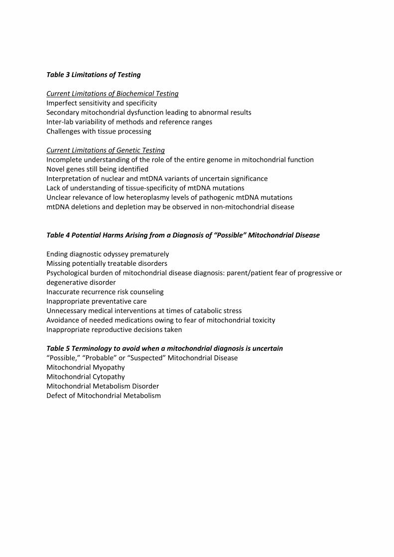
Table 3 Limitations of Testing
Current Limitations of Biochemical TestingImperfect sensitivity and specificitySecondary mitochondrial dysfunction leading to abnormal resultsInter-lab variability of methods and reference rangesChallenges with tissue processing
Current Limitations of Genetic TestingIncomplete understanding of the role of the entire genome in mitochondrial functionNovel genes still being identifiedInterpretation of nuclear and mtDNA variants of uncertain significanceLack of understanding of tissue-specificity of mtDNA mutationsUnclear relevance of low heteroplasmy levels of pathogenic mtDNA mutationsmtDNA deletions and depletion may be observed in non-mitochondrial disease
Table 4 Potential Harms Arising from a Diagnosis of “Possible” Mitochondrial Disease
Ending diagnostic odyssey prematurelyMissing potentially treatable disordersPsychological burden of mitochondrial disease diagnosis: parent/patient fear of progressive ordegenerative disorderInaccurate recurrence risk counselingInappropriate preventative careUnnecessary medical interventions at times of catabolic stressAvoidance of needed medications owing to fear of mitochondrial toxicityInappropriate reproductive decisions taken
Table 5 Terminology to avoid when a mitochondrial diagnosis is uncertain“Possible,” “Probable” or “Suspected” Mitochondrial DiseaseMitochondrial MyopathyMitochondrial CytopathyMitochondrial Metabolism DisorderDefect of Mitochondrial Metabolism
1
Supplementary Material
Biochemical Testing Concerns
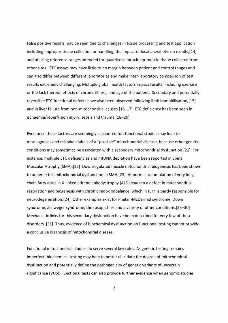
Once a mitochondrial disease is suspected, most testing protocols begin with assessing lactic
acid, plasma or CSF amino acids and urine organic acids. Elevations of plasma lactic acid and
certain amino acids (such as alanine, glycine, proline or threonine) or urinary organic acids
(such as malate, fumarate or 3-methylglutaconic acid) may indicate mitochondrial dysfunction.
Concerns regarding their sensitivity and specificity have been discussed previously, and they
remain imperfect biomarkers in assessing mitochondrial dysfunction. However they continue to
be routinely obtained since they are relatively inexpensive and may be used to guide the need
for further testing.[1] Some have reported that when collected properly and considered in the
correct clinical context, elevations in lactate may selectively have specificity as high as 83-
100%[2–4] although this is not always the case.[5] Nonetheless, permanent urinary excretion
of 3-methylglutaconic acid, if associated with a suggestive clinical picture, is usually a good
indicator of PMD.[6] FGF21 and GDF15 are cytokines more recently associated with
mitochondrial dysfunction in cases of myopathy but also have less than optimal sensitivity and
specificity, often being elevated in non-mitochondrial diseases including diabetes,
hypothyroidism, overfeeding, sepsis and other genetic diseases.[2, 7–13]
While evaluation of suspected mitochondrial disease now often quickly moves to molecular
genetic testing, functional studies of the mitochondrial electron transport chain (ETC) are still
important in many situations. However, in many centers they are no longer the first line test
when mitochondrial disease is suspected. Functional assays include ETC enzymology via
spectrophotometry, measurement of the presence of selected protein components within
complexes and super-complexes via western blots and blue-native gel electrophoresis, and
oxygen consumption rates using various substrates and inhibitors.
2
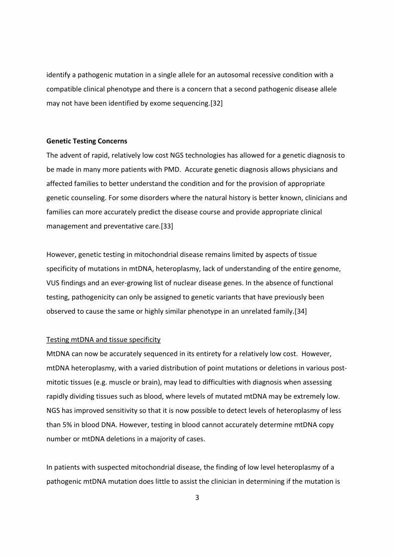
False positive results may be seen due to challenges in tissue processing and test application
including improper tissue collection or handling, the impact of local anesthetic on results,[14]
and utilizing reference ranges intended for quadriceps muscle for muscle tissue collected from
other sites. ETC assays may have little to no margin between patient and control ranges and
can also differ between different laboratories and make inter-laboratory comparison of test
results extremely challenging. Multiple global health factors impact results, including exercise
or the lack thereof, effects of chronic illness, and age of the patient. Secondary and potentially
reversible ETC functional defects have also been observed following limb immobilisation,[15]
and in liver failure from non-mitochondrial causes.[16, 17] ETC deficiency has been seen in
ischaemia/reperfusion injury, sepsis and trauma.[18–20]
Even once these factors are seemingly accounted for, functional studies may lead to
misdiagnoses and mistaken labels of a “possible” mitochondrial disease, because other genetic
conditions may sometimes be associated with a secondary mitochondrial dysfunction.[21] For
instance, multiple ETC deficiencies and mtDNA depletion have been reported in Spinal
Muscular Atrophy (SMA).[22] Downregulated muscle mitochondrial biogenesis has been shown
to underlie this mitochondrial dysfunction in SMA.[23] Abnormal accumulation of very long-
chain fatty acids in X-linked adrenoleukodystrophy (ALD) leads to a defect in mitochondrial
respiration and biogenesis with chronic redox imbalance, which in turn is partly responsible for
neurodegeneration.[24] Other examples exist for Phelan-McDermid syndrome, Down
syndrome, Zellweger syndrome, the rasopathies and a variety of other conditions.[25–30]
Mechanistic links for this secondary dysfunction have been described for very few of these
disorders .[31] Thus, evidence of biochemical dysfunction on functional testing cannot provide
a conclusive diagnosis of mitochondrial disease.
Functional mitochondrial studies do serve several key roles. As genetic testing remains
imperfect, biochemical testing may help to better elucidate the degree of mitochondrial
dysfunction and potentially define the pathogenicity of genetic variants of uncertain
significance (VUS). Functional tests can also provide further evidence when genomic studies
3
identify a pathogenic mutation in a single allele for an autosomal recessive condition with a
compatible clinical phenotype and there is a concern that a second pathogenic disease allele
may not have been identified by exome sequencing.[32]
Genetic Testing Concerns
The advent of rapid, relatively low cost NGS technologies has allowed for a genetic diagnosis to
be made in many more patients with PMD. Accurate genetic diagnosis allows physicians and
affected families to better understand the condition and for the provision of appropriate
genetic counseling. For some disorders where the natural history is better known, clinicians and
families can more accurately predict the disease course and provide appropriate clinical
management and preventative care.[33]
However, genetic testing in mitochondrial disease remains limited by aspects of tissue
specificity of mutations in mtDNA, heteroplasmy, lack of understanding of the entire genome,
VUS findings and an ever-growing list of nuclear disease genes. In the absence of functional
testing, pathogenicity can only be assigned to genetic variants that have previously been
observed to cause the same or highly similar phenotype in an unrelated family.[34]
Testing mtDNA and tissue specificity
MtDNA can now be accurately sequenced in its entirety for a relatively low cost. However,
mtDNA heteroplasmy, with a varied distribution of point mutations or deletions in various post-
mitotic tissues (e.g. muscle or brain), may lead to difficulties with diagnosis when assessing
rapidly dividing tissues such as blood, where levels of mutated mtDNA may be extremely low.
NGS has improved sensitivity so that it is now possible to detect levels of heteroplasmy of less
than 5% in blood DNA. However, testing in blood cannot accurately determine mtDNA copy
number or mtDNA deletions in a majority of cases.
In patients with suspected mitochondrial disease, the finding of low level heteroplasmy of a
pathogenic mtDNA mutation does little to assist the clinician in determining if the mutation is
4
clinically relevant and if so, how the prognosis is affected. Low level heteroplasmy in blood
does not exclude a pathogenic level of heteroplasmy in another tissue, especially if the
patient’s symptoms are primarily muscle or brain related. In disorders that have been well
characterized with clear heteroplasmy:phenotype relationships (e.g. MERRF m.8344G>A,
MELAS m.3243A>G, NARP m.8993T>C/G) the finding of low level heteroplasmy (< 5 %) in blood
is not always likely to be associated with neurological disease; however, assessment in other
tissues is still recommended if the phenotype is compatible.
Incidental findings of mtDNA mutations at low level heteroplasmy are not uncommon,
especially since ~ 1/200 asymptomatic people may carry a low level heteroplasmic pathogenic
mtDNA variant in blood.[35–37] Such variants are now even detected in patients tested by
WES in whom mitochondrial disease may not have been strongly on the differential diagnosis
list. It is quite easy to mistakenly attribute clinical relevance to low-level heteroplasmic mtDNA
mutations due to a clinician’s anchoring and confirmation bias even though it may not be the
actual cause of a given patient’s constellation of medical problems.
Assessing mtDNA in other tissues such as muscle, liver or urinary epithelial cells may help when
pathogenic mutations are not detected in blood or only low levels of a pathogenic mutation are
found. MtDNA point mutation heteroplasmy analysis in urine provides another non-invasive,
reliable and relatively inexpensive methodology that has been validated against skeletal muscle
heteroplasmy, although testing is not available on a clinical basis in all regions.[38] Some
mtDNA point mutations and large-scale or multiple deletions may only be detected in muscle or
liver in some patients. Long-range PCR is the preferred method for detecting deletions as
Southern blot analysis lacks sufficient sensitivity to detect low levels of heteroplasmic deletions.
Southern blot analysis remains useful for clarifying the type of rearrangement in patients with
duplicated or deleted mtDNA.[39] Interpretation is complicated, as normal aging may lead to a
low level of multiple mtDNA deletions in tissues including muscle, and accumulation of mtDNA
deletions may be accelerated in other muscle disorders, particularly sporadic inclusion body
myositis.
5
MtDNA copy number analysis for mtDNA depletion is also not yet routinely measurable by NGS
or accurately quantifiable or always represented in blood. Such testing may become viable and
cost effective via whole genome sequencing (WGS) or other NGS approaches in the near future.
Currently, the most widely used approach if mtDNA depletion is suspected is to perform a
quantitative real-time PCR assay, preferably in an affected tissue, although unaffected tissue
(e.g. skeletal muscle in POLG-related Alpers-Huttenlocher syndrome) may still demonstrate a
significant, albeit clinically silent, mtDNA depletion. Interpretation of mtDNA copy number data
is critically dependent on appropriate age and tissue matched normal control ranges,[40] which
can be difficult to obtain, thereby limiting applicability. Results may be equivocal even when
normal control ranges are available. To complicate matters, mtDNA depletion has been
identified in non-mitochondrial diseases including desminopathies, Parkinson disease, age-
related changes in paraspinal muscles and as a consequence of antiretroviral therapy.[41–45]
Heteroplasmic variants
As mtDNA has a higher mutation rate than the nuclear genome, many individuals have rare or
private mtDNA polymorphisms. Determining the pathogenicity of these polymorphisms
remains challenging. Interpreting the genetic results in the light of clinical and laboratory
findings and the family history may be helpful, but still may not provide a conclusive answer.
MtDNA haplotyping may assist in establishing pathogenicity,[46] and the same mutation arising
independently several times and co-segregating with clinical features may support a causal
role.[47]
Even when pathogenicity is suspected, phenotypic variability remains pronounced. This is the
case for many of the less common or novel pathogenic mutations in the mt-tRNA genes.
Distinguishing novel pathogenic mutations from benign polymorphisms remains difficult.
Research based testing to interrogate the pathogenicity of heteroplasmic mutations – via single
fibre and/or transmitochondrial cybrid studies – remains costly and labour intensive,[48] and is
not possible for most patients.

6
Testing nuclear genes
Since the mitochondrial genome only encodes 13 mitochondrial proteins (and 24 tRNA and
rRNA genes required for the intramitochondrial synthesis of these 13 proteins), nuclear genes
encode almost the entire mitochondrial proteome. New genes impacting mitochondrial
function and leading to human disease continue to be discovered. Many of the known disease-
causing genes may be tested via gene panels and whole exome sequencing (WES) although the
ability to detect a clearly pathogenic mutation in suspected PMD is imperfect, with a reported
diagnostic yield ranging from 25-75%, dependent upon study design.[49–53]
WGS is beginning to be utilized beyond research-based testing but our current understanding
remains limited. Many ongoing WGS analyses are restricted to assessing variants in the
approximately 2% of the genome that represents the exome, owing to a very limited
understanding of the consequences of sequence variants in other regions (promoters, deep
intronic regions and other non-coding regions). Much of what the genome holds in regard to
causing primary mitochondrial disease remains to be seen. This lack of a complete
understanding of and the inability to meaningfully analyze variants in all the genomic elements
involved in mitochondrial function greatly contributes to our inability to make genetic
diagnoses in 100% of cases with suspected PMD.
Using current approaches, many suspected cases, both pediatric and adult, may still remain
without a genetic diagnosis following WES or WGS.[54, 55] This figure will decrease as
sequencing quality, bioinformatic analysis, and our ability to interpret variants throughout the
genome improves. In addition, supplementary approaches such as RNA sequencing of the
transcriptome and proteomics and metabolomics approaches are proving helpful in increasing
the genetic diagnosis rate.[5, 55]
Nuclear gene variants

7
It is now well known that when analysing large numbers of genes by WES or WGS, tens of
thousands of coding sequence variants will be identified in any given individual. Even after
bioinformatic filtering of common benign polymorphisms, hundreds of putative significant
variants remain. Interpreting these variants constitutes a huge challenge. In practice, one or
more VUS is routinely identified. A multi-disciplinary team approach is necessary for optimal
interpretation, bringing together clinical, biochemical, histological and genetic expertise. In the
case of known mitochondrial disease genes, concordance with clinical and biochemical features
can be complicated by weak genotype-phenotype correlation and the continuing widening of
phenotype associated with a particular gene as more cases are identified. Further studies,
including RNA sequencing or protein functional studies, may be necessary although these
follow-up studies are often only available in a research setting.
For nuclear VUS, variant prioritisation following bioinformatic analysis[56–58] and database
interrogation (e.g. 1000 Genomes,[55] ClinVar,[59] ExAC/gnomAD,[60] Medical Genome
Reference Bank, MITOMAP,[61] MSeqDR,[62]), as well as classification using the American
College of Medical Genetics (ACMG) criteria,[34] a search of known attributes of the affected
gene and its transcriptional/translational products can inform an approach toward confirmation
of pathogenicity.
The simplest approach for ascribing conditional pathogenicity is investigating segregation of a
variant with disease in family members. This may be difficult when considering multisystemic
mitochondrial diseases that can display high levels of phenotypic heterogeneity for the same
gene or variant.[63] If familial segregation information is inconclusive or cannot be obtained,
investigating larger cohorts of phenotypically similar patients for the same genotype may
provide support for pathogenicity. As mentioned above, this may be difficult in the absence of a
consistent genotype-phenotype correlation and additionally in the setting of a rare disease. In
any case, segregation data must be accompanied by empirical evidence of a functional defect,
requiring a synthesis of clinical tests and laboratory experiments to establish a pathological
basis for the VUS. This may require a research laboratory with specialist interests in the gene or
8
disease in question and can be aided by the availability of patient-derived tissue samples or cell
lines for investigations.
Validation of VUS pathogenicity is determined by the nature of the variant in question
(missense, nonsense, splicing, indel, copy number variation, structural variant, etc.), the
affected gene and its associated function and the biological/pathological relevance.
Experimental evidence supporting pathogenicity (in addition to clinical presentation,
phenotype/genotype correlation and theoretical evidence) could include imaging studies,
biomarker measurements, or functional approaches in patient-derived tissue and cell lines or
model organisms.
Functional biochemical testing in muscle or other tissues may help better ascertain the
relevance of selected VUS and remains a necessary tool in some patients. Interpreting VUS
remains an ongoing challenge although there are guidelines on how to better categorize
these.[34] These are important considerations for accurate diagnosis and counselling of
patients with PMD disorders, particularly at a time when reproductive choice has been
expanded to include preimplantation genetic diagnosis and more recently mitochondrial
donation.
mtDNA VUS
Novel mtDNA variants pose unique challenges for clinical correlation and interpretation given
the challenges of performing functional validation studies, broad heterogeneity of PMD clinical
phenotypes, and unknown threshold effects necessary for a given variant to manifest disease in
particular organs. However, additional information at both bioinformatic and clinical levels can
be readily attained to further understand the potential functional impact of a given mtDNA
variant.
Several readily available bioinformatics tools now exist that can be used to efficiently gather
information about novel mtDNA variants. For example, mvTool,[64] hosted on the

9
Mitochondrial Disease Sequence Data Resource (MSeqDR[62]), provides population level
frequency data for mtDNA variants, a compilation of in silico variant pathogenicity predictor
tools, and a collection of expert curated pathogenicity data from multiple sources including
MITOMAP[61] and HmtDB.[65]
Guidelines and recommendations for mtDNA variants curation have also been developed by an
expert panel to aid clinicians and molecular geneticists in proper interpretation. (Standards and
guidelines for mitochondrial DNA variant interpretation. [Elizabeth M. McCormick et al.. Human
Mutation 2018 (accepted; under revision)]
Valuable information may also be gained from pursuing diagnostic testing of the mtDNA variant
in question in multiple tissues from both the proband and family members, although this is not
always possible due to family preferences and insurance restrictions. A higher level of
heteroplasmy in more clinically symptomatic tissues provides strong supporting evidence for
the likely pathogenicity of a given mtDNA variant.
Traditionally, the gold-standard definitive study to assess pathogenicity of a heteroplasmic
mtDNA mutation was to generate a transmitochondrial cell line, by fusion of an enucleated
patient cell with a rho zero cell lacking mtDNA, to determine whether a biochemical defect is
transferred with the patient mtDNA.[66]
10
REFERENCES
1 Parikh S, Goldstein A, Koenig MK, Scaglia F, Enns GM, Saneto R, Anselm I, Cohen BH, Falk
MJ, Greene C, Gropman AL, Haas R, Hirano M, Morgan P, Sims K, Tarnopolsky M, Van
Hove JLK, Wolfe L, DiMauro S. Diagnosis and management of mitochondrial disease: a
consensus statement from the Mitochondrial Medicine Society. Genet Med
2015;17:689–701.
2 Suomalainen A, Elo JM, Pietiläinen KH, Hakonen AH, Sevastianova K, Korpela M, Isohanni
P, Marjavaara SK, Tyni T, Kiuru-Enari S, Pihko H, Darin N, Õunap K, Kluijtmans LAJ, Paetau
A, Buzkova J, Bindoff LA, Annunen-Rasila J, Uusimaa J, Rissanen A, Yki-Järvinen H, Hirano
M, Tulinius M, Smeitink J, Tyynismaa H. FGF-21 as a biomarker for muscle-manifesting
mitochondrial respiratory chain deficiencies: A diagnostic study. Lancet Neurol
2011;10:806–18.
3 Davis RL, Liang C, Edema-Hildebrand F, Riley C, Needham M, Sue CM. Fibroblast growth
factor 21 is a sensitive biomarker of mitochondrial disease. Neurology Published Online
First: 18 October 2013. doi:10.1212/01.wnl.0000436068.43384.ef
4 Tarnopolsky M, Stevens L, MacDonald JR, Rodriguez C, Mahoney D, Rush J, Maguire J.
Diagnostic utility of a modified forearm ischemic exercise test and technical issues
relevant to exercise testing. Muscle Nerve 2003;27:359–66.
5 Rahman J, Rahman S. Mitochondrial medicine in the omics era. Lancet (London, England)
2018;391:2560–74.
6 Wortmann SB, Kluijtmans LA, Rodenburg RJ, Sass JO, Nouws J, van Kaauwen EP, Kleefstra
T, Tranebjaerg L, de Vries MC, Isohanni P, Walter K, Alkuraya FS, Smuts I, Reinecke CJ,
van der Westhuizen FH, Thorburn D, Smeitink JA, Morava E, Wevers RA. 3-
Methylglutaconic aciduria--lessons from 50 genes and 977 patients. J Inherit Metab Dis
2013;36:913–21.
7 Kim KH, Kim SH, Min YK, Yang HM, Lee JB, Lee MS. Acute exercise induces FGF21
expression in mice and in healthy humans. PLoS One 2013;8:e63517.
8 Gariani K, Drifte G, Dunn-Siegrist I, Pugin J, Jornayvaz FR. Increased FGF21 plasma levels
in humans with sepsis and SIRS. Endocr Connect 2013;2:146–53.
11
9 Salehi MH, Kamalidehghan B, Houshmand M, Aryani O, Sadeghizadeh M, Mossalaeie
MM. Association of fibroblast growth factor (FGF-21) as a biomarker with primary
mitochondrial disorders, but not with secondary mitochondrial disorders (Friedreich
Ataxia). Mol Biol Rep 2013;40:6495–9.
10 Lee Y, Park YJ, Ahn HY, Lim JA, Park KU, Choi SH, Park do J, Oh BC, Jang HC, Yi KH. Plasma
FGF21 levels are increased in patients with hypothyroidism independently of lipid profile.
Endocr J 2013;60:977–83.
11 Suomalainen A, Elo JM, Pietilainen KH, Hakonen AH, Sevastianova K, Korpela M, Isohanni
P, Marjavaara SK, Tyni T, Kiuru-Enari S, Pihko H, Darin N, Ounap K, Kluijtmans LA, Paetau
A, Buzkova J, Bindoff LA, Annunen-Rasila J, Uusimaa J, Rissanen A, Yki-Jarvinen H, Hirano
M, Tulinius M, Smeitink J, Tyynismaa H. FGF-21 as a biomarker for muscle-manifesting
mitochondrial respiratory chain deficiencies: a diagnostic study. Lancet Neurol
2011;10:806–18.
12 Davis RL, Liang C, Sue CM. A comparison of current serum biomarkers as diagnostic
indicators of mitochondrial diseases. Neurology 2016;86:2010–5.
13 Yatsuga S, Fujita Y, Ishii A, Fukumoto Y, Arahata H, Kakuma T, Kojima T, Ito M, Tanaka M,
Saiki R, Koga Y. Growth differentiation factor 15 as a useful biomarker for mitochondrial
disorders. Ann Neurol 2015;78:814–23.
14 Janssen AJ, Smeitink JA, van den Heuvel LP. Some practical aspects of providing a
diagnostic service for respiratory chain defects. Ann Clin Biochem 2003;40:3–8.
15 Abadi A, Glover EI, Isfort RJ, Raha S, Safdar A, Yasuda N, Kaczor JJ, Melov S, Hubbard A,
Qu X, Phillips SM, Tarnopolsky M. Limb immobilization induces a coordinate down-
regulation of mitochondrial and other metabolic pathways in men and women. PLoS One
2009;4:e6518.
16 McKiernan P, Ball S, Santra S, Foster K, Fratter C, Poulton J, Craig K, McFarland R, Rahman
S, Hargreaves I, Gupte G, Sharif K, Taylor RW. Incidence of Primary Mitochondrial Disease
in Children Younger Than 2 Years Presenting With Acute Liver Failure. J Pediatr
Gastroenterol Nutr 2016;63:592–7.
17 Lane M, Boczonadi V, Bachtari S, Gomez-Duran A, Langer T, Griffiths A, Kleinle S, Dineiger
12
C, Abicht A, Holinski-Feder E, Schara U, Gerner P, Horvath R. Mitochondrial dysfunction
in liver failure requiring transplantation. J Inherit Metab Dis 2016;39:427–36.
18 Murphy MP. Understanding and preventing mitochondrial oxidative damage. Biochem
Soc Trans 2016;44:1219–26.
19 Stepien KM, Heaton R, Rankin S, Murphy A, Bentley J, Sexton D, Hargreaves IP. Evidence
of Oxidative Stress and Secondary Mitochondrial Dysfunction in Metabolic and Non-
Metabolic Disorders. J Clin Med 2017;6:71.
20 Kilbaugh TJ, Karlsson M, Byro M, Bebee A, Ralston J, Sullivan S, Duhaime AC, Hansson MJ,
Elmér E, Margulies SS. Mitochondrial bioenergetic alterations after focal traumatic brain
injury in the immature brain. Exp Neurol 2015;271:136–44.
21 Pyle A, Nightingale HJ, Griffin H, Abicht A, Kirschner J, Baric I, Cuk M, Douroudis K, Feder
L, Kratz M, Czermin B, Kleinle S, Santibanez-Koref M, Karcagi V, Holinski-Feder E,
Chinnery PF, Horvath R. Respiratory chain deficiency in nonmitochondrial disease. Neurol
Genet 2015;1. doi:10.1212/NXG.0000000000000006
22 Berger A, Mayr JA, Meierhofer D, Fötschl U, Bittner R, Budka H, Grethen C, Huemer M,
Kofler B, Sperl W. Severe depletion of mitochondrial DNA in spinal muscular atrophy.
Acta Neuropathol 2003;105:245–51.
23 Ripolone M, Ronchi D, Violano R, Vallejo D, Fagiolari G, Barca E, Lucchini V, Colombo I,
Villa L, Berardinelli A, Balottin U, Morandi L, Mora M, Bordoni A, Fortunato F, Corti S,
Parisi D, Toscano A, Sciacco M, DiMauro S, Comi GP, Moggio M, Di Mauro S, Comi GP,
Moggio M. Impaired muscle mitochondrial biogenesis and myogenesis in spinal muscular
atrophy. JAMA Neurol 2015;72:666–75.
24 Fourcade S, Ferrer I, Pujol A. Oxidative stress, mitochondrial and proteostasis
malfunction in adrenoleukodystrophy: A paradigm for axonal degeneration. Free Radic.
Biol. Med. 2015;88:18–29.
25 Frye RE, Cox D, Slattery J, Tippett M, Kahler S, Granpeesheh D, Damle S, Legido A,
Goldenthal MJ. Mitochondrial Dysfunction may explain symptom variation in Phelan-
McDermid Syndrome. Sci Rep 2016;6. doi:10.1038/srep19544
26 Hui J, Kirby DM, Thorburn DR, Boneh A. Decreased activities of mitochondrial respiratory
13
chain complexes in non-mitochondrial respiratory chain diseases. Dev Med Child Neurol
2006;48:132–6.
27 Niyazov DM, Kahler SG, Frye RE. Primary Mitochondrial Disease and Secondary
Mitochondrial Dysfunction: Importance of Distinction for Diagnosis and Treatment. Mol.
Syndromol. 2016;7:122–37.
28 Garcia-Cazorla A, Serrano M, Perez-Duenas B, Gonzalez V, Ormazabal A, Pineda M,
Fernandez-Alvarez E, Campistol JM, Artuch RM. Secondary abnormalities of
neurotransmitters in infants with neurological disorders. Dev Med Child Neurol
2007;49:740–4.
29 Prince J, Jia S, Båve U, Annerén G, Oreland L. Mitochondrial enzyme deficiencies in
Down’s syndrome. J Neural Transm - Park Dis Dement Sect 1994;8:171–81.
30 Kleefstra T, Wortmann SB, Rodenburg RJT, Bongers EMHF, Hadzsiev K, Noordam C, van
den Heuvel LP, Nillesen WM, Hollody K, Gillessen-Kaesbach G, Lammens M, Smeitink
JAM, van der Burgt I, Morava E. Mitochondrial dysfunction and organic aciduria in five
patients carrying mutations in the Ras-MAPK pathway. Eur J Hum Genet 2011;19:138–44.
31 Panneman DM, Smeitink JA, Rodenburg RJ. Mining for mitochondrial mechanisms: linking
known syndromes to mitochondrial function. Clin Genet Published Online First: 2017.
doi:10.1111/cge.13094
32 Simon MT, Ng BG, Friederich MW, Wang RY, Boyer M, Kircher M, Collard R, Buckingham
KJ, Chang R, Shendure J, Nickerson DA, Bamshad MJ, Van Hove JLK, Freeze HH, Abdenur
JE. Activation of a cryptic splice site in the mitochondrial elongation factor GFM1 causes
combined OXPHOS deficiency. Mitochondrion 2017;34:84–90.
33 Parikh S, Goldstein A, Karaa A, Koenig MK, Anselm I, Brunel-Guitton C, Christodoulou J,
Cohen BH, Dimmock D, Enns GM, Falk MJ, Feigenbaum A, Frye RE, Ganesh J, Griesemer
D, Haas R, Horvath R, Korson M, Kruer MC, Mancuso M, McCormack S, Raboisson MJ,
Reimschisel T, Salvarinova R, Saneto RP, Scaglia F, Shoffner J, Stacpoole PW, Sue CM,
Tarnopolsky M, Van Karnebeek C, Wolfe LA, Cunningham ZZ, Rahman S, Chinnery PF.
Patient care standards for primary mitochondrial disease: a consensus statement from
the Mitochondrial Medicine Society. Genet Med Published Online First: 27 July 2017.
14
doi:10.1038/gim.2017.107
34 Richards S, Aziz N, Bale S, Bick D, Das S, Gastier-Foster J, Grody WW, Hegde M, Lyon E,
Spector E, Voelkerding K, Rehm HL. Standards and guidelines for the interpretation of
sequence variants: A joint consensus recommendation of the American College of
Medical Genetics and Genomics and the Association for Molecular Pathology. Genet Med
2015;17:405–24.
35 Elliott HR, Samuels DC, Eden JA, Relton CL, Chinnery PF. Pathogenic mitochondrial DNA
mutations are common in the general population. Am J Hum Genet 2008;83:254–60.
36 Bitner-Glindzicz M, Pembrey M, Duncan A, Heron J, Ring SM, Hall A, Rahman S.
Prevalence of Mitochondrial 1555A→G Mutation in European Children. N Engl J Med
Published Online First: 2009. doi:10.1056/NEJMc0806396
37 Manwaring N, Jones MM, Wang JJ, Rochtchina E, Howard C, Mitchell P, Sue CM.
Population prevalence of the MELAS A3243G mutation. Mitochondrion 2007;7:230–3.
38 Poulton J, Morten KJ, Weber K, Brown GK, Bindoff L. Are duplications of mitochondrial
DNA characteristic of Kearns-Sayre syndrome? Hum Mol Genet 1994;3:947–51.
39 Poulton J, Holt IJ. Mitochondrial DNA: Does more lead to less? Nat Genet 1994;8:313–5.
40 Calvo SE, Clauser KR, Mootha VK. MitoCarta2.0: An updated inventory of mammalian
mitochondrial proteins. Nucleic Acids Res 2016;44:D1251–7.
41 McCormick EM, Kenyon L, Falk MJ. Desmin common mutation is associated with multi-
systemic disease manifestations and depletion of mitochondria and mitochondrial DNA.
Front Genet 2015;6:1–5.
42 Scruggs ER, Dirks Naylor AJ. Mechanisms of zidovudine-induced mitochondrial toxicity
and myopathy . Pharmacology 2008;82:83–8.
43 Pyle A, Anugrha H, Kurzawa-Akanbi M, Yarnall A, Burn D, Hudson G. Reduced
mitochondrial DNA copy number is a biomarker of Parkinson’s disease. Neurobiol Aging
2015;38:216.e7-216.e10.
44 Campbell GR, Reeve A, Ziabreva I, Polvikoski TM, Taylor RW, Reynolds R, Turnbull DM,
Mahad DJ. Mitochondrial DNA deletions and depletion within paraspinal muscles.
Neuropathol Appl Neurobiol 2013;39:377–89.
15
45 Poulton J, Sewry C, Potter CG, Bougeron T, Chretien D, Wijburg FA, Morten KJ, Brown G.
Variation in mitochondrial DNA levels in muscle from normal controls. Is depletion of
mtDNA in patients with mitochondrial myopathy a distinct clinical syndrome? J Inherit
Metab Dis Published Online First: 1995. doi:10.1007/BF00711367
46 Yarham JW, Al-Dosary M, Blakely EL, Alston CL, Taylor RW, Elson JL, McFarland R. A
comparative analysis approach to determining the pathogenicity of mitochondrial tRNA
mutations. Hum Mutat 2011;32:1319–25.
47 Dombi E, Diot A, Morten K, Carver J, Lodge T, Fratter C, Ng YS, Liao C, Muir R, Blakely EL,
Hargreaves I, Al-Dosary M, Sarkar G, Hickman SJ, Downes SM, Jayawant S, Yu-Wai-Man P,
Taylor RW, Poulton J. The m.13051G>A mitochondrial DNA mutation results in variable
neurology and activated mitophagy. Neurology. 2016;86:1921–3.
48 Caporali L, Iommarini L, La Morgia C, Olivieri A, Achilli A, Maresca A, Valentino ML,
Capristo M, Tagliavini F, Del Dotto V, Zanna C, Liguori R, Barboni P, Carbonelli M, Cocetta
V, Montopoli M, Martinuzzi A, Cenacchi G, De Michele G, Testa F, Nesti A, Simonelli F,
Porcelli AM, Torroni A, Carelli V. Peculiar combinations of individually non-pathogenic
missense mitochondrial DNA variants cause low penetrance Leber’s hereditary optic
neuropathy. PLoS Genet 2018;14. doi:10.1371/journal.pgen.1007210
49 Calvo SE, Compton AG, Hershman SG, Lim SC, Lieber DS, Tucker EJ, Laskowski A, Garone
C, Liu S, Jaffe DB, Christodoulou J, Fletcher JM, Bruno DL, Goldblatt J, Dimauro S,
Thorburn DR, Mootha VK. Molecular diagnosis of infantile mitochondrial disease with
targeted next-generation sequencing. Sci Transl Med 2012;4:118ra10.
50 Wortmann SB, Koolen DA, Smeitink JA, van den Heuvel L, Rodenburg RJ. Whole exome
sequencing of suspected mitochondrial patients in clinical practice. J Inherit Metab Dis
2015;38:437–43.
51 Taylor RW, Pyle A, Griffin H, Blakely EL, Duff J, He L, Smertenko T, Alston CL, Neeve VC,
Best A, Yarham JW, Kirschner J, Schara U, Talim B, Topaloglu H, Baric I, Holinski-Feder E,
Abicht A, Czermin B, Kleinle S, Morris AAM, Vassallo G, Gorman GS, Ramesh V, Turnbull
DM, Santibanez-Koref M, McFarland R, Horvath R, Chinnery PF. Use of whole-exome
sequencing to determine the genetic basis of multiple mitochondrial respiratory chain

16
complex deficiencies. Jama 2014;312:68–77.
52 Lieber DS, Calvo SE, Shanahan K, Slate NG, Liu S, Hershman SG, Gold NB, Chapman BA,
Thorburn DR, Berry GT, Schmahmann JD, Borowsky ML, Mueller DM, Sims KB, Mootha
VK. Targeted exome sequencing of suspected mitochondrial disorders. Neurology
2013;80:1762–70.
53 Kohda M, Tokuzawa Y, Kishita Y, Nyuzuki H, Moriyama Y, Mizuno Y, Hirata T, Yatsuka Y,
Yamashita-Sugahara Y, Nakachi Y, Kato H, Okuda A, Tamaru S, Borna NN, Banshoya K,
Aigaki T, Sato-Miyata Y, Ohnuma K, Suzuki T, Nagao A, Maehata H, Matsuda F, Higasa K,
Nagasaki M, Yasuda J, Yamamoto M, Fushimi T, Shimura M, Kaiho-Ichimoto K, Harashima
H, Yamazaki T, Mori M, Murayama K, Ohtake A, Okazaki Y. A Comprehensive Genomic
Analysis Reveals the Genetic Landscape of Mitochondrial Respiratory Chain Complex
Deficiencies. PLoS Genet 2016;12. doi:10.1371/journal.pgen.1005679
54 Kremer LS, Bader DM, Mertes C, Kopajtich R, Pichler G, Iuso A, Haack TB, Graf E,
Schwarzmayr T, Terrile C, Koňaříkova E, Repp B, Kastenmüller G, Adamski J, Lichtner P,
Leonhardt C, Funalot B, Donati A, Tiranti V, Lombes A, Jardel C, Gläser D, Taylor RW,
Ghezzi D, Mayr JA, Rötig A, Freisinger P, Distelmaier F, Strom TM, Meitinger T, Gagneur J,
Prokisch H. Genetic diagnosis of Mendelian disorders via RNA sequencing. Nat Commun
2017;8. doi:10.1038/ncomms15824
55 1000 Genomes Project Consortium, Auton A, Brooks LD, Durbin RM, Garrison EP, Kang
HM, Korbel JO, Marchini JL, McCarthy S, McVean GA, Abecasis GR. A global reference for
human genetic variation. Nature 2015;526:68–74.
56 Van der Auwera GA, Carneiro MO, Hartl C, Poplin R, del Angel G, Levy-Moonshine A,
Jordan T, Shakir K, Roazen D, Thibault J, Banks E, Garimella K V., Altshuler D, Gabriel S,
DePristo MA. From fastQ data to high-confidence variant calls: The genome analysis
toolkit best practices pipeline. Curr Protoc Bioinforma Published Online First: 2013.
doi:10.1002/0471250953.bi1110s43
57 Paila U, Chapman BA, Kirchner R, Quinlan AR. GEMINI: Integrative Exploration of Genetic
Variation and Genome Annotations. PLoS Comput Biol 2013;9.
doi:10.1371/journal.pcbi.1003153
17
58 McLaren W, Gil L, Hunt SE, Riat HS, Ritchie GRS, Thormann A, Flicek P, Cunningham F. The
Ensembl Variant Effect Predictor. Genome Biol 2016;17. doi:10.1186/s13059-016-0974-4
59 Landrum MJ, Lee JM, Benson M, Brown G, Chao C, Chitipiralla S, Gu B, Hart J, Hoffman D,
Hoover J, Jang W, Katz K, Ovetsky M, Riley G, Sethi A, Tully R, Villamarin-Salomon R,
Rubinstein W, Maglott DR. ClinVar: Public archive of interpretations of clinically relevant
variants. Nucleic Acids Res 2016;44:D862–8.
60 Lek M, Karczewski KJ, Minikel E V., Samocha KE, Banks E, Fennell T, O’Donnell-Luria AH,
Ware JS, Hill AJ, Cummings BB, Tukiainen T, Birnbaum DP, Kosmicki JA, Duncan LE,
Estrada K, Zhao F, Zou J, Pierce-Hoffman E, Berghout J, Cooper DN, Deflaux N, DePristo
M, Do R, Flannick J, Fromer M, Gauthier L, Goldstein J, Gupta N, Howrigan D, Kiezun A,
Kurki MI, Moonshine AL, Natarajan P, Orozco L, Peloso GM, Poplin R, Rivas MA, Ruano-
Rubio V, Rose SA, Ruderfer DM, Shakir K, Stenson PD, Stevens C, Thomas BP, Tiao G,
Tusie-Luna MT, Weisburd B, Won HH, Yu D, Altshuler DM, Ardissino D, Boehnke M,
Danesh J, Donnelly S, Elosua R, Florez JC, Gabriel SB, Getz G, Glatt SJ, Hultman CM,
Kathiresan S, Laakso M, McCarroll S, McCarthy MI, McGovern D, McPherson R, Neale
BM, Palotie A, Purcell SM, Saleheen D, Scharf JM, Sklar P, Sullivan PF, Tuomilehto J,
Tsuang MT, Watkins HC, Wilson JG, Daly MJ, MacArthur DG. Analysis of protein-coding
genetic variation in 60,706 humans. Nature 2016;536:285–91.
61 Lott MT, Leipzig JN, Derbeneva O, Michael Xie H, Chalkia D, Sarmady M, Procaccio V,
Wallace DC. MtDNA variation and analysis using Mitomap and Mitomaster. Curr Protoc
Bioinforma Published Online First: 2013. doi:10.1002/0471250953.bi0123s44
62 Shen L, Diroma MA, Gonzalez M, Navarro-Gomez D, Leipzig J, Lott MT, van Oven M,
Wallace DC, Muraresku CC, Zolkipli-Cunningham Z, Chinnery PF, Attimonelli M, Zuchner
S, Falk MJ, Gai X. MSeqDR: A Centralized Knowledge Repository and Bioinformatics Web
Resource to Facilitate Genomic Investigations in Mitochondrial Disease. Hum Mutat
2016;37:540–8.
63 Crimmins D, Morris JG, Walker GL, Sue CM, Byrne E, Stevens S, Jean-Francis B, Yiannikas
C, Pamphlett R. Mitochondrial encephalomyopathy: variable clinical expression within a
single kindred. J Neurol Neurosurg Psychiatry 1993;56:900–5.
18
64 Shen L, Attimonelli M, Bai R, Lott MT, Wallace DC, Falk MJ, Gai X. MSeqDR mvTool: A
mitochondrial DNA Web and API resource for comprehensive variant annotation,
universal nomenclature collation, and reference genome conversion. Hum. Mutat. 2018.
doi:10.1002/humu.23422
65 Clima R, Preste R, Calabrese C, Diroma MA, Santorsola M, Scioscia G, Simone D, Shen L,
Gasparre G, Attimonelli M. HmtDB 2016: Data update, a better performing query system
and human mitochondrial DNA haplogroup predictor. Nucleic Acids Res 2017;45:D698–
706.
66 King MP, Attardi G. Human cells lacking mtDNA: Repopulation with exogenous
mitochondria by complementation. Science (80- ) Published Online First: 1989.
doi:10.1126/science.2814477

1
Case 1 Basal ganglia disease
Two sisters from a consanguineous family had a severe neurodegenerative disorder,
characterized by infantile onset of profound intellectual disability, failure to thrive associated
with severe feeding difficulties, seizures and dystonia. Pertinent investigations in the older
proband included normal urine amino and organic acids, plasma acylcarnitines and blood
lactate. CSF lactate was also normal. Nerve conduction studies were suggestive of an axonal
neuropathy. Brain magnetic resonance imaging (MRI) at 13 months was notable for bilateral
basal ganglia hyperintensities and cerebral atrophy. Muscle histology was nonspecifically
abnormal. Muscle complex IV activity was 48% of the control mean relative to citrate synthase,
and in liver was 32% of normal relative to citrate synthase. She died at 26 months of age. Her
younger sister exhibited a very similar onset and course, but has survived, currently aged 13
years, albeit with profound intellectual disability, upper airway abnormalities and poor growth
requiring gastrostomy feeding. A brain MRI at seven months of age also showed bilateral
symmetrical basal ganglia hyperintensities. She did not have any biopsies but her serum FGF21
was elevated at 1400 pg/ml (normal < 300). Together, these features were highly suggestive of
an underlying mitochondrial electron transport chain (ETC) disorder primarily affecting complex
IV. However whole genome sequencing revealed a homozygous variant in the SLC39A8 gene
(Riley et al 2017), which was recently identified to cause a manganese transporter disorder also
impacting glycosylation. (Boycott et al 2015). Manganese is a cofactor for manganese
superoxide dismutase, without which there is an increase in oxidative stress and decrease in
ROS scavenging. (Riley et al 2017 PMID: 27995398) Subsequent analysis of serum transferrin
isoforms revealed a profile consistent with the expected type II congenital disorder of
glycosylation pattern.
Case 2 Ophthalmoplegia and myopathy
Two brothers (68 and 80 years old) from a non-consanguineous family were evaluated for
progressive ptosis, ophthalmoplegia, dysphagia and mild limb-girdle muscle weakness since
their 50s. Pedigree analysis was suggestive of maternally inherited autosomal dominant
disease, with no clinical anticipation. Blood tests, including creatine kinase (CK) and lactate,
2
were normal in both brothers. Electromyography (EMG) studies revealed myopathic changes,
and a muscle biopsy performed in the older brother (at the age of 70 years) showed rare ragged
red and cytochrome c oxidase (COX) negative fibers (5% for both). Long-range PCR analysis
revealed multiple mtDNA deletions in muscle. However, next generation sequencing (NGS)
analysis of genes involved in mtDNA maintenance was negative. Further analysis of the PABPN1
gene revealed expansion of 13 GCN trinucleotide repeat in the first exon, thus shifting the
diagnosis to oculopharyngeal muscular dystrophy (OPMD). OPMD and myotonic dystrophy are
two genetic illnesses in addition to many mitochondrial disorders where ptosis, incomplete
ophthalmoplegia, dysphagia and myopathy are common features. As NGS technology
improves, the nucleotide repeat disorders will be more easily identified.
Case 3 Lactic acidosis and atrophy of the putamen
A one-year-old boy was evaluated for hypotonia and motor regression. He was normal at birth
but late in the first year of life he was found to have lost motor tone. He had undergone
extensive biochemical evaluation prior to his initial assessment at the specialist center. Physical
examination was normal other than extreme hypotonia. He had no speech but was attentive
and smiled socially. On direct enquiry, mother had not observed any crystals in his urine. MRI
brain showed mild parenchymal atrophy and volume loss bilaterally in the putamen and
thalamus. Blood lactic acid was elevated at 6 mmol/L. A muscle biopsy was performed,
showing normal histology but the enzymatic activity of complex I (I+III assay) was 25% of the
mean control. He was treated with coenzyme Q10, levocarnitine and a B vitamin complex for
presumed complex I disease. His examination over the next few years showed him to be an
alert and visually interactive child who did not develop language function and had continued
severe hypotonia with progressive but mild dystonia. At the age of 5 years, he arrived for a
routine examination wearing gloves, to protect his fingers from a recent onset of biting
behavior (self-mutilation). The uric acid level was elevated and hypoxanthine phosphoribosyl-
transferase enzyme activity was less than 10% of control values. Genetic testing was not
performed but the patient’s clinical course followed that typical of Lesch-Nyhan disease. The
history of progressive hypotonia with evolving dystonia, language delay, lactic acidosis, deep
3
gray matter loss and generalized brain parenchymal volume loss, together with the low
complex I ETC activity seemed to fit together with a primary mitochondrial disorder. With our
current knowledge regarding altered concentration of nucleotide pools affecting mtDNA
replication, it seems likely that the abnormal purine concentrations could result in mtDNA
depletion, although this was not confirmed in this patient.
Case 4 Cognitive decline with ataxia and dystonia
The patient, a 20-year-old male, was born at term (weight 3.01kg) to unrelated parents
following an uncomplicated pregnancy and normal delivery. He displayed mild early gross
motor development delay (crawled at 14 months, walked at 18 months) but language
development was normal.
He first presented to the paediatric service at the age of 6 years with an episode suggestive of a
nocturnal tonic-clonic seizure. A history of ‘blank spells’ from age 6 months was noted by his
mother. Neurological examination was normal but an electroencephalogram (EEG)
demonstrated bilateral independent foci in both temporal regions and he was commenced on
carbamazepine. His school performance was above the average expected for his age group. He
remained under follow up and these events were eventually deemed non-epileptic, and
carbamazepine was weaned.
Between the ages of 10 and 12 years, there was emerging concern that the quality of his
school-work had declined. However, neuropsychological assessment at age 12 years
demonstrated that he was still working within the normal expected range, albeit at the lower
end (10th centile for verbal comprehension; 5th centile perceptual reasoning; 55th centile for
working memory). However, by the age of 14 years there was a clear and objective
deterioration. He had also started tripping frequently; was struggling to button, zip or tie laces;
his speech had declined; and there was prominent daytime somnolence.
4
On clinical examination there were no dysmorphic or neuro-cutaneous signs. Cranial nerve
examination, including eye movements, was normal. Tone was normal, but reflexes were brisk
throughout, plantar responses were down-going. No sensory abnormalities were evident. He
had a clumsy gait, with an inability to tandem walk, mild dystonic posturing and subtle
choreiform movements of the upper limbs. Cerebellar dysarthria, and finger-nose, heel-shin
and truncal ataxia were noted. Cognitive assessment revealed emotional lability and poverty of
mentation.
MRI brain demonstrated bilateral symmetrical increased signal in the white matter adjacent to
the posterior horns of the lateral ventricles. Routine laboratory tests were normal including
ferritin, transferrin isoforms, thyroid function, cholesterol, triglycerides, lipids, copper,
ceruloplasmin, lysosomal enzymes, very long chain fatty acids, vitamin E, biotinidase,
intermediate metabolites, lactate, pyruvate, ammonia, urate, amino acids, urinary organic acids
and mucopolysaccharide screen. Genetic tests for DRPLA, Friedreich ataxia, spinocerebellar
ataxias (SCA1, 2, 3, 6, 7 and 17) and Huntington disease were negative. No concerning features
were identified on slit lamp examination. Visual evoked potentials (VEP) and electroretinogram
(ERG) were normal.
At age 15 years, the patient was referred to a regional neuro-genetic clinic where a
mitochondrial aetiology was explored, given the clinical features and imaging findings. Thiamine
was commenced but without clinical improvement. Muscle biopsy was undertaken for ETC
enzyme analysis which identified a complex I deficiency (0.051 U/UCS; normal 0.104+0.036)
with low complex I:II ratio (0.05;, normal 0.52-0.95). Muscle histochemistry was normal. Array
CGH was normal and further genetic testing for mutations in POLG, MTATP6, MTATP8 and
MTTK (associated with MERRF phenotypes) were all negative.
At clinical review at 18 years of age, symptoms had progressed and the patient was having
increasing problems with falls, speech, and cognitive decline. There was a history suggestive of

5
mild cataplexy with heightened emotion and giggling triggering brief head drop. Examination
revealed a mixed dystonic-ataxic gait, myoclonic jerks and a supranuclear gaze palsy.
Given the clinical features, a sample for plasma oxysterols was sent. Cholestane-3b, 5a, 6b-triol
was found to be elevated at 55.4 ng/ml (normal range 8.1-37.7 ng/ml) and pathogenic
compound heterozygous mutations were identified in NPC1 (c.3182T>C;p.lle1061Thr and
c.3566A>G;p.Glu1189Gly) which confirmed the diagnosis of Niemann-Pick disease type C. The
patient was referred for commencement of miglustat therapy.
Photograph and video of the patient at 18 years of age.
Case 5: mtDNA pathogenic mutation with low level heteroplasmy in blood
A 43-year-old Romanian woman presented with normal early development except for lifelong
clumsiness and progressive multi-systemic disease since her 20s. Her disease was progressive
and severe, including complex epilepsy since age 20 with generalized seizures and then
myoclonic jerks with diffuse slowing and multi-focal periodic lateralizing epileptiform
discharges on electroencephalogram, mild cortical atrophy on computed tomography (CT) at
age 40 and more diffuse cortical atrophy on brain MRI at the time of her first stroke at age 42.
She had recurrent acute "metabolic" strokes in the posterior parietal and medial occipital-
6
temporal cortex at age 42 with residual hemiplegia, hypertonia, walker or wheelchair
dependence, dysarthria, and permanent left peripheral vision loss. She developed cognitive
decline from her mid-30s including psychosis, delusions and functioning estimated at a second-
grade level. She also had migraines. Additional systemic manifestations include vertical and
horizontal ophthalmoplegia, bilateral progressive sensorineural hearing loss onset at 30, left
ventricular hypertrophy (LVH), a patent foramen ovale, nausea and vomiting with stress,
irritable bowel syndrome, gastroesophageal reflux disease, bilateral renal cysts, and early
morning carbohydrate craving and cloudy cognition that improved with eating. Testing showed
elevated lactate intermittently in blood (4 mM; normal < 2.2) and CSF (4.2 mM; normal < 2.2),
and mildly elevated glutaryl dicarboxylic acid on plasma acylcarnitine analysis.
Her family history was significant in that her mother had type II diabetes diagnosed at age 56
and an episode of paralysis at age 56, poor balance with apparent peripheral neuropathy,
cortical atrophy, sudden complete hair loss in her 30s with thinner hair regrowth over time, and
sensorineural hearing loss onset in her 70s. Her maternal grandmother had extreme fatigue.
Muscle biopsy at age 42 showed mild myofiber size variation, hypertrophic and atrophic fibers,
one necrotic fiber, rare degenerating but no regenerating fibers, an unquantified number of
ragged red fibers, targetoid and small dark fibers on NADH-TR staining, significant type II > I
fiber atrophy, increased SDH positive fibers, and 5-10% COX negative fibers. No biochemical or
genetic testing was performed on muscle.
A low level (8%) of the mitochondrial DNA (mtDNA) mutation m.3243A>G that causes
Mitochondrial Encephalopathy Lactic Acidosis and Stroke-like episodes (MELAS) syndrome was
detected in her blood. This same variant was subsequently found by highly sensitive ARMS
qPCR analysis to be at similarly low levels (4% in blood, 7% in urine) in her mother. Urine
heteroplasmy testing in the proband revealed 60% m.3243A>G mutant heteroplasmy load,
which was clinically significant and confirmed the diagnosis of MELAS spectrum disorder.

7
Typical heteroplasmy levels in symptomatic patients are in the 25-90% range. However, a wide
range of multi-systemic diseases that poorly fit into a pre-defined clinical syndrome can often
be seen.[1, 2] It is well-known that the m.3243A>G mutation load is selected against and
decreases over time in blood at the same time it may persist or increase in other tissues such as
urine, which reflects shed renal cells.[3–6] A software tool to model the expected tissue
heteroplasmy levels in individuals with the m.3243A>G mutation in blood is available at the
Newcastle Wellcome Trust website (www.newcastle-mitochondria.com/).
Case 6: mtDNA VUS
A 19-year-old Caucasian man presented with progressive multi-system involvement. Several
features were attributed to a congenital open neural tube defect (L5/S1 myelomeningocele)
including complete paraplegia of lower extremities, lack of bladder and bowel control, and
Chiari malformation requiring shunt placement. However, additional multisystem involvement
unexplained by the myelomeningocele was ultimately found to be related to a novel
pathogenic mtDNA variant (Schrier et al., 2012). As previously reported, these manifestations
included developmental delay and intellectual disability, hypotonia, bilateral severe mixed
hearing loss, moderate to severe LVH, Wolff-Parkinson-White syndrome, severe retinal
dystrophy, bilateral dense nuclear cataracts requiring extraction, hypopituitarism, growth
deficiency, type II diabetes mellitus, hypogonadism, truncal obesity, and osteopenia. Family
history was non-contributory. The proband’s mother was healthy until her death in a motor
vehicle accident in her early 30s.
Metabolic screening studies showed persistently elevated blood alanine (492 nmol/mL and
708.9 nmol/mL; normal 88-440) and intermittently elevated blood lactate (1.54 mM and 2.8
mM; normal <2). Skeletal muscle biopsy findings in biceps (proband recently received botox
injections to both quadriceps) were consistent with a mitochondrial myopathy (unquantified
number of ragged red fibers and absence of COX staining in scattered fibers; electron
microscopy revealing irregular branched mitochondria, focal mitochondrial collections, and
8
mitochondrial lipid deposits). ETC enzyme activities were within normal limits and citrate
synthase was increased (159% of mean).
Clinical molecular testing available at the time (2007-2008) was unrevealing, including
karyotype, microarray, BBS1 and BBS2 gene sequencing, mtDNA common point mutation and
deletion analysis in blood, and mtDNA deletion and duplication analysis in muscle. A
homoplasmic novel variant, m.12264C>T in MTTS2 encoding mitochondrial tRNA-serine 2, was
identified in muscle by mtDNA whole genome sequencing. Research-based mtDNA
heteroplasmy quantitation was performed in blood, buccal, muscle, and extracted cataract
samples from the proband and blood and buccal samples from the proband’s father and
maternal grandmother. The variant was found to be homoplasmic in the proband’s muscle,
extracted cataract, and buccal sample, and heteroplasmic in blood (34%). His father did not
have the mtDNA variant. His maternal grandmother was found to have the m.12264C>T variant
at 1% heteroplasmy level in blood and 18% heteroplasmy level in her buccal sample. His mother
was deceased and a sample was not available for testing.
Case 7: OPA1 VUS
A 67-year-old Caucasian male presented with fatigue, disturbed sleep, muscle pain, weakness
and numbness. Clinical examination revealed lower limb distal muscle weakness and wasting,
peripheral neuropathy, mild sensorineural hearing loss, mild bilateral ptosis, dyspnea,
dysphagia and dysarthria with bulbar and respiratory muscle weakness, as well as dyslipidaemia
and elevated transaminase levels. A muscle biopsy showed histological changes consistent with
mitochondrial myopathy, including increased subsarcolemmal staining with Gomori trichrome
and COX-negative fibres (quantity not known). The family history was unremarkable and did not
indicate a likely mode of inheritance.
Serum levels of FGF21 were elevated at 798, 691 and 812 pg/ml (control range < 350 pg/ml on
longitudinal sampling, potentially supporting a muscle-manifesting mitochondrial disease. Long-
range PCR of the mitochondrial genome showed a 16.5kb amplicon with no evidence of large-
9
scale deletions. Deep sequencing of the entire mtDNA amplified by long-range PCR revealed no
significant variants. Bioinformatic analysis of known mitochondrial and neuromuscular disease
genes from whole genome sequencing (WGS) of blood genomic DNA revealed a heterozygous,
in-frame, 18 nucleotide deletion in OPA1 (NM_015560.2(OPA1):c.113_130del18
(p.Arg38_Ser43del)) that was confirmed by Sanger sequencing on a separate blood sample. The
deletion locates to exon 2 of OPA1 and potentially interferes with the mitochondrial targeting
sequence of the protein. There are reports of at least 9 other individuals with the same deletion
in ClinVar, however it is classified as a VUS owing to conflicting descriptions of associated
pathogenicity and phenotype
(https://www.ncbi.nlm.nih.gov/clinvar/variation/214916/#supporting-observations).
OPA1 encodes a mitochondrial inner-membrane protein with dynamin-related GTPase activity
that is required for maintenance of cristae structure (in doing so regulating apoptosis), the
mitochondrial reticulum (by mediating mitochondrial fusion) and the mitochondrial genome.
Autosomal dominant pathogenic variants in OPA1 (OMIM 605290) are associated with Optic
Atrophy 1 (Phenotype MIM 165500), an optic neuropathy affecting retinal ganglion cells and
leading to blindness, and also dominant optic atrophy plus (Phenotype MIM 125250), which has
systemic manifestations (e.g. hearing loss, myopathy and neuropathy) in addition to optic
atrophy. As the patient described here had no evidence of optic atrophy he was not initially
considered for OPA1 genetic testing. However, his symptoms do fit with the extraocular
manifestations of dominant optic atrophy plus.
Case 8: Spasticity and mitochondrial dysfunction
The now 43-year old patient presented in the early 1990s, during late adolescence, with new
onset academic difficulties in high school. Over the next few years she had difficulty with
ambulation. Her examination when first evaluated about 8 years after her symptoms began
revealed mild cognitive dysfunction, dysarthria, spasticity and neuropathy. Her initial brain MRI
was normal but her biochemical evaluation revealed consistently elevated pipecolic acid levels
(range of 2.7-4.7 uM (control 0.7-2.5). Detailed evaluation for known peroxisomal disorders
10
was performed with the advice and assistance of experts in this group of disorders, and was not
revealing. A liver biopsy was performed in 1999 to confirm what was believed to be a
peroxisomal disorder. The EM of the liver demonstrated normal peroxisomal appearances but
massive and abnormally shaped mitochondria with paracrystalline inclusions. A subsequent
muscle biopsy showed ragged blue fibers (amount not specified) and a biochemical defect in
ETC Complex I, I+III and III (amounts not specified). The patient did not have consistent
abnormalities in lactic acid, blood amino acids or urine organic acids, but because of the
histopathology and biochemical findings, the working diagnosis was believed to be a
mitochondrial disease. Over the next 13 years the patient underwent genetic testing in a
sequential fashion that included screening for common mtDNA mutations, Southern blot for
mtDNA deletions, whole genome mtDNA sequencing, and sequencing of numerous individual
nuclear genes. This testing did not reveal any mutations that would explain her phenotype. Her
clinical symptoms worsened and the patient currently has difficulty speaking in sentences
because of both cognitive limitations and dysarthria. She spends most of her day in a
wheelchair, with a limited ability to ambulate. In addition to the spasticity and neuropathy her
clinical findings now include retinal degeneration, scoliosis, pes cavus, myopathy, ataxia and
parkinsonism (stooped posture, masked facies and bradykinesia). The brain MRI shows
moderate generalized volume loss. In 2012 sequence analysis of a large panel of nuclear-
encoded mitochondrial genes (> 250 genes) was normal. In late 2013 WES was reported to
show a number of possible candidate mutations. These included compound heterozygous
mutations in TIMM44. Both mutations were predicted in silico to be pathogenic, but this
mitochondrial gene had not been reported to be associated with any clinical disease. In
addition, a nonsense mutation was found in SPG11, which was also present in the patient’s
father, but not mother. Because only one mutation was identified in SPG11 (a gene whose
defects typically cause autosomal recessive disease) and a phenotype that partially matched
the phenotype (no cerebellar atrophy or thinning of the corpus callosum was identified), both
SPG11 and TIMM44 remained candidate genes. Further genetic evaluation was delayed
because of costs and the need for insurance approval to pay for the testing but in 2015 del/dup
analysis of SPG11 was performed and revealed the previously identified c.1744G>T
11
(p.Glu5882*) mutation, also identified in the father, as well as partial deletion of the gene
including exons 17 and 18, which was found in her mother a year later. The gene product of
SPG11 controls the assembly of the ribosomes in the mitochondria and is associated with the
clinical illness autosomal recessive spastic paraplegia type 11 (OMIM 604360), which is a good
fit for this patient’s illness. The liver pathology and biochemistry reported in this case suggest
this gene, and disease, could be considered as a primary mitochondrial disorder, although it is
not known if the mutations in TIMM44 play a role in the patient’s phenotype. Future
technological advances may help to address this question as the patient and her family wish to
pursue this question. Of note, her GDF15 is elevated at 955 pg/mL (control range < 750). This
case illustrates a two-decade journey through technological advancements that eventually led
to the diagnosis, possibly led astray by unexpected laboratory findings. Had the initial
evaluation not identified the consistent elevation in pipecolic acid, the liver biopsy would not
have been performed and the subsequent evaluation would likely not have led down a
mitochondrial pathway. An explanation for the elevation in pipecolic acid remains unclear. If
the initial clinical suspicion led towards a spastic paraparesis evaluation, the results of genetic
testing would have likely stumbled across the same technological issues, specifically early
massively parallel sequencing failed to identify many deletions, duplications or insertions.
12
REFERENCES
1 Chin J, Marotta R, Chiotis M, Allan EH, Collins SJ. Detection rates and phenotypic
spectrum of m.3243A>G in the MT-TL1 gene: A molecular diagnostic laboratory
perspective. Mitochondrion 2014;17:34–41.
2 Kaufmann P, Engelstad K, Wei Y, Kulikova R, Oskoui M, Battista V, Koenigsberger DY,
Pascual JM, Sano M, Hirano M, DiMauro S, Shungu DC, Mao X, De Vivo DC. Protean
phenotypic features of the A3243G mitochondrial DNA mutation. Arch Neurol
2009;66:85–91.
3 McDonnell MT, Schaefer AM, Blakely EL, McFarland R, Chinnery PF, Turnbull DM, Taylor
RW. Nonivasive diagnosis of the 3243A > G mitochondrial DNA mutation using urinary
epithelial cells. Eur J Hum Genet 2004;12:778–81.
4 Shanske S, Pancrudo J, Kaufmann P, Engelstad K, Jhung S, Lu J, Naini A, DiMauro S, De
Vivo DC. Varying loads of the mitochondrial DNA A3243G mutation in different tissues:
Implications for diagnosis. Am J Med Genet 2004;130 A:134–7.
5 Whittaker RG, Blackwood JK, Alston CL, Blakely EL, Elson JL, McFarland R, Chinnery PF,
Turnbull DM, Taylor RW. Urine heteroplasmy is the best predictor of clinical outcome in
the m.3243A>G mtDNA mutation. Neurology 2009;72:568–9.
6 Rahman S, Poulton J, Marchington D, Suomalainen A. Decrease of 3243 A-->G mtDNA
mutation from blood in MELAS syndrome: a longitudinal study. Am J Hum Genet
2001;68:238–40.