A devised strategy for tracheal extubation for predicted ...
4
CASE REPORT Open Access A devised strategy for tracheal extubation for predicted difficult airway in a child with unilateral vocal cord paralysis: a case report Mariko Nagata, Yasuyo Shimomura, Yoshitaka Hara, Tomoyuki Nakamura, Seiko Hayakawa, Hidefumi Komura, Junpei Shibata, Chizuru Yamashita and Osamu Nishida * Abstract Background: Extubation is a more challenging medical practice than intubation, and countermeasures against it are similar to those described in the Difficult Intubation Guidelines, but problems cannot be overcome by completely the same methods. We predicted difficult extubation in a pediatric patient with left recurrent laryngeal nerve paralysis and devised an extubation method. Case presentation: The patient was a 2-year-and-8-month-old boy scheduled for cleft palate repair. Concomitant cardiac anomaly and first and second branchial arch syndrome-associated facial malformations, such as mandibular micrognathia and auricular malformation, were observed. He had a past medical history of difficult intubation and respiratory arrest on a catheter test under intravenous sedation at 4 months old. Left recurrent laryngeal nerve paralysis was discovered on preoperative examination of the cleft palate, based on which difficulty in postoperative extubation was predicted. A catheter for tracheal tube exchange proposed by the extubation guidelines of the Difficult Airway Society (DAS) was placed, endoscopic examination was performed while inducing spontaneous breathing and swallowing reflex by an otolaryngologist, and the tube was removed while movement of the tissue around the glottis was visually evaluated. The patient was managed in an ICU after extubation, and both the systemic and respiratory conditions were favorable. Conclusions: Extubation and airway management could be safely performed by devising extubation while conforming to the DAS guidelines. Keywords: Vocal cord paralysis, Vocal fold paralysis, Difficult tracheal extubation, Airway exchange catheter, Endoscope examination Background Extubation is a more challenging medical practice than intubation, and countermeasures against it are similar to those described in the Difficult Intubation Guidelines, but problems cannot be overcome by completely the same methods. Extubation is difficult because it is neces- sary to consider the airway conditions including excess laryngeal reflex, reduced airway reflex, laryngeal reflex dysfunction, atelectasis, reduced functional residual capacity, and the influence of the surgical procedure, such as airway injury [1]. We encountered a pediatric patient scheduled for palatoplasty in whom recurrent la- ryngeal nerve paralysis was discovered before surgery, and we devised the extubation strategy. The patient was a 2 year and 8 months old boy. The height was 74 cm; body weight, 10.8 kg; born at 39 weeks of gestation through normal delivery; birth weight, 2534 g. Complications were cardiac anomaly and first and second branchial arch syndrome (mandibular micro- gnathia was noted but trismus was absent, left auricular dysplasia syndrome). The patient had past surgical his- tories at other hospitals of pulmonary artery banding (PAB) at 1 month old, ventricular switch operation, * Correspondence: [email protected] The abstract of this report was presented at the 36th Annual Meeting of the Japan Society for Clinical Anesthesia (2016, Kochi, JAPAN). Department of Anesthesiology and Critical Care Medicine, Fujita Health University School of Medicine, 1-98 Dengakugakubo, Kutsukake-cho, Toyoake, Aichi 470-1192, Japan © The Author(s). 2017 Open Access This article is distributed under the terms of the Creative Commons Attribution 4.0 International License (http://creativecommons.org/licenses/by/4.0/), which permits unrestricted use, distribution, and reproduction in any medium, provided you give appropriate credit to the original author(s) and the source, provide a link to the Creative Commons license, and indicate if changes were made. Nagata et al. JA Clinical Reports (2017) 3:21 DOI 10.1186/s40981-017-0091-8
Transcript of A devised strategy for tracheal extubation for predicted ...

Nagata et al. JA Clinical Reports (2017) 3:21 DOI 10.1186/s40981-017-0091-8
CASE REPORT Open Access
A devised strategy for tracheal extubationfor predicted difficult airway in a child withunilateral vocal cord paralysis: a case report
Mariko Nagata, Yasuyo Shimomura, Yoshitaka Hara, Tomoyuki Nakamura, Seiko Hayakawa, Hidefumi Komura,Junpei Shibata, Chizuru Yamashita and Osamu Nishida*Abstract
Background: Extubation is a more challenging medical practice than intubation, and countermeasures against itare similar to those described in the Difficult Intubation Guidelines, but problems cannot be overcome bycompletely the same methods. We predicted difficult extubation in a pediatric patient with left recurrent laryngealnerve paralysis and devised an extubation method.
Case presentation: The patient was a 2-year-and-8-month-old boy scheduled for cleft palate repair. Concomitantcardiac anomaly and first and second branchial arch syndrome-associated facial malformations, such as mandibularmicrognathia and auricular malformation, were observed. He had a past medical history of difficult intubation andrespiratory arrest on a catheter test under intravenous sedation at 4 months old. Left recurrent laryngeal nerveparalysis was discovered on preoperative examination of the cleft palate, based on which difficulty in postoperativeextubation was predicted. A catheter for tracheal tube exchange proposed by the extubation guidelines of theDifficult Airway Society (DAS) was placed, endoscopic examination was performed while inducing spontaneousbreathing and swallowing reflex by an otolaryngologist, and the tube was removed while movement of the tissuearound the glottis was visually evaluated. The patient was managed in an ICU after extubation, and both thesystemic and respiratory conditions were favorable.
Conclusions: Extubation and airway management could be safely performed by devising extubation whileconforming to the DAS guidelines.
Keywords: Vocal cord paralysis, Vocal fold paralysis, Difficult tracheal extubation, Airway exchange catheter,Endoscope examination
BackgroundExtubation is a more challenging medical practice thanintubation, and countermeasures against it are similar tothose described in the Difficult Intubation Guidelines,but problems cannot be overcome by completely thesame methods. Extubation is difficult because it is neces-sary to consider the airway conditions including excesslaryngeal reflex, reduced airway reflex, laryngeal reflexdysfunction, atelectasis, reduced functional residual
* Correspondence: [email protected] abstract of this report was presented at the 36th Annual Meeting of theJapan Society for Clinical Anesthesia (2016, Kochi, JAPAN).Department of Anesthesiology and Critical Care Medicine, Fujita HealthUniversity School of Medicine, 1-98 Dengakugakubo, Kutsukake-cho,Toyoake, Aichi 470-1192, Japan
© The Author(s). 2017 Open Access This articleInternational License (http://creativecommons.oreproduction in any medium, provided you givthe Creative Commons license, and indicate if
capacity, and the influence of the surgical procedure,such as airway injury [1]. We encountered a pediatricpatient scheduled for palatoplasty in whom recurrent la-ryngeal nerve paralysis was discovered before surgery,and we devised the extubation strategy.The patient was a 2 year and 8 months old boy. The
height was 74 cm; body weight, 10.8 kg; born at 39 weeksof gestation through normal delivery; birth weight,2534 g. Complications were cardiac anomaly and firstand second branchial arch syndrome (mandibular micro-gnathia was noted but trismus was absent, left auriculardysplasia syndrome). The patient had past surgical his-tories at other hospitals of pulmonary artery banding(PAB) at 1 month old, ventricular switch operation,
is distributed under the terms of the Creative Commons Attribution 4.0rg/licenses/by/4.0/), which permits unrestricted use, distribution, ande appropriate credit to the original author(s) and the source, provide a link tochanges were made.

Nagata et al. JA Clinical Reports (2017) 3:21 Page 2 of 4
closure of atrial septal defect, and removal of PAB at4 months old, and pulmonary arterial angioplasty at1 year and 3 months old. Palatoplasty is normally per-formed at 1–1.5 years old, but heart surgery preceded inthis patient.After heart surgery, the patient was brought to the
pediatric department of our hospital before cleft palate sur-gery at 1.5 years old. Since he had a past medical history ofrespiratory arrest and difficult intubation during cardiaccatheterization under sedation at 4 months old, endoscopicexamination was performed by an otolaryngologist suspect-ing laryngomalacia. Laryngomalacia was excluded by thisexamination, but no mobility of the left vocal cord was ob-served, and the patient was diagnosed with left recurrent la-ryngeal nerve paralysis. Endoscopic examination wasrepeated at 2 years and 6 months old, but no improvementof left vocal cord paralysis was observed.
Case presentationThe following were the risk factors of the patient forpredicted difficult airway: (1) Maxillofacial anomaly dueto first and second branchial arch syndrome. (2) Historyof respiratory arrest and difficult intubation during car-diac catheterization under sedation at 4 months old. (3)A diagnosis of left recurrent laryngeal nerve paralysis at1.5 years by an otolaryngologist.To deal with expected risks of this patient, a video la-
ryngoscope, laryngeal mask, and tracheotomy kit wereprepared in the operative room for difficult airway
Fig. 1 Anesthesia record. Details of events (1–6). (1) Glossoptosis was causeresolved by airway intubation. (2) Initiation of spontaneous breathing. (3) Aand the tracheal tube was removed. (4) An otolaryngologist confirmed theairway tissue was noted, and AEC was removed. (5) Spontaneous breathingThread to pull the tongue was placed on the tongue tip during surgery, anwas observed in an ICU. Dexmedetomidine was administered for sedation,patient was transferred to a general ward. (△) Operation room in/out. (×) ACompletion of induction. (◎) Surgery start/completion. (E) Endotracheal ex
management (DAM). In addition, endoscopic examin-ation of tissue around the glottis, tracheotomy, and post-operative ICU management were planned for extubationon the assumption of hemorrhage in the oral cavity andstenosis of the pharyngeal cavity accompanying cleft pal-ate closure surgery and aggravation of the respiratorycondition due to tracheal intubation-induced vocal cordedema and right (healthy side) recurrent laryngeal nerveparalysis.On endoscopic examination, observation of vocal cord
mobility is necessary to evaluate recurrent laryngealnerve paralysis. Observation of the moving larynx andglottis in the presence of spontaneous breathing by anotolaryngologist using a bronchoscope after surgery wasplanned, but there are risks of breath holding and laryn-geal spasm in intratracheal operation under sedation.Thus, referring to the extubation guidelines [1] of theDifficult Airway Society (DAS), extubation using a cath-eter for tracheal tube exchange (airway exchange cath-eter (AEC)) was planned. DAM was also prepared, andan anesthesiologist was added.The course of anesthesia is shown in Fig. 1. After se-
curing a route of drip infusion, anesthesia was rapidlyinduced with midazolame after sedation with oxygenand sevoflurane. Glossoptosis occurred and ventilationbecame difficult after sedation, but ventilation becameeasy by oral insertion of oropharyngeal airway, androcuronium bromide was administered. The larynxcould be easily exposed. For the tracheal tube, Polar™
d by initiation of sedation, and ventilation became difficult. It wasEC (Cook Airway Exchanger Catheter™ 8.0 Fr) was inserted and placed,glottis by endoscopy. No problem with the vocal cord or upperwas favorable, but airway obstruction by glossoptosis was noted.d obstruction was improved by pulling. (6) The postoperative coursebut no problem with the respiratory condition occurred, and thenesthesia start/completion. (T) Endotracheal intubation. (▼)tubation
Nagata et al. JA Clinical Reports (2017) 3:21 Page 3 of 4
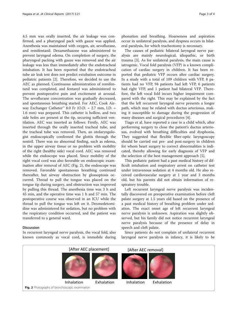
4.5 mm was orally inserted, the air leakage was con-firmed, and a pharyngeal pack with gauze was applied.Anesthesia was maintained with oxygen, air, sevoflurane,and remifentanil. Dexamethasone was administered toprevent laryngeal edema. On completion of surgery, thepharyngeal packing with gauze was removed and the airleakage was less than immediately after the endotrachealintubation. It has been reported that the endotrachealtube air leak test does not predict extubation outcome inpediatric patients [2]. Therefore, we decided to use theAEC as planned. Continuous administration of remifen-tanil was completed, and fentanyl was administered toprevent postoperative pain and excitement at arousal.The sevoflurane concentration was gradually decreased,and spontaneous breathing started. For AEC, Cook Air-way Exchanger Catheter™ 8.0 Fr (O.D. = 2.7 mm, I.D. =1.6 mm) was prepared. This catheter is hollow, and twoside holes are present at the tip, securing sufficient ven-tilation. AEC was inserted as follows: Firstly, AEC wasinserted through the orally inserted tracheal tube, andthe tracheal tube was removed. Then, an otolaryngolo-gist endoscopically confirmed the glottis through thenostril. There was no abnormal finding, such as edema,in the upper airway tissue or no problem with mobilityof the right (healthy side) vocal cord. AEC was removedwhile the endoscope was placed. Since mobility of theright vocal cord was also favorable on endoscopic exam-ination after removal of AEC (Fig. 2), the endoscope wasremoved. Favorable spontaneous breathing continuedthereafter, but airway obstruction by glossoptosis oc-curred. Thread to pull the tongue was placed on thetongue tip during surgery, and obstruction was improvedby pulling this thread. The anesthesia time was 3 h and45 min, and the operative time was 1 h and 57 min. Thepostoperative course was observed in an ICU while thethread to pull the tongue was left on it. Dexmedetomi-dine was administered for sedation, but no problem withthe respiratory condition occurred, and the patient wastransferred to a general ward.
DiscussionIn recurrent laryngeal nerve paralysis, the vocal fold, alsoknown commonly as vocal cord, is immobile during
Fig. 2 Photographs of bronchoscopic examination
phonation and breathing. Hoarseness and aspirationoccur in unilateral paralysis, and dyspnea occurs in bilat-eral paralysis, for which tracheotomy is necessary.The causes of pediatric bilateral laryngeal nerve par-
alysis are mainly neurological, idiopathic, or birthtrauma [3]. As for unilateral paralysis, the main cause isiatrogenic. Vocal fold paralysis (VFP) is a known compli-cation of cardiac surgery in children. It has been re-ported that pediatric VFP occurs after cardiac surgery.In a study with a total of 109 children with VFP, 8 pa-tients had no VFP, 94 patients had left VFP, 6 patientshad right VFP, and 1 patient had bilateral VFP. There-fore, the left vocal fold incurs higher impairment com-pared with the right. This may be explained by the factthat the left recurrent laryngeal nerve presents a longerpath, which may be related with ductus arteriosus, mak-ing it susceptible to damage during the progression ofmany diseases and surgical procedures [4].Tiago et al. have reported a case in a child which, after
performing surgery to close the patient’s ductus arterio-sus, evolved with breathing difficulties and dysphonia.They suggested that flexible fiber-optic laryngoscopyshould be carried out pre- and post-surgery in childrenfor whom heart surgery to correct abnormalities is indi-cated, thereby allowing for early diagnosis of VFP andthe selection of the best management approach [5].This pediatric patient had a past medical history of dif-
ficult intubation and respiratory arrest on catheter testunder intravenous sedation at 4 months old. He also re-ceived cardiovascular surgery at 1 year and 3 monthsold, but his parents did not obtain information of re-spiratory trouble.Left recurrent laryngeal nerve paralysis was inciden-
tally discovered on preoperative examination before cleftpalate surgery at 1.5 years old based on the presence ofa past medical history of breathing problem under sed-ation. The exact onset age of left recurrent laryngealnerve paralysis is unknown. Aspiration was slightly ob-served, but his family did not notice recurrent laryngealnerve paralysis because of the presence of delay inspeech and cleft palate.Since patients do not complain of unilateral recurrent
laryngeal nerve paralysis in infancy, it is likely to be

Nagata et al. JA Clinical Reports (2017) 3:21 Page 4 of 4
overlooked. In such cases, the possibility of recurrent la-ryngeal nerve paralysis should be taken into consider-ation from before surgery.In addition, in cleft palate closure surgery, not only
tracheal intubation but also pressing of the tracheal tubeby a Dingman mouth gag may damage the recurrent la-ryngeal nerve. In unilateral recurrent laryngeal nerveparalysis, such as the present case, if the nerve on thehealthy side is damaged, the condition becomes bilateralparalysis resulting in dyspnea. Thus, a careful extubationplan is needed.Various countermeasures against difficult intubation,
such as DAM, have been investigated, but no closeinvestigation was performed with regard to extuba-tion. DAS published the extubation guidelines in 2012[1]. Patients likely to develop complication accom-panying extubation are regarded as the high riskgroup, and the algorithm is comprised of fourprocedures.
Step 1: Plan extubation. Assess the airway and generalrisk factors because the ability to oxygenate is uncertainand reintubation may be difficult, and/or general riskfactors may be present. We assessed the risk factors atextubation in the present case, such as maxillofacialanomaly (first and second branchial arch syndrome),hemorrhage in the oral cavity, and stenosis of thepharyngeal cavity accompanying cleft palate closuresurgery, as well as aggravation of the respiratorycondition due to tracheal intubation-induced vocal cordedema and right (healthy side) recurrent laryngealnerve paralysis.Step 2: Prepare for extubation. DAM was also preparedand an anesthesiologist was added.Step 3: Perform extubation. There is a key question, “isit safe to remove the tube?” If the answer is “Yes”,proceed to “Awake extubation” or “Advancedtechniques.” Use of laryngeal mask, continuousadministration of remifentanil, and extubation withAEC are proposed. If the answer is “No,”“Postponement of extubation” timing and“Tracheotomy” are proposed. We chose “Advancedtechniques” and planned bronchoscopic examinationduring extubation to evaluate recurrent laryngeal nerveparalysis, referring to the DAS extubation guidelines.To evaluate mobility of the vocal cord, intratrachealoperation with an endoscope while inducing thespontaneous breathing and the swallowing reflex isnecessary, but these may cause breath holding andlaryngeal spasm. Thus, in the present case, the AECwas placed following the method proposed by theguidelines, and extubation was performed while anotolaryngologist endoscopically evaluated the movingglottis.
Step 4: Post-extubation care: recovery and follow-up.After extubation, the patient was managed in the ICU,and the systemic and respiratory conditions were favor-able in the postoperative course.
By devising extubation while conforming to the DASguidelines, extubation and airway management could besafely performed.
ConclusionsExtubation and airway management could be safely per-formed by devising extubation while conforming to theDAS guidelines.
AbbreviationsAEC: Airway exchange catheter; DAM: Difficult airway management;DAS: Difficult Airway Society; PAB: Pulmonary artery banding; VFP: Vocal foldparalysis
Authors’ contributionsMN, YS, and YH drafted the manuscript. YS and TN provided the detail ideafor the perioperative management and the anesthetic care. SH and CYperformed the anesthetic preoperative evaluation. JS and HK provided thepostoperative management in ICU. ON supervised the treatment and helpeddraft the manuscript. All authors read and approved the final manuscript.
Competing interestsThe authors declare that they have no competing interests.
Consent for publicationWritten informed consent was obtained from the legal representatives(parents) of the pediatric patient for the publication of this case report andany accompanying images. A copy of the written consent is available forreview by the Editor-in-Chief of this journal.
Ethics approval and consent to participateNot applicable.
Publisher’s NoteSpringer Nature remains neutral with regard to jurisdictional claims inpublished maps and institutional affiliations.
Received: 27 December 2016 Accepted: 26 April 2017
References1. Popat M, Mitchell V, Dravid R, Patel A, Swampillai C, Higgs A, et al. Difficult
Airway Society Guidelines for the management of tracheal extubation.Anaesthesia. 2012;67(3):318–40.
2. Wratney AT, Benjamin DK, Slonim AD, He J, Hamel DS, Cheifetz IM. Theendotracheal tube air leak test does not predict extubation outcome incritically ill pediatric patients. Pediatr Crit Care Med. 2008;9(5):490–6.
3. Daya H, Hosni A, Bejar-Solar I, Evans JN, Bailey CM. Pediatric vocal foldparalysis: a long-term retrospective study. Arch Otolaryngol Head Neck Surg.2000;126(1):21–5.
4. Truong MT, Messner AH, Kerschner JE, Scholes M, Wong-Dominguez J,Milczuk HA, et al. Pediatric vocal fold paralysis after cardiac surgery: rate ofrecovery and sequelae. Otolaryngol Head Neck Surg. 2007;137(5):780–4.
5. Tiago RS, Patrocínio SJ, dos Anjos PS, Ribeiro JT, Gil FM, Denunci FV. Vocalfold paralysis in children: diagnostic and management from a case report.Braz J Otorhinolaryngol. 2005;71(3):382–5.