A DEFINITIVE MANDIBULAR GUIDE FLANGE PROSTHESIS … Report.pdf · An interim removable partial...
6
Case Report A DEFINITIVE MANDIBULAR GUIDE FLANGE PROSTHESIS FOLLOWING HEMIMANDIBULECTOMY: A CLINICAL REPORT Puja Hazari, * Ajay V Gaikwad ** * Senior Lecturer, Department of Prosthodontics, RKDF Dental College, Bhopal, Madhya Pradesh, India ** Reader, Department of Prosthodontics, RKDF Dental College, Bhopal, Madhya Pradesh, India ________________________________________________________________________ ABSTRACT Loss of continuity of the mandible destroys the balance and symmetry of mandibular function, leading to altered mandibular movements, disfigurement, difficult in swallowing, impaired speech and articulation and deviation of the residual fragment towards the surgical side. Upon opening the mouth, this deviation increases, leading to the opening and closing in the angular pathway. A corrective device named "Guide Flange Prosthesis" is indicated to limit that clinical manifestation. A new possibility for treating hemimandibulectomy patients is using an only one device both for Physiotherapy and Mastication. KEYWORDS: Flange prosthesis; Mandibular defects; Mandibular resection; Refractory cast INTRODUCTION Neoplasms which are associated directly or indirectly with the mandible usually require surgical removal of the lesion and extensive resection of the bone. [1,2] Loss of the proprioceptive sense of occlusion following hemimandibulectomy leads to the uncoordinated, less precise movements of the mandible. [3] The basic rehabilitation objective is to re-educate mandibular muscles to re-establish an acceptable occlusal relationship (physio-therapeutic function) for residual mandible, so that patient could control adequately and repeatedly opening and closing mandibular movements. [4] Cantor and Curtis have classified the mandibular defects into 6 categories . [5] Class I: Mandibular resection involving alveolar defect with preservation of mandibular continuity (Fig. 1a). Class II: Resection defects involve loss of mandibular continuity distal to the canine area (Fig. 1b). Class III: Resection defect involves loss up to the mandibular midline region (Fig. 1c) Class IV: Resection defect involves the lateral aspect of the mandible, but are augmented to maintain pseudoarticulation of bone and soft tissues in the region of the ascending ramus (Fig. 1d). Class V: Resection defect involves the symphysis and parasymphysis region only, augmented to preserve bilateral temporomandibular articulations (Fig. 1e). Class VI: Similar to class V, except that the mandibular continuity is not restored (Fig. 1f). [6] Numerous prosthetic methods can be employed to reduce or minimize deviation and improve functions such as Maxillo-mandibular fixation, Implant supported prosthesis, Removable mandibular guide flange prosthesis and palatal based guidance restoration. [6,7] CASE REPORT A female patient, 36 years of age, visited the Department of prosthodontics, peoples college of dental sciences and research centre, Bhopal. The chief complaint was the unaesthetic appearance because of hemimandibulectomy, 2 years back due to squamous cell carcinoma. Intra oral examination revealed a complete absence of mandibular left segment. The defect crossed the midline and hence could be classified as Cantor and Curtis classification-III (Fig. 2a & Fig. 2 b). An interim removable partial denture followed by a definitive cast partial denture with a guiding flange appliance was planned for this patient. For the interim prosthesis primary impression was made in alginate, followed by a dual arch impression for final cast (Fig. 3a). Jaw relation was recorded (Fig. 3b). Teeth arrangement and try in was done (Fig. 3c) and the interim prosthesis was delivered after application of tissue conditioner to the intaglio surface (Fig. 3d & Fig. 3e). For the definitive prosthesis the diagnostic cast was surveyed (Fig. 4a). Mouth preparation Received : 02‑08‑13 Review completed : 10‑10‑13 Accepted : 24‑11‑13 IJOCR Jan - Mar 2014; Volume 2 Issue 1 39
Transcript of A DEFINITIVE MANDIBULAR GUIDE FLANGE PROSTHESIS … Report.pdf · An interim removable partial...
Case Report
A DEFINITIVE MANDIBULAR GUIDE FLANGE PROSTHESIS FOLLOWING HEMIMANDIBULECTOMY: A CLINICAL REPORT
Puja Hazari, * Ajay V Gaikwad **
* Senior Lecturer, Department of Prosthodontics, RKDF Dental College, Bhopal, Madhya Pradesh, India** Reader, Department of Prosthodontics, RKDF Dental College, Bhopal, Madhya Pradesh, India
________________________________________________________________________
ABSTRACT
Loss of continuity of the mandible destroys the
balance and symmetry of mandibular function,
leading to altered mandibular movements,
disfigurement, difficult in swallowing,
impaired speech and articulation and deviation
of the residual fragment towards the surgical
side. Upon opening the mouth, this deviation
increases, leading to the opening and closing in
the angular pathway. A corrective device
named "Guide Flange Prosthesis" is indicated
to limit that clinical manifestation. A new
possibility for treating hemimandibulectomy
patients is using an only one device both for
Physiotherapy and Mastication.
KEYWORDS: Flange prosthesis; Mandibular
defects; Mandibular resection; Refractory cast
INTRODUCTION
Neoplasms which are associated directly or
indirectly with the mandible usually require
surgical removal of the lesion and extensive
resection of the bone.[1,2]
Loss of the
proprioceptive sense of occlusion following
hemimandibulectomy leads to the uncoordinated,
less precise movements of the mandible.[3]
The
basic rehabilitation objective is to re-educate
mandibular muscles to re-establish an acceptable
occlusal relationship (physio-therapeutic
function) for residual mandible, so that patient
could control adequately and repeatedly opening
and closing mandibular movements.[4]
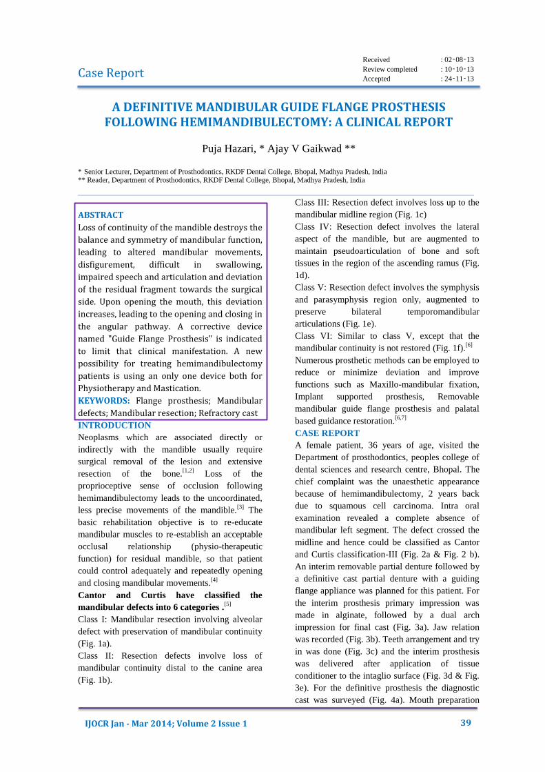
Cantor and Curtis have classified the
mandibular defects into 6 categories .[5]
Class I: Mandibular resection involving alveolar
defect with preservation of mandibular continuity
(Fig. 1a).
Class II: Resection defects involve loss of
mandibular continuity distal to the canine area
(Fig. 1b).
Class III: Resection defect involves loss up to the
mandibular midline region (Fig. 1c)
Class IV: Resection defect involves the lateral
aspect of the mandible, but are augmented to
maintain pseudoarticulation of bone and soft
tissues in the region of the ascending ramus (Fig.
1d).
Class V: Resection defect involves the symphysis
and parasymphysis region only, augmented to
preserve bilateral temporomandibular
articulations (Fig. 1e).
Class VI: Similar to class V, except that the
mandibular continuity is not restored (Fig. 1f).[6]
Numerous prosthetic methods can be employed to
reduce or minimize deviation and improve
functions such as Maxillo-mandibular fixation,
Implant supported prosthesis, Removable
mandibular guide flange prosthesis and palatal
based guidance restoration.[6,7]
CASE REPORT
A female patient, 36 years of age, visited the
Department of prosthodontics, peoples college of
dental sciences and research centre, Bhopal. The
chief complaint was the unaesthetic appearance
because of hemimandibulectomy, 2 years back
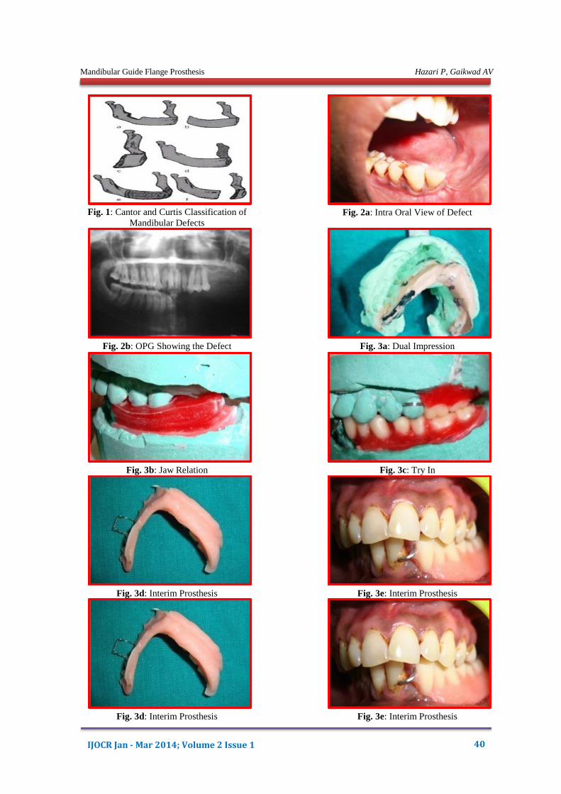
due to squamous cell carcinoma. Intra oral
examination revealed a complete absence of
mandibular left segment. The defect crossed the
midline and hence could be classified as Cantor
and Curtis classification-III (Fig. 2a & Fig. 2 b).
An interim removable partial denture followed by
a definitive cast partial denture with a guiding
flange appliance was planned for this patient. For
the interim prosthesis primary impression was
made in alginate, followed by a dual arch
impression for final cast (Fig. 3a). Jaw relation
was recorded (Fig. 3b). Teeth arrangement and try
in was done (Fig. 3c) and the interim prosthesis
was delivered after application of tissue
conditioner to the intaglio surface (Fig. 3d & Fig.
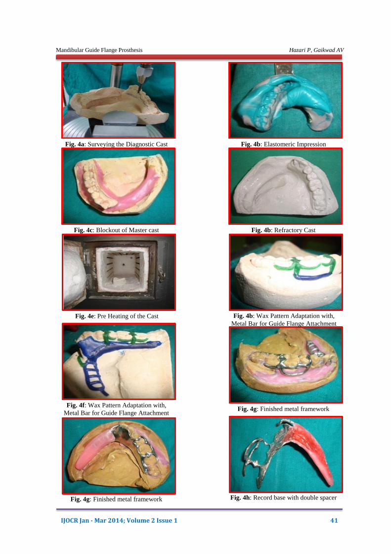
3e). For the definitive prosthesis the diagnostic
cast was surveyed (Fig. 4a). Mouth preparation
Received : 02‑08‑13
Review completed : 10‑10‑13
Accepted : 24‑11‑13
IJOCR Jan - Mar 2014; Volume 2 Issue 1 39
Mandibular Guide Flange Prosthesis Hazari P, Gaikwad AV
Fig. 1: Cantor and Curtis Classification of
Mandibular Defects
Fig. 2a: Intra Oral View of Defect
IJOCR Jan - Mar 2014; Volume 2 Issue 1 40
Fig. 2b: OPG Showing the Defect Fig. 3a: Dual Impression
Fig. 3b: Jaw Relation Fig. 3c: Try In
Fig. 3d: Interim Prosthesis Fig. 3e: Interim Prosthesis
Fig. 3d: Interim Prosthesis Fig. 3e: Interim Prosthesis
Mandibular Guide Flange Prosthesis Hazari P, Gaikwad AV
Fig. 4a: Surveying the Diagnostic Cast Fig. 4b: Elastomeric Impression
Fig. 4c: Blockout of Master cast Fig. 4b: Refractory Cast
Fig. 4e: Pre Heating of the Cast Fig. 4b: Wax Pattern Adaptation with,
Metal Bar for Guide Flange Attachment
Fig. 4f: Wax Pattern Adaptation with,
Metal Bar for Guide Flange Attachment Fig. 4g: Finished metal framework
Fig. 4g: Finished metal framework Fig. 4h: Record base with double spacer
IJOCR Jan - Mar 2014; Volume 2 Issue 1 41
Mandibular Guide Flange Prosthesis Hazari P, Gaikwad AV
Fig. 4i: Impression with Tissue
Conditioner
Fig. 4j: Impression with Light body
Fig. 4k: Sectioned Master cast Fig. 4l: Checking the Adaptation
Fig. 4m: Serrations for Better
Interlocking
Fig. 4n: Altered Cast
Fig. 4o: Neutral Zone Recorded Fig. 4p: Try-In
Fig. 4q: Articulation of Maxillary and
Mandibular Casts
Fig. 4r: Guide Flange Prosthesis
IJOCR Jan - Mar 2014; Volume 2 Issue 1 42

Mandibular Guide Flange Prosthesis Hazari P, Gaikwad AV
was done. Final impression was made in light
body and the master cast was poured (Fig. 4b).
Block out of the master cast was done (Fig. 4c).
The master cast was duplicated in agar and
refractory cast was poured (Fig. 4d). Preheating
of the cast was done in the furnace (Fig. 4e). The
preheated refractory cast was soaked in molten
bees wax for strengthening. The preheated
refractory cast was soaked in molten bees wax for
strengthening. On the refractory cast the wax
pattern was adapted. A metal bar was attached to
the pattern for the attachment of the guiding
flange (Fig. 4f). The sprues were attached, pattern
was invested and casting was done. Once the
casting was retrieved, it was finished and polished
(Fig. 4g). To obtain the altered cast a record base
was made on the minor connector with the double
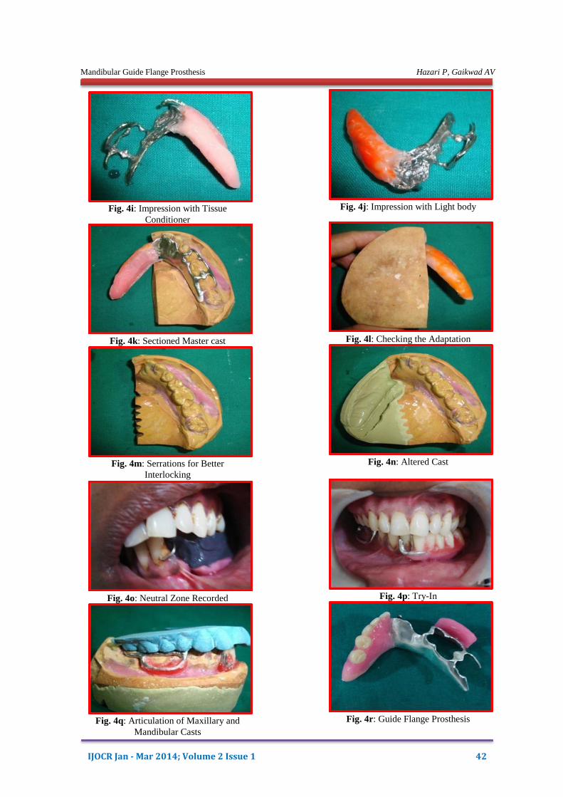
spacer (Fig. 4h). The final impression of the
edentulous, resected, soft tissue region was taken
by tissue conditioner (Fig. 4i). Over the tissue
conditioner a light body wash was taken (Fig. 4j).
The master cast was sectioned (Fig. 4k). The fit of
the prosthesis was rechecked (Fig. 4l). For better
interlocking of the two sections serrations were
made on the cast (Fig. 4m). An altered cast was
made (Fig. 4n). This cast was duplicated and a
permanent record base was made. Jaw relation
was recorded. Neutral zone was recorded to
enhance stability (Fig. 4o). Teeth arrangement
and try in was done (Fig. 4p). The maxillary cast
was placed on the mandibular cast (Fig. 4q). For
the fabrication of the guiding flange, wax
extension was made on the maxillary molars (Fig.
4r). The complete assembly was cured. Final
prosthesis was delivered (Fig. 4s). Marked
improvement was noted in esthetics of the patient
(Fig. 4t).
DISCUSSION
This clinical report illustrates the prosthetic
management of a patient who underwent
mandibular resection. The earlier the mandibular
guidance therapy is initiated in the course of
treatment the more successful the patients
definitive occlusal relationship is restored.[1]
The
basic rehabilitation objective in this case was to
re-educate mandibular muscles to re-establish an
acceptable occlusal relationship (physio-
therapeutic function) for residual mandible and to
restore the mastication.[8]
It also aided by
controlling adequate and repeated opening and
closing mandibular movements.[9]
The most
common treatment modalities for such patients
are maxilla mandibular fixation, implant
supported prosthesis, removable mandibular
guide flange prosthesis and palatal based
guidance restorations. An implant supported
prosthesis was not considered since no bone graft
was used.[6]
The mandibular guide flange device
for hemimandibulectomy patients presenting
good natural teeth on the residual mandible fits
Fig. 4s: Final Prosthesis in place
Fig. 4t: Comparison of patient face with Pre and Post Treatment
IJOCR Jan - Mar 2014; Volume 2 Issue 1 43

Mandibular Guide Flange Prosthesis Hazari P, Gaikwad AV
generally over that teeth (base-plate) and has a
guide plane (flange splint) extending into the
maxillary buccal vestibule, and which rides on the
buccal surfaces of several of the maxillary teeth:
this is the mechanical system preventing the
mandible from turning toward the resected
side.[6,9]
Every patient should maintain centric
occlusion for mastication, and this may be
accomplished by a guide plane.[10]
Using only
one prosthetic device as that proposed in this
work permits patients by guide flange to re-
educate mandibular muscles and removing the
same structure to eat. In this way patients are not
obliged to use one device for the physiotherapic
step and a second different device to eat. The
prosthetic device proposed was easy to make and
repair, comfortable to wear, also without guide
flange inserted, easy to clean and functional for
patient's disease so that expected results are
obtained.[4]
CONCLUSION
This sort of device permits the use the same
prosthesis both for eating and for mechanical
correction of mandibular deviation. A common
feature among all removable resection prosthesis
is that all framework designs should be dictated
by basic prosthodontic designs such as Broad
stress distribution, Cross arch stabilization. A
Rigid major connector stabilizing and retaining
components at locations within the arch to
minimize dislodgement and replacement of tooth
position that optimize prosthesis.
BIBLIOGRAPHY
1. Beumer J III, Curtis TA, Marunick MT.
Maxillofacial Rehabilitation. Prosthodontic
and Surgical consideration. St. Louis:
Ishiyaku. Euro America. 1996, p. 113-224.
2. Taylor td. Clinical maxillofacial prosthetics.
Quintessence Publication Co, Illunios :1997,
p. 171-188.
3. Sahu S. Mandibular Guide Flange Prosthesis
Following mandibular resection: A Clinical
Report. JCDR. 2010;4:3266-70.
4. Branchi R, Fancelli V, Desalvador A,
Durval E. A clinical report for corrective
mandibular movement therapy.
http://www.odontostudio.net/pub003n.htm
5. Fonsica RJ, Davis WH. Reconstruction
Preprosthetic oral and maxillofacial surgery,
2nd
Ed. WB Saunders Company: p. 1063-7.
6. Shetty P, Baliga M, Rodrigues S, Dixit S
Prosthetic management following
mandibular resection: A clinical report. J
Nepal Dent Assoc. 2009;10:1:57-60.
7. Penn M, Grossman Y, Shifman A, Shlomo
T. Implant retained feeding aid prosthesis
for a patient following total glossectomy and
laryngectomy: A clinical report. J Prosthet
Dent. 2007;97:261-5.
8. Desjardins RP. Occlusal considerations for
the partial mandibulectomy patient. J
Prosthet Dent. 1979;41:308-15.
9. Desjardins RP, Laney WR. Prosthetic
rehabilitation after cancer resection in the
head and neck. Surg Clin North Am.
1977;57:809-22.
10. Banerjee R, Banerjee S. Guiding Flange
Prosthesis for a Patient with a Hemi-
Mandibulectomy Defect: A clinical report.
JCDR. 2010;4:2347-2353.
Source of Support: Nil
Conflict of Interest: Nil
IJOCR Jan - Mar 2014; Volume 2 Issue 1 44