A cyclic di-GMP-specific phosphodiesterase, PdeB that...
36
1 A cyclic di-GMP-specific phosphodiesterase, PdeB that 1 regulates Shewanella oneidensis MR-1 motility and biofilm 2 formation 3 4 Lily Chao 1,‡ , Shauna Rakshe 2,‡ , Maija Leff 3 , and Alfred M. Spormann 1,2,3,* 5 Departments of Civil & Environmental Engineering 1 , Chemical Engineering 2 , and 6 Biological Sciences 3 , Stanford University, Stanford, California 94305-5429 7 8 9 Running title: Biofilm formation and sulfur assimilation in Shewanella oneidensis MR-1 10 ‡ These authors contributed equally to this work 11 *Corresponding author's address: 12 James H. Clark Center, E250 13 Stanford University 14 Stanford, CA 94305-5429 15 Phone: 650.723.3668, Fax: 650.724.4927, e-mail: [email protected] 16 17 18 Copyright © 2013, American Society for Microbiology. All Rights Reserved. J. Bacteriol. doi:10.1128/JB.00498-13 JB Accepts, published online ahead of print on 21 June 2013 on April 27, 2018 by guest http://jb.asm.org/ Downloaded from
Transcript of A cyclic di-GMP-specific phosphodiesterase, PdeB that...
1
A cyclic di-GMP-specific phosphodiesterase, PdeB that 1
regulates Shewanella oneidensis MR-1 motility and biofilm 2
formation 3
4
Lily Chao1,‡, Shauna Rakshe2,‡, Maija Leff3, and Alfred M. Spormann1,2,3,* 5
Departments of Civil & Environmental Engineering1, Chemical Engineering2, and 6
Biological Sciences3, Stanford University, Stanford, California 94305-5429 7
8
9
Running title: Biofilm formation and sulfur assimilation in Shewanella oneidensis MR-1 10
‡These authors contributed equally to this work 11
*Corresponding author's address: 12
James H. Clark Center, E250 13
Stanford University 14
Stanford, CA 94305-5429 15
Phone: 650.723.3668, Fax: 650.724.4927, e-mail: [email protected] 16
17
18
Copyright © 2013, American Society for Microbiology. All Rights Reserved.J. Bacteriol. doi:10.1128/JB.00498-13 JB Accepts, published online ahead of print on 21 June 2013
on April 27, 2018 by guest
http://jb.asm.org/
Dow
nloaded from

2
ABSTRACT 19
The respiratorily versatile γ-proteobacterium, Shewanella oneidensis MR-1, forms 20
biofilms on mineral surfaces through a process controlled by the cyclic di-nucleotide 21
messenger c-di-GMP. Cellular concentrations of c-di-GMP are maintained by proteins 22
containing GGDEF and EAL domains, which encode diguanylate cyclases for c-di-GMP 23
synthesis and phosphodiesterases for c-di-GMP hydroysis, respectively. The S. 24
oneidensis MR-1 genome encodes several GGDEF- and EAL-domain proteins (50 and 25
31, respectively), with a significant fraction (~10) predicted to be multi-domain (e.g. 26
GGDEF-EAL) enzymes containing an additional Per-Arnt-Sim (PAS) sensor domain. 27
However, the biochemical activities and physiological functions of these multi-domain 28
enzymes remain largely unknown. Here, we present genetic and biochemical analyses of 29
a predicted PAS-GGDEF-EAL domain-containing protein, SO0437, named here PdeB. 30
A pdeB deletion mutant exhibited decreased swimming motility and increased biofilm 31
formation under rich growth medium conditions, which was consistent with an increase 32
in intracellular c-di-GMP. A mutation inactivating the EAL domain also produced 33
similar swimming and biofilm phenotypes indicating that the increase in c-di-GMP was 34
likely due to a loss in phosphodiesterase activity. Therefore, we also examined the 35
enzymatic activity of purified PdeB, and found that the protein exhibited 36
phosphodiesterase activity via the EAL domain. No diguanylate cyclase activity was 37
observed. In addition to the motility and biofilm phenotypes, transcriptional profiling by 38
DNA microarray analysis of biofilms of pdeB (in-frame deletion and EAL) mutant cells 39
revealed that expression of genes involved in sulfate uptake and assimilation were 40
repressed. Addition of sulfate to the growth medium resulted in significantly less motile 41
on April 27, 2018 by guest
http://jb.asm.org/
Dow
nloaded from

3
pdeB mutants. Together, these results indicate a link between c-di-GMP metabolism, S. 42
oneidensis MR-1 biofilm development, and sulfate uptake/assimilation. 43
44
on April 27, 2018 by guest
http://jb.asm.org/
Dow
nloaded from

4
INTRODUCTION 45
The bacterial second messenger cyclic di-GMP (c-di-GMP) has become the focus 46
of intense research for its role in controlling biofilm formation, motility, virulence, and 47
cell cycle progression (1-13). The modes of its regulatory activity are diverse, and this 48
regulation can occur at the transcriptional (e.g. FleQ (14)), posttranscriptional (e.g. 49
GEMM riboswitch (15)), or posttranslational (e.g. cellulose synthase (16-18)) level, via 50
binding to specific effector proteins whose (regulatory) activity is modulated by c-di-51
GMP. Thus far, most physiological processes that have been demonstrated to be 52
regulated by c-di-GMP do not control essential cellular functions, and no control of 53
central metabolic processes has been reported. 54
C-di-GMP is synthesized by diguanylate cyclases (DGC), characterized by a 55
canonical GGDEF amino acid sequence motif, by condensation of two molecules of 56
GTP, and hydrolyzed by phosphodiesterases (PDE), characterized by conserved EAL or 57
HD-GYP amino acid sequence motifs, to pGpG or two molecules of GMP, respectively 58
(7, 19-22). Based on the amino acid sequences conserved in DGCs and PDEs, bacterial 59
genomes are predicted to have highly variable numbers of genes encoding these enzymes, 60
and their abundance appears to roughly correlate with genome size and environmental 61
versatility of the microorganism (23, 24). The complexity of the c-di-GMP regulatory 62
network is indicated further by the presence of multiple multi-domain proteins containing 63
both GGDEF and EAL or HD-GYP domains, as well as additional sensor domains such 64
as Che, PAS and NIT domains (23). The presence of the associated sensor domains 65
suggest that c-di-GMP signaling might target physiological processes other than those 66
currently known. When both GGDEF and EAL domains are present, one of the two 67
on April 27, 2018 by guest
http://jb.asm.org/
Dow
nloaded from
5
domains is often either inactive, or acts as a regulatory domain that changes the activity 68
of the other domain upon allosteric activation by GTP or c-di- GMP (6, 25). For example, 69
the GGDEF domain of PdeA from Caulobacter crescentus binds GTP and allosterically 70
activates the EAL domain; the GGDEF domain of in this protein has the slightly altered 71
amino acid sequence GEDEF and is incapable of catalyzing the formation of c-di-GMP 72
(25). 73
Shewanella oneidensis MR-1 is a gram-negative and respiratorily versatile γ-74
proteobacterium that is of considerable interest for applications in bioremediation and 75
microbial fuel cells, partly because of its ability to form biofilms on mineral surfaces (26-76
28). Biofilm formation in S. oneidensis MR-1, which has been studied in detail, is also 77
controlled by c-di-GMP (29-31), but little is known about the metabolism and targets of 78
c-di-GMP in this microorganism in general. The S. oneidensis MR-1 genome is predicted 79
to encode several GGDEF- and EAL-domain proteins (50 and 31, respectively), with a 80
significant fraction predicted as multi-domain (e.g. GGDEF-EAL domain-containing) 81
enzymes. Ten of those multi-domain proteins contain additional Per-Arnt-Sim (PAS) 82
sensory domains, which are important signaling modules previously shown to respond to 83
changes in environmental or cellular cues including light, redox state, and oxygen in a 84
wide range of organisms. Alternatively, PAS domains may mediate protein-protein 85
interactions or bind other small ligands (32, 33). In this work, we describe the 86
characterization of the PAS-GGDEF-EAL protein SO0437, renamed PdeB for 87
Phosphodiesterase Biofilm protein. We demonstrate that PdeB is a c-di-GMP-88
hydrolyzing phosphodiesterase that affects S. oneidensis MR-1 biofilm formation and is 89
linked to the regulation of sulfate uptake and assimilation. 90
91
on April 27, 2018 by guest
http://jb.asm.org/
Dow
nloaded from

6
MATERIALS AND METHODS 92
Growth conditions and media. The strains used in this study are summarized in Table 93
1. Escherichia coli and S. oneidensis MR-1 strains were grown at 37ºC and 30ºC, 94
respectively. 95
Construction of the mutants was carried out in Luria-Bertani (LB) or minimal medium 96
(4M) [485 µM CaCl2 · 2H2O, 5 µM CoCl2, 0.2 µM CuSO4 · 5H2O, 57 µM H3BO3, 1.27 97
mM K2HPO4, 0.73 mM KH2PO4, 1.0 mM MgSO4 · 7H2O, 1.3 µM MnSO4, 67.2 µM 98
Na2EDTA, 3.9 µM Na2MoO4 · 2H2O, 1.5 µM Na2SeO4, 150 mM NaCl, 2 mM NaHCO3, 99
5 µM NiCl2 · 5H2O, 1 µM ZnSO4, 9 mM (NH4)2SO4, 20 mM lactate, and 5 mM HEPES 100
(pH 7.4)]. 101
Swim plate experiments were carried out in LB or lactate medium (LM) [0.02% 102
w/v yeast extract, 0.01% w/v peptone, 10 mM HEPES (pH 7.4), 10 mM NaHCO3, 0.5 103
mM lactate] plates solidified with 0.25% w/v agar. All swim plate experiments were 104
conducted in quadruplicate. 105
Flow chamber-grown biofilm experiments were carried out as previously 106
described (30) in LM medium. All biofilm characterizations were conducted in triplicate. 107
Samples collected for microarray and RT-qPCR analysis were collected from 108
flow chamber-grown biofilms (see “RNA purification and cDNA synthesis,” below). 109
E. coli cultures for protein expression were carried out in maximal induction 110
media (MIM) [3.2% tryptone, 2% yeast extract, 33.5 mM Na2HPO4 17.5 mM KH2HPO4, 111
5.6 mM NaCl, 18.7 mM NH4Cl, 1 mM MgSO4, 0.1 mM CaCl2]. 112
113
on April 27, 2018 by guest
http://jb.asm.org/
Dow
nloaded from

7
Strain construction in S. oneidensis MR-1. All genetic work was carried out according 114
to standard protocols. Kits for isolation and/or purification of DNA were obtained from 115
Promega, and enzymes were purchased from New England Biolabs (NEB). See Table 1 116
for strains used, Table S1 for plasmids used, and Table S2 for primers used in this study. 117
AS579 served as the parental strain for construction of the pdeB deletion mutant 118
(ΔpdeB). ΔpdeB was constructed as previously described (34). Briefly, the upstream and 119
downstream regions of the pdeB open reading frame were PCR-amplified from wild type 120
(WT, AS579) genomic DNA and subsequently joined using overlap extension PCR. The 121
fusion product was ligated into pDS3.0 via the SmaI restriction site and transformed into 122
E. coli S17-λpir. The resulting plasmid, pDS3.0-ΔpdeB, was also verified by sequencing. 123
Following confirmation, pDS3.0-ΔpdeB was transformed into AS579 through bi-124
parental mating on a LB agar plate. After 8 h incubation, the mating mix was 125
resuspended in 4M liquid media and subsequently plated on 4M agar plates containing 5 126
µg/mL gentamycin. Colonies were screened for integration of pDS3.0-ΔpdeB into the 127
chromosome using PCR primers flanking the recombination region. The strain with 128
pDS3.0-ΔpdeB integrated into the chromosome was grown in LB medium (without 129
NaCl), and then plated on LB (without NaCl) agar plates supplemented with 10% 130
sucrose. The resulting colonies were patched onto LB plates containing 10 µg/mL 131
gentamycin to confirm the loss of plasmid. The gene deletion was confirmed by PCR 132
and DNA sequencing. 133
To complement the mutant, the wild type gene was reintroduced at the 134
chromosomal pdeB locus by gene replacement as an exact restoration of the original 135
gene. This was done similarly to the method described above, except that the pdeB wild 136
on April 27, 2018 by guest
http://jb.asm.org/
Dow
nloaded from

8
type gene and its flanking regions were cloned into pDS132 resulting in pDS132-pdeB 137
(35). The mating was performed using E. coli strain WM3064 and the mating mix was 138
plated on LB plates containing 15 µg/mL chloramphenicol to select for integration of the 139
plasmid into the chromosome. 140
The pdeB EAL domain mutant (E634A), pdeBeal, was constructed by generating 141
the mutation in pDS132-pdeB via QuikChange site-directed mutagenesis (Agilent). The 142
mutated pdeB allele was then introduced into the ΔpdeB mutant at the pdeB locus as for 143
the complementation strain above. 144
For biofilm studies, the strains were labeled with constitutively expressed green 145
fluorescent protein (GFP), using the Tn7 delivery system as previously described (30). 146
Briefly, GFP-expressing strains were constructed by tri-parental mating of the S. 147
oneidensis MR-1 strain with AS262 and AS392 harboring the Tn7-egfp plasmid. The 148
resulting gentamycin-resistant strains exhibited similar growth and biofilm phenotypes to 149
the untagged strains. 150
151
Image acquisition and processing. Microscopic visualization of biofilms was performed 152
on an upright Zeiss LSM510 confocal laser scanning microscope (Carl Zeiss, Jena, 153
Germany) equipped with the following objectives: x10/0.3 Plan-Neofluar, x20/0.5W 154
Achroplan, and x40/1.2W C-Apochromat. Biofilm parameters, such as biofilm mass and 155
average biofilm thickness, were quantified with the COMSTAT program (36). Image 156
data obtained were further processed by using the IMARIS software package (Bitplane 157
AG, Zürich, Switzerland). 158
159
on April 27, 2018 by guest
http://jb.asm.org/
Dow
nloaded from

9
Expression and Purification of MBP−His6−Fusion Protein. The pLIC-HMK vector 160
(gift from J. Berger) was used to express PdeB protein (residues 260-856) and its EAL 161
domain mutant (E634A) form as fusion proteins with a N-terminal maltose-binding 162
protein (MBP) tag and a C-terminal His6 tag. Site-directed mutations in PdeB were 163
generated using QuikChange site-directed mutagenesis (Agilent). The sequence changes 164
were confirmed by DNA sequencing. 165
The pLIC-pdeB plasmid was transformed into E. coli Tuner(DE3)pLysS cells 166
(EMD Chemicals) for protein expression. A single colony was used to inoculate LB 167
medium containing 50 µg/mL kanamycin and then grown with shaking at 37 °C. After 168
overnight growth, two shaker flasks containing 1 L MIM medium containing 0.5% 169
glucose and 50 µg/mL kanamycin were inoculated with 10 mL of the overnight culture. 170
The culture was grown at 250 rpm at 37°C to an OD600 of 0.3. The cultures were then 171
cooled to 16°C and expression was induced by the addition of 0.5 mM isopropyl-β-d-172
thiogalactopyranoside (IPTG). The cultures were shaken for another 18 h before the cells 173
were harvested by centrifugation. The cells were then resuspended in buffer A [10 mL of 174
50 mM Na2HPO4 (pH 8.0), 300 mM NaCl, 5 mM β-mercaptoethanol, and 5% glycerol] 175
containing 100 µM phenylmethylsulfonyl fluoride (PMSF), 1 mM benzamidine, and 176
DNaseI before being stored at -80 °C. 177
Nickel and amylose affinity chromatography were used for protein purification. 178
The frozen cell suspensions were thawed, lysed with a French pressure cell press (SLM-179
AMINCO), and pelleted at 140000g for 45 min. After centrifugation, the supernatant was 180
applied to a 1 mL column of Ni-NTA Agarose resin (Qiagen) equilibrated with buffer A. 181
The column was washed with five column volumes of buffer A and one column volume 182
on April 27, 2018 by guest
http://jb.asm.org/
Dow
nloaded from

10
of buffer A containing 40 mM imidazole and 1 M NaCl. Bound protein was eluted with 183
500 mM imidazole in buffer A (buffer B). The eluted protein was then applied to a 1 mL 184
column of amylose resin (New England Biolabs) equilibrated with buffer B. The column 185
was washed with five column volumes of buffer B and five column volumes of buffer C 186
[10 mM MOPS (pH 7.6), 150 mM NaCl, 10 mM MgCl2, and 10% glycerol] containing 5 187
mM BME. Bound protein was eluted with 15 mM maltose in buffer C. The eluted 188
protein was concentrated to <1 mL using a Vivaspin-6 10K filter (Vivascience) before it 189
was buffer-exchanged with buffer C and 5 mM dithiothreitol (DTT) using a PD-10 190
column (GE Healthcare), and stored at −80 °C. Purity of the protein was assessed by 191
sodium dodecyl sulfate–polyacrylamide gel electrophoresis (SDS-PAGE). 192
193
Diguanylate cyclase and phosphodiesterase assays. Assays were carried out in buffer 194
C containing 1 mM DTT. Diguanylate cyclase (DGC) and phosphodiesterase (PDE) 195
activities were assessed by incubating enzyme with α-32P-labeled GTP ( [α-32P]GTP; 196
Perkin-Elmer) and α-32P-labeled c-di-GMP (see below for preparation), respectively, at 197
30°C. The reactions were quenched at various time points with 12.5 mM EDTA (pH 8.0) 198
and/or boiled for 2.5 min, and centrifuged before 1 µL of the supernatant was spotted 199
onto polyethyleneimine-cellulose thin-layer chromatography plates (PEI-TLC; Merck 200
KGaA, Darmstadt, Germany). The PEI-TLC plates were developed in a 1.5:1.0 mixture 201
of 1.5 M KH2PO4 (pH 3.6) and 4.0 M (NH4)2SO4 (pH 3.6), air-dried, exposed to a 202
PhosphorImager screen (GE Healthcare), and scanned on a Typhoon Imager (GE 203
Healthcare). Under these conditions pGpG migrates with Rf = 0.34 and c-di-GMP with Rf 204
on April 27, 2018 by guest
http://jb.asm.org/
Dow
nloaded from

11
= 0.18. The spot intensities were quantified using ImageQuant version 5.2 (Molecular 205
Dynamics). 206
Radioactively labeled c-di-GMP was enzymatically synthesized from [α-207
32P]GTP using the TM1788 DGC from T. maritima (tDGC), which was expressed and 208
purified as previously described (37). Three-hour reactions incubated at 30°C were 209
boiled for 5 min and centrifuged to remove the precipitated tDGC, and the supernatant 210
was applied to a 10K Ultrafree 0.5 mL filter (Millipore) to remove any leftover protein. 211
The flow-through was then used in PDE activity assays as described above. 212
213
RNA purification and cDNA synthesis. Total RNA was purified from 24 h S. 214
oneidensis MR-1 biofilms using Trizol Reagent (Invitrogen). Cells from LM-grown flow 215
chamber biofilms were harvested and lysed in Trizol, and total RNA was isolated 216
according to the recommendations of the manufacturer. DNA was digested with 217
amplification grade DNaseI (Invitrogen). The digests were cleaned using the RNeasy 218
Mini Kit (Invitrogen) and the integrity of the RNA samples was evaluated using a 2100 219
BioAnalyzer (Agilent). cDNA was synthesized using the RevertAid First Strand cDNA 220
Synthesis Kit and random hexamer primer (Fermentas). 221
222
Gene expression profiling. Labeling of cDNA with cy3-dUTP or cy5-dUTP dyes 223
(Invitrogen) and microarray hybridization were performed according to the Agilent Gene 224
Expression oligo microarray protocol (Two Color Microarray-Based Gene Expression 225
Analysis, Agilent Technologies). A dye swap was performed to account for dye 226
incorporation artifacts. Competitive cDNA hybridization was performed on an Agilent 227
on April 27, 2018 by guest
http://jb.asm.org/
Dow
nloaded from

12
44,000-probe custom oligonucleotide DNA microarray. Each gene in the S. oneidensis 228
MR-1 genome was represented by three unique probes, for a total of 13,180 60-mer 229
probes representing 4,648 genes. Microarrays were scanned with a GenePix 400A 230
instrument (Axon Instruments), using the GENEPIX 5.0 software. Normalization was 231
executed with the Agilent feature extraction software. Microarray data were analyzed 232
using GeneSpring Software (Agilent) and filtering based on p<0.05 and fold change of 2 233
or greater. Data are based on one microarray experiment performed with three biological 234
replicates each of wild type (WT) and the ΔpdeB mutant. 235
236
Quantitative PCR. Quantitative analyses of transcripts were carried out with IQ 237
SYBR Green Supermix (Biorad) and iCycler iQ Real Time PCR Detection System 238
(Biorad). The genes examined and primers used are listed in Table S1. Samples were 239
analyzed in triplicate. Data analysis was performed as previously described (38). 240
SO0011 (gyrB) and SO1126 (dnaK) were used for normalization; data for gyrB is shown 241
(Table 3). 242
243
RESULTS & DISCUSSION 244
A gene search (Pfam entries: GGDEF, EAL, and PAS) against the S. oneidensis 245
MR-1 genome database (Joint Genome Institute Integrated Microbial Genomes, as of 246
May 2011) identified a gene, SO0437, which we named here pdeB, as encoding a 247
putative diguanylate cyclase/phosphodiesterase (DGC/PDE) with a PAS sensor domain 248
(Swiss-Prot; Figure 1A). Primary amino acid sequence analysis indicated that both 249
GGDEF and EAL domains appeared to be intact and that the protein also contained two 250
on April 27, 2018 by guest
http://jb.asm.org/
Dow
nloaded from

13
transmembrane segments (positions 7-29 and 230-252; TMHMM2) suggesting that PdeB 251
is a transmembrane protein. 252
In order to gain insights into the in vivo function of PdeB, an in-frame 253
chromosomal deletion of pdeB (ΔpdeB, AS979) was constructed. We observed no 254
difference in growth rate between the ΔpdeB and wild type (WT) strains in either rich or 255
minimal media (data not shown). However, the ΔpdeB mutant was observed to be less 256
motile than WT when grown on rich agar plates (Figure 2A and 2C), though not on 257
minimal media (Figure 2B). The swim diameter of the mutant was ~50% that of WT. 258
This motility phenotype was rescued when a wild type copy of the pdeB allele was 259
reintroduced at the chromosomal locus by gene replacement (pdeB+; Figure 2). We also 260
analyzed the ΔpdeB mutant under hydrodynamic biofilm conditions. As Figure 3 shows, 261
ΔpdeB formed thicker biofilms with approximately twice as much biomass as WT when 262
grown under semi-rich media conditions (Table 2). These observations suggest that a 263
pdeB deletion mutant results in an increase in intracellular c-di-GMP concentration; an 264
increase has been previously shown to result in thicker S. oneidensis MR-1 biofilms (29). 265
The motility and biofilm phenotypes of the ΔpdeB mutant suggested that there 266
was an increase in intracellular c-di-GMP concentration in S. oneidensis MR-1. In order 267
to determine the domain(s) of the PdeB protein that may contribute to this intracellular 268
increase in c-di-GMP, we examined whether PdeB showed PDE and/or DGC activity in 269
vitro and therefore whether the ΔpdeB mutant phenotype was due to a c-di-GMP-related 270
activity of PdeB. Briefly, a truncated form of PdeB (residues 260-856) containing the 271
PAS, GGDEF, and EAL domains but lacking the transmembrane segment was expressed 272
and purified with a N-terminal MBP tag as well as a C-terminal His6 tag (MBP-PdeBeal-273
on April 27, 2018 by guest
http://jb.asm.org/
Dow
nloaded from

14
His6 ) (Figure 1B). The MBP tag allowed for higher yield expression of soluble protein 274
and purification by amylose-affinity chromatography, whereas the C-terminal His6 tag 275
allowed for purification by nickel-affinity chromatography. All protein constructs made 276
in this study were purified to >95% homogeneity as assessed by SDS-PAGE (data not 277
shown). DGC and PDE activities were assayed by incubating the purified protein with 278
radioactively labeled substrates (e.g., α-32P- GTP, 32P-c-di-GMP) and then by separating 279
the reaction products on a TLC plate. Incubation of PdeB with α-32P-GTP did not result 280
in formation of a detectable (limit ~2x10-18 mol) TLC spot corresponding to c-di-GMP or 281
pGpG (data not shown) suggesting that the purified protein did not carry DGC activity. 282
In contrast, after a 30 min incubation when assaying for PDE activity using 32P-c-di-GMP 283
as substrate, >50% of the radioactivity was recovered at a spot corresponding to pGpG, 284
indicating that PdeB exhibited PDE activity in vitro (Figure 4). We estimate the specific 285
activity of this PdeB construct to be approximately 0.6 μmol c-di-GMP hydrolyzed per 286
mg protein per minute. Additionally, a PdeB protein carrying a E634A mutation in the 287
EAL domain, MBP-PdeBeal-His6, was also purified and assayed for PDE activity. As 288
shown in Figure 4, the alanine substitution of the glutamate residue in the EAL motif was 289
sufficient to eliminate the in vitro PDE activity of PdeB. Also with this protein, no DGC 290
activity was observed (data not shown). Together, these biochemical data demonstrate 291
that PdeB has PDE activity and likely acts as a phosphodiesterase in vivo. Although 292
amino acid sequence comparisons with other biochemically characterized DGCs suggest 293
that PdeB had an intact GGDEF domain, no in vitro DGC activity was observed even 294
when the EAL domain was inactivated. It is possible that the GGDEF domain of PdeB 295
has no catalytic but a regulatory role and is involved in the allosteric regulation of the 296
on April 27, 2018 by guest
http://jb.asm.org/
Dow
nloaded from

15
EAL domain. It is also possible that the removal of N-terminal transmembrane portion of 297
the protein and/or the addition of the N-terminal MBP-tag resulted in the loss or 298
inhibition of DGC activity. Further experiments would be required to resolve this issue. 299
In order to determine whether the observed PDE activity of PdeB was relevant to 300
the observed in vivo ΔpdeB phenotypes, we also constructed the corresponding EAL 301
mutant (E634A, pdeBeal, AS981 and AS984 for GFP-tagged construct) and compared its 302
swimming motility and biofilm phenotypes to ΔpdeB. The mutated allele was introduced 303
at the chromosomal locus of the ΔpdeB mutant through homologous recombination. The 304
pdeBeal mutant displayed both swimming motility and biofilm phenotypes that were 305
similar to those of the ΔpdeB mutant (Figures 2 and 3), demonstrating that all of these 306
observed phenotypes were consistent with PdeB acting as a phosphodiesterase in vivo. 307
In addition to the EAL and GGDEF domains, PdeB also contains a predicted PAS 308
sensor domain. Many GGDEF and EAL domains are associated with putative sensor 309
domains for perceiving various environmental signals (23). However, the 310
characterization of these sensor domains and their associated signals has yet to be 311
examined in S. oneidensis MR-1. Found in all domains of life, PAS domains are best 312
known for perceiving changes in light, oxygen concentration, and redox potential via a 313
linked flavin or heme cofactor (32, 39). However, sequence analyses of the PAS domain 314
associated with PdeB suggest that it contained no conserved residues for binding either a 315
flavin or heme cofactor and was therefore unlikely to be involved in sensing changes in 316
oxygen or redox (39). Indeed, no flavin or heme was found associated with the purified 317
PdeB protein and no differences in motility and biofilm phenotype were observed in the 318
pdeB mutant under aerobic and anaerobic growth conditions. 319
on April 27, 2018 by guest
http://jb.asm.org/
Dow
nloaded from

16
Therefore, in order to identify potential environmental signals that PdeB may 320
perceive, we also examined the global transcriptional profiles of ΔpdeB and wild type 321
cells in biofilms under semi-rich growth conditions by competitive hybridization of total 322
labeled RNA using custom-designed DNA microarrays. Briefly, total RNA was 323
extracted from 24 h biofilms of each strain, reverse transcribed, and subjected to 324
microarray analysis. The DNA microarray included three probes targeting each and 325
every gene of the S. oneidensis MR-1 genome. While most genes in the mutant did not 326
show a significant (FC > 2, p < 0.05) difference in expression, we found that a set of 327
genes related to sulfate uptake and assimilation was differentially expressed in ΔpdeB 328
relative to WT (Table S2). These genes include those that encode some of the subunits of 329
the major sulfate ABC transporter (cysT-2 and cysP) required for uptake of sulfate, as 330
well as several enzymes that catalyze the activation of sulfate and its assimilation into 331
cysteine (cysK and cysM, cysteine synthase subunit A and B; cysN and cysD, sulfate 332
adenyltransferase subunit 1 and 2) (Figure S1). 333
We also used quantitative PCR analyses to accurately determine that these genes 334
were down-regulated (~2-fold) in ΔpdeB versus the WT strain (Table 3). The expression 335
of these genes was also measured in the pdeBeal mutant. Interestingly, we observed 336
similar differences in expression between ΔpdeB and pdeBeal biofilms suggesting that c-337
di-GMP metabolism and sulfate uptake and assimilation may be linked (Figure 3 and 5; 338
Table 3) 339
Because we observed the down-regulation of genes involved in sulfate 340
transport/assimilation from cells harvested from ΔpdeB and pdeBeal mutant biofilms, we 341
hypothesized that PdeB activity may be controlled by sulfate. In order to test this, we 342
on April 27, 2018 by guest
http://jb.asm.org/
Dow
nloaded from

17
examined the swimming motility phenotype of the mutants when the growth medium was 343
amended with additional sulfate (e.g., Na2SO4 and NH4SO4). Sulfate was added to semi-344
rich media (LM) instead of limiting it in defined minimal media (4M) because no 345
detectable swimming motility phenotype was observed in 4M which contained ~9-10 346
mM sulfate (Figure 2B). Swimming motility was tested in the WT, the ΔpdeB mutant, 347
the mutant complemented with the WT allele (pdeB+), and the pdeBeal mutant. Sulfate 348
(40 mM Na2SO4 or NH4SO4) was added to the swim plates and the relative difference 349
between the WT and mutant motility was measured, where relative difference represents 350
the difference between the WT and mutant swimming diameters divided by the WT 351
swimming diameter. In the absence of additional sulfate, the swimming diameters of the 352
ΔpdeB mutant and the pdeBeal mutant were about 85% of that of WT (Figure 5A and 5C). 353
However, in the presence of additional sulfate, the swimming diameters of the ΔpdeB 354
mutant and the pdeBeal mutant were only about 50% of that of the WT (Figure 5B and 355
5C), indicating that the motility phenotype was exacerbated by the addition of sulfate. 356
Because the swimming motility phenotype was observed only in undefined growth media 357
(LM and LB), it is possible that sulfate in conjunction with other organic sulfur 358
compounds may have contributed to the swimming defect. Although the concentration of 359
sulfate used in this assay was relatively high and non-physiological, this concentration 360
did not affect growth rate (data not shown), indicating that the observed effect is not due 361
to an unspecific inhibition by sulfate. 362
In addition to the swimming motility assay, we also investigated the effect of 363
sulfate addition on biofilms of ΔpdeB and pdeBeal cells. Unfortunately, the results for 364
these experiments were inconclusive (data not shown). Additional salt in the growth 365
on April 27, 2018 by guest
http://jb.asm.org/
Dow
nloaded from

18
medium results in fragile S. oneidensis MR-1 biofilms, which easily fell apart under 366
hydrodynamic flow conditions, thereby making it difficult to obtain reproducible results 367
by COMSTAT analysis. 368
Thus far, it was unclear what the signal input (if any) into PdeB was, and to what 369
degree the PAS domain was involved in sensing that signal. As stated earlier, we did not 370
find evidence for the presence of a heme or flavin cofactor bound to the PAS domain of 371
PdeB during purification and the amino acid sequence of the PAS domain did not contain 372
predicted heme- or flavin-binding residues. Therefore, we tested whether addition of 373
sulfate (1:1 or 1:10 protein to Na2SO4 ratio) to the in vitro PDE assay changed the PDE 374
activity of PdeB; notably, no change in overall specific activity was observed (data not 375
shown). It is possible that ligand binding to the PAS domain requires the presence of the 376
N-terminus of PdeB that was not present in our purified construct (PdeB residues 260-377
856). It is also possible that another intermediate (e.g., from the sulfate assimilation 378
pathway) is responsible for regulating the enzymatic activity of PdeB. Alternatively, the 379
PAS domain may not bind a ligand but rather mediate protein-protein interactions (40). 380
In summary, we have identified a c-di-GMP-hydrolyzing enzyme, PdeB, whose 381
phosphodiesterase activity was involved in S. oneidensis MR-1 biofilm formation. 382
Furthermore, biochemical and genetic experiments also suggest that PdeB was linked to 383
the regulation of sulfate transport and assimilation. Together, these observations indicate 384
that c-di-GMP metabolism is not only linked to S. oneidensis MR-1 biofilm development 385
but also to sulfate transport and assimilation. However, the details of the signaling 386
pathways involved remain to be elucidated. To our knowledge, regulation of the sulfate 387
on April 27, 2018 by guest
http://jb.asm.org/
Dow
nloaded from

19
transport and assimilation genes is a previously unknown target of c-di-GMP signaling 388
and biofilm formation. 389
390
Acknowledgements 391
This work was funded by DOE Grant DE-FG02-07ER64386 to AMS through the 392
Shewanella Federation as well as a NSF Graduate Research Fellowship to SR. We thank 393
Ian Marshall for help with the design of the DNA microarray and with statistical analysis 394
of the COMSTAT biofilm data. We also thank the J. Berger laboratory at UC Berkeley 395
for the gift of the pLIC-HMK plasmid. 396
397
on April 27, 2018 by guest
http://jb.asm.org/
Dow
nloaded from

20
REFERENCES 398
1. Boehm, A., S. Steiner, F. Zaehringer, A. Casanova, F. Hamburger, D. Ritz, 399
W. Keck, M. Ackermann, T. Schirmer, and U. Jenal. 2009. Second messenger 400
signalling governs Escherichia coli biofilm induction upon ribosomal stress. Mol. 401
Microbiol. 72:1500-1516. 402
2. Cotter, P. A., and S. Stibitz. 2007. c-di-GMP-mediated regulation of virulence 403
and biofilm formation. Curr Opin Microbiol 10:17-23. 404
3. Dow, J. M., Y. Fouhy, J. F. Lucey, and R. P. Ryan. 2006. The HD-GYP 405
domain, cyclic di-GMP signaling, and bacterial virulence to plants. Mol. Plant. 406
Microbe Interact. 19:1378-1384. 407
4. Fineran, P. C., N. R. Williamson, K. S. Lilley, and G. P. Salmond. 2007. 408
Virulence and prodigiosin antibiotic biosynthesis in Serratia are regulated 409
pleiotropically by the GGDEF/EAL domain protein, PigX. J Bacteriol 189:7653-410
7662. 411
5. Jenal, U. 2004. Cyclic di-guanosine-monophosphate comes of age: a novel 412
secondary messenger involved in modulating cell surface structures in bacteria? 413
Curr Opin Microbiol 7:185-191. 414
6. Jenal, U., and J. Malone. 2006. Mechanisms of cyclic-di-GMP signaling in 415
bacteria. Annu. Rev. Genet. 40:385-407. 416
7. Paul, R., S. Weiser, N. C. Amiot, C. Chan, T. Schirmer, B. Giese, and U. 417
Jenal. 2004. Cell cycle-dependent dynamic localization of a bacterial response 418
regulator with a novel di-guanylate cyclase output domain. Genes Dev. 18:715-419
727. 420
on April 27, 2018 by guest
http://jb.asm.org/
Dow
nloaded from

21
8. Romling, U., and D. Amikam. 2006. Cyclic di-GMP as a second messenger. 421
Curr. Opin. Microbiol. 9:218-228. 422
9. Romling, U., M. Gomelsky, and M. Y. Galperin. 2005. C-di-GMP: the dawning 423
of a novel bacterial signalling system. Mol. Microbiol. 57:629-639. 424
10. Ryan, R. P., Y. Fouhy, J. F. Lucey, and J. M. Dow. 2006. Cyclic di-GMP 425
signaling in bacteria: recent advances and new puzzles. J Bacteriol 188:8327-426
8334. 427
11. Ryan, R. P., Y. Fouhy, J. F. Lucey, B. L. Jiang, Y. Q. He, J. X. Feng, J. L. 428
Tang, and J. M. Dow. 2007. Cyclic di-GMP signalling in the virulence and 429
environmental adaptation of Xanthomonas campestris. Mol Microbiol 63:429-430
442. 431
12. Tamayo, R., J. T. Pratt, and A. Camilli. 2007. Roles of cyclic diguanylate in 432
the regulation of bacterial pathogenesis. Annual review of microbiology 61:131-433
148. 434
13. Wolfe, A. J., and K. L. Visick. 2008. Get the message out: cyclic-di-GMP 435
regulates multiple levels of flagellum-based motility. J Bacteriol 190:463-475. 436
14. Hickman, J. W., and C. S. Harwood. 2008. Identification of FleQ from 437
Pseudomonas aeruginosa as a c-di-GMP-responsive transcription factor. Mol 438
Microbiol 69:376-389. 439
15. Sudarsan, N., E. R. Lee, Z. Weinberg, R. H. Moy, J. N. Kim, K. H. Link, and 440
R. R. Breaker. 2008. Riboswitches in eubacteria sense the second messenger 441
cyclic di-GMP. Science 321:411-413. 442
on April 27, 2018 by guest
http://jb.asm.org/
Dow
nloaded from

22
16. Amikam, D., and M. Y. Galperin. 2006. PilZ domain is part of the bacterial c-443
di-GMP binding protein. Bioinformatics 22:3-6. 444
17. Ross, P., H. Weinhouse, Y. Aloni, D. Michaeli, P. Weinberger-Ohana, R. 445
Mayer, S. Braun, E. de Vroom, G. A. van der Marel, J. H. van Boom, and M. 446
Benziman. 1987. Regulation of cellulose synthesis in Acetobacter xylinum by 447
cyclic diguanylic acid. Nature 325:279-281. 448
18. Weinhouse, H., S. Sapir, D. Amikam, Y. Shilo, G. Volman, P. Ohana, and M. 449
Benziman. 1997. c-di-GMP-binding protein, a new factor regulating cellulose 450
synthesis in Acetobacter xylinum. FEBS Lett. 416:207-211. 451
19. Ryjenkov, D. A., M. Tarutina, O. V. Moskvin, and M. Gomelsky. 2005. 452
Cyclic diguanylate is a ubiquitous signaling molecule in bacteria: insights into 453
biochemistry of the GGDEF protein domain. J. Bacteriol. 187:1792-1798. 454
20. Christen, M., B. Christen, M. Folcher, A. Schauerte, and U. Jenal. 2005. 455
Identification and characterization of a cyclic di-GMP-specific phosphodiesterase 456
and its allosteric control by GTP. J. Biol. Chem. 280:30829-30837. 457
21. Schmidt, A. J., D. A. Ryjenkov, and M. Gomelsky. 2005. The ubiquitous 458
protein domain EAL is a cyclic diguanylate-specific phosphodiesterase: 459
enzymatically active and inactive EAL domains. J. Bacteriol. 187:4774-4781. 460
22. Ryan, R. P., Y. Fouhy, J. F. Lucey, L. C. Crossman, S. Spiro, Y. W. He, L. H. 461
Zhang, S. Heeb, M. Camara, P. Williams, and J. M. Dow. 2006. Cell-cell 462
signaling in Xanthomonas campestris involves an HD-GYP domain protein that 463
functions in cyclic di-GMP turnover. Proc. Natl. Acad. Sci. U.S.A. 103:6712-464
6717. 465
on April 27, 2018 by guest
http://jb.asm.org/
Dow
nloaded from
23
23. Galperin, M. Y., A. N. Nikolskaya, and E. V. Koonin. 2001. Novel domains of 466
the prokaryotic two-component signal transduction systems. FEMS Microbiol. 467
Lett. 203:11-21. 468
24. Seshasayee, A. S., G. M. Fraser, and N. M. Luscombe. 2010. Comparative 469
genomics of cyclic-di-GMP signalling in bacteria: post-translational regulation 470
and catalytic activity. Nucleic Acids Res 38:5970-5981. 471
25. Christen, B., M. Christen, R. Paul, F. Schmid, M. Folcher, P. Jenoe, M. 472
Meuwly, and U. Jenal. 2006. Allosteric control of cyclic di-GMP signaling. J 473
Biol Chem 281:32015-32024. 474
26. Gorby, Y. A., S. Yanina, J. S. McLean, K. M. Rosso, D. Moyles, A. 475
Dohnalkova, T. J. Beveridge, I. S. Chang, B. H. Kim, K. S. Kim, D. E. Culley, 476
S. B. Reed, M. F. Romine, D. A. Saffarini, E. A. Hill, L. Shi, D. A. Elias, D. 477
W. Kennedy, G. Pinchuk, K. Watanabe, S. Ishii, B. Logan, K. H. Nealson, 478
and J. K. Fredrickson. 2006. Electrically conductive bacterial nanowires 479
produced by Shewanella oneidensis strain MR-1 and other microorganisms. Proc. 480
Natl. Acad. Sci. U.S.A. 103:11358-11363. 481
27. Hau, H. H., and J. A. Gralnick. 2007. Ecology and biotechnology of the genus 482
Shewanella. Annual review of microbiology 61:237-258. 483
28. Marsili, E., D. B. Baron, I. D. Shikhare, D. Coursolle, J. A. Gralnick, and D. 484
R. Bond. 2008. Shewanella secretes flavins that mediate extracellular electron 485
transfer. Proc Natl Acad Sci U S A 105:3968-3973. 486
29. Thormann, K. M., S. Duttler, R. M. Saville, M. Hyodo, S. Shukla, Y. 487
Hayakawa, and A. M. Spormann. 2006. Control of formation and cellular 488
on April 27, 2018 by guest
http://jb.asm.org/
Dow
nloaded from

24
detachment from Shewanella oneidensis MR-1 biofilms by cyclic di-GMP. J. 489
Bacteriol. 188:2681-2691. 490
30. Thormann, K. M., R. M. Saville, S. Shukla, D. A. Pelletier, and A. M. 491
Spormann. 2004. Initial Phases of biofilm formation in Shewanella oneidensis 492
MR-1. J. Bacteriol. 186:8096-8104. 493
31. Saville, R. M., N. Dieckmann, and A. M. Spormann. 2010. Spatiotemporal 494
activity of the mshA gene system in Shewanella oneidensis MR-1 biofilms. FEMS 495
Microbiol Lett 308:76-83. 496
32. Taylor, B. L., and I. B. Zhulin. 1999. PAS domains: internal sensors of oxygen, 497
redox potential, and light. Microbiol. Mol. Biol. Rev. 63:479-506. 498
33. Zhulin, I. B., B. L. Taylor, and R. Dixon. 1997. PAS domain S-boxes in 499
Archaea, Bacteria and sensors for oxygen and redox. Trends Biochem. Sci. 500
22:331-333. 501
34. Thormann, K. M., R. M. Saville, S. Shukla, and A. M. Spormann. 2005. 502
Induction of rapid detachment in Shewanella oneidensis MR-1 biofilms. J. 503
Bacteriol. 187:1014-1021. 504
35. Philippe, N., J. P. Alcaraz, E. Coursange, J. Geiselmann, and D. Schneider. 505
2004. Improvement of pCVD442, a suicide plasmid for gene allele exchange in 506
bacteria. Plasmid 51:246-255. 507
36. Heydorn, A., A. T. Nielsen, M. Hentzer, C. Sternberg, M. Givskov, B. K. 508
Ersboll, and S. Molin. 2000. Quantification of biofilm structures by the novel 509
computer program COMSTAT. Microbiology 146 ( Pt 10):2395-2407. 510
on April 27, 2018 by guest
http://jb.asm.org/
Dow
nloaded from

25
37. Rao, F., S. Pasunooti, Y. Ng, W. Zhuo, L. Lim, A. W. Liu, and Z. X. Liang. 511
2009. Enzymatic synthesis of c-di-GMP using a thermophilic diguanylate cyclase. 512
Anal. Biochem. 389:138-142. 513
38. Livak, K. J., and T. D. Schmittgen. 2001. Analysis of relative gene expression 514
data using real-time quantitative PCR and the 2(-Delta Delta C(T)) Method. 515
Methods 25:402-408. 516
39. Henry, J. T., and S. Crosson. 2011. Ligand-binding PAS domains in a genomic, 517
cellular, and structural context. Annual review of microbiology 65:261-286. 518
40. Anantharaman, V., E. V. Koonin, and L. Aravind. 2001. Regulatory potential, 519
phyletic distribution and evolution of ancient, intracellular small-molecule-520
binding domains. J. Mol. Biol. 307:1271-1292. 521
41. Miller, V. L., and J. J. Mekalanos. 1988. A novel suicide vector and its use in 522
construction of insertion mutations: osmoregulation of outer membrane proteins 523
and virulence determinants in Vibrio cholerae requires toxR. J. Bacteriol. 524
170:2575-2583. 525
42. Saltikov, C. W., and D. K. Newman. 2003. Genetic identification of a 526
respiratory arsenate reductase. Proc Natl Acad Sci U S A 100:10983-10988. 527
43. Simon, R., U. Priefer, and A. Puhler. 1983. A Broad Host Range Mobilization 528
System for In vivo Genetic Engineering - Transposon Mutagenesis in Gram-529
Negative Bacteria. Bio-Technol 1:784-791. 530
44. Muller, J., M. C. Miller, A. T. Nielsen, G. K. Schoolnik, and A. M. 531
Spormann. 2007. vpsA- and luxO-independent biofilms of Vibrio cholerae. 532
FEMS Microbiol Lett 275:199-206. 533
on April 27, 2018 by guest
http://jb.asm.org/
Dow
nloaded from

26
45. Myers, C. R., and K. H. Nealson. 1988. Bacterial manganese reduction and 534
growth with manganese oxide as the sole electron acceptor. Science 240:1319-535
1321. 536
537
538
on April 27, 2018 by guest
http://jb.asm.org/
Dow
nloaded from

27
FIGURES 539
540
Figure 1. Forms of PdeB used in this study. (A) Predicted domain structure of PdeB 541
(Swiss-Prot). The small black rectangles represent the predicted transmembrane regions 542
(THMM). (B) The PdeB(260-856) purified protein (MBP, maltose-binding protein). (C) 543
Mutant version of PdeB with PDE activity inactivated (PdeBeal, E634A). Black triangle 544
represents mutated amino acid residue. 545
546
547
548
PAS GGDEF EAL
amino acids 260-856
PAS GGDEF EALMBP
PdeB
PAS GGDEF EAL
His-tag
PdeBeal
E634A
(A)
(B)
(C)
on April 27, 2018 by guest
http://jb.asm.org/
Dow
nloaded from

28
549
Figure 2. Swimming motility patterns for pdeB mutants and wild type. Swimming 550
motility of WT (top) and ΔpdeB (bottom) cultures were assessed in (A) LB or (B) 4M + 551
20 mM fumarate. Plates contained 0.25% agar. LB cultures were incubated at 30°C for 552
48 hours and 4M cultures were incubated at room temperature for 48 hours, 553
anaerobically. (C) LM swim plate incubated at 30°C for 48 hours. 554
555
on April 27, 2018 by guest
http://jb.asm.org/
Dow
nloaded from

29
556
Figure 3. Biofilms of ΔpdeB and pdeBeal mutants. Biofilms were grown in 557
hydrodynamic flow chambers with LM media with for 15-16 h at 30°C before imaging. 558
Quantification of biofilms is shown in Table 2. 559
560
on April 27, 2018 by guest
http://jb.asm.org/
Dow
nloaded from

30
561
562
Figure 4. Cyclic-di-GMP-specific phosphodiesterase activity of PdeB. (A) P32-c-di-563
GMP was incubated with (1 µM) or without MBP-PdeB(260-856)-His6 at 30ºC for 30 564
min before 1 µL of sample was resolved on PEI-cellulose TLC plate. -, no enzyme; 565
PdeB, MBP-PdeB(260-856)-His6 ; PdeBeal , E634A mutant. 566
567
568
on April 27, 2018 by guest
http://jb.asm.org/
Dow
nloaded from
31
569
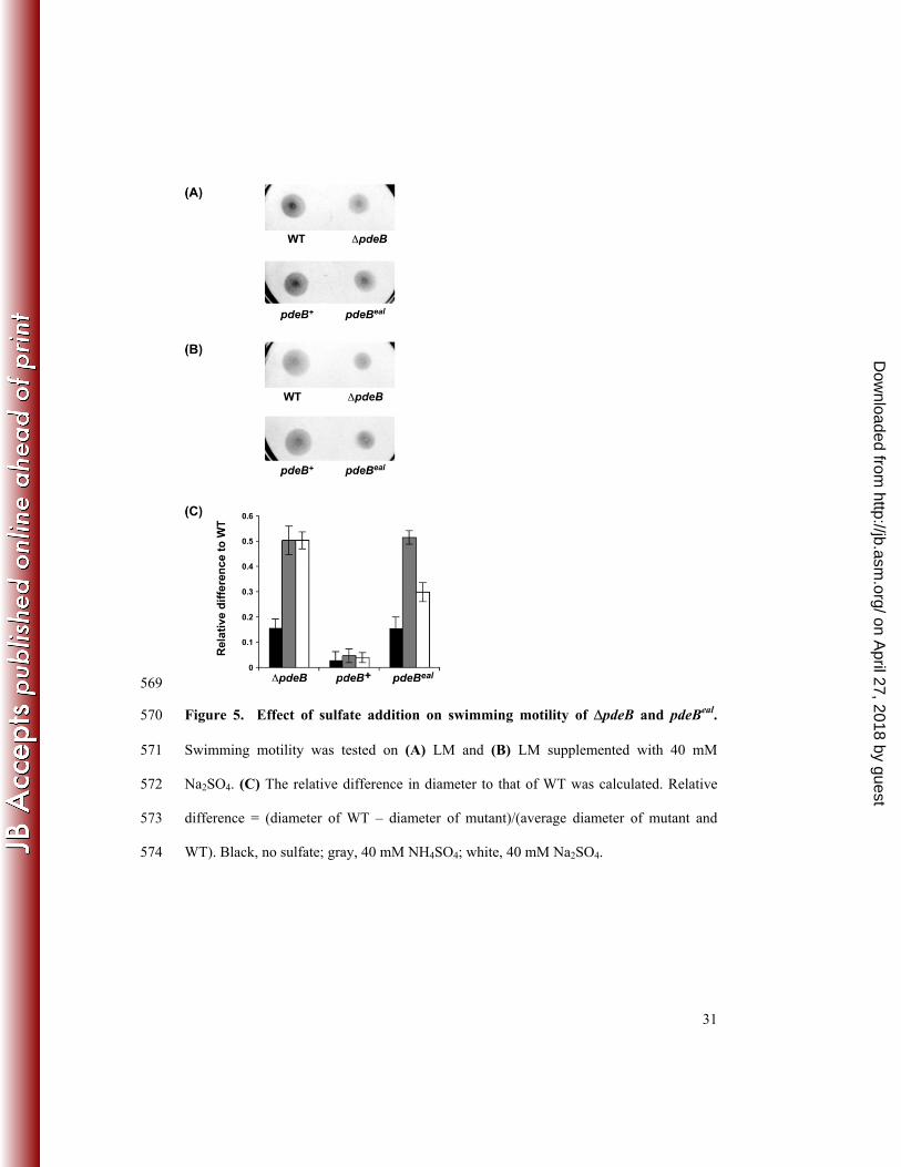
Figure 5. Effect of sulfate addition on swimming motility of ΔpdeB and pdeBeal. 570
Swimming motility was tested on (A) LM and (B) LM supplemented with 40 mM 571
Na2SO4. (C) The relative difference in diameter to that of WT was calculated. Relative 572
difference = (diameter of WT – diameter of mutant)/(average diameter of mutant and 573
WT). Black, no sulfate; gray, 40 mM NH4SO4; white, 40 mM Na2SO4. 574
on April 27, 2018 by guest
http://jb.asm.org/
Dow
nloaded from
32
TABLES 575
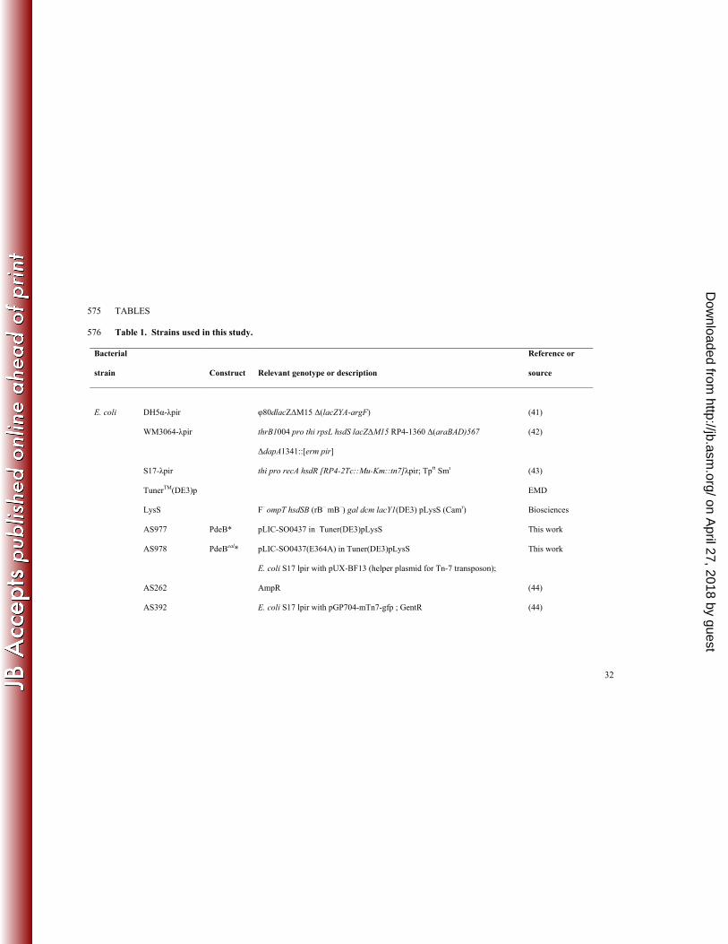
Table 1. Strains used in this study. 576
Bacterial
strain Construct Relevant genotype or description
Reference or
source
E. coli DH5α-λpir φ80dlacZΔM15 Δ(lacZYA-argF) (41)
WM3064-λpir thrB1004 pro thi rpsL hsdS lacZΔM15 RP4-1360 Δ(araBAD)567
ΔdapA1341::[erm pir]
(42)
S17-λpir thi pro recA hsdR [RP4-2Tc::Mu-Km::tn7]λpir; Tprr Smr (43)
TunerTM(DE3)p
LysS F– ompT hsdSB (rB– mB–) gal dcm lacY1(DE3) pLysS (Camr)
EMD
Biosciences
AS977 PdeB* pLIC-SO0437 in Tuner(DE3)pLysS This work
AS978 PdeBeal* pLIC-SO0437(E364A) in Tuner(DE3)pLysS This work
AS262
E. coli S17 lpir with pUX-BF13 (helper plasmid for Tn-7 transposon);
AmpR (44)
AS392 E. coli S17 lpir with pGP704-mTn7-gfp ; GentR (44)
on April 27, 2018 by guest
http://jb.asm.org/
Dow
nloaded from

33
S.
oneidensis AS579 WT Shewanella oneidensis MR-1 (PNNL strain) (45)
AS979 ΔpdeB AS579 ΔpdeB (markerless in-frame deletion) This work
AS980 pdeB+ AS579 ΔpdeB + pdeB+ (chromosomal replacement) This work
AS981 pdeBeal AS579 ΔpdeB + pdeBeal (E634A) This work
AS982 WT AS579 chromosomally tagged with gfp; Gentr This work
AS983 ΔpdeB AS979 chromosomally tagged with gfp; Gentr This work
AS984 pdeBeal AS981 chromosomally tagged with gfp; Gentr This work
*refers to protein 577
578
579
on April 27, 2018 by guest
http://jb.asm.org/
Dow
nloaded from

34
Table 2. COMSTAT analysis of S. oneidensis MR-1 biofilms. 580
Strain Biomass (µm3/µm2)a Thickness (µm) a
WT 7.15 ± 1.46 10.18 ± 2.43
ΔpdeB 13.81 ± 1.20 22.77 ± 3.13
pdeBeal 12.33 ± 1.59 21.35 ± 3.27
a Results were quantified from images taken from three flow chamber replicates. Error represents one standard deviation. 581
582
583
584
585
586
on April 27, 2018 by guest
http://jb.asm.org/
Dow
nloaded from

35
Table 3. Summary of quantitative PCR results.a 587
Locus Gene FC in ΔpdeB b FC in pdeBeal
SO2903 cysK 0.66 ± 0.03 0.65 ± 0.04
SO3598 cysM 0.52 ± 0.03 0.49 ± 0.03
SO3599 cysP 0.65 ± 0.05 0.47 ± 0.04
SO3727 cysD 0.39 ± 0.04 0.32 ± 0.03
SO4653 cysT-2 0.27 ± 0.02 0.27 ± 0.02
a Samples were grown in biofilms with LM media for approximately 24 h before harvest. 588
b Results represent average of triplicate samples. FC represents gene expression in mutant normalized to that of WT. Error represents 589
one standard deviation.590
on April 27, 2018 by guest
http://jb.asm.org/
Dow
nloaded from