A comparative perspective on lipid storage in animals · Journal of Cell Science A comparative...
12
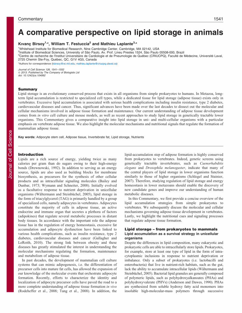
Journal of Cell Science A comparative perspective on lipid storage in animals Kıvanc ¸ Birsoy 1, *, William T. Festuccia 2 and Mathieu Laplante 3, * 1 Whitehead Institute for Biomedical Research, Nine Cambridge Center, Cambridge, MA 02142, USA 2 Institute of Biomedical Sciences, University of Sa ˜ o Paulo, Av. Prof. Lineu Prestes 1524, Sa ˜ o Paulo 05508-000, Brazil 3 Centre de recherche de l’Institut Universitaire de Cardiologie et de Pneumologie de Que ´ bec (CRIUCPQ), Faculte ´ de Me ´ decine, Universite ´ Laval, 2725 Chemin Ste-Foy, Que ´ bec, QC, G1V 4G5, Canada *Authors for correspondence ([email protected]; [email protected]) Journal of Cell Science 126, 1541–1552 ß 2013. Published by The Company of Biologists Ltd doi: 10.1242/jcs.104992 Summary Lipid storage is an evolutionary conserved process that exists in all organisms from simple prokaryotes to humans. In Metazoa, long- term lipid accumulation is restricted to specialized cell types, while a dedicated tissue for lipid storage (adipose tissue) exists only in vertebrates. Excessive lipid accumulation is associated with serious health complications including insulin resistance, type 2 diabetes, cardiovascular diseases and cancer. Thus, significant advances have been made over the last decades to dissect out the molecular and cellular mechanisms involved in adipose tissue formation and maintenance. Our current understanding of adipose tissue development comes from in vitro cell culture and mouse models, as well as recent approaches to study lipid storage in genetically tractable lower organisms. This Commentary gives a comparative insight into lipid storage in uni- and multi-cellular organisms with a particular emphasis on vertebrate adipose tissue. We also highlight the molecular mechanisms and nutritional signals that regulate the formation of mammalian adipose tissue. Key words: Adipocyte stem cell, Adipose tissue, Invertebrate fat, Lipid storage, Nutrients Introduction Lipids are a rich source of energy, yielding twice as many calories per gram than do sugars owing to their high-energy bonds (Drewnowski, 1992). In addition to serving as an energy source, lipids are also used as building blocks for membrane biosynthesis, as precursors for the synthesis of other cellular products and as intracellular signaling molecules (Bailey and Dunbar, 1973; Wymann and Schneiter, 2008). Initially evolved as a facultative response to nutrient deprivation in unicellular organisms (Wa ¨ltermann and Steinbu ¨chel, 2005), lipid storage in the form of triacylglycerol (TAG) is primarily handled by a group of specialized cells, namely adipocytes in vertebrates. Adipocytes constitute the majority of cells in adipose tissue, an active endocrine and immune organ that secretes a plethora of factors (adipokines) that regulate several metabolic processes in distant body tissues. In accordance with the important role the adipose tissue has in the regulation of energy homeostasis, excessive fat accumulation and adipocyte dysfunction have been linked to various health complications, such as insulin resistance, type 2 diabetes, cardiovascular diseases and cancer (Gallagher and LeRoith, 2010). The strong link between obesity and these diseases has greatly stimulated the interest in understanding the molecular mechanisms regulating the formation, maintenance and metabolism of adipose tissue. In past decades, the development of mammalian cell culture systems that can mimic adipogenesis, i.e. the differentiation of precursor cells into mature fat cells, has allowed the expansion of our knowledge of the molecular events that orchestrate adipocyte formation. Recently, efforts to characterize the identity and localization of adipocyte precursor cells have paved the road to a more complete understanding of adipose tissue formation in vivo (Rodeheffer et al., 2008; Tang et al., 2008). In addition, the lipid-accumulation step of adipose formation is highly conserved from prokaryotes to vertebrates. Indeed, genetic screens using genetically tractable invertebrates, such as Caenorhabditis elegans and Drosophila melanogaster, indicate that many of the central players of lipid storage in lower organisms function similarly to those of higher organisms (Schlegel and Stainier, 2007). Therefore, studying regulation of lipid storage and energy homeostasis in lower metazoans should enable the discovery of new candidate genes and improve our understanding of human metabolic diseases. In this Commentary, we first provide a concise overview of the lipid accumulation strategies from simple prokaryotes to vertebrates. We subsequently discuss the molecular and cellular mechanisms governing adipose tissue development in vertebrates. Lastly, we highlight the nutritional cues and signaling processes that regulate adipose tissue formation. Lipid storage – from prokaryotes to mammals Lipid accumulation as a survival strategy in unicellular organisms Despite the differences in lipid composition, many eukaryotic and prokaryotic cells are able to intracellularly store lipids. Prokaryotes, for example, store at least one type of lipid in the form of intra- cytoplasmic inclusions in response to nutrient deprivation or imbalance. Only a subset of prokaryotes (i.e. lactobacilli and enterobacteria) that live in nutrient-rich habitats, such as the gut, lack the ability to accumulate intracellular lipids (Wa ¨ltermann and Steinbu ¨chel, 2005). Bacterial lipid granules are generally composed of polymeric lipids, such as polyhydroxyalkanoates (PHAs) and polyhydroxyvalerate (PHVs) (Anderson and Dawes, 1990). PHAs are synthesized from soluble hydroxy fatty acid monomers into insoluble high-molecular-mass polymers through successive Commentary 1541
Transcript of A comparative perspective on lipid storage in animals · Journal of Cell Science A comparative...
Journ
alof
Cell
Scie
nce
A comparative perspective on lipid storage in animals
Kıvanc Birsoy1,*, William T. Festuccia2 and Mathieu Laplante3,*1Whitehead Institute for Biomedical Research, Nine Cambridge Center, Cambridge, MA 02142, USA2Institute of Biomedical Sciences, University of Sao Paulo, Av. Prof. Lineu Prestes 1524, Sao Paulo 05508-000, Brazil3Centre de recherche de l’Institut Universitaire de Cardiologie et de Pneumologie de Quebec (CRIUCPQ), Faculte de Medecine, Universite Laval,2725 Chemin Ste-Foy, Quebec, QC, G1V 4G5, Canada
*Authors for correspondence ([email protected]; [email protected])
Journal of Cell Science 126, 1541–1552� 2013. Published by The Company of Biologists Ltddoi: 10.1242/jcs.104992
SummaryLipid storage is an evolutionary conserved process that exists in all organisms from simple prokaryotes to humans. In Metazoa, long-term lipid accumulation is restricted to specialized cell types, while a dedicated tissue for lipid storage (adipose tissue) exists only in
vertebrates. Excessive lipid accumulation is associated with serious health complications including insulin resistance, type 2 diabetes,cardiovascular diseases and cancer. Thus, significant advances have been made over the last decades to dissect out the molecular andcellular mechanisms involved in adipose tissue formation and maintenance. Our current understanding of adipose tissue development
comes from in vitro cell culture and mouse models, as well as recent approaches to study lipid storage in genetically tractable lowerorganisms. This Commentary gives a comparative insight into lipid storage in uni- and multi-cellular organisms with a particularemphasis on vertebrate adipose tissue. We also highlight the molecular mechanisms and nutritional signals that regulate the formation of
mammalian adipose tissue.
Key words: Adipocyte stem cell, Adipose tissue, Invertebrate fat, Lipid storage, Nutrients
IntroductionLipids are a rich source of energy, yielding twice as many
calories per gram than do sugars owing to their high-energy
bonds (Drewnowski, 1992). In addition to serving as an energy
source, lipids are also used as building blocks for membrane
biosynthesis, as precursors for the synthesis of other cellular
products and as intracellular signaling molecules (Bailey and
Dunbar, 1973; Wymann and Schneiter, 2008). Initially evolved
as a facultative response to nutrient deprivation in unicellular
organisms (Waltermann and Steinbuchel, 2005), lipid storage in
the form of triacylglycerol (TAG) is primarily handled by a group
of specialized cells, namely adipocytes in vertebrates. Adipocytes
constitute the majority of cells in adipose tissue, an active
endocrine and immune organ that secretes a plethora of factors
(adipokines) that regulate several metabolic processes in distant
body tissues. In accordance with the important role the adipose
tissue has in the regulation of energy homeostasis, excessive fat
accumulation and adipocyte dysfunction have been linked to
various health complications, such as insulin resistance, type 2
diabetes, cardiovascular diseases and cancer (Gallagher and
LeRoith, 2010). The strong link between obesity and these
diseases has greatly stimulated the interest in understanding the
molecular mechanisms regulating the formation, maintenance
and metabolism of adipose tissue.
In past decades, the development of mammalian cell culture
systems that can mimic adipogenesis, i.e. the differentiation of
precursor cells into mature fat cells, has allowed the expansion of
our knowledge of the molecular events that orchestrate adipocyte
formation. Recently, efforts to characterize the identity and
localization of adipocyte precursor cells have paved the road to a
more complete understanding of adipose tissue formation in vivo
(Rodeheffer et al., 2008; Tang et al., 2008). In addition, the
lipid-accumulation step of adipose formation is highly conserved
from prokaryotes to vertebrates. Indeed, genetic screens using
genetically tractable invertebrates, such as Caenorhabditis
elegans and Drosophila melanogaster, indicate that many of
the central players of lipid storage in lower organisms function
similarly to those of higher organisms (Schlegel and Stainier,
2007). Therefore, studying regulation of lipid storage and energy
homeostasis in lower metazoans should enable the discovery of
new candidate genes and improve our understanding of human
metabolic diseases.
In this Commentary, we first provide a concise overview of the
lipid accumulation strategies from simple prokaryotes to
vertebrates. We subsequently discuss the molecular and cellular
mechanisms governing adipose tissue development in vertebrates.
Lastly, we highlight the nutritional cues and signaling processes
that regulate adipose tissue formation.
Lipid storage – from prokaryotes to mammalsLipid accumulation as a survival strategy in unicellular
organisms
Despite the differences in lipid composition, many eukaryotic and
prokaryotic cells are able to intracellularly store lipids. Prokaryotes,
for example, store at least one type of lipid in the form of intra-
cytoplasmic inclusions in response to nutrient deprivation or
imbalance. Only a subset of prokaryotes (i.e. lactobacilli and
enterobacteria) that live in nutrient-rich habitats, such as the gut,
lack the ability to accumulate intracellular lipids (Waltermann and
Steinbuchel, 2005). Bacterial lipid granules are generally composed
of polymeric lipids, such as polyhydroxyalkanoates (PHAs) and
polyhydroxyvalerate (PHVs) (Anderson and Dawes, 1990). PHAs
are synthesized from soluble hydroxy fatty acid monomers into
insoluble high-molecular-mass polymers through successive
Commentary 1541

Journ
alof
Cell
Scie
nce
elongation reactions catalyzed by a key enzyme only found inbacteria, PHA synthase (Sim et al., 1997). Phasins are the most
abundant proteins in these intracellular inclusions, contributing upto 5% of the total cellular protein in bacteria (Steinbuchel et al.,1995; Wieczorek et al., 1995). In prokaryotes, phasin expressionand lipid accumulation are tightly coupled and regulated by the
cellular concentration of PhaR, a transcriptional repressor that cansimultaneously bind to lipids and DNA elements (Maehara et al.,2002; Potter et al., 2002; York et al., 2002; Yamada et al., 2007).
Phasins stabilize lipid granules and prevent their fusion, a role thatlater in evolution is assumed by lipid droplet (LD)-associatedproteins in eukaryotes. The most abundant and conserved
eukaryotic LD proteins are those of the PAT family [perilipin,adipose differentiation-related protein (ADRP), tail-interactingprotein of 47 kDa (TIP47)] (Miura et al., 2002; Yamaguchi,2007). PAT-domain-containing proteins can be found early in
evolution in organisms as primitive as some fungal species andarthropods (i.e. D. melanogaster and Dictyostelium discoideum;Wang and St Leger, 2007; Bickel et al., 2009) (Box 1).
Although most prokaryotes store carbon in the form of PHAs,some rare prokaryotes (i.e. Mycobacterium, Rhodococcus andDietzia) can accumulate lipids in the form of TAGs (Waltermann
and Steinbuchel, 2005). Prokaryotic TAGs are synthesized by abifunctional enzyme that shares no identity to diacylglycerolacyltransferases 1 (DGAT1) and DGAT2, which catalyze thefinal and the dedicated enzymatic step in TAG synthesis in
eukaryotes (Table 1; Box 2) (Alvarez and Steinbuchel, 2002).TAG accumulation in these organisms is induced duringthe stationary phase upon nutrient restriction (Silva et al.,
2010) and is also exploited as a survival strategy in the courseof pathogenesis. For example, Mycobacterium tuberculosis
accumulates lipids during the dormant phase of tuberculosis, a
state in which the bacteria can live for long periods of time untilthe host immune system is weakened (Daniel et al., 2011).Bacteria lacking the ability to accumulate lipids are more
sensitive to antibiotic treatment, suggesting that inhibition oflipid accumulation could represent a possible strategy toovercome antibiotic resistance (Baek et al., 2011).
Similar to prokaryotes, simple eukaryotes also store fatty acids
under conditions of nutrient limitation, but only in TAG form.Eukaryotic LDs are composed of a hydrophobic core containingTAGs and/or sterol esters that are surrounded by a monolayer of
phospholipids and specialized proteins (Walther and Farese,2012). The composition and size of LDs (0.1 mm in yeast to over200 mm in white adipocytes) can vary according to the cell type
and the external nutritional cues (Table 1) (Walther and Farese,2009). Even though LDs are generally regarded as independentorganelles, it has been shown that they can be connected to theendoplasmic reticulum (ER) through a membrane bridge
(Jacquier et al., 2011). This is consistent with the widelyaccepted hypothesis that LDs are formed in ER and bud from it(Czabany et al., 2007), which is in contrast to the mechanism in
prokaryotes, where these droplets appear to be formed from theplasma membrane (Waltermann et al., 2005). Owing to thefunctional and structural interaction, membrane constituents
of LDs and ER can move bidirectionally between bothcompartments by diffusion (Jacquier et al., 2011). The size ofLDs is also dynamic and oscillates during cell growth as a
reflection of the balance between TAG synthesis and breakdown(Mullner and Daum, 2004). During proliferation of yeast, TAGsare hydrolyzed by lipases (Tgl3, Tgl4 and Tgl5) and the resulting
fatty acids are used for the synthesis of membrane lipids(Athenstaedt and Daum, 2005; Rajakumari et al., 2008). This
increase in TAG hydrolysis during cell proliferation is, at least inpart, promoted by the activation of TGL4 by the cell-cycle-related cyclin-dependent kinase 1 (Cdk1, also known as Cdc28)
(Kurat et al., 2009). In mammals, the lipases adipose triglyceridelipase (ATGL, also known as PNPLA2), hormone sensitive lipase(HSL, also known as LIPE) and monoglyceride lipase (MGL orMGLL) catalyze the sequential hydrolysis of TAG to
diacylglycerol (DAG), monoacylglycerol (MAG), and glyceroland fatty acids, respectively (Zechner et al., 2012). ATGL, whichwas discovered less than a decade ago (Zimmermann et al.,
2004), belongs to the patatin-domain-containing family ofproteins that are well conserved throughout evolution. ATGLorthologs are found in essentially all eukaryotic species,
including vertebrates, invertebrates, plants and fungi (Zechneret al., 2012). In accordance with such a high degree ofconservation, expression of murine ATGL in yeast that are
Box 1. Lipid droplet proteins
Similar to phasins in prokaryotes, eukaryotic LDs contain ‘lipid
droplet proteins’ that protect the core lipids from random
degradation. In most but not all yeast species (e.g. M.
anisopliae) (Wang and St Leger, 2007), homologs of classical
metazoan LD proteins, such as PAT family proteins (Perilipin,
ADRP and TIP47), have not yet been discovered. However,
several comprehensive yeast proteome studies have
characterized the LD proteome and identified proteins that are
similar to the mammalian counterparts and have roles in lipid
metabolism, protein transport and signaling processes
(Athenstaedt et al., 1999; Grillitsch et al., 2011). Even though
homologs of many LD proteins including seipin, leipin and caveolin
exist in worms, no perilipin homolog has been identified in
nematode species, suggesting a great structural and functional
divergence in these proteins in worms (Mak, 2012). Proteomics
studies of fly LDs have also identified LD-associated proteins that
are involved in lipid metabolism and transport, intracellular
trafficking and RNA metabolism (Beller et al., 2006).
Furthermore, insects express two PAT family proteins, lipid
storage droplet-1 (Lsd-1) and Lsd-2. Although these proteins are
localized on the LD surface, they have both opposing and redundant
functions (Bi et al., 2012). Lsd-1 modulates the accessibility of
lipases, such as Drosophila hormone sensitive lipase (Hsl), to the
LD and stabilizes the structure of the LD (Arrese et al., 2008; Beller
et al., 2010). Lsd-1 mutant flies are hyperphagic and obese (Beller
et al., 2010). Conversely, loss of Lsd-2 results in a decrease in fat
mass, whereas ectopic expression of Lsd-2 leads to a dose-
dependent increase in TAG storage (Gronke et al., 2003).
Reflecting the adaptation of multicellular organisms to nutritional
needs, vertebrate LD proteins diversified and are specialized to
specific tissues and organs. In addition to perilipin (PLIN1) and
adipose differentiation-related protein (ADRP or PLIN2), three
additional members of the PAT family of LD proteins, PLIN3–
PLIN5 (also known as TIP47, S3-12 and OXPAT, respectively), are
present in vertebrates. They are expressed in variety of organs with
different temporal and spatial patterns, reflecting their tissue-
specific roles (Dalen et al., 2004; Minnaard et al., 2009; Wang
and Sztalryd, 2011). In addition to members of the PAT family, the
repertoire of LD proteins in vertebrates also includes the cell-death-
inducing DNA fragmentation factor-like effector (CIDE) domain
family (CIDEA, CIDEB and CIDEC/FSP27), adding more
complexity to LD regulation (Wu et al., 2008).
Journal of Cell Science 126 (7)1542
Journ
alof
Cell
Scie
nce
deficient in Tgl4 restores their reduced TAG hydrolysis to normal
levels (Kurat et al., 2006).
Specification of lipid-storing cell types in metazoa
In invertebrates, specific cell types assume the role of long-term
storage of lipids. For example, C. elegans accumulate lipids mostly
in their intestinal, as well as skin-like, epidermal cells (Nelson et al.,
2002; Mullaney and Ashrafi, 2009; O’Rourke et al., 2009). The fat-
accumulating organ is much more developed and specialized in
arthropods, especially in insects. Here, a specialized organ, often
called adipose body or fat body, simultaneously exerts both liver and
adipose tissue functions, suggesting that a separation of these
metabolic functions to different organs occurred later during the
evolution of vertebrates (Arrese and Soulages, 2010). The insect fat
body coordinates metamorphosis and reproduction mainly by
storing and secreting compounds that regulate developmental
processes (Arrese and Soulages, 2010).
The investigation of the mechanisms regulating lipid
metabolism in invertebrates, such as C. elegans and D.
melanogaster, has been proven useful for the study of
adipocyte biology in higher organisms. Indeed, many conserved
proteins discovered in invertebrates have been shown to exert
similar functions in mammals. Adipose (adp) is one such
evolutionary conserved gene, encoding a protein with a WD40
domain that regulates chromatin dynamics and gene transcription
(Hader et al., 2003). adp was initially cloned from obese flies
(Doane, 1960), and loss of its function promotes TAG storage in
flies and worms, pointing to the crucial role this gene plays in
energy homeostasis (Hader et al., 2003; Suh et al., 2007). Similar
to in invertebrate models, mice that are heterozygous for Wdtc1,
the homolog of adp, are obese and insulin resistant, which
provides further support for a conserved role of this protein in
modulating lipid accumulation (Suh et al., 2007).
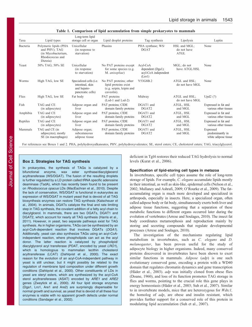
Table 1. Comparison of lipid accumulation from simple prokaryotes to mammals
Taxa Lipid typesLong-term lipid
storage cell or organ Lipid droplet proteins Tag synthesis Lipolysis Leptin
Bacteria Polymeric lipids (PHAand PHV); TAG(in Mycobacterium,Rhodococcus andDietzia)
Unicellular(in response tostarvation)
Phasins PHA synthase; WS/DGAT
HSL and MGL;do not haveATGL
None
Yeast 50% TAG; 50% SE Unicellular(in responseto starvation)
No PAT proteins exceptfor some species (e.g.M. anisopliae)
Acyl-CoAdependent (Dga1);acyl-CoA independent(Lro1)
MGL; do nothave ATGL/HSL
None
Worms High TAG, low SE Specialized cells (i.e.intestinal, skinand hepato-pancreatic cells)
No PAT proteins; otherlipid proteins exist(e.g. seipin, leipin andcaveolin).
Y53G8B.2 ATGL and HSL;do not have MGL
None
Flies High TAG, low SE Fat body PAT proteins(Lsd-1 and Lsd-2)
Midway ATGL and HSL;do not have MGL
Upd2 (?)
Fish TAG and CE(in adipocytes)
Adipose organ andliver
PAT proteins; CIDEdomain family proteins
DGAT1 andDGAT2
ATGL, HSLand MGL
Expressed in fat andvarious other tissues
Amphibia TAG and CE(in adipocytes)
Adipose organ andliver
PAT proteins; CIDEdomain family proteins
DGAT1 andDGAT2
ATGL, HSLand MGL
Expressed in fat andvarious other tissues
Reptiles TAG and CE(in adipocytes)
Adipose organ andliver
PAT proteins; CIDEdomain family proteins
DGAT1 andDGAT2
ATGL, HSLand MGL
Expressed in fat andvarious other tissues
Mammals TAG and CE (inadipocytes); mostlySE (in macrophages)
Adipose organ;subcutaneousadipose tissue
PAT proteins; CIDEdomain family proteins
DGAT1 andDGAT2
ATGL, HSLand MGL
Expressedpredominantly inwhite adipose tissue
For references see Boxes 1 and 2. PHA, polyhydroxyalkanoates, PHV, polyhydroxyvalerates; SE, sterol esters; CE, cholesterol esters; TAG, triacylglycerol.
Box 2. Strategies for TAG synthesis
In prokaryotes, the synthesis of TAGs is catalyzed by a
bifunctional enzyme, wax ester synthase/diacylglecerol
acyltransferase (WS/DGAT). The fusion of the resulting droplets
is further regulated by a LD protein called tRNA specific adenosine
deaminase (TadA), which has recently been found to be present
on Rhodococcus opacus LDs (MacEachran et al., 2010). Despite
the lack of conservation, WS/DGAT is functional in eukaryotes as
expression of WS/DGAT in mutant yeast that is devoid of all TAG
biosynthesis enzymes can restore TAG synthesis (Kalscheuer et
al., 2004). In animals, DGATs catalyze the final and rate limiting
step in TAG synthesis, the covalent addition of a fatty acyl chain to
diacylglcerol. In mammals, there are two DGATs, DGAT1 and
DGAT2, which account for nearly all TAG synthesis (Harris et al.,
2011). However, in yeast, two separate pathways exist for TAG
synthesis. As in higher organisms, TAGs can be synthesized by an
acyl-CoA-dependent reaction that involves DGATs (DGA1).
Additionally, yeast can also synthesize TAGs using an acyl-CoA-
independent reaction, where phospholipids can act as the acyl
donor. The latter reaction is catalyzed by phospholipid/
diacylglycerol acyl transferase (PDAT, encoded by yeast LRO1),
which is homologous to mammalian lecithin cholesterol
acyltransferase (LCAT) (Dahlqvist et al., 2000). The exact
reason for the evolution of an acyl-CoA-independent pathway in
yeast is still unclear, but it might possibly be related to the
regulation of membrane lipid composition under different growth
conditions (Dahlqvist et al., 2000). Other constituents of LDs in
yeast are steryl esters, which are synthesized by the acyl-CoA
sterol acyltransferases (ASAT) encoded by ARE1 and ARE2
genes (Zweytick et al., 2000). All four lipid storage enzymes
(Dga1, Lro1, Are1 and Are2) are surprisingly dispensable for
normal growth and survival, as yeast that is devoid of all these four
enzymes is viable with no apparent growth defects under normal
conditions (Sandager et al., 2002).
Lipid storage in animals 1543

Journ
alof
Cell
Scie
nce
Among the advantages of using invertebrate models to studylipid metabolism are their convenience and feasibility for genetic
and RNA interference (RNAi) screens. Vital dye staining ofneutral lipids and buoyancy-based screening methods have beenextremely useful to identify genes that affect lipid storage inworms and flies (Ashrafi et al., 2003; Pospisilik et al., 2010; Reis
et al., 2010). Such studies identified many genes that are relatedto lipid metabolism, feeding behavior, nutrient sensing and LDformation. RNAi screens in flies suggest that ,1.5% of the fly
genome contributes to LD formation and function (Guo et al.,2008). One gene found in these screens is FLD1, the yeasthomolog of human seipin (Fei et al., 2008). Seipin loss is
associated with congenital lipodystrophy in humans (Magre et al.,2001), a phenotype that might be related to its role inadipogenesis, and LD formation and maintenance (Chen et al.,2009; Chen et al., 2012). Other examples include genes encoding
hedgehog, sterol regulatory element binding protein-1 (SREBP1)and CCAAT-enhancer-binding protein (C/EBP), which havefunctionally similar roles in mammals (McKay et al., 2003; Yang
et al., 2006; Pospisilik et al., 2010). Altogether, these resultsdemonstrate that despite seemingly different forms of fat storage,invertebrate models are still instrumental in the identification of
pathways that regulate fat accumulation in higher organisms andthat many genes that are related to lipid metabolism and storageare functionally conserved throughout evolution.
Starting with primitive vertebrates (jawless vertebrates such aslampreys), lipid-storing cells evolved into a tissue that hasdistinct functions and a specialized location (Gelman et al.,2009). In fish, amphibian and reptiles, adipose tissue is mainly
found in intra-abdominal regions and subcutaneous fat tissue ismostly non-existent. This intra-abdominal location of fat isconsistent with the hypothesis that adipose tissue should be at the
center of gravity, since it is highly plastic and changes its massrapidly (Pond, 1992). Furthermore, adipose tissue in lowerexothermic animals is usually small because the liver performs
the major lipid-storing function. Interestingly, in these animals,the liver secretes many of the mammalian orthologs of theadipokines, such as adiponectin and leptin (Huising et al., 2006;Nishio et al., 2008; Pfundt et al., 2009).
In mammals, adipose tissue has developed to its most evolvedand complex form, with a concomitant decrease in hepatic lipidstores. Unlike exothermic vertebrates and invertebrates,
mammalian adipose tissue is widely distributed throughout thebody, although it is not clear why such a wide fat distribution hasbeen selected by evolution. Even though the presence of fat
depots in sub-dermal and subcutaneous areas appear to providethermal insulation, the observation that there is very littledifference in fat distribution between mammals living in tropical
versus arctic regions is not concordant with this hypothesis(Pond, 1992). Other selective pressures, such as sexual selectionand immune function of adipose tissue, might be importantfactors driving fat distribution in mammals. Sex hormones indeed
affect fat distribution, as evidenced by the increase insubcutaneous adipose tissue in males treated with estrogen(Elbers et al., 1999).
Body fat distribution is a heritable trait in mammals,suggesting that different developmental genetic programs mightexist for different fat depots (Bouchard and Tremblay, 1997;
Nelson et al., 2000; Baker et al., 2005). Indeed, each adiposedepot has a unique gene expression signature during development(Linder et al., 2004; Hishikawa et al., 2005). These signatures are
retained when progenitors from different depots are cultured
in vitro, suggesting an intrinsic program rather than a niche-dependent effect (Gesta et al., 2006; Macotela et al., 2012).For example, patterning genes, including a number of
homeobox genes (Hoxa5, Hoxc8 and Hoxc9) and Tbx15 haveroles in the patterning of the adipose depot specific program(Gesta et al., 2006). Furthermore, different fat depots responddifferently to nutritional status. In obesity, subcutaneous
adipose tissue expands through hyperplasia, and adipocyteprogenitors are more abundant, in contrast to visceral fat,which grows mainly by hypertrophy (Joe et al., 2009). Taken
together, these findings suggest that distinct molecularand cellular programs have evolved to determine the fatdistribution in mammals.
Adipose tissue as an active endocrine and immune organ
In addition to its energy-storing role, adipose tissue is an activeendocrine and immune organ. Adipocytes secrete a number of
adipokines that by acting on the central nervous system andperipheral tissues regulate adiposity, glucose homeostasis, foodintake, blood pressure, fibrinolysis, inflammation, lipidmetabolism and angiogenesis (Alvarez-Llamas et al., 2007).
Besides proteins, adipocytes also secrete lipids (lipokines) thatcan communicate with distant organs to maintain systemicmetabolic homeostasis (Cao et al., 2008).
Leptin is a major adipokine that is primarily produced bymature fat cells and whose expression is highly correlated withthe degree of adiposity (Maffei et al., 1995; Birsoy et al., 2008b).
Increase in fat mass in mammals causes an increase in the amountof leptin, which provides an afferent signal to the hypothalamusto decrease food intake and increase energy expenditure, thuskeeping the body weight stable (Friedman and Halaas, 1998).
Even though leptin is also expressed in other vertebrates, itsadipose-specific expression and primary role as the pivotalregulator of energy homeostasis appears to be specific to
mammals (Table 1). Indeed, in contrast to its adipose-restrictedexpression in mammals, leptin is expressed in several tissues infish, including in liver, the biliary system and intestine (Pfundt
et al., 2009; Russo et al., 2011; Tinoco et al., 2012). In addition,the anorexigenic effect of leptin appears to be only partiallyconserved in frogs, rainbow trout, carp and lizards (Niewiarowskiet al., 2000; Crespi and Denver, 2006; Murashita et al., 2008; Li
et al., 2010). Recombinant leptin also affects metamorphosis,suggesting a pleiotropic role for leptin in the early developmentof exothermic vertebrates (Crespi and Denver, 2006). Analogous
to the endocrine function of adipose tissue, a similar energyhomeostasis circuit is believed to be present in lower organisms.Recently, Unpaired 2 (Upd2), a cytokine produced by fat body,
has been shown to exert leptin-like effects in flies throughGABAergic neurons (Rajan and Perrimon, 2012). NeuropeptideY, dopamine and serotonin are all present in flies and worms, and
modulate feeding behaviors, further confirming the existence ofan energy homeostasis circuitry in lower organisms (de Bono andBargmann, 1998; Srinivasan et al., 2008; Hong et al., 2012).
Adipose tissue also acts as a systemic immune organ and
displays significant intrinsic inflammatory properties. Adipocytesreact to infections through the activation of multipleinflammatory signal transduction cascades, and the secretion of
potent inflammatory cytokines, such as interleukin 6 (IL6) andtumor necrosis factor a (TNFa) (Hotamisligil et al., 1993; Wellenand Hotamisligil, 2003). Interestingly, either bacterial infections
Journal of Cell Science 126 (7)1544

Journ
alof
Cell
Scie
nce
or exposure to lipopolysaccharide (LPS) and/or cytokines can
promote adipose tissue lipolysis and reduce TAG uptake by
adipocytes (Feingold et al., 1994; Penfornis and Marette, 2005;
Zu et al., 2009). Such a lipolytic response to bacterial infections
is also present in flies (Dionne et al., 2006). The rise in
circulating levels of lipids following infection is thought to
support the immune system by facilitating energy production and
by neutralizing infectious agents. The intricate balance between
metabolic and immune pathways is deregulated in obesity, as an
increase in adipose tissue mass results in systemic inflammation
and insulin resistance (Hotamisligil, 2010). Adipose tissue
inflammation in obesity mimics the ‘danger signals’ that arise
from bacterial infections and this condition promotes the
secretion of several cytokines that produce an immune reaction
known to cause hyperglycemia and hypertriglyceridemia (Wellen
and Hotamisligil, 2003). This immune function of adipose tissue
is reminiscent of the immune properties of insect fat body, which
expresses the pathogen pattern recognition Toll-like receptors
(TLRs) that are required to induce the secretion of antimicrobial
peptides upon exposure to microorganisms (Manfruelli et al.,
1999; Jang et al., 2006).
Fat tissue development and nutritionalregulationFormation of a fat cell in vertebrates
Adipocyte formation occurs through two stages: (1) the
commitment of stem cells into adipocyte precursors and (2) the
terminal differentiation of preadipocytes into mature fat cells. Most
of our knowledge of the formation of vertebrate fat cells comes from
in vitro studies using embryonic stem cells (ESCs), mesenchymal
stem cells (MSCs) and 3T3L1 fibroblasts (preadipocytes).
Additionally, differentiation of fish and carp preadipocytes can be
induced with a similar generic differentiation cocktail to that used
for mammalian preadipocyte differentiation (Vegusdal et al., 2003;
Li, 2012) Although these in vitro models do not completely
recapitulate in vivo adipogenesis, these systems have allowed the
identification of key regulators of the adipocyte differentiation
process such as bone morphogenetic protein (BMP) 2 and 4 (Bowers
et al., 2006; Huang et al., 2009), Wnt (Ross et al., 2000; Bennett
et al., 2002), transforming growth factor b (TGFb) (Ignotz and
Massague, 1985), fibroblast growth factor (FGF) 1 and 2 (Widberg
et al., 2009; Xiao et al., 2010) and retinoblastoma protein (Rb) (Calo
et al., 2010) in the early steps of adipocyte commitment (Fig. 1A;
A
B
Day 8DaDaDDDDaDaDaDaDaDaDaDaDaDaDaaaaaaDaDaDDDDaDaDaDDDDaDaDaDaaDDaDaDaDDaDaDaDaDaDaDaDaDDaDaaDDaDaaDaaDDaDaaaaDaaaaaaDaDaaaaaaaaDaDaaaaaaaDaDaDaaaaaaaaaaaaaDaaaaDaaDDaaDDDDDaaaaaaaDDayyyyyyyyy yy yyyyyyyyy yyyyyyyy yy yyyyy yyyyyyyyyyyyyyyyyyy y yyyyyyyyy yyyyyyyyyyyyy yyyyyyyyyyyy yyyyyyyyyyy 8888888888888888888888888888888888888888888888888888888888888888
NucleusLipids
InsulinDexaIBMX
Day 0 Day 2 Day 8
Insulin
In vitro differentiation – 3T3-L1 cells
NucleusLipidsVasculature
Nursing/feeding
E15.5 Adult
In vivo differentiation – mouse
P0
C/EBPα
PPARγC/EBPβC/EBPδ
KLF4KROX20
GATA2/3 KLF2FOXO1
KLF5/15
CREB
CHOPAdipogenesisLipogenesisAdipokinesWnt
Commitment PreadipocytesMSCs Preadipocytes Adipocytes
Differentiation
Preadiposedetermination
WNT5AWNT10B
BMP2/4
TGFβ
Rb
ZFP423
ZFP521
EBF1FGF1/2
KLF3
IRF3/4
GR
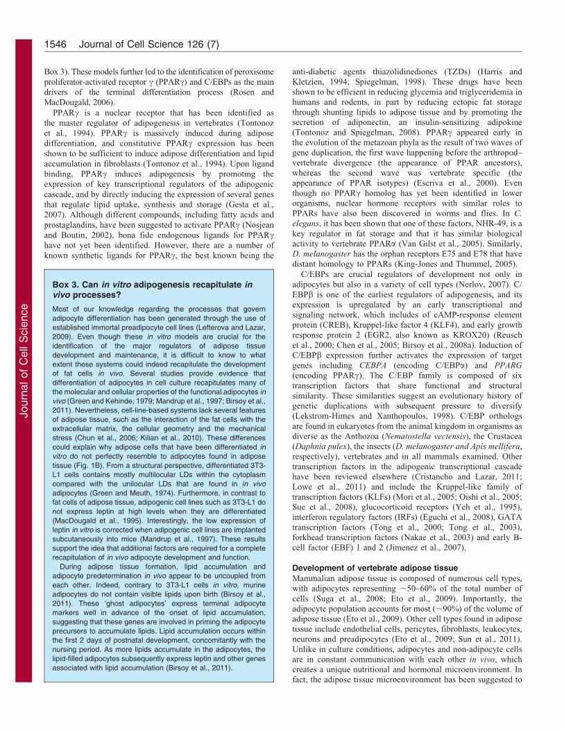
Fig. 1. Overview of the transcriptional cascade regulating the commitment and the differentiation of adipocytes. (A) The commitment and the differentiation of
mesenchymal stem cells (MSCs) into mature adipocytes follows a well-orchestrated transcriptional program. Although the molecular mechanisms regulating the early
events in preadipocyte determination are not perfectly elucidated, key players have been identified. BMP2 and BMP4, Wnt and TGFb signaling represent some of the
pathways involved in the commitment of the MSCs to the adipocyte lineage. The transcriptional events that culminate into the activation of PPARc and the terminal
differentiation of adipocytes are well defined, as presented here. GR, glucocorticoid receptor; CHOP, C/EBP-homologous protein (also known as DDIT3). (B) The
preadipocyte cell line 3T3-L1 has been extensively used for the study of adipogenesis in vitro. At 2 days post-confluence, 3T3-L1 cells are treated with a differentiation
cocktail containing insulin, dexamethasone (Dexa) and 3-isobutyl-1-methylxanthine (IBMX), required to activate PPARc, before the medium is changed and
supplemented only with insulin for the rest of the protocol. Upon differentiation, 3T3-L1 cells mostly accumulate lipids in multilocular LDs. In vivo, mice are born with
no apparent adipose tissue but with committed adipocytes that do not contain any triglycerides. These ‘ghost’ adipocytes start to accumulate lipids when the animal is
exposed to maternal milk and food. In contrast to differentiated 3T3-L1 cells, adipocytes isolated from mouse store triglycerides in a large and unique LD.
Lipid storage in animals 1545
Journ
alof
Cell
Scie
nce
Box 3). These models further led to the identification of peroxisome
proliferator-activated receptor c (PPARc) and C/EBPs as the main
drivers of the terminal differentiation process (Rosen and
MacDougald, 2006).
PPARc is a nuclear receptor that has been identified as
the master regulator of adipogenesis in vertebrates (Tontonoz
et al., 1994). PPARc is massively induced during adipose
differentiation, and constitutive PPARc expression has been
shown to be sufficient to induce adipose differentiation and lipid
accumulation in fibroblasts (Tontonoz et al., 1994). Upon ligand
binding, PPARc induces adipogenesis by promoting the
expression of key transcriptional regulators of the adipogenic
cascade, and by directly inducing the expression of several genes
that regulate lipid uptake, synthesis and storage (Gesta et al.,
2007). Although different compounds, including fatty acids and
prostaglandins, have been suggested to activate PPARc (Nosjean
and Boutin, 2002), bona fide endogenous ligands for PPARchave not yet been identified. However, there are a number of
known synthetic ligands for PPARc, the best known being the
anti-diabetic agents thiazolidinediones (TZDs) (Harris andKletzien, 1994; Spiegelman, 1998). These drugs have been
shown to be efficient in reducing glycemia and triglyceridemia inhumans and rodents, in part by reducing ectopic fat storagethrough shunting lipids to adipose tissue and by promoting thesecretion of adiponectin, an insulin-sensitizing adipokine
(Tontonoz and Spiegelman, 2008). PPARc appeared early inthe evolution of the metazoan phyla as the result of two waves ofgene duplication, the first wave happening before the arthropod–
vertebrate divergence (the appearance of PPAR ancestors),whereas the second wave was vertebrate specific (theappearance of PPAR isotypes) (Escriva et al., 2000). Even
though no PPARc homolog has yet been identified in lowerorganisms, nuclear hormone receptors with similar roles toPPARs have also been discovered in worms and flies. In C.
elegans, it has been shown that one of these factors, NHR-49, is a
key regulator in fat storage and that it has similar biologicalactivity to vertebrate PPARa (Van Gilst et al., 2005). Similarly,D. melanogaster has the orphan receptors E75 and E78 that have
distant homology to PPARs (King-Jones and Thummel, 2005).
C/EBPs are crucial regulators of development not only inadipocytes but also in a variety of cell types (Nerlov, 2007). C/
EBPb is one of the earliest regulators of adipogenesis, and itsexpression is upregulated by an early transcriptional andsignaling network, which includes of cAMP-response element
protein (CREB), Kruppel-like factor 4 (KLF4), and early growthresponse protein 2 (EGR2, also known as KROX20) (Reuschet al., 2000; Chen et al., 2005; Birsoy et al., 2008a). Induction ofC/EBPb expression further activates the expression of target
genes including CEBPA (encoding C/EBPa) and PPARG
(encoding PPARc). The C/EBP family is composed of sixtranscription factors that share functional and structural
similarity. These similarities suggest an evolutionary history ofgenetic duplications with subsequent pressure to diversify(Lekstrom-Himes and Xanthopoulos, 1998). C/EBP orthologs
are found in eukaryotes from the animal kingdom in organisms asdiverse as the Anthozoa (Nematostella vectensis), the Crustacea(Daphnia pulex), the insects (D. melanogaster and Apis mellifera,respectively), vertebrates and in all mammals examined. Other
transcription factors in the adipogenic transcriptional cascadehave been reviewed elsewhere (Cristancho and Lazar, 2011;Lowe et al., 2011) and include the Kruppel-like family of
transcription factors (KLFs) (Mori et al., 2005; Oishi et al., 2005;Sue et al., 2008), glucocorticoid receptors (Yeh et al., 1995),interferon regulatory factors (IRFs) (Eguchi et al., 2008), GATA
transcription factors (Tong et al., 2000; Tong et al., 2003),forkhead transcription factors (Nakae et al., 2003) and early B-cell factor (EBF) 1 and 2 (Jimenez et al., 2007).
Development of vertebrate adipose tissue
Mammalian adipose tissue is composed of numerous cell types,with adipocytes representing ,50–60% of the total number of
cells (Suga et al., 2008; Eto et al., 2009). Importantly, theadipocyte population accounts for most (,90%) of the volume ofadipose tissue (Eto et al., 2009). Other cell types found in adipose
tissue include endothelial cells, pericytes, fibroblasts, leukocytes,neurons and preadipocytes (Eto et al., 2009; Sun et al., 2011).Unlike in culture conditions, adipocytes and non-adipocyte cells
are in constant communication with each other in vivo, whichcreates a unique nutritional and hormonal microenvironment. Infact, the adipose tissue microenvironment has been suggested to
Box 3. Can in vitro adipogenesis recapitulate invivo processes?
Most of our knowledge regarding the processes that govern
adipocyte differentiation has been generated through the use of
established immortal preadipocyte cell lines (Lefterova and Lazar,
2009). Even though these in vitro models are crucial for the
identification of the major regulators of adipose tissue
development and maintenance, it is difficult to know to what
extent these systems could indeed recapitulate the development
of fat cells in vivo. Several studies provide evidence that
differentiation of adipocytes in cell culture recapitulates many of
the molecular and cellular properties of the functional adipocytes in
vivo (Green and Kehinde, 1979; Mandrup et al., 1997; Birsoy et al.,
2011). Nevertheless, cell-line-based systems lack several features
of adipose tissue, such as the interaction of the fat cells with the
extracellular matrix, the cellular geometry and the mechanical
stress (Chun et al., 2006; Kilian et al., 2010). These differences
could explain why adipose cells that have been differentiated in
vitro do not perfectly resemble to adipocytes found in adipose
tissue (Fig. 1B). From a structural perspective, differentiated 3T3-
L1 cells contains mostly multilocular LDs within the cytoplasm
compared with the unilocular LDs that are found in in vivo
adipocytes (Green and Meuth, 1974). Furthermore, in contrast to
fat cells of adipose tissue, adipogenic cell lines such as 3T3-L1 do
not express leptin at high levels when they are differentiated
(MacDougald et al., 1995). Interestingly, the low expression of
leptin in vitro is corrected when adipogenic cell lines are implanted
subcutaneously into mice (Mandrup et al., 1997). These results
support the idea that additional factors are required for a complete
recapitulation of in vivo adipocyte development and function.
During adipose tissue formation, lipid accumulation and
adipocyte predetermination in vivo appear to be uncoupled from
each other. Indeed, contrary to 3T3-L1 cells in vitro, murine
adipocytes do not contain visible lipids upon birth (Birsoy et al.,
2011). These ‘ghost adipocytes’ express terminal adipocyte
markers well in advance of the onset of lipid accumulation,
suggesting that these genes are involved in priming the adipocyte
precursors to accumulate lipids. Lipid accumulation occurs within
the first 2 days of postnatal development, concomitantly with the
nursing period. As more lipids accumulate in the adipocytes, the
lipid-filled adipocytes subsequently express leptin and other genes
associated with lipid accumulation (Birsoy et al., 2011).
Journal of Cell Science 126 (7)1546

Journ
alof
Cell
Scie
nce
promote leukocyte recruitment, which has been implicated inseveral inflammatory conditions including tumor formation
(Anderson et al., 2010; Park and Scherer, 2012).
Adipose tissue has the ability to expand in adulthood to copewith periods of chronic positive energy balance. In adult humans,adipocyte turnover rates vary greatly but are estimated to be
,10% per year (Spalding et al., 2008). A recent study suggeststhat 1–5% of murine adipocytes are replaced each day(Rigamonti et al., 2011). Furthermore, lipids in the adipocytes
are also constantly renewed so that TAGs are replaced six timesduring the average 10-year lifespan of one fat cell (Arner et al.,2011). Given that adipocytes are terminally differentiated and
post-mitotic, progenitor cells must exist to expand adipose tissue.However, we still have limited information about the identity,location and origin of adipocyte progenitor cells. Accordingly,two recent studies independently suggest that perivascular cells
of murine adipose tissue that express the surface markers CD34,CD24, SCA1, CD140a and CD140b are putative adipocyteprogenitor cells (Rodeheffer et al., 2008; Tang et al., 2008).
These cells have a high adipogenic potential in vitro and in vivo
and express the zinc finger protein 423 (ZFP423), a transcriptionfactor involved in preadipocyte determination in vivo (Gupta
et al., 2010; Gupta et al., 2012). Recently it has been shown thatanother transcription factor, zinc finger protein 521 (ZFP521),can regulate adipocyte development by inhibiting the expression
of ZFP423 (Kang et al., 2012).
In addition to the classical lipid-storing unilocular adipocyte,white adipose depots are also composed of multilocularadipocytes that express uncoupling protein 1 (UCP1) and have
higher oxidative and thermogenic capacities upon b-adrenergicstimulation than regular adipocytes (Wu et al., 2013). These cells,known as ‘brite’ or ‘beige’ adipocytes, arise from a different
progenitor cell than the classical white and brown adipocytes(Wu et al., 2012). More recently, bi-potent adipocyte progenitorcells that express PDGFRa, CD34 and SCA1 were characterized.
Such cells have been shown to differentiate into white unilocularadipocytes upon high-fat feeding or brown adipocyte (UCP1-positive cells) upon b-adrenergic activation (Lee et al., 2012).Whether these cells are progenitors of white, beige, brown or
other uncharacterized adipocytes remains to be investigated.
Normal adipose tissue development is tightly coupled to theformation of new blood vessels (Crandall et al., 1997; Rupnick
et al., 2002; Fukumura et al., 2003). Adequate adipose tissuevascularization and blood flow are crucial to support adipocyteswith nutrients and oxygen, and to maximize the efficiency of
lipid uptake and/or release in response to variations in thenutritional state. Pharmacological inhibition of angiogenesis isassociated with a significant reduction in adiposity in both miceand monkeys (Brakenhielm et al., 2004; Cao, 2010; Barnhart
et al., 2011; Sun et al., 2012). Interestingly, in pathologicalconditions such as obesity, adipose tissue grows independently ofvascularization, leading to a poor oxygen and nutrient supply to
adipocytes (Hosogai et al., 2007; Rausch et al., 2008; Yin et al.,2009; Kim et al., 2012). These findings indicate that adipocytesunder normal conditions interact with different cells and are
exposed to a distinct nutritional milieu than those that comprisethe adipose tissue of obese patients. Taking into account thatnutrients are important modulators of adipose tissue growth, as
further discussed in the next section, elucidation of differences ofadipocyte milieu and thus nutrient availability in health anddisease are of great importance.
Nutritional cues affecting adipose tissue formation
Adipose tissue has an outstanding capacity to modulate its sizeand function in response to nutritional cues. Extreme variations in
adipose tissue mass are observed between under and overfedanimals. Indeed, studies have shown that morbidly obese humanscan lose as much as 30–40% of their body weight over the first
year after undergoing bariatric surgery, a type of intervention thatreduces food intake and in some case nutrient absorption (Shahet al., 2006). Such a massive reduction in adipose tissue mass is
also observed when hyperphagia associated with leptindeficiency is corrected by leptin administration in mice andhumans (Halaas et al., 1995; Farooqi et al., 1999). The close
relationship between adipose tissue accumulation and nutrientavailability strongly suggests the existence of regulatorymechanisms that allow the rapid adjustment of adipocytefunction and metabolism to the nutritional status.
One important protein that has emerged as a key regulator ofadipogenesis and adipocyte maintenance in response to nutrientintake is the serine/threonine protein kinase AKT (also known as
PKB), which is well conserved from yeast to mammals (Vivancoand Sawyers, 2002). Loss of AKT in mice causes dwarfism andcompletely blocks adipogenesis (Peng et al., 2003), whereas its
constitutive activation is sufficient to induce the differentiation of3T3-L1 cells into mature adipocytes (Magun et al., 1996).Moreover, mutation in AKT2, one of the three AKT isoforms,leads to partial lipodystrophy in humans (George et al., 2004).
The rise in insulin following a meal induces the activationof phosphoinositide 3-kinase (PI3K) in adipocytes, whichdrives phosphatidylinositol (3,4,5)-triphosphate [PtdIns(3,4,5)P3]
production and activates AKT (Fig. 2). When active, AKT inducesthe translocation of the glucose transporter protein 4 (GLUT4) tothe cell membrane, thereby promoting the uptake of glucose, an
important substrate for lipid synthesis. Akt also activatesSREBP1, a key transcription factor controlling lipidmetabolism (Yecies et al., 2011). The concomitant rise in
glucose uptake and the increased SREBP1 activity play a crucialrole in promoting lipid accumulation in response to insulin.In addition to lipogenesis, AKT also directly regulatesthe adipogenic cascade by exacerbating PPARc activity.
Specifically, AKT phosphorylates GATA2 and FOXO1, whichpromotes the exclusion of these proteins from the nucleus andinduces PPARc expression and activity (Dowell et al., 2003;
Nakae et al., 2003; Menghini et al., 2005; Armoni et al., 2006;Fan et al., 2009).
The mammalian (or mechanistic) target of rapamycin (mTOR)
is a conserved kinase that senses and integrates a variety ofenvironmental cues to regulate cell and/or organ growth, andhomeostasis (Laplante and Sabatini, 2012). mTOR is aconstituent of two well-characterized protein complexes,
mTOR complex 1 (mTORC1) and mTORC2. From yeast tomammals, nutrients activate mTOR signaling, which in turntriggers the activation of several anabolic processes that are
required for cell growth and proliferation (Laplante and Sabatini,2009). With the emergence of metazoans, the mTOR pathwaybecame wired to signaling pathways that are activated by growth
factors, such as insulin (Laplante and Sabatini, 2012).Importantly, AKT plays a central role in relaying insulinsignaling to mTORC1 (Laplante and Sabatini, 2012). Several
pieces of evidence indicate that mTORC1 activation by nutrientsand/or growth factors is an essential molecular event required foradipose tissue maintenance and growth (Fig. 2). The increase in
Lipid storage in animals 1547

Journ
alof
Cell
Scie
nce
adiposity found upon high-fat feeding, leptin deficiency or
pharmacological PPARc activation is associated with a marked
activation of mTORC1 in adipose tissue (Um et al., 2004;
Blanchard et al., 2012). Furthermore, pharmacological or genetic
inhibition of mTORC1 is associated with a reduction in adipose
tissue mass due to both reduced adipocyte size and number
(Polak et al., 2008; Houde et al., 2010). mTORC1 appears to
affect adiposity by modulating several processes that are
involved in adipocyte formation (Fig. 2). mTORC1 regulates
the commitment of embryonic stem cells to early adipocyte
progenitors by activating S6 kinase 1 (S6K1) (Carnevalli et al.,
2010). Furthermore, several studies indicate that mTORC1
activation is also a fundamental step in the terminal
differentiation of preadipocytes. mTORC1 overactivation is
associated with an increase in adipogenesis in vitro, whereas
the complete inhibition of mTORC1 by rapamycin or genetic
deletion of RAPTOR, a key mTORC1 component, blocks this
process (Cho et al., 2004; Kim and Chen, 2004; Polak et al.,
2008; Yu et al., 2008; Zhang et al., 2009). mTORC1, through its
positive impact on protein translation, promotes the terminal
differentiation of adipocytes by regulating the expression of key
adipogenic regulators such as PPARc, CEBPd and C/EBPa (Le
Bacquer et al., 2007). Furthermore, mTORC1 also controls lipid
synthesis by directly activating SREBP1 (Bakan and Laplante,
2012).
Taken together, these studies indicate that mTORC1 is a strong
positive regulator of fat cell formation. Nevertheless, a recent
report from our laboratory suggests that excessive mTORC1
signaling can also have a negative impact on fat cell development
(Laplante et al., 2012). We showed that mice overexpressing
DEPTOR (DEP domain containing mTOR-interacting protein), a
protein that reduces mTOR signaling, unexpectedly accumulate
more adipose tissue than control mice. Interestingly, we observed
that the Deptor locus is part of a quantitative trait locus (QTL)
that has been linked to obesity in mice, and that DEPTOR
expression is elevated in adipose tissue of obese humans
(Laplante et al., 2012). From a mechanistic perspective,
DEPTOR promotes adipogenesis by dampening mTORC1
activity, without blocking it. Because mTORC1 overactivation
blocks AKT signaling through several negative-feedback loops
(Laplante and Sabatini, 2012), we proposed that elevated
DEPTOR expression might be required to preserve the pro-
adipogenic functions of AKT. These results support a
fundamental role of the well-conserved AKT–mTORC1 axis in
controlling lipid storage and adipocyte development in response
to the nutrients and growth factors.
Concluding remarksStudying lipid accumulation and/or adipogenesis from lower
organisms to mammals has expanded the comprehension of the
molecular mechanisms governing adipocyte formation and
metabolism. The identification and localization of fat cell
precursors along with the discovery of PPARc and several pro-
and anti-adipogenic proteins that regulate adipocyte terminal
differentiation represent some of the key discoveries in adipocyte
biology. Despite these advances, we should note that many
important questions regarding adipocyte development and
function still remain unanswered and are likely to be the source
of extensive investigation over the years to come. For example,
although we now have a better understanding of the identity and
the origin of the adipocytes that form adipose tissue, we still have
very limited knowledge of the molecular mechanisms that
regulate the recruitment of new adipocytes during adipose
tissue development and expansion. Do the signals regulating
adipocyte commitment come from circulating growth factors
and/or nutrients, or emerge directly from enlarged fat cells? How
Food consumption
Adipocyte commitment
mTORC1
AKT
Glucose
Insulin
Nutrients
IRS PI3KGLUT4 Glucose Lipogenic
substrates
TSC1/2
RHEB
S6K1 4E-BP1
eIF4E Proteinsynthesis
SREBP1
Lipogenicgenes
Lipogenesis
PPAR ligands Adipogenesis
FOXO1
PPARγ Adipogenicgenes
Circulation
Cytoplasm
DEPTOR
GLUT4
GATA2
Fig. 2. Regulation of lipogenesis and
adipogenesis in mammalian cells by
nutrients and insulin. Food consumption
leads to a rise in the circulation levels of
nutrients and glucose, and promotes the
secretion of insulin from the pancreas. The
activation of PI3K by insulin promotes
AKT and mTORC1 activity, which in turn
regulate key complementary steps
involved in the promotion of lipogenesis
and adipogenesis. Circulating levels of
nutrients are sensed by mTORC1 and
contribute to the full activation of this
protein complex. The coordinated
activation of AKT and mTORC1 is
essential to drive glucose uptake and
metabolism, lipogenic gene expression,
PPARc expression and activation, and
adipogenic gene expression, which are all
crucial steps in adipocyte formation.
Journal of Cell Science 126 (7)1548
Journ
alof
Cell
Scie
nce
do these signals turn on PPARc expression during the early steps
of preadipocyte commitment? It is well appreciated that
mammalian fat cells express certain factors, such as leptin, to a
degree that is proportional to the amount lipid stored in the cells.
This suggests that a lipid-sensing mechanism could exist in
adipocytes, similar to other nutrient-sensing pathways. The
identity of this putative lipid-sensing mechanism in vertebrate
adipocytes is still unknown. Identifying such a mechanism is
clinically very relevant as this could help the development of new
therapeutic avenues to treat obesity and its complications.
AcknowledgementsWe thank Aysu Uygur and Ozge Ceyhan for their helpful commentson the manuscript.
FundingK.B. is a fellow of the Jane Coffin Childs Memorial Fund forMedical Research. The activities of M.L. are funded by the CanadianInstitute of Health Research, the Natural Sciences and EngineeringResearch Council of Canada, the Fonds de Recherche du Quebec-Sante, the Canadian Liver Foundation and the Fondation de l’Institutuniversitaire de cardiologie et de pneumologie de Quebec. W.T.F. isfunded by Fundacao de Amparo a Pesquisa do Estado de Sao Paulo(FAPESP) [grant numbers 2009/15354-7 and 2010/52191-6].
ReferencesAlvarez, H. M. and Steinbuchel, A. (2002). Triacylglycerols in prokaryotic
microorganisms. Appl. Microbiol. Biotechnol. 60, 367-376.
Alvarez-Llamas, G., Szalowska, E., de Vries, M. P., Weening, D., Landman, K.,
Hoek, A., Wolffenbuttel, B. H., Roelofsen, H. and Vonk, R. J. (2007).
Characterization of the human visceral adipose tissue secretome. Mol. Cell.
Proteomics 6, 589-600.
Anderson, A. J. and Dawes, E. A. (1990). Occurrence, metabolism, metabolic role, and
industrial uses of bacterial polyhydroxyalkanoates. Microbiol. Rev. 54, 450-472.
Anderson, E. K., Gutierrez, D. A. and Hasty, A. H. (2010). Adipose tissue recruitment
of leukocytes. Curr. Opin. Lipidol. 21, 172-177.
Armoni, M., Harel, C., Karni, S., Chen, H., Bar-Yoseph, F., Ver, M. R., Quon, M. J.
and Karnieli, E. (2006). FOXO1 represses peroxisome proliferator-activated
receptor-gamma1 and -gamma2 gene promoters in primary adipocytes. A novel
paradigm to increase insulin sensitivity. J. Biol. Chem. 281, 19881-19891.
Arner, P., Bernard, S., Salehpour, M., Possnert, G., Liebl, J., Steier, P., Buchholz,
B. A., Eriksson, M., Arner, E., Hauner, H. et al. (2011). Dynamics of human
adipose lipid turnover in health and metabolic disease. Nature 478, 110-113.
Arrese, E. L. and Soulages, J. L. (2010). Insect fat body: energy, metabolism, and
regulation. Annu. Rev. Entomol. 55, 207-225.
Arrese, E. L., Mirza, S., Rivera, L., Howard, A. D., Chetty, P. S. and Soulages, J. L.
(2008). Expression of lipid storage droplet protein-1 may define the role of AKH as a
lipid mobilizing hormone in Manduca sexta. Insect Biochem. Mol. Biol. 38, 993-1000.
Ashrafi, K., Chang, F. Y., Watts, J. L., Fraser, A. G., Kamath, R. S., Ahringer,
J. and Ruvkun, G. (2003). Genome-wide RNAi analysis of Caenorhabditis elegans
fat regulatory genes. Nature 421, 268-272.
Athenstaedt, K. and Daum, G. (2005). Tgl4p and Tgl5p, two triacylglycerol lipases of
the yeast Saccharomyces cerevisiae are localized to lipid particles. J. Biol. Chem. 280,
37301-37309.
Athenstaedt, K., Zweytick, D., Jandrositz, A., Kohlwein, S. D. and Daum, G. (1999).
Identification and characterization of major lipid particle proteins of the yeast
Saccharomyces cerevisiae. J. Bacteriol. 181, 6441-6448.
Baek, S. H., Li, A. H. and Sassetti, C. M. (2011). Metabolic regulation of
mycobacterial growth and antibiotic sensitivity. PLoS Biol. 9, e1001065.
Bailey, J. M. and Dunbar, L. M. (1973). Essential fatty acid requirements of cells in
tissue culture: a review. Exp. Mol. Pathol. 18, 142-161.
Bakan, I. and Laplante, M. (2012). Connecting mTORC1 signaling to SREBP-1
activation. Curr. Opin. Lipidol. 23, 226-234.
Baker, M., Gaukrodger, N., Mayosi, B. M., Imrie, H., Farrall, M., Watkins, H.,
Connell, J. M., Avery, P. J. and Keavney, B. (2005). Association between common
polymorphisms of the proopiomelanocortin gene and body fat distribution: a family
study. Diabetes 54, 2492-2496.
Barnhart, K. F., Christianson, D. R., Hanley, P. W., Driessen, W. H., Bernacky,
B. J., Baze, W. B., Wen, S., Tian, M., Ma, J., Kolonin, M. G. et al. (2011). A
peptidomimetic targeting white fat causes weight loss and improved insulin resistance
in obese monkeys. Sci. Transl. Med. 3, 108ra112.
Beller, M., Riedel, D., Jansch, L., Dieterich, G., Wehland, J., Jackle, H. and
Kuhnlein, R. P. (2006). Characterization of the Drosophila lipid droplet
subproteome. Mol. Cell. Proteomics 5, 1082-1094.
Beller, M., Bulankina, A. V., Hsiao, H. H., Urlaub, H., Jackle, H. and Kuhnlein,
R. P. (2010). PERILIPIN-dependent control of lipid droplet structure and fat storagein Drosophila. Cell Metab. 12, 521-532.
Bennett, C. N., Ross, S. E., Longo, K. A., Bajnok, L., Hemati, N., Johnson, K. W.,
Harrison, S. D. and MacDougald, O. A. (2002). Regulation of Wnt signaling duringadipogenesis. J. Biol. Chem. 277, 30998-31004.
Bi, J., Xiang, Y., Chen, H., Liu, Z., Gronke, S., Kuhnlein, R. P. and Huang,
X. (2012). Opposite and redundant roles of the two Drosophila perilipins in lipidmobilization. J. Cell Sci. 125, 3568-3577.
Bickel, P. E., Tansey, J. T. and Welte, M. A. (2009). PAT proteins, an ancient familyof lipid droplet proteins that regulate cellular lipid stores. Biochim. Biophys. Acta
1791, 419-440.
Birsoy, K., Chen, Z. and Friedman, J. (2008a). Transcriptional regulation ofadipogenesis by KLF4. Cell Metab. 7, 339-347.
Birsoy, K., Soukas, A., Torrens, J., Ceccarini, G., Montez, J., Maffei, M., Cohen, P.,
Fayzikhodjaeva, G., Viale, A., Socci, N. D. et al. (2008b). Cellular programcontrolling the recovery of adipose tissue mass: An in vivo imaging approach. Proc.
Natl. Acad. Sci. USA 105, 12985-12990.
Birsoy, K., Berry, R., Wang, T., Ceyhan, O., Tavazoie, S., Friedman, J. M. andRodeheffer, M. S. (2011). Analysis of gene networks in white adipose tissuedevelopment reveals a role for ETS2 in adipogenesis. Development 138, 4709-4719.
Blanchard, P. G., Festuccia, W. T., Houde, V. P., St-Pierre, P., Brule, S., Turcotte,V., Cote, M., Bellmann, K., Marette, A. and Deshaies, Y. (2012). Majorinvolvement of mTOR in the PPARc-induced stimulation of adipose tissue lipiduptake and fat accretion. J. Lipid Res. 53, 1117-1125.
Bouchard, C. and Tremblay, A. (1997). Genetic influences on the response of body fatand fat distribution to positive and negative energy balances in human identical twins.J. Nutr. 127 Suppl., 943S-947S.
Bowers, R. R., Kim, J. W., Otto, T. C. and Lane, M. D. (2006). Stable stem cellcommitment to the adipocyte lineage by inhibition of DNA methylation: role of theBMP-4 gene. Proc. Natl. Acad. Sci. USA 103, 13022-13027.
Brakenhielm, E., Cao, R., Gao, B., Angelin, B., Cannon, B., Parini, P. and Cao,
Y. (2004). Angiogenesis inhibitor, TNP-470, prevents diet-induced and geneticobesity in mice. Circ. Res. 94, 1579-1588.
Calo, E., Quintero-Estades, J. A., Danielian, P. S., Nedelcu, S., Berman, S. D. andLees, J. A. (2010). Rb regulates fate choice and lineage commitment in vivo. Nature
466, 1110-1114.
Cao, Y. (2010). Adipose tissue angiogenesis as a therapeutic target for obesity andmetabolic diseases. Nat. Rev. Drug Discov. 9, 107-115.
Cao, H., Gerhold, K., Mayers, J. R., Wiest, M. M., Watkins, S. M. and Hotamisligil,
G. S. (2008). Identification of a lipokine, a lipid hormone linking adipose tissue tosystemic metabolism. Cell 134, 933-944.
Carnevalli, L. S., Masuda, K., Frigerio, F., Le Bacquer, O., Um, S. H., Gandin, V.,
Topisirovic, I., Sonenberg, N., Thomas, G. and Kozma, S. C. (2010). S6K1 plays acritical role in early adipocyte differentiation. Dev. Cell 18, 763-774.
Chen, Z., Torrens, J. I., Anand, A., Spiegelman, B. M. and Friedman, J. M. (2005).Krox20 stimulates adipogenesis via C/EBPbeta-dependent and -independent mechanisms.Cell Metab. 1, 93-106.
Chen, W., Yechoor, V. K., Chang, B. H., Li, M. V., March, K. L. and Chan,
L. (2009). The human lipodystrophy gene product Berardinelli-Seip congenitallipodystrophy 2/seipin plays a key role in adipocyte differentiation. Endocrinology
150, 4552-4561.
Chen, W., Chang, B., Saha, P., Hartig, S. M., Li, L., Reddy, V. T., Yang, Y.,Yechoor, V., Mancini, M. A. and Chan, L. (2012). Berardinelli-seip congenitallipodystrophy 2/seipin is a cell-autonomous regulator of lipolysis essential foradipocyte differentiation. Mol. Cell. Biol. 32, 1099-1111.
Cho, H. J., Park, J., Lee, H. W., Lee, Y. S. and Kim, J. B. (2004). Regulation ofadipocyte differentiation and insulin action with rapamycin. Biochem. Biophys. Res.
Commun. 321, 942-948.
Chun, T. H., Hotary, K. B., Sabeh, F., Saltiel, A. R., Allen, E. D. and Weiss, S. J.(2006). A pericellular collagenase directs the 3-dimensional development of whiteadipose tissue. Cell 125, 577-591.
Crandall, D. L., Hausman, G. J. and Kral, J. G. (1997). A review of themicrocirculation of adipose tissue: anatomic, metabolic, and angiogenic perspectives.Microcirculation 4, 211-232.
Crespi, E. J. and Denver, R. J. (2006). Leptin (ob gene) of the South African clawedfrog Xenopus laevis. Proc. Natl. Acad. Sci. USA 103, 10092-10097.
Cristancho, A. G. and Lazar, M. A. (2011). Forming functional fat: a growingunderstanding of adipocyte differentiation. Nat. Rev. Mol. Cell Biol. 12, 722-734.
Czabany, T., Athenstaedt, K. and Daum, G. (2007). Synthesis, storage anddegradation of neutral lipids in yeast. Biochim. Biophys. Acta 1771, 299-309.
Dahlqvist, A., Stahl, U., Lenman, M., Banas, A., Lee, M., Sandager, L., Ronne,
H. and Stymne, S. (2000). Phospholipid:diacylglycerol acyltransferase: an enzymethat catalyzes the acyl-CoA-independent formation of triacylglycerol in yeast andplants. Proc. Natl. Acad. Sci. USA 97, 6487-6492.
Dalen, K. T., Schoonjans, K., Ulven, S. M., Weedon-Fekjaer, M. S., Bentzen, T. G.,Koutnikova, H., Auwerx, J. and Nebb, H. I. (2004). Adipose tissue expression ofthe lipid droplet-associating proteins S3-12 and perilipin is controlled by peroxisomeproliferator-activated receptor-gamma. Diabetes 53, 1243-1252.
Daniel, J., Maamar, H., Deb, C., Sirakova, T. D. and Kolattukudy, P. E. (2011).Mycobacterium tuberculosis uses host triacylglycerol to accumulate lipid droplets andacquires a dormancy-like phenotype in lipid-loaded macrophages. PLoS Pathog. 7,e1002093.
Lipid storage in animals 1549

Journ
alof
Cell
Scie
nce
de Bono, M. and Bargmann, C. I. (1998). Natural variation in a neuropeptide Y
receptor homolog modifies social behavior and food response in C. elegans. Cell 94,
679-689.
Dionne, M. S., Pham, L. N., Shirasu-Hiza, M. and Schneider, D. S. (2006). Akt and
FOXO dysregulation contribute to infection-induced wasting in Drosophila. Curr.
Biol. 16, 1977-1985.
Doane, W. W. (1960). Developmental physiology of the mutant female sterile(2)adipose
of Drosophila melanogaster. II. Effects of altered environment and residual genome
on its expression. J. Exp. Zool. 145, 23-41.
Dowell, P., Otto, T. C., Adi, S. and Lane, M. D. (2003). Convergence of peroxisome
proliferator-activated receptor gamma and Foxo1 signaling pathways. J. Biol. Chem.
278, 45485-45491.
Drewnowski, A. (1992). Sensory properties of fats and fat replacements. Nutr. Rev. 50,
17-20.
Eguchi, J., Yan, Q. W., Schones, D. E., Kamal, M., Hsu, C. H., Zhang, M. Q.,
Crawford, G. E. and Rosen, E. D. (2008). Interferon regulatory factors are
transcriptional regulators of adipogenesis. Cell Metab. 7, 86-94.
Elbers, J. M., Asscheman, H., Seidell, J. C. and Gooren, L. J. (1999). Effects of sex
steroid hormones on regional fat depots as assessed by magnetic resonance imaging in
transsexuals. Am. J. Physiol. 276, E317-E325.
Escriva, H., Delaunay, F. and Laudet, V. (2000). Ligand binding and nuclear receptor
evolution. Bioessays 22, 717-727.
Eto, H., Suga, H., Matsumoto, D., Inoue, K., Aoi, N., Kato, H., Araki, J. and
Yoshimura, K. (2009). Characterization of structure and cellular components of
aspirated and excised adipose tissue. Plast. Reconstr. Surg. 124, 1087-1097.
Fan, W., Imamura, T., Sonoda, N., Sears, D. D., Patsouris, D., Kim, J. J. and
Olefsky, J. M. (2009). FOXO1 transrepresses peroxisome proliferator-activated
receptor gamma transactivation, coordinating an insulin-induced feed-forward
response in adipocytes. J. Biol. Chem. 284, 12188-12197.
Farooqi, I. S., Jebb, S. A., Langmack, G., Lawrence, E., Cheetham, C. H., Prentice,
A. M., Hughes, I. A., McCamish, M. A. and O’Rahilly, S. (1999). Effects of
recombinant leptin therapy in a child with congenital leptin deficiency. N. Engl.
J. Med. 341, 879-884.
Fei, W., Shui, G., Gaeta, B., Du, X., Kuerschner, L., Li, P., Brown, A. J., Wenk,
M. R., Parton, R. G. and Yang, H. (2008). Fld1p, a functional homologue of human
seipin, regulates the size of lipid droplets in yeast. J. Cell Biol. 180, 473-482.
Feingold, K. R., Marshall, M., Gulli, R., Moser, A. H. and Grunfeld, C. (1994).
Effect of endotoxin and cytokines on lipoprotein lipase activity in mice. Arterioscler.
Thromb. 14, 1866-1872.
Friedman, J. M. and Halaas, J. L. (1998). Leptin and the regulation of body weight in
mammals. Nature 395, 763-770.
Fukumura, D., Ushiyama, A., Duda, D. G., Xu, L., Tam, J., Krishna, V., Chatterjee,
K., Garkavtsev, I. and Jain, R. K. (2003). Paracrine regulation of angiogenesis and
adipocyte differentiation during in vivo adipogenesis. Circ. Res. 93, e88-e97.
Gallagher, E. J. and LeRoith, D. (2010). Insulin, insulin resistance, obesity, and
cancer. Curr. Diab. Rep. 10, 93-100.
Gelman, S., Cohen, A. H. and Sanovich, E. (2009). Developmental changes in the
ultrastructure of the lamprey lateral line nerve during metamorphosis. J. Morphol.
270, 815-824.
George, S., Rochford, J. J., Wolfrum, C., Gray, S. L., Schinner, S., Wilson, J. C.,
Soos, M. A., Murgatroyd, P. R., Williams, R. M., Acerini, C. L. et al. (2004). A
family with severe insulin resistance and diabetes due to a mutation in AKT2. Science
304, 1325-1328.
Gesta, S., Bluher, M., Yamamoto, Y., Norris, A. W., Berndt, J., Kralisch, S.,
Boucher, J., Lewis, C. and Kahn, C. R. (2006). Evidence for a role of
developmental genes in the origin of obesity and body fat distribution. Proc. Natl.
Acad. Sci. USA 103, 6676-6681.
Gesta, S., Tseng, Y. H. and Kahn, C. R. (2007). Developmental origin of fat: tracking
obesity to its source. Cell 131, 242-256.
Green, H. and Kehinde, O. (1979). Formation of normally differentiated subcutaneous
fat pads by an established preadipose cell line. J. Cell. Physiol. 101, 169-171.
Green, H. and Meuth, M. (1974). An established pre-adipose cell line and its
differentiation in culture. Cell 3, 127-133.
Grillitsch, K., Connerth, M., Kofeler, H., Arrey, T. N., Rietschel, B., Wagner, B.,
Karas, M. and Daum, G. (2011). Lipid particles/droplets of the yeast Saccharomyces
cerevisiae revisited: lipidome meets proteome. Biochim. Biophys. Acta 1811, 1165-
1176.
Gronke, S., Beller, M., Fellert, S., Ramakrishnan, H., Jackle, H. and Kuhnlein, R. P.
(2003). Control of fat storage by a Drosophila PAT domain protein. Curr. Biol. 13,
603-606.
Guo, Y., Walther, T. C., Rao, M., Stuurman, N., Goshima, G., Terayama, K., Wong,
J. S., Vale, R. D., Walter, P. and Farese, R. V. (2008). Functional genomic screen
reveals genes involved in lipid-droplet formation and utilization. Nature 453, 657-
661.
Gupta, R. K., Arany, Z., Seale, P., Mepani, R. J., Ye, L., Conroe, H. M., Roby, Y. A.,
Kulaga, H., Reed, R. R. and Spiegelman, B. M. (2010). Transcriptional control of
preadipocyte determination by Zfp423. Nature 464, 619-623.
Gupta, R. K., Mepani, R. J., Kleiner, S., Lo, J. C., Khandekar, M. J., Cohen, P.,
Frontini, A., Bhowmick, D. C., Ye, L., Cinti, S. et al. (2012). Zfp423 expression
identifies committed preadipocytes and localizes to adipose endothelial and
perivascular cells. Cell Metab. 15, 230-239.
Hader, T., Muller, S., Aguilera, M., Eulenberg, K. G., Steuernagel, A., Ciossek, T.,
Kuhnlein, R. P., Lemaire, L., Fritsch, R., Dohrmann, C. et al. (2003). Control oftriglyceride storage by a WD40/TPR-domain protein. EMBO Rep. 4, 511-516.
Halaas, J. L., Gajiwala, K. S., Maffei, M., Cohen, S. L., Chait, B. T., Rabinowitz, D.,
Lallone, R. L., Burley, S. K. and Friedman, J. M. (1995). Weight-reducing effects
of the plasma protein encoded by the obese gene. Science 269, 543-546.
Harris, P. K. and Kletzien, R. F. (1994). Localization of a pioglitazone response
element in the adipocyte fatty acid-binding protein gene. Mol. Pharmacol. 45, 439-445.
Harris, C. A., Haas, J. T., Streeper, R. S., Stone, S. J., Kumari, M., Yang, K., Han,
X., Brownell, N., Gross, R. W., Zechner, R. et al. (2011). DGAT enzymes are
required for triacylglycerol synthesis and lipid droplets in adipocytes. J. Lipid Res. 52,657-667.
Hishikawa, D., Hong, Y. H., Roh, S. G., Miyahara, H., Nishimura, Y., Tomimatsu,
A., Tsuzuki, H., Gotoh, C., Kuno, M., Choi, K. C. et al. (2005). Identification ofgenes expressed differentially in subcutaneous and visceral fat of cattle, pig, and
mouse. Physiol. Genomics 21, 343-350.
Hong, S. H., Lee, K. S., Kwak, S. J., Kim, A. K., Bai, H., Jung, M. S., Kwon, O. Y.,
Song, W. J., Tatar, M. and Yu, K. (2012). Minibrain/Dyrk1a regulates food intakethrough the Sir2-FOXO-sNPF/NPY pathway in Drosophila and mammals. PLoS
Genet. 8, e1002857.
Hosogai, N., Fukuhara, A., Oshima, K., Miyata, Y., Tanaka, S., Segawa, K.,
Furukawa, S., Tochino, Y., Komuro, R., Matsuda, M. et al. (2007). Adipose tissuehypoxia in obesity and its impact on adipocytokine dysregulation. Diabetes 56, 901-
911.
Hotamisligil, G. S. (2010). Endoplasmic reticulum stress and the inflammatory basis of
metabolic disease. Cell 140, 900-917.
Hotamisligil, G. S., Shargill, N. S. and Spiegelman, B. M. (1993). Adipose expression
of tumor necrosis factor-alpha: direct role in obesity-linked insulin resistance. Science
259, 87-91.
Houde, V. P., Brule, S., Festuccia, W. T., Blanchard, P. G., Bellmann, K., Deshaies,
Y. and Marette, A. (2010). Chronic rapamycin treatment causes glucose intoleranceand hyperlipidemia by upregulating hepatic gluconeogenesis and impairing lipid
deposition in adipose tissue. Diabetes 59, 1338-1348.
Huang, H., Song, T. J., Li, X., Hu, L., He, Q., Liu, M., Lane, M. D. and Tang, Q. Q.
(2009). BMP signaling pathway is required for commitment of C3H10T1/2pluripotent stem cells to the adipocyte lineage. Proc. Natl. Acad. Sci. USA 106,
12670-12675.
Huising, M. O., Geven, E. J., Kruiswijk, C. P., Nabuurs, S. B., Stolte, E. H.,
Spanings, F. A., Verburg-van Kemenade, B. M. and Flik, G. (2006). Increasedleptin expression in common Carp (Cyprinus carpio) after food intake but not after
fasting or feeding to satiation. Endocrinology 147, 5786-5797.
Ignotz, R. A. and Massague, J. (1985). Type beta transforming growth factor controls
the adipogenic differentiation of 3T3 fibroblasts. Proc. Natl. Acad. Sci. USA 82, 8530-8534.
Jacquier, N., Choudhary, V., Mari, M., Toulmay, A., Reggiori, F. and Schneiter,
R. (2011). Lipid droplets are functionally connected to the endoplasmic reticulum in
Saccharomyces cerevisiae. J. Cell Sci. 124, 2424-2437.
Jang, I. H., Chosa, N., Kim, S. H., Nam, H. J., Lemaitre, B., Ochiai, M., Kambris,
Z., Brun, S., Hashimoto, C., Ashida, M. et al. (2006). A Spatzle-processing enzymerequired for toll signaling activation in Drosophila innate immunity. Dev. Cell 10, 45-
55.
Jimenez, M. A., Akerblad, P., Sigvardsson, M. and Rosen, E. D. (2007). Critical rolefor Ebf1 and Ebf2 in the adipogenic transcriptional cascade. Mol. Cell. Biol. 27, 743-
757.
Joe, A. W., Yi, L., Even, Y., Vogl, A. W. and Rossi, F. M. (2009). Depot-specific
differences in adipogenic progenitor abundance and proliferative response to high-fatdiet. Stem Cells 27, 2563-2570.
Kalscheuer, R., Luftmann, H. and Steinbuchel, A. (2004). Synthesis of novel lipids inSaccharomyces cerevisiae by heterologous expression of an unspecific bacterial
acyltransferase. Appl. Environ. Microbiol. 70, 7119-7125.
Kang, S., Akerblad, P., Kiviranta, R., Gupta, R. K., Kajimura, S., Griffin, M. J.,
Min, J., Baron, R. and Rosen, E. D. (2012). Regulation of early adiposecommitment by Zfp521. PLoS Biol. 10, e1001433.
Kilian, K. A., Bugarija, B., Lahn, B. T. and Mrksich, M. (2010). Geometric cues fordirecting the differentiation of mesenchymal stem cells. Proc. Natl. Acad. Sci. USA
107, 4872-4877.
Kim, J. E. and Chen, J. (2004). Regulation of peroxisome proliferator-activatedreceptor-gamma activity by mammalian target of rapamycin and amino acids in
adipogenesis. Diabetes 53, 2748-2756.
Kim, D. H., Gutierrez-Aguilar, R., Kim, H. J., Woods, S. C. and Seeley, R. J. (2012).
Increased adipose tissue hypoxia and capacity for angiogenesis and inflammation inyoung diet-sensitive C57 mice compared with diet-resistant FVB mice. Int. J. Obes.
(Lond). [Epub ahead of print] doi: 10.1038/ijo.2012.141.
King-Jones, K. and Thummel, C. S. (2005). Nuclear receptors – a perspective from
Drosophila. Nat. Rev. Genet. 6, 311-323.
Kurat, C. F., Natter, K., Petschnigg, J., Wolinski, H., Scheuringer, K., Scholz, H.,
Zimmermann, R., Leber, R., Zechner, R. and Kohlwein, S. D. (2006). Obeseyeast: triglyceride lipolysis is functionally conserved from mammals to yeast. J. Biol.
Chem. 281, 491-500.
Kurat, C. F., Wolinski, H., Petschnigg, J., Kaluarachchi, S., Andrews, B., Natter,
K. and Kohlwein, S. D. (2009). Cdk1/Cdc28-dependent activation of the major
Journal of Cell Science 126 (7)1550

Journ
alof
Cell
Scie
nce
triacylglycerol lipase Tgl4 in yeast links lipolysis to cell-cycle progression. Mol. Cell
33, 53-63.
Laplante, M. and Sabatini, D. M. (2009). mTOR signaling at a glance. J. Cell Sci. 122,3589-3594.
Laplante, M. and Sabatini, D. M. (2012). mTOR signaling in growth control anddisease. Cell 149, 274-293.
Laplante, M., Horvat, S., Festuccia, W. T., Birsoy, K., Prevorsek, Z., Efeyan, A. andSabatini, D. M. (2012). DEPTOR cell-autonomously promotes adipogenesis, and itsexpression is associated with obesity. Cell Metab. 16, 202-212.
Le Bacquer, O., Petroulakis, E., Paglialunga, S., Poulin, F., Richard, D., Cianflone,K. and Sonenberg, N. (2007). Elevated sensitivity to diet-induced obesity and insulinresistance in mice lacking 4E-BP1 and 4E-BP2. J. Clin. Invest. 117, 387-396.
Lee, Y. H., Petkova, A. P., Mottillo, E. P. and Granneman, J. G. (2012). In vivoidentification of bipotential adipocyte progenitors recruited by b3-adrenoceptoractivation and high-fat feeding. Cell Metab. 15, 480-491.
Lefterova, M. I. and Lazar, M. A. (2009). New developments in adipogenesis. Trends
Endocrinol. Metab. 20, 107-114.
Lekstrom-Himes, J. and Xanthopoulos, K. G. (1998). Biological role of the CCAAT/enhancer-binding protein family of transcription factors. J. Biol. Chem. 273, 28545-28548.
Li, Y. (2012). Establishment and evaluation of a new model for studying lipogenesis ingrass carp (Ctenopharyngodon idella) preadipocytes. In Vitro Cell. Dev. Biol. Anim.
48, 37-42.
Li, G. G., Liang, X. F., Xie, Q., Li, G., Yu, Y. and Lai, K. (2010). Gene structure,recombinant expression and functional characterization of grass carp leptin. Gen.
Comp. Endocrinol. 166, 117-127.
Linder, K., Arner, P., Flores-Morales, A., Tollet-Egnell, P. and Norstedt, G. (2004).Differentially expressed genes in visceral or subcutaneous adipose tissue of obesemen and women. J. Lipid Res. 45, 148-154.
Lowe, C. E., O’Rahilly, S. and Rochford, J. J. (2011). Adipogenesis at a glance.J. Cell Sci. 124, 2681-2686.
MacDougald, O. A., Hwang, C. S., Fan, H. and Lane, M. D. (1995). Regulatedexpression of the obese gene product (leptin) in white adipose tissue and 3T3-L1adipocytes. Proc. Natl. Acad. Sci. USA 92, 9034-9037.
MacEachran, D. P., Prophete, M. E. and Sinskey, A. J. (2010). The Rhodococcusopacus PD630 heparin-binding hemagglutinin homolog TadA mediates lipid bodyformation. Appl. Environ. Microbiol. 76, 7217-7225.
Macotela, Y., Emanuelli, B., Mori, M. A., Gesta, S., Schulz, T. J., Tseng, Y. H. and
Kahn, C. R. (2012). Intrinsic differences in adipocyte precursor cells from differentwhite fat depots. Diabetes 61, 1691-1699.
Maehara, A., Taguchi, S., Nishiyama, T., Yamane, T. and Doi, Y. (2002). A repressorprotein, PhaR, regulates polyhydroxyalkanoate (PHA) synthesis via its directinteraction with PHA. J. Bacteriol. 184, 3992-4002.
Maffei, M., Halaas, J., Ravussin, E., Pratley, R. E., Lee, G. H., Zhang, Y., Fei, H.,
Kim, S., Lallone, R., Ranganathan, S. et al. (1995). Leptin levels in human androdent: measurement of plasma leptin and ob RNA in obese and weight-reducedsubjects. Nat. Med. 1, 1155-1161.
Magre, J., Delepine, M., Khallouf, E., Gedde-Dahl, T., Jr, Van Maldergem, L.,
Sobel, E., Papp, J., Meier, M., Megarbane, A., Bachy, A. et al.; BSCL WorkingGroup (2001). Identification of the gene altered in Berardinelli-Seip congenitallipodystrophy on chromosome 11q13. Nat. Genet. 28, 365-370.
Magun, R., Burgering, B. M., Coffer, P. J., Pardasani, D., Lin, Y., Chabot, J. andSorisky, A. (1996). Expression of a constitutively activated form of protein kinase B(c-Akt) in 3T3-L1 preadipose cells causes spontaneous differentiation. Endocrinology
137, 3590-3593.
Mak, H. Y. (2012). Lipid droplets as fat storage organelles in Caenorhabditis elegans:thematic review series: lipid droplet synthesis and metabolism: from yeast to man.J. Lipid Res. 53, 28-33.
Mandrup, S., Loftus, T. M., MacDougald, O. A., Kuhajda, F. P. and Lane, M. D.
(1997). Obese gene expression at in vivo levels by fat pads derived from s.c.implanted 3T3-F442A preadipocytes. Proc. Natl. Acad. Sci. USA 94, 4300-4305.
Manfruelli, P., Reichhart, J. M., Steward, R., Hoffmann, J. A. and Lemaitre,
B. (1999). A mosaic analysis in Drosophila fat body cells of the control ofantimicrobial peptide genes by the Rel proteins Dorsal and DIF. EMBO J. 18, 3380-3391.
McKay, R. M., McKay, J. P., Avery, L. and Graff, J. M. (2003). C elegans: a modelfor exploring the genetics of fat storage. Dev. Cell 4, 131-142.
Menghini, R., Marchetti, V., Cardellini, M., Hribal, M. L., Mauriello, A., Lauro, D.,
Sbraccia, P., Lauro, R. and Federici, M. (2005). Phosphorylation of GATA2 by Aktincreases adipose tissue differentiation and reduces adipose tissue-related inflammation: anovel pathway linking obesity to atherosclerosis. Circulation 111, 1946-1953.
Minnaard, R., Schrauwen, P., Schaart, G., Jorgensen, J. A., Lenaers, E., Mensink,
M. and Hesselink, M. K. (2009). Adipocyte differentiation-related protein andOXPAT in rat and human skeletal muscle: involvement in lipid accumulation andtype 2 diabetes mellitus. J. Clin. Endocrinol. Metab. 94, 4077-4085.
Miura, S., Gan, J. W., Brzostowski, J., Parisi, M. J., Schultz, C. J., Londos, C.,Oliver, B. and Kimmel, A. R. (2002). Functional conservation for lipid storagedroplet association among Perilipin, ADRP, and TIP47 (PAT)-related proteins inmammals, Drosophila, and Dictyostelium. J. Biol. Chem. 277, 32253-32257.
Mori, T., Sakaue, H., Iguchi, H., Gomi, H., Okada, Y., Takashima, Y., Nakamura,
K., Nakamura, T., Yamauchi, T., Kubota, N. et al. (2005). Role of Kruppel-likefactor 15 (KLF15) in transcriptional regulation of adipogenesis. J. Biol. Chem. 280,12867-12875.
Mullaney, B. C. and Ashrafi, K. (2009). C. elegans fat storage and metabolic
regulation. Biochim. Biophys. Acta 1791, 474-478.
Mullner, H. and Daum, G. (2004). Dynamics of neutral lipid storage in yeast. Acta
Biochim. Pol. 51, 323-347.
Murashita, K., Uji, S., Yamamoto, T., Rønnestad, I. and Kurokawa, T. (2008).
Production of recombinant leptin and its effects on food intake in rainbow trout
(Oncorhynchus mykiss). Comp. Biochem. Physiol. B Biochem. Mol. Biol. 150, 377-
384.
Nakae, J., Kitamura, T., Kitamura, Y., Biggs, W. H., 3rd, Arden, K. C. and Accili, D.
(2003). The forkhead transcription factor Foxo1 regulates adipocyte differentiation.
Dev. Cell 4, 119-129.
Nelson, T. L., Vogler, G. P., Pedersen, N. L., Hong, Y. and Miles, T. P. (2000).
Genetic and environmental influences on body fat distribution, fasting insulin levels
and CVD: are the influences shared? Twin Res. 3, 43-50.
Nelson, M. M., Leighton, D. L., Phleger, C. F. and Nichols, P. D. (2002). Comparison
of growth and lipid composition in the green abalone, Haliotis fulgens, provided
specific macroalgal diets. Comp. Biochem. Physiol. B Biochem. Mol. Biol. 131, 695-
712.
Nerlov, C. (2007). The C/EBP family of transcription factors: a paradigm for interaction
between gene expression and proliferation control. Trends Cell Biol. 17, 318-324.
Niewiarowski, P. H., Balk, M. L. and Londraville, R. L. (2000). Phenotypic effects of
leptin in an ectotherm: a new tool to study the evolution of life histories and
endothermy? J. Exp. Biol. 203, 295-300.
Nishio, S., Gibert, Y., Bernard, L., Brunet, F., Triqueneaux, G. and Laudet,
V. (2008). Adiponectin and adiponectin receptor genes are coexpressed during
zebrafish embryogenesis and regulated by food deprivation. Dev. Dyn. 237, 1682-
1690.
Nosjean, O. and Boutin, J. A. (2002). Natural ligands of PPARgamma: are
prostaglandin J(2) derivatives really playing the part? Cell. Signal. 14, 573-583.
O’Rourke, E. J., Soukas, A. A., Carr, C. E. and Ruvkun, G. (2009). C. elegans major
fats are stored in vesicles distinct from lysosome-related organelles. Cell Metab. 10,
430-435.
Oishi, Y., Manabe, I., Tobe, K., Tsushima, K., Shindo, T., Fujiu, K., Nishimura, G.,
Maemura, K., Yamauchi, T., Kubota, N. et al. (2005). Kruppel-like transcription
factor KLF5 is a key regulator of adipocyte differentiation. Cell Metab. 1, 27-39.
Park, J. and Scherer, P. E. (2012). Adipocyte-derived endotrophin promotes malignant
tumor progression. J. Clin. Invest. 122, 4243-4256.
Penfornis, P. and Marette, A. (2005). Inducible nitric oxide synthase modulates
lipolysis in adipocytes. J. Lipid Res. 46, 135-142.
Peng, X. D., Xu, P. Z., Chen, M. L., Hahn-Windgassen, A., Skeen, J., Jacobs, J.,
Sundararajan, D., Chen, W. S., Crawford, S. E., Coleman, K. G. et al. (2003).
Dwarfism, impaired skin development, skeletal muscle atrophy, delayed bone
development, and impeded adipogenesis in mice lacking Akt1 and Akt2. Genes Dev.
17, 1352-1365.
Pfundt, B., Sauerwein, H. and Mielenz, M. (2009). Leptin mRNA and protein
immunoreactivity in adipose tissue and liver of rainbow trout (Oncorhynchus mykiss)
and immunohistochemical localization in liver. Anat. Histol. Embryol. 38, 406-410.
Polak, P., Cybulski, N., Feige, J. N., Auwerx, J., Ruegg, M. A. and Hall, M. N.
(2008). Adipose-specific knockout of raptor results in lean mice with enhanced
mitochondrial respiration. Cell Metab. 8, 399-410.
Pond, C. M. (1992). An evolutionary and functional view of mammalian adipose tissue.
Proc. Nutr. Soc. 51, 367-377.
Pospisilik, J. A., Schramek, D., Schnidar, H., Cronin, S. J., Nehme, N. T., Zhang, X.,
Knauf, C., Cani, P. D., Aumayr, K., Todoric, J. et al. (2010). Drosophila genome-
wide obesity screen reveals hedgehog as a determinant of brown versus white adipose
cell fate. Cell 140, 148-160.
Potter, M., Madkour, M. H., Mayer, F. and Steinbuchel, A. (2002). Regulation of
phasin expression and polyhydroxyalkanoate (PHA) granule formation in Ralstonia
eutropha H16. Microbiology 148, 2413-2426.
Rajakumari, S., Grillitsch, K. and Daum, G. (2008). Synthesis and turnover of non-
polar lipids in yeast. Prog. Lipid Res. 47, 157-171.
Rajan, A. and Perrimon, N. (2012). Drosophila cytokine unpaired 2 regulates
physiological homeostasis by remotely controlling insulin secretion. Cell 151, 123-
137.
Rausch, M. E., Weisberg, S., Vardhana, P. and Tortoriello, D. V. (2008). Obesity in
C57BL/6J mice is characterized by adipose tissue hypoxia and cytotoxic T-cell
infiltration. Int. J. Obes. (Lond.) 32, 451-463.
Reis, T., Van Gilst, M. R. and Hariharan, I. K. (2010). A buoyancy-based screen of
Drosophila larvae for fat-storage mutants reveals a role for Sir2 in coupling fat storage
to nutrient availability. PLoS Genet. 6, e1001206.
Reusch, J. E., Colton, L. A. and Klemm, D. J. (2000). CREB activation induces
adipogenesis in 3T3-L1 cells. Mol. Cell. Biol. 20, 1008-1020.
Rigamonti, A., Brennand, K., Lau, F. and Cowan, C. A. (2011). Rapid cellular
turnover in adipose tissue. PLoS ONE 6, e17637.
Rodeheffer, M. S., Birsoy, K. and Friedman, J. M. (2008). Identification of white
adipocyte progenitor cells in vivo. Cell 135, 240-249.
Rosen, E. D. and MacDougald, O. A. (2006). Adipocyte differentiation from the inside
out. Nat. Rev. Mol. Cell Biol. 7, 885-896.
Ross, S. E., Hemati, N., Longo, K. A., Bennett, C. N., Lucas, P. C., Erickson, R. L.
and MacDougald, O. A. (2000). Inhibition of adipogenesis by Wnt signaling.
Science 289, 950-953.
Lipid storage in animals 1551

Journ
alof
Cell
Scie
nce
Rupnick, M. A., Panigrahy, D., Zhang, C. Y., Dallabrida, S. M., Lowell, B. B.,
Langer, R. and Folkman, M. J. (2002). Adipose tissue mass can be regulated
through the vasculature. Proc. Natl. Acad. Sci. USA 99, 10730-10735.
Russo, F., De Girolamo, P., Neglia, S., Gargiulo, A., Arcamone, N., Gargiulo, G. and
Varricchio, E. (2011). Immunohistochemical and immunochemical characterizationof the distribution of leptin-like proteins in the gastroenteric tract of two teleosts
(Dicentrarchus labrax and Carassius auratus L.) with different feeding habits.
Microsc. Res. Tech. 74, 714-719.
Sandager, L., Gustavsson, M. H., Stahl, U., Dahlqvist, A., Wiberg, E., Banas, A.,
Lenman, M., Ronne, H. and Stymne, S. (2002). Storage lipid synthesis is non-essential in yeast. J. Biol. Chem. 277, 6478-6482.
Schlegel, A. and Stainier, D. Y. (2007). Lessons from ‘lower’ organisms: what worms,flies, and zebrafish can teach us about human energy metabolism. PLoS Genet. 3,
e199.
Shah, M., Simha, V. and Garg, A. (2006). Review: long-term impact of bariatricsurgery on body weight, comorbidities, and nutritional status. J. Clin. Endocrinol.
Metab. 91, 4223-4231.
Silva, R. A., Grossi, V., Olivera, N. L. and Alvarez, H. M. (2010). Characterization of
indigenous Rhodococcus sp. 602, a strain able to accumulate triacylglycerides fromnaphthyl compounds under nitrogen-starved conditions. Res. Microbiol. 161, 198-
207.
Sim, S. J., Snell, K. D., Hogan, S. A., Stubbe, J., Rha, C. and Sinskey, A. J. (1997).
PHA synthase activity controls the molecular weight and polydispersity ofpolyhydroxybutyrate in vivo. Nat. Biotechnol. 15, 63-67.
Spalding, K. L., Arner, E., Westermark, P. O., Bernard, S., Buchholz, B. A.,
Bergmann, O., Blomqvist, L., Hoffstedt, J., Naslund, E., Britton, T. et al. (2008).Dynamics of fat cell turnover in humans. Nature 453, 783-787.
Spiegelman, B. M. (1998). PPAR-gamma: adipogenic regulator and thiazolidinedionereceptor. Diabetes 47, 507-514.
Srinivasan, S., Sadegh, L., Elle, I. C., Christensen, A. G., Faergeman, N. J. and
Ashrafi, K. (2008). Serotonin regulates C. elegans fat and feeding through
independent molecular mechanisms. Cell Metab. 7, 533-544.
Steinbuchel, A., Aerts, K., Babel, W., Follner, C., Liebergesell, M., Madkour, M. H.,
Mayer, F., Pieper-Furst, U., Pries, A., Valentin, H. E. et al. (1995). Considerations
on the structure and biochemistry of bacterial polyhydroxyalkanoic acid inclusions.Can. J. Microbiol. 41 Suppl. 1, 94-105.
Sue, N., Jack, B. H., Eaton, S. A., Pearson, R. C., Funnell, A. P., Turner, J., Czolij,
R., Denyer, G., Bao, S., Molero-Navajas, J. C. et al. (2008). Targeted disruption of
the basic Kruppel-like factor gene (Klf3) reveals a role in adipogenesis. Mol. Cell.
Biol. 28, 3967-3978.
Suga, H., Matsumoto, D., Inoue, K., Shigeura, T., Eto, H., Aoi, N., Kato, H., Abe,
H. and Yoshimura, K. (2008). Numerical measurement of viable and nonviableadipocytes and other cellular components in aspirated fat tissue. Plast. Reconstr. Surg.
122, 103-114.
Suh, J. M., Zeve, D., McKay, R., Seo, J., Salo, Z., Li, R., Wang, M. and Graff, J. M.
(2007). Adipose is a conserved dosage-sensitive antiobesity gene. Cell Metab. 6, 195-207.
Sun, K., Kusminski, C. M. and Scherer, P. E. (2011). Adipose tissue remodeling andobesity. J. Clin. Invest. 121, 2094-2101.
Sun, K., Wernstedt Asterholm, I., Kusminski, C. M., Bueno, A. C., Wang, Z. V.,
Pollard, J. W., Brekken, R. A. and Scherer, P. E. (2012). Dichotomous effects ofVEGF-A on adipose tissue dysfunction. Proc. Natl. Acad. Sci. USA 109, 5874-5879.
Tang, W., Zeve, D., Suh, J. M., Bosnakovski, D., Kyba, M., Hammer, R. E.,
Tallquist, M. D. and Graff, J. M. (2008). White fat progenitor cells reside in the
adipose vasculature. Science 322, 583-586.
Tinoco, A. B., Nisembaum, L. G., Isorna, E., Delgado, M. J. and de Pedro, N. (2012).
Leptins and leptin receptor expression in the goldfish (Carassius auratus). Regulation
by food intake and fasting/overfeeding conditions. Peptides 34, 329-335.
Tong, Q., Dalgin, G., Xu, H., Ting, C. N., Leiden, J. M. and Hotamisligil, G. S.
(2000). Function of GATA transcription factors in preadipocyte-adipocyte transition.Science 290, 134-138.
Tong, Q., Tsai, J. and Hotamisligil, G. S. (2003). GATA transcription factors and fatcell formation. Drug News Perspect. 16, 585-588.
Tontonoz, P. and Spiegelman, B. M. (2008). Fat and beyond: the diverse biology of
PPARgamma. Annu. Rev. Biochem. 77, 289-312.
Tontonoz, P., Hu, E. and Spiegelman, B. M. (1994). Stimulation of adipogenesis in
fibroblasts by PPAR gamma 2, a lipid-activated transcription factor. Cell 79, 1147-1156.
Um, S. H., Frigerio, F., Watanabe, M., Picard, F., Joaquin, M., Sticker, M.,
Fumagalli, S., Allegrini, P. R., Kozma, S. C., Auwerx, J. et al. (2004). Absence of
S6K1 protects against age- and diet-induced obesity while enhancing insulinsensitivity. Nature 431, 200-205.
Van Gilst, M. R., Hadjivassiliou, H., Jolly, A. and Yamamoto, K. R. (2005). Nuclear
hormone receptor NHR-49 controls fat consumption and fatty acid composition in C.elegans. PLoS Biol. 3, e53.
Vegusdal, A., Sundvold, H., Gjøen, T. and Ruyter, B. (2003). An in vitro method forstudying the proliferation and differentiation of Atlantic salmon preadipocytes. Lipids
38, 289-296.
Vivanco, I. and Sawyers, C. L. (2002). The phosphatidylinositol 3-Kinase AKT
pathway in human cancer. Nat. Rev. Cancer 2, 489-501.
Waltermann, M. and Steinbuchel, A. (2005). Neutral lipid bodies in prokaryotes:recent insights into structure, formation, and relationship to eukaryotic lipid depots.J. Bacteriol. 187, 3607-3619.
Waltermann, M., Hinz, A., Robenek, H., Troyer, D., Reichelt, R., Malkus, U., Galla,
H. J., Kalscheuer, R., Stoveken, T., von Landenberg, P. et al. (2005). Mechanismof lipid-body formation in prokaryotes: how bacteria fatten up. Mol. Microbiol. 55,750-763.
Walther, T. C. and Farese, R. V., Jr (2009). The life of lipid droplets. Biochim.
Biophys. Acta 1791, 459-466.Walther, T. C. and Farese, R. V., Jr (2012). Lipid droplets and cellular lipid
metabolism. Annu. Rev. Biochem. 81, 687-714.Wang, C. and St Leger, R. J. (2007). The Metarhizium anisopliae perilipin homolog
mpl1 regulates lipid metabolism, appressorial turgor pressure, and virulence. J. Biol.
Chem. 282, 21110-21115.Wang, H. and Sztalryd, C. (2011). Oxidative tissue: perilipin 5 links storage with the
furnace. Trends Endocrinol. Metab. 22, 197-203.Wellen, K. E. and Hotamisligil, G. S. (2003). Obesity-induced inflammatory changes
in adipose tissue. J. Clin. Invest. 112, 1785-1788.Widberg, C. H., Newell, F. S., Bachmann, A. W., Ramnoruth, S. N., Spelta, M. C.,
Whitehead, J. P., Hutley, L. J. and Prins, J. B. (2009). Fibroblast growth factorreceptor 1 is a key regulator of early adipogenic events in human preadipocytes. Am.
J. Physiol. Endocrinol. Metab. 296, E121-E131.Wieczorek, R., Pries, A., Steinbuchel, A. and Mayer, F. (1995). Analysis of a 24-
kilodalton protein associated with the polyhydroxyalkanoic acid granules inAlcaligenes eutrophus. J. Bacteriol. 177, 2425-2435.
Wu, C., Zhang, Y., Sun, Z. and Li, P. (2008). Molecular evolution of Cide familyproteins: novel domain formation in early vertebrates and the subsequent divergence.BMC Evol. Biol. 8, 159.
Wu, J., Bostrom, P., Sparks, L. M., Ye, L., Choi, J. H., Giang, A. H., Khandekar,
M., Virtanen, K. A., Nuutila, P., Schaart, G. et al. (2012). Beige adipocytes are adistinct type of thermogenic fat cell in mouse and human. Cell 150, 366-376.
Wu, J., Cohen, P. and Spiegelman, B. M. (2013). Adaptive thermogenesis inadipocytes: is beige the new brown? Genes Dev. 27, 234-250.
Wymann, M. P. and Schneiter, R. (2008). Lipid signalling in disease. Nat. Rev. Mol.
Cell Biol. 9, 162-176.Xiao, L., Sobue, T., Esliger, A., Kronenberg, M. S., Coffin, J. D., Doetschman,
T. and Hurley, M. M. (2010). Disruption of the Fgf2 gene activates the adipogenicand suppresses the osteogenic program in mesenchymal marrow stromal stem cells.Bone 47, 360-370.
Yamada, M., Yamashita, K., Wakuda, A., Ichimura, K., Maehara, A., Maeda, M.and Taguchi, S. (2007). Autoregulator protein PhaR for biosynthesis ofpolyhydroxybutyrate [P(3HB)] possibly has two separate domains that bind to thetarget DNA and P(3HB): functional mapping of amino acid residues responsible forDNA binding. J. Bacteriol. 189, 1118-1127.
Yamaguchi, T. (2007). [PAT family: lipid droplet-associated proteins that regulate fatstorage and lipolysis]. Seikagaku 79, 162-166.
Yang, F., Vought, B. W., Satterlee, J. S., Walker, A. K., Jim Sun, Z. Y., Watts, J. L.,DeBeaumont, R., Saito, R. M., Hyberts, S. G., Yang, S. et al. (2006). An ARC/Mediator subunit required for SREBP control of cholesterol and lipid homeostasis.Nature 442, 700-704.
Yecies, J. L., Zhang, H. H., Menon, S., Liu, S., Yecies, D., Lipovsky, A. I., Gorgun,
C., Kwiatkowski, D. J., Hotamisligil, G. S., Lee, C. H. et al. (2011). Akt stimulateshepatic SREBP1c and lipogenesis through parallel mTORC1-dependent andindependent pathways. Cell Metab. 14, 21-32.
Yeh, W. C., Cao, Z., Classon, M. and McKnight, S. L. (1995). Cascade regulation ofterminal adipocyte differentiation by three members of the C/EBP family of leucinezipper proteins. Genes Dev. 9, 168-181.
Yin, J., Gao, Z., He, Q., Zhou, D., Guo, Z. and Ye, J. (2009). Role of hypoxia inobesity-induced disorders of glucose and lipid metabolism in adipose tissue. Am. J.
Physiol. Endocrinol. Metab. 296, E333-E342.York, G. M., Stubbe, J. and Sinskey, A. J. (2002). The Ralstonia eutropha PhaR
protein couples synthesis of the PhaP phasin to the presence of polyhydroxybutyratein cells and promotes polyhydroxybutyrate production. J. Bacteriol. 184, 59-66.
Yu, W., Chen, Z., Zhang, J., Zhang, L., Ke, H., Huang, L., Peng, Y., Zhang, X., Li,S., Lahn, B. T. et al. (2008). Critical role of phosphoinositide 3-kinase cascade inadipogenesis of human mesenchymal stem cells. Mol. Cell. Biochem. 310, 11-18.
Zechner, R., Zimmermann, R., Eichmann, T. O., Kohlwein, S. D., Haemmerle,
G., Lass, A. and Madeo, F. (2012). FAT SIGNALS – lipases and lipolysis in lipidmetabolism and signaling. Cell Metab. 15, 279-291.
Zhang, H. H., Huang, J., Duvel, K., Boback, B., Wu, S., Squillace, R. M., Wu, C. L.
and Manning, B. D. (2009). Insulin stimulates adipogenesis through the Akt-TSC2-mTORC1 pathway. PLoS ONE 4, e6189.
Zimmermann, R., Strauss, J. G., Haemmerle, G., Schoiswohl, G., Birner-Gruenberger, R., Riederer, M., Lass, A., Neuberger, G., Eisenhaber,
F., Hermetter, A. et al. (2004). Fat mobilization in adipose tissue is promoted byadipose triglyceride lipase. Science 306, 1383-1386.
Zu, L., He, J., Jiang, H., Xu, C., Pu, S. and Xu, G. (2009). Bacterial endotoxinstimulates adipose lipolysis via toll-like receptor 4 and extracellular signal-regulatedkinase pathway. J. Biol. Chem. 284, 5915-5926.
Zweytick, D., Leitner, E., Kohlwein, S. D., Yu, C., Rothblatt, J. and Daum,G. (2000). Contribution of Are1p and Are2p to steryl ester synthesis in the yeastSaccharomyces cerevisiae. Eur. J. Biochem. 267, 1075-1082.
Journal of Cell Science 126 (7)1552