A combined digital and stereophotogrammetric technique for ...
8
CLINICAL RESEARCH A combined digital and stereophotogrammetric technique for rehabilitation with immediate loading of complete-arch, implant-supported prostheses: A randomized controlled pilot clinical trial María Peñarrocha-Diago, PhD, a José Carlos Balaguer-Martí, DDS, b David Peñarrocha-Oltra, PhD, c José Francisco Balaguer-Martínez, PhD, d Miguel Peñarrocha-Diago, PhD, e and Rubén Agustín-Panadero, PhD f Stereophotogrammetry could be incorporated into dental practice, where it can be used for digital impressions in res- torations involving complete- arch, implant-supported fixed prostheses. 1 In conventional digital impression techniques, as the number of implants to be included in the impression increases, precision decreases, since the individual measure- ment error for each of them is cumulative. 2 Computer-assisted design and computer-assisted manufacturing (CAD-CAM) processing can reduce human error and improve the fit of the prosthesis, 3-5 but impressions still have a margin of error in the position of the implants, 2 particularly with complete-arch restorations. Such problems are reduced with photogrammetry 6 because the discrepancies with this technique are small; they a Assistant Professor, Valencia University Medical and Dental School, Valencia, Spain. b Postgraduate student, Department of Stomatology, Valencia University Medical and Dental School, Valencia, Spain. c Associate Lecturer, Department of Stomatology, Valencia University Medical and Dental School, Valencia, Spain. d Associate Professor, Valencia University Medical and Dental School, Valencia, Spain. e Chairman and Director, Department of Oral Surgery and Implantology, Valencia University Medical and Dental School, Valencia, Spain. f Associate Professor, Department of Stomatology, Valencia University Medical and Dental School, Valencia, Spain. ABSTRACT Statement of problem. Traditional impressions for complete-arch restorations are complex and time-consuming, and they can be uncomfortable for the patient. New digital techniques such as stereophotogrammetry may mitigate this. Purpose. The purpose of this randomized controlled pilot clinical trial was to compare the patient and dentist satisfaction and work times of traditional impressions (control group) and digital impressions with stereophotogrammetry in complete-arch, implant-supported prostheses. Success rates, implant survival, marginal bone loss around the dental implants, and prosthesis survival were also analyzed. Material and methods. This randomized controlled pilot clinical trial included 18 participants who received 131 dental implants. Implant impressions in the experimental group were made with stereophotogrammetry (8 participants with 66 implants), while traditional impressions were made in the control group (10 participants with 65 implants). Working times were measured in minutes starting from removal of the healing abutments to their replacement after the impression. Patient and dentist satisfaction was analyzed using a questionnaire with a visual analog scale, and implant success was assessed using the Buser success criteria. Prosthesis survival was defined as the presence of the prosthesis in the mouth, without screw loosening or fracture. Results. The work times were 15.6 (experimental group) and 20.5 minutes (control group) (P<.001). The patient satisfaction scores were 8.8 in the experimental and 7.9 in the control group (P=.02). The dentist satisfaction scores were 9.1 in the experimental group and 8.5 in the control group (P=.03). The implant success rate was 100% in both groups. Marginal bone loss was 0.6 ±0.5 mm (experi- mental group) and 0.6 ±0.2 mm (control group) (P=.72). Conclusions. Digital impressions using stereophotogrammetry may be an alternative to tradi- tional impressions. Patient and dentist satisfaction improved, and the work time was reduced in the experimental group. No statistically significant differences were found in terms of the implant success rate, implant survival, marginal bone loss, or prosthesis survival between the 2 groups. (J Prosthet Dent 2017;-:---) THE JOURNAL OF PROSTHETIC DENTISTRY 1
Transcript of A combined digital and stereophotogrammetric technique for ...
CLINICAL RESEARCH
aAssistant PrbPostgraduatcAssociate LedAssociate PreChairman anfAssociate Pr
THE JOURNA
A combined digital and stereophotogrammetric technique forrehabilitation with immediate loading of complete-arch,
implant-supported prostheses: A randomized controlled pilotclinical trial
María Peñarrocha-Diago, PhD,a José Carlos Balaguer-Martí, DDS,b David Peñarrocha-Oltra, PhD,c
José Francisco Balaguer-Martínez, PhD,d Miguel Peñarrocha-Diago, PhD,e and Rubén Agustín-Panadero, PhDf
ABSTRACTStatement of problem. Traditional impressions for complete-arch restorations are complex andtime-consuming, and they can be uncomfortable for the patient. New digital techniques such asstereophotogrammetry may mitigate this.
Purpose. The purpose of this randomized controlled pilot clinical trial was to compare the patientand dentist satisfaction and work times of traditional impressions (control group) and digitalimpressions with stereophotogrammetry in complete-arch, implant-supported prostheses. Successrates, implant survival, marginal bone loss around the dental implants, and prosthesis survival werealso analyzed.
Material and methods. This randomized controlled pilot clinical trial included 18 participants whoreceived 131 dental implants. Implant impressions in the experimental group were made withstereophotogrammetry (8 participants with 66 implants), while traditional impressions were madein the control group (10 participants with 65 implants). Working times were measured in minutesstarting from removal of the healing abutments to their replacement after the impression. Patientand dentist satisfaction was analyzed using a questionnaire with a visual analog scale, and implantsuccess was assessed using the Buser success criteria. Prosthesis survival was defined as thepresence of the prosthesis in the mouth, without screw loosening or fracture.
Results. The work times were 15.6 (experimental group) and 20.5 minutes (control group) (P<.001).The patient satisfaction scores were 8.8 in the experimental and 7.9 in the control group (P=.02). Thedentist satisfaction scores were 9.1 in the experimental group and 8.5 in the control group (P=.03).The implant success rate was 100% in both groups. Marginal bone loss was 0.6 ±0.5 mm (experi-mental group) and 0.6 ±0.2 mm (control group) (P=.72).
Conclusions. Digital impressions using stereophotogrammetry may be an alternative to tradi-tional impressions. Patient and dentist satisfaction improved, and the work time was reduced inthe experimental group. No statistically significant differences were found in terms of theimplant success rate, implant survival, marginal bone loss, or prosthesis survival between the 2groups. (J Prosthet Dent 2017;-:---)
Stereophotogrammetry couldbe incorporated into dentalpractice, where it can be usedfor digital impressions in res-torations involving complete-arch, implant-supported fixedprostheses.1 In conventionaldigital impression techniques,as the number of implants tobe included in the impressionincreases, precision decreases,since the individual measure-ment error for each of them iscumulative.2 Computer-assisteddesign and computer-assistedmanufacturing (CAD-CAM)processing can reduce humanerror and improve the fit of theprosthesis,3-5 but impressionsstill have a margin of error inthe position of the implants,2
particularly with complete-archrestorations. Such problems arereduced with photogrammetry6
because the discrepancies withthis technique are small; they
ofessor, Valencia University Medical and Dental School, Valencia, Spain.e student, Department of Stomatology, Valencia University Medical and Dental School, Valencia, Spain.cturer, Department of Stomatology, Valencia University Medical and Dental School, Valencia, Spain.ofessor, Valencia University Medical and Dental School, Valencia, Spain.d Director, Department of Oral Surgery and Implantology, Valencia University Medical and Dental School, Valencia, Spain.ofessor, Department of Stomatology, Valencia University Medical and Dental School, Valencia, Spain.
L OF PROSTHETIC DENTISTRY 1
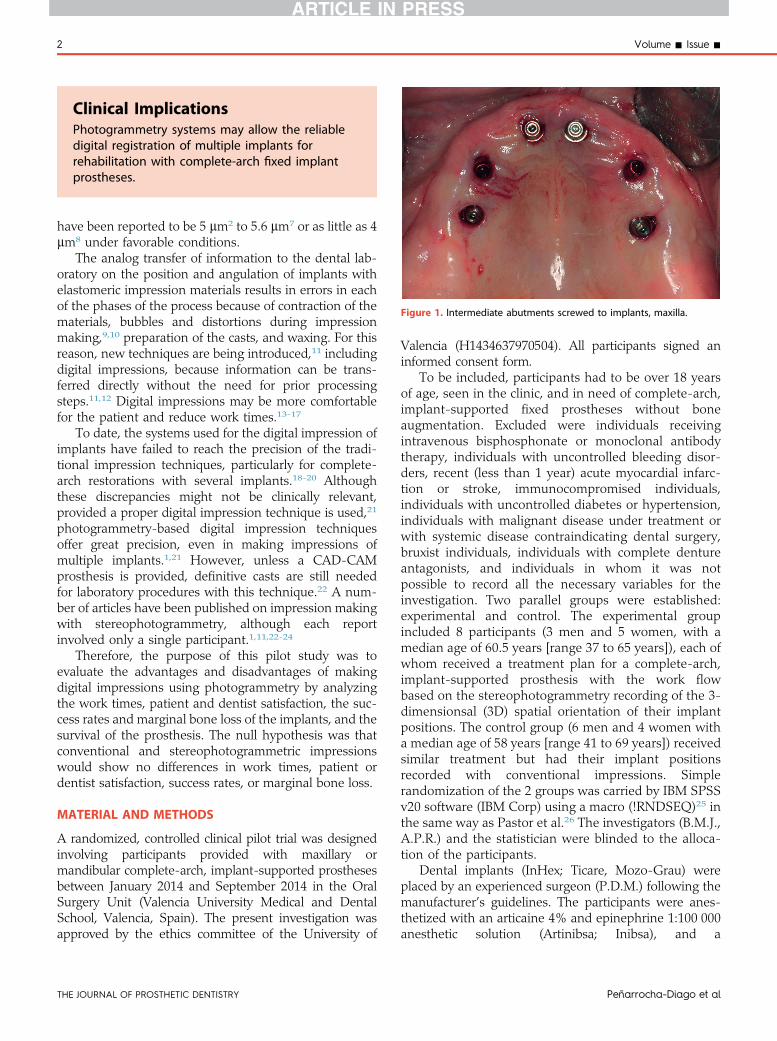
Figure 1. Intermediate abutments screwed to implants, maxilla.
Clinical ImplicationsPhotogrammetry systems may allow the reliabledigital registration of multiple implants forrehabilitation with complete-arch fixed implantprostheses.
2 Volume - Issue -
have been reported to be 5 mm2 to 5.6 mm7 or as little as 4mm8 under favorable conditions.
The analog transfer of information to the dental lab-oratory on the position and angulation of implants withelastomeric impression materials results in errors in eachof the phases of the process because of contraction of thematerials, bubbles and distortions during impressionmaking,9,10 preparation of the casts, and waxing. For thisreason, new techniques are being introduced,11 includingdigital impressions, because information can be trans-ferred directly without the need for prior processingsteps.11,12 Digital impressions may be more comfortablefor the patient and reduce work times.13-17
To date, the systems used for the digital impression ofimplants have failed to reach the precision of the tradi-tional impression techniques, particularly for complete-arch restorations with several implants.18-20 Althoughthese discrepancies might not be clinically relevant,provided a proper digital impression technique is used,21
photogrammetry-based digital impression techniquesoffer great precision, even in making impressions ofmultiple implants.1,21 However, unless a CAD-CAMprosthesis is provided, definitive casts are still neededfor laboratory procedures with this technique.22 A num-ber of articles have been published on impression makingwith stereophotogrammetry, although each reportinvolved only a single participant.1,11,22-24
Therefore, the purpose of this pilot study was toevaluate the advantages and disadvantages of makingdigital impressions using photogrammetry by analyzingthe work times, patient and dentist satisfaction, the suc-cess rates and marginal bone loss of the implants, and thesurvival of the prosthesis. The null hypothesis was thatconventional and stereophotogrammetric impressionswould show no differences in work times, patient ordentist satisfaction, success rates, or marginal bone loss.
MATERIAL AND METHODS
A randomized, controlled clinical pilot trial was designedinvolving participants provided with maxillary ormandibular complete-arch, implant-supported prosthesesbetween January 2014 and September 2014 in the OralSurgery Unit (Valencia University Medical and DentalSchool, Valencia, Spain). The present investigation wasapproved by the ethics committee of the University of
THE JOURNAL OF PROSTHETIC DENTISTRY
Valencia (H1434637970504). All participants signed aninformed consent form.
To be included, participants had to be over 18 yearsof age, seen in the clinic, and in need of complete-arch,implant-supported fixed prostheses without boneaugmentation. Excluded were individuals receivingintravenous bisphosphonate or monoclonal antibodytherapy, individuals with uncontrolled bleeding disor-ders, recent (less than 1 year) acute myocardial infarc-tion or stroke, immunocompromised individuals,individuals with uncontrolled diabetes or hypertension,individuals with malignant disease under treatment orwith systemic disease contraindicating dental surgery,bruxist individuals, individuals with complete dentureantagonists, and individuals in whom it was notpossible to record all the necessary variables for theinvestigation. Two parallel groups were established:experimental and control. The experimental groupincluded 8 participants (3 men and 5 women, with amedian age of 60.5 years [range 37 to 65 years]), each ofwhom received a treatment plan for a complete-arch,implant-supported prosthesis with the work flowbased on the stereophotogrammetry recording of the 3-dimensionsal (3D) spatial orientation of their implantpositions. The control group (6 men and 4 women witha median age of 58 years [range 41 to 69 years]) receivedsimilar treatment but had their implant positionsrecorded with conventional impressions. Simplerandomization of the 2 groups was carried by IBM SPSSv20 software (IBM Corp) using a macro (!RNDSEQ)25 inthe same way as Pastor et al.26 The investigators (B.M.J.,A.P.R.) and the statistician were blinded to the alloca-tion of the participants.
Dental implants (InHex; Ticare, Mozo-Grau) wereplaced by an experienced surgeon (P.D.M.) following themanufacturer’s guidelines. The participants were anes-thetized with an articaine 4% and epinephrine 1:100 000anesthetic solution (Artinibsa; Inibsa), and a
Peñarrocha-Diago et al
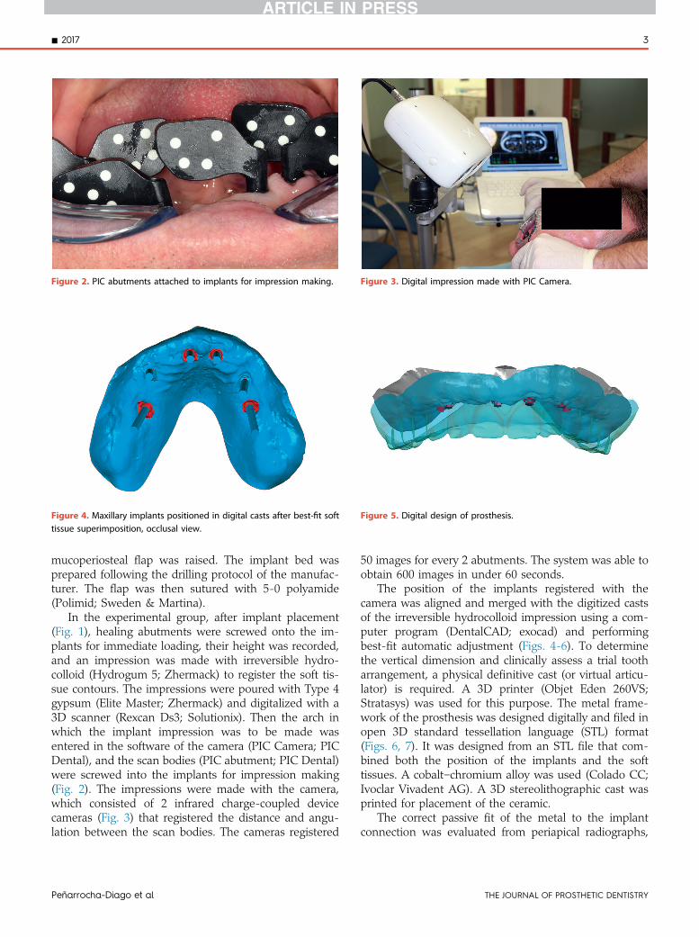
Figure 2. PIC abutments attached to implants for impression making. Figure 3. Digital impression made with PIC Camera.
Figure 4. Maxillary implants positioned in digital casts after best-fit softtissue superimposition, occlusal view.
Figure 5. Digital design of prosthesis.
- 2017 3
mucoperiosteal flap was raised. The implant bed wasprepared following the drilling protocol of the manufac-turer. The flap was then sutured with 5-0 polyamide(Polimid; Sweden & Martina).
In the experimental group, after implant placement(Fig. 1), healing abutments were screwed onto the im-plants for immediate loading, their height was recorded,and an impression was made with irreversible hydro-colloid (Hydrogum 5; Zhermack) to register the soft tis-sue contours. The impressions were poured with Type 4gypsum (Elite Master; Zhermack) and digitalized with a3D scanner (Rexcan Ds3; Solutionix). Then the arch inwhich the implant impression was to be made wasentered in the software of the camera (PIC Camera; PICDental), and the scan bodies (PIC abutment; PIC Dental)were screwed into the implants for impression making(Fig. 2). The impressions were made with the camera,which consisted of 2 infrared charge-coupled devicecameras (Fig. 3) that registered the distance and angu-lation between the scan bodies. The cameras registered
Peñarrocha-Diago et al
50 images for every 2 abutments. The system was able toobtain 600 images in under 60 seconds.
The position of the implants registered with thecamera was aligned and merged with the digitized castsof the irreversible hydrocolloid impression using a com-puter program (DentalCAD; exocad) and performingbest-fit automatic adjustment (Figs. 4-6). To determinethe vertical dimension and clinically assess a trial tootharrangement, a physical definitive cast (or virtual articu-lator) is required. A 3D printer (Objet Eden 260VS;Stratasys) was used for this purpose. The metal frame-work of the prosthesis was designed digitally and filed inopen 3D standard tessellation language (STL) format(Figs. 6, 7). It was designed from an STL file that com-bined both the position of the implants and the softtissues. A cobaltechromium alloy was used (Colado CC;Ivoclar Vivadent AG). A 3D stereolithographic cast wasprinted for placement of the ceramic.
The correct passive fit of the metal to the implantconnection was evaluated from periapical radiographs,
THE JOURNAL OF PROSTHETIC DENTISTRY
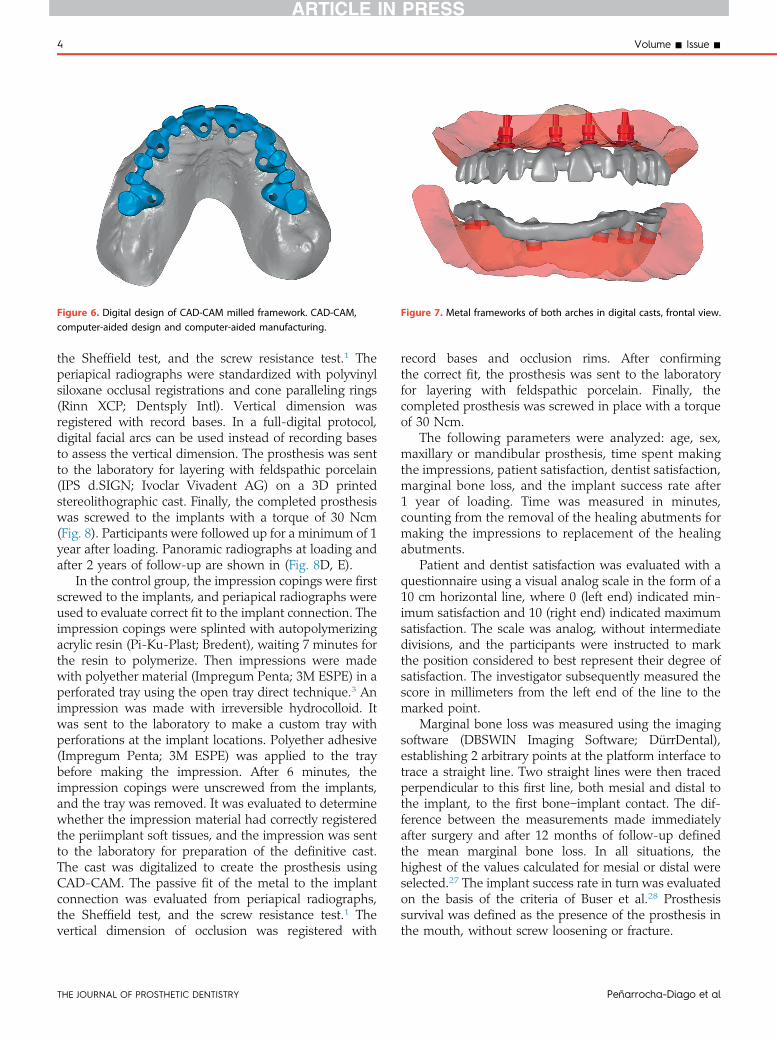
Figure 6. Digital design of CAD-CAM milled framework. CAD-CAM,computer-aided design and computer-aided manufacturing.
Figure 7. Metal frameworks of both arches in digital casts, frontal view.
4 Volume - Issue -
the Sheffield test, and the screw resistance test.1 Theperiapical radiographs were standardized with polyvinylsiloxane occlusal registrations and cone paralleling rings(Rinn XCP; Dentsply Intl). Vertical dimension wasregistered with record bases. In a full-digital protocol,digital facial arcs can be used instead of recording basesto assess the vertical dimension. The prosthesis was sentto the laboratory for layering with feldspathic porcelain(IPS d.SIGN; Ivoclar Vivadent AG) on a 3D printedstereolithographic cast. Finally, the completed prosthesiswas screwed to the implants with a torque of 30 Ncm(Fig. 8). Participants were followed up for a minimum of 1year after loading. Panoramic radiographs at loading andafter 2 years of follow-up are shown in (Fig. 8D, E).
In the control group, the impression copings were firstscrewed to the implants, and periapical radiographs wereused to evaluate correct fit to the implant connection. Theimpression copings were splinted with autopolymerizingacrylic resin (Pi-Ku-Plast; Bredent), waiting 7 minutes forthe resin to polymerize. Then impressions were madewith polyether material (Impregum Penta; 3M ESPE) in aperforated tray using the open tray direct technique.3 Animpression was made with irreversible hydrocolloid. Itwas sent to the laboratory to make a custom tray withperforations at the implant locations. Polyether adhesive(Impregum Penta; 3M ESPE) was applied to the traybefore making the impression. After 6 minutes, theimpression copings were unscrewed from the implants,and the tray was removed. It was evaluated to determinewhether the impression material had correctly registeredthe periimplant soft tissues, and the impression was sentto the laboratory for preparation of the definitive cast.The cast was digitalized to create the prosthesis usingCAD-CAM. The passive fit of the metal to the implantconnection was evaluated from periapical radiographs,the Sheffield test, and the screw resistance test.1 Thevertical dimension of occlusion was registered with
THE JOURNAL OF PROSTHETIC DENTISTRY
record bases and occlusion rims. After confirmingthe correct fit, the prosthesis was sent to the laboratoryfor layering with feldspathic porcelain. Finally, thecompleted prosthesis was screwed in place with a torqueof 30 Ncm.
The following parameters were analyzed: age, sex,maxillary or mandibular prosthesis, time spent makingthe impressions, patient satisfaction, dentist satisfaction,marginal bone loss, and the implant success rate after1 year of loading. Time was measured in minutes,counting from the removal of the healing abutments formaking the impressions to replacement of the healingabutments.
Patient and dentist satisfaction was evaluated with aquestionnaire using a visual analog scale in the form of a10 cm horizontal line, where 0 (left end) indicated min-imum satisfaction and 10 (right end) indicated maximumsatisfaction. The scale was analog, without intermediatedivisions, and the participants were instructed to markthe position considered to best represent their degree ofsatisfaction. The investigator subsequently measured thescore in millimeters from the left end of the line to themarked point.
Marginal bone loss was measured using the imagingsoftware (DBSWIN Imaging Software; DürrDental),establishing 2 arbitrary points at the platform interface totrace a straight line. Two straight lines were then tracedperpendicular to this first line, both mesial and distal tothe implant, to the first boneeimplant contact. The dif-ference between the measurements made immediatelyafter surgery and after 12 months of follow-up definedthe mean marginal bone loss. In all situations, thehighest of the values calculated for mesial or distal wereselected.27 The implant success rate in turn was evaluatedon the basis of the criteria of Buser et al.28 Prosthesissurvival was defined as the presence of the prosthesis inthe mouth, without screw loosening or fracture.
Peñarrocha-Diago et al
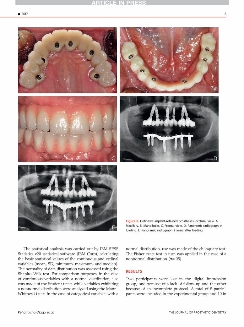
Figure 8. Definitive implant-retained prostheses, occlusal view. A,Maxillary. B, Mandibular. C, Frontal view. D, Panoramic radiograph atloading. E, Panoramic radiograph 2 years after loading.
- 2017 5
The statistical analysis was carried out by IBM SPSSStatistics v20 statistical software (IBM Corp), calculatingthe basic statistical values of the continuous and ordinalvariables (mean, SD, minimum, maximum, and median).The normality of data distribution was assessed using theShapiro-Wilk test. For comparison purposes, in the caseof continuous variables with a normal distribution, usewas made of the Student t test, while variables exhibitinga nonnormal distribution were analyzed using the Mann-Whitney U test. In the case of categorical variables with a
Peñarrocha-Diago et al
normal distribution, use was made of the chi-square test.The Fisher exact test in turn was applied in the case of anonnormal distribution (a=.05).
RESULTS
Two participants were lost in the digital impressiongroup, one because of a lack of follow-up and the otherbecause of an incomplete protocol. A total of 8 partici-pants were included in the experimental group and 10 in
THE JOURNAL OF PROSTHETIC DENTISTRY
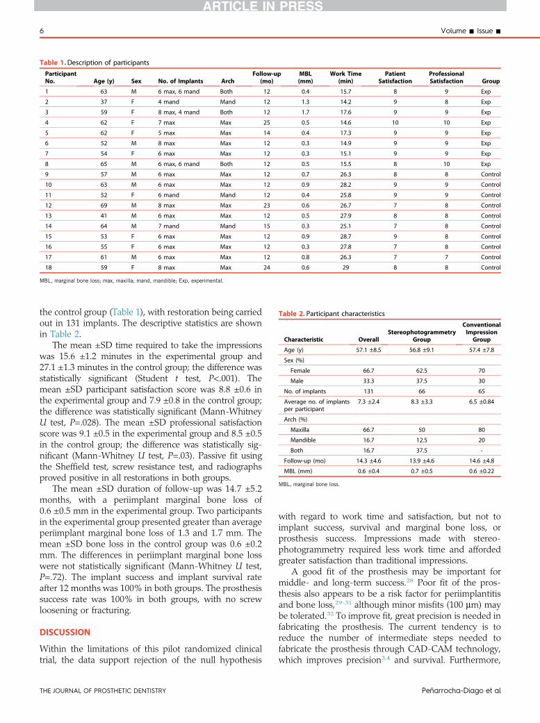
Table 1.Description of participants
ParticipantNo. Age (y) Sex No. of Implants Arch
Follow-up(mo)
MBL(mm)
Work Time(min)
PatientSatisfaction
ProfessionalSatisfaction Group
1 63 M 6 max, 6 mand Both 12 0.4 15.7 8 9 Exp
2 37 F 4 mand Mand 12 1.3 14.2 9 8 Exp
3 59 F 8 max, 4 mand Both 12 1.7 17.6 9 9 Exp
4 62 F 7 max Max 25 0.5 14.6 10 10 Exp
5 62 F 5 max Max 14 0.4 17.3 9 9 Exp
6 52 M 8 max Max 12 0.3 14.9 9 9 Exp
7 54 F 6 max Max 12 0.3 15.1 9 9 Exp
8 65 M 6 max, 6 mand Both 12 0.5 15.5 8 10 Exp
9 57 M 6 max Max 12 0.7 26.3 8 8 Control
10 63 M 6 max Max 12 0.9 28.2 9 9 Control
11 52 F 6 mand Mand 12 0.4 25.8 9 9 Control
12 69 M 8 max Max 23 0.6 26.7 7 8 Control
13 41 M 6 max Max 12 0.5 27.9 8 8 Control
14 64 M 7 mand Mand 15 0.3 25.1 7 8 Control
15 53 F 6 max Max 12 0.9 28.7 9 8 Control
16 55 F 6 max Max 12 0.3 27.8 7 8 Control
17 61 M 6 max Max 12 0.8 26.3 7 7 Control
18 59 F 8 max Max 24 0.6 29 8 8 Control
MBL, marginal bone loss; max, maxilla; mand, mandible; Exp, experimental.
Table 2. Participant characteristics
Characteristic OverallStereophotogrammetry
Group
ConventionalImpression
Group
Age (y) 57.1 ±8.5 56.8 ±9.1 57.4 ±7.8
Sex (%)
Female 66.7 62.5 70
Male 33.3 37.5 30
No. of implants 131 66 65
Average no. of implantsper participant
7.3 ±2.4 8.3 ±3.3 6.5 ±0.84
Arch (%)
Maxilla 66.7 50 80
Mandible 16.7 12.5 20
Both 16.7 37.5 -
Follow-up (mo) 14.3 ±4.6 13.9 ±4.6 14.6 ±4.8
MBL (mm) 0.6 ±0.4 0.7 ±0.5 0.6 ±0.22
MBL, marginal bone loss.
6 Volume - Issue -
the control group (Table 1), with restoration being carriedout in 131 implants. The descriptive statistics are shownin Table 2.
The mean ±SD time required to take the impressionswas 15.6 ±1.2 minutes in the experimental group and27.1 ±1.3 minutes in the control group; the difference wasstatistically significant (Student t test, P<.001). Themean ±SD participant satisfaction score was 8.8 ±0.6 inthe experimental group and 7.9 ±0.8 in the control group;the difference was statistically significant (Mann-WhitneyU test, P=.028). The mean ±SD professional satisfactionscore was 9.1 ±0.5 in the experimental group and 8.5 ±0.5in the control group; the difference was statistically sig-nificant (Mann-Whitney U test, P=.03). Passive fit usingthe Sheffield test, screw resistance test, and radiographsproved positive in all restorations in both groups.
The mean ±SD duration of follow-up was 14.7 ±5.2months, with a periimplant marginal bone loss of0.6 ±0.5 mm in the experimental group. Two participantsin the experimental group presented greater than averageperiimplant marginal bone loss of 1.3 and 1.7 mm. Themean ±SD bone loss in the control group was 0.6 ±0.2mm. The differences in periimplant marginal bone losswere not statistically significant (Mann-Whitney U test,P=.72). The implant success and implant survival rateafter 12 months was 100% in both groups. The prosthesissuccess rate was 100% in both groups, with no screwloosening or fracturing.
DISCUSSION
Within the limitations of this pilot randomized clinicaltrial, the data support rejection of the null hypothesis
THE JOURNAL OF PROSTHETIC DENTISTRY
with regard to work time and satisfaction, but not toimplant success, survival and marginal bone loss, orprosthesis success. Impressions made with stereo-photogrammetry required less work time and affordedgreater satisfaction than traditional impressions.
A good fit of the prosthesis may be important formiddle- and long-term success.28 Poor fit of the pros-thesis also appears to be a risk factor for periimplantitisand bone loss,29-31 although minor misfits (100 mm) maybe tolerated.32 To improve fit, great precision is needed infabricating the prosthesis. The current tendency is toreduce the number of intermediate steps needed tofabricate the prosthesis through CAD-CAM technology,which improves precision3,4 and survival. Furthermore,
Peñarrocha-Diago et al
- 2017 7
the periimplant marginal bone loss is similar to whentransepithelial abutments are used.5
To date, no reliable digital methods have been availablefor making complete-arch implant impressions, with thecumulative error observed to increase with the number ofimplants.20 Lee et al9 analyzed a system for making digitalimpressions over implants and found that traditional im-pressions with elastomeric materials offered greater pre-cision than digital impressions along the long axis of theimplant, with similar performance in terms of the rest ofthe studied magnitudes. Stereophotogrammetry hasrecently been used for making intraoral digital impres-sions.1,11 This system affords very high precision, with anerror of under 5 mm in in vitro studies2,7 and of less than 10mm in in vivo studies, independent of the number of im-plants involved.1,11 This allows shortening of the chain oferror, thanks to the direct digital transfer of the informationfrom the impressions to drilling of the working cast.13
Material contraction due to polymerization or CAD-CAMalignment is thereby prevented. However, previous pub-lished articles about stereophotogrammetry were in vitro oronly included a single participant. Furthermore, thesestudies concentrated on accuracy, survival, and marginalperiimplant bone loss, without considering other impor-tant variables such as work time and satisfaction that wereassessed in this study.
The time needed for impression making in the pre-sent study was shorter with the stereophotogrammetrytechnique, coinciding with the observations of otherauthors who likewise recorded shorter times with thedigital impressions compared with traditional impres-sions;13-16 however, Wismeijer et al17 recorded theopposite results in their study. An explanation for thisparadoxical result is that Wismeijer et al used a closedtray impression technique without splinting the implants.Digital impressions do not use materials that need topolymerize, reducing working time. In the present study,the conventional impression times were much longerthan in the aforementioned studies. A possible expla-nation for this difference is that the impressions weremade in participants undergoing restoration withcomplete-arch prostheses instead of crowns and short-span fixed dental prostheses. Furthermore, the studiesthat compared digital and conventional impressionsfailed to specify whether the implants were splintedbefore impression making.13,17 Splinting prolongs thetime needed to make an implant impression by at least15 minutes because it is necessary to wait for the resin topolymerize.
Reported satisfaction was high in both groups,thanks to the reduction of participant discomfort asso-ciated with impressions using elastomeric materials. Theexperimental group yielded significantly higher satis-faction scores among both the participants and the
Peñarrocha-Diago et al
professionals, in agreement with the observations ofother investigators.13,14,17 The improved satisfactionmay be due to the reduced scanning time, and place-ment of the scan bodies could be better tolerated thanelastomeric materials.
In the present study, no statistically significant dif-ferences were found in periimplant marginal bone loss, inimplant success and survival rates, or in prosthesis sur-vival between the stereophotogrammetry and conven-tional impression techniques. A possible explanation maybe that prosthesis fit was similar in both techniques,9 andminor misfits moreover do not affect the long-termoutcome.33
The limitations of the present investigation include thelack of soft tissue reproduction with the camera system:the cast must be scanned, or an intraoral digital scannermust be used. When the implants have greater proximityor converge, scanning can be done in separate phases,keeping attached at least one PIC abutment alreadyregistered in the first phase. Then the PIC abutment inproximity to another implant can be placed in the adjacentimplant to register it in a second scanning phase. This is apilot study on work time and satisfaction using the tech-nique. Further studies are needed to analyze in-mouthprecision, with the inclusion of larger samples and thequantification of the fit in clinical conditions.
CONCLUSIONS
Within the limitations of this pilot randomized clinicaltrial, the following conclusions were drawn:
1. The time needed for impression making was shorterwith the stereophotogrammetry technique.
2. Patient and dentist satisfaction was greater with thestereophotogrammetry technique.
3. Stereophotogrammetric and traditional impressionsshowed no differences in implant survival, marginalbone loss of the implants, or prosthesis survival after1 year of follow-up.
REFERENCES
1. Peñarrocha-Oltra D, Agustín-Panadero R, Bagán L, Giménez B,Peñarrocha M. Impression of multiple implants using photogrammetry:description of technique and case presentation. Med Oral Patol Oral CirBucal 2014;19:366-71.
2. Giménez B, Özcan M, Martínez-Rus F, Pradíes G. Accuracy of a digitalimpression system based on active wavefront sampling technology for im-plants considering operator experience, implant angulation, and depth. ClinImplant Dent Relat Res 2015;17:54-64.
3. de França DG, Morais MH, das Neves FD, Barbosa G. Influence of CAD/CAM on the fit accuracy of implant-supported zirconia and cobalt-chromiumfixed dental prostheses. J Prosthet Dent 2015;113:22-8.
4. Abduo J. Fit of CAD/CAM implant frameworks: a comprehensive review.J Oral Implantol 2012;40:758-66.
5. Kapos T, Evans C. CAD/CAM technology for implant abutments, crowns,and superstructures. Int J Oral Maxillofac Implants 2014;29:117-36.
6. Ortorp A, Jemt T, Bäck T. Photogrammetry and conventional impressions forrecording implant positions: a comparative laboratory study. Clin ImplantDent Relat Res 2005;7:43-50.
THE JOURNAL OF PROSTHETIC DENTISTRY
8 Volume - Issue -
7. Bergin JM, Rubenstein JE, Mancl L, Brudvik JS, Raigrodski AJ. An in vitrocomparison of photogrammetric and conventional complete-arch implantimpression techniques. J Prosthet Dent 2013;110:243-51.
8. Rivara F, Lumetti S, Calciolari E, Toffoli A, Forlani G, Manfredi E.Photogrammetric method to measure the discrepancy between clinicaland software-designed positions of implants. J Prosthet Dent 2016;115:703-11.
9. Lee SJ, Betensky RA, Gianneschi GE, Gallucci GO. Accuracy of digital versusconventional implant impressions. Clin Oral Implants Res 2014;26:715-9.
10. Paniz G, Stellini E, Meneghello R, Cerardi A, Gobbato EA, Bressan E. Theprecision of fit of cast and milled full-arch implant-supported restorations. IntJ Oral Maxillofac Implants 2013;28:687-93.
11. Pradíes G, Ferreiroa A, Özcan M, Giménez B, Martínez-Rus F. Using ster-eophotogrammetric technology for obtaining intraoral digital impressions ofimplants. J Am Dent Assoc 2014;145:338-44.
12. Papaspyridakos P, Chen CJ, Chuang SK, Weber HP, Gallucci GO.A systematic review of biologic and technical complications with fixedimplant rehabilitations for edentulous patients. Int J Oral Maxillofac Implants2012;27:102-10.
13. Koch GK, Gallucci GO, Lee SJ. Accuracy in the digital workflow: from dataacquisition to the digitally milled cast. J Prosthet Dent 2016;115:749-54.
14. Joda T, Brägger U. Patient-centered outcomes comparing digital and con-ventional implant impression procedures: a randomized crossover trial. ClinOral Implants Res 2016;27:e185-9.
15. Patzelt SBM, Lamprinos C, Stampf S, Att W. The time efficiency of intraoralscanners: an in vitro comparative study. J Am Dent Assoc 2014;145:542-51.
16. Lee SJ, Gallucci GO. Digital vs. conventional implant impressions: efficiencyoutcomes. Clin Oral Implants Res 2013;24:111-5.
17. Wismeijer D, Mans R, Van Genuchten M, Reijers HA. Patients’ preferenceswhen comparing analogue implant impressions using a polyether impressionmaterial versus digital impressions (Intraoral Scan) of dental implants. ClinOral Implants Res 2013;25:1113-8.
18. Howell KJ, McGlumphy EA, Drago C, Knapik G. Comparison of the accuracyof Biomet 3i Encode Robocast Technology and conventional implantimpression techniques. Int J Oral Maxillofac Implants 2013;28:228-40.
19. Al-Abdullah K, Zandparsa R, Finkelman M, Hirayama H. An in vitro com-parison of the accuracy of implant impressions with coded healing abutmentsand different implant angulations. J Prosthet Dent 2013;110:90-100.
20. Park JI, Yoon TH. A three-dimensional image-superimposition CAD/CAMtechnique to record the position and angulation of the implant abutmentscrew access channel. J Prosthet Dent 2013;109:57-60.
21. Ender A, Mehl A. Influence of scanning strategies on the accuracy of digitalintraoral scanning systems. Int J Comput Dent 2013;16:11-21.
22. Sánchez-Monescillo A, Sánchez-Turrión A, Vellon-Domarco E, Salinas-Goodier C, Prados-Frutos JC. Photogrammetry impression technique: a casehistory report. Int J Prosthodont 2016;29:71-3.
THE JOURNAL OF PROSTHETIC DENTISTRY
23. Peñarrocha-Oltra D, Agustín-Panadero R, Pradíes G, Gomar-Vercher S,Peñarrocha-Diago M. Maxillary full-arch immediately loaded implant-supported fixed prosthesis designed and produced by photogrammetry anddigital printing: a clinical report. J Prosthodont 2017;26:75-81.
24. Agustín-Panadero R, Peñarrocha-Oltra D, Gomar-Vercher S, Peñarrocha-Diago M. Stereophotogrammetry for recording the position of multiple im-plants: technical description. Int J Prosthodont 2015;28:631-6.
25. Doménech JM, Granero R. Extraction of N random integers from LN to HN.Macro RNDI for SPSS statistics: exhaustive sampling [computer program].Version 2011.09.09. Barcelona: Universitat Autònoma de Barcelona (UAB);2011. Available at: http://www.metodo.uab.cat/macros.htm. AccessedFebruary 2, 2015.
26. Pastor MA, López-Roig S, Lledó A, Peñacoba C, Velasco L, Schweiger-Gallo I. Combining motivational and volitional strategies to promoteunsupervised walking in patients with fibromyalgia: study protocol for arandomized controlled trial. Trials 2014;15:120.
27. Boronat A, Peñarrocha M, Carrillo C, Marti E. Marginal bone loss indental implants subjected to early loading (6 to 8 weeks postplacement)with a retrospective short-term follow-up. J Oral Maxillofac Surg 2008;66:246-50.
28. Buser D, Weber HP, Lang NP. Tissue integration of non-submerged im-plants. 1-year results of a prospective study with 100 ITI hollow-cylinder andhollow-screw implants. Clin Oral Implants Res 1990;1:33-40.
29. Brånemark PI. Osseointegration and its experimental background. J ProsthetDent 1983;50:399-410.
30. Saaby M, Karring E, Schou S, Isidor F. Factors influencing severity of peri-implantitis. Clin Oral Implants Res 2016;27:7-12.
31. Jokstad A, Shokati B. New 3D technologies applied to assess the long-termclinical effects of misfit of the full jaw fixed prosthesis on dental implants. ClinOral Implants Res 2015;26:1129-34.
32. Chen CJ, Papaspyridakos P, Guze K, Singh M, Weber HP, Gallucci G. Effectof misfit of cement-retained implant single crowns on crestal bone changes.Int J Prosthodont 2013;26:135-7.
33. Jemt T, Book K. Prosthesis misfit and marginal bone loss in edentulousimplant patients. Int J Oral Maxillofac Implants 1996;11:620-5.
Corresponding author:Dr Miguel Peñarrocha-DiagoClínicas OdontológicasGascó Oliag 1ValenciaSPAINEmail: [email protected]
Copyright © 2017 by the Editorial Council for The Journal of Prosthetic Dentistry.
Peñarrocha-Diago et al