A CLASS OF ITS OWN: FUNCTION-DISCOVERY OF HYDY,
205
A CLASS OF ITS OWN: FUNCTION-DISCOVERY OF HYDY, A NOVEL CLASS OF [FEFE]-HYDROGENASES A Dissertation by DARRELL WAYNE MARTIN JR Submitted to the Office of Graduate and Professional Studies of Texas A&M University in partial fulfillment of the requirements for the degree of DOCTOR OF PHILOSOPHY Chair of Committee, David P. Barondeau Committee Members, Marcetta Y. Darensbourg Paul A. Lindahl Frank M. Raushel Head of Department, Simon W. North August 2016 Major Subject: Chemistry Copyright 2016 Darrell Wayne Martin Jr
Transcript of A CLASS OF ITS OWN: FUNCTION-DISCOVERY OF HYDY,

A CLASS OF ITS OWN: FUNCTION-DISCOVERY OF HYDY,
A NOVEL CLASS OF [FEFE]-HYDROGENASES
A Dissertation
by
DARRELL WAYNE MARTIN JR
Submitted to the Office of Graduate and Professional Studies of
Texas A&M University
in partial fulfillment of the requirements for the degree of
DOCTOR OF PHILOSOPHY
Chair of Committee, David P. Barondeau
Committee Members, Marcetta Y. Darensbourg
Paul A. Lindahl
Frank M. Raushel
Head of Department, Simon W. North
August 2016
Major Subject: Chemistry
Copyright 2016 Darrell Wayne Martin Jr

ii
ABSTRACT
Hydrogen has received widespread attention as a potential energy carrier due to
its high energy content and clean combustion product H2O. [FeFe]-H2ases exhibit the
highest H2 production rates utilizing a complex iron sulfur cofactor, called the H-cluster,
that requires three biosynthetic maturation proteins. Mutagenesis studies of conserved
residues surrounding the H-cluster led to variants with either decreased overall activity
or minimal H-cluster incorporation. These studies underscore the importance of the
protein matrix in tuning active site chemistry. Here, we investigated a new class of
enzymes consisting of N-terminal [FeFe]-H2ase fused to a C-terminal rubrerythrin
domain (named HydY). Using protein film electrochemistry and colorimetric assays,
HydY was found to function differently than standard [FeFe]-H2ases: it exhibits strong
product inhibition for H+ reduction, a low KM for H2 oxidation, bias toward H2 oxidation
and significant overpotential. We hypothesized that the altered reactivity for HydY was
due to hydrogen bonds from conserved residue substitutions vital for H-cluster
coordination. Consistent with this hypothesis, HydY variants result in enzymes with
catalytic properties more similar to traditional [FeFe]-H2ases. In addition, the C-terminal
domain (CTD) of HydY efficiently reduces H2O2 to H2O. Electronic absorbance, EPR
and Mössbauer spectroscopic studies of the CTD are consistent with a rubrerythrin di-
iron active site with flanking mononuclear iron sites. A 1.77 Å crystal structure of the
CTD reveals a domain swapped dimer in which ligands for a modified di-iron
rubrerythrin active site are provided by residues across the dimer interface. Further, our

iii
results indicate that electrons generated by the oxidation of H2 are transferred to the
CTD, presumably for H2O2 reduction. This is the first example of H2-dependent
peroxidase. We hypothesize that evolution ‘tuned’ HydY to favor H2 oxidation and that
HydY has a protective role in anaerobic bacteria that allows survival upon transient
oxygen exposure. Additional bioinformatics indicate that HydY belongs to a broader
class of [FeFe]-H2ases that also use H2 as a reductant for various substrates. Overall,
these studies identify determinants for controlling active site chemistry of [FeFe]-H2ases
that may lead to improved design of biomimetic compounds with implications in energy
production.

iv
DEDICATION
This dissertation is dedicated to my loving, patient and supportive wife Elizabeth,
from whom I received constant encouragement and support kept me motivated to
complete my Ph.D.

v
ACKNOWLEDGEMENTS
I would like to acknowledge my advisor Dr. David Barondeau. He taught me
how to think critically and deeply, how to analyze a scientific idea to the core, to
understand the fundamentals of a research project. He taught me how to be a scientist,
and for that I will always be thankful and grateful. I would also I would like to thank my
committee members, Dr. Marcetta Darensbourg, Dr. Paul Lindahl and Dr. Frank Raushel
for their helpful suggestions, support and guidance throughout my career at Texas A&M.
I would like to thank my colleagues from the Barondeau lab, past and present, for
their helpful discussions and suggestions during my research endeavors. Specifically, I
would like to thank Seth Cory for his expertise with protein crystallography and my
previous undergraduate students Samuel Choi, Eric Redundo and Nathan Winser.
I must also acknowledge my invaluable and excellent collaborators. Mössbauer
was collected and interpreted by Josh Wofford and Dr. Paul Lindahl, PFE and protein
reconstitution with synthetic [2Fe]H was conducted by Garrett Williams in Dr. Anne
Jones lab at ASU and computational modeling was started by Dr. Haixai Li in Dr.
Michael Hall’s lab. I was lucky enough to travel to ASU to learn PFE, and I must again
thank both my advisor and Dr. Jones for the opportunity and experience. My research is
indebted to all my collaborators. Thank you also to Dr. James Sacchetini for allowing
me to use his Mosquito robot and x-ray equipment to set up crystal trays and crystal
screening. Thank you also to Stanford Synchrotron Radiation Laboratory for providing
opportunities to collect crystallography data at their synchrotron.

vi
Finally, I would like to thank my friends, family and again my wife, Elizabeth,
for their amazing support.

vii
TABLE OF CONTENTS
Page
ABSTRACT .............................................................................................................. ii
DEDICATION .......................................................................................................... iv
ACKNOWLEDGEMENTS ...................................................................................... v
TABLE OF CONTENTS .......................................................................................... vii
LIST OF FIGURES ................................................................................................... x
LIST OF TABLES .................................................................................................... xxii
CHAPTER I THE ANAEROBIC WAY OF LIFE ................................................... 1
Introduction ....................................................................................................... 1 H2 evolving microbes: Anaerobic metabolism.......................................... 2 H2 consuming bacteria .............................................................................. 5
Diversity of hydrogenases ................................................................................. 6 Structure and function of [Fe]-H2ases ....................................................... 7
Structure and function of [NiFe]-H2ases ................................................... 8 Structure and function of [FeFe]-H2ases ................................................... 12
Electrochemistry of hydrogenases ..................................................................... 21 Basics of protein film electrochemistry .................................................... 21
Protein film electrochemistry with hydrogenases ..................................... 26 Biosynthesis of hydrogenases ........................................................................... 30
Anaerobes and the problem with molecular oxygen ......................................... 33 Rubrerythrins ............................................................................................. 40
CHAPTER II EXPERIMENTAL SECTION FOR SUBSEQUENT
CHAPTERS .......................................................................................................... 51
Experimental methods for Chapter III ............................................................... 51 Reagents and general procedures .............................................................. 51 Cloning and protein expression ................................................................. 51
Protein purification .................................................................................... 54 EPR spectroscopy ...................................................................................... 56 Mössbauer spectroscopy ........................................................................... 56 H2 oxidation/H2 production assays ............................................................ 57 Oxygen inactivation assay ......................................................................... 57

viii
H2O2/O2 reduction steady-state measurements ......................................... 58 Reconstitution of hydrogenases with synthetic H-cluster ......................... 58 Electrochemistry........................................................................................ 59 H2-dependent reduction of CTD ............................................................... 60 Sequence alignments ................................................................................. 60
Experimental section for Chapter IV ................................................................. 61 Protein preparation .................................................................................... 61 Solution activity measurements ................................................................ 61 FTIR .......................................................................................................... 61 Protein-film electrochemistry .................................................................... 62 Computational modeling ........................................................................... 62
Experimental methods for Chapter V ................................................................ 62 Sequence alignments ................................................................................. 62 Protein purification .................................................................................... 63 Crystallization and data collection ............................................................ 63 Data collection, processing, structure determination ................................ 64
Experimental methods for Chapter VI .............................................................. 64 Sequence similarity network generation ................................................... 64 Sequence alignments ................................................................................. 65
CHAPTER III A CLASS OF ITS OWN: HydY, FROM CLOSTRIDIUM
SYMBIOSUM, IS A H2-DEPENDENT PEROXIDASE .................................... 66
Introduction ....................................................................................................... 66
Results ............................................................................................................... 68 Spectroscopic and functional characterization of the CTD ....................... 71 Functional characterization of CTD .......................................................... 80 Spectroscopic and functional characterization of the NTD ...................... 81 Electrochemical investigation of HydY
s and NTD
e/s ................................ 83
H2-dependent reduction of the CTD by NTDe .......................................... 89
Bioinformatic analysis of HydY homologs ............................................... 89 Discussion ......................................................................................................... 94
CHAPTER IV INVESTIGATION OF SECOND-SHELL CONTRIBUTIONS
FOR HYDY CATALYSIS ................................................................................... 99
Introduction ....................................................................................................... 99 Results ............................................................................................................... 101
FTIR Spectroscopy .................................................................................... 101
Solution characterization of NTD substitutions ........................................ 104 Interrogation of NTD constructs by PFE .................................................. 106 Computational investigation for hydrophilic active site substitutions ...... 108
Discussion ......................................................................................................... 110

ix
CHAPTER V STRUCTURAL INVESTIGATION OF CTD ................................... 113
Introduction ....................................................................................................... 113 Results ............................................................................................................... 117
Sequence alignment of rubrerythrins ........................................................ 117 Overall fold ............................................................................................... 120 Subunit interactions ................................................................................... 122 The diiron site............................................................................................ 124 Solvent canyon .......................................................................................... 127
Discussion ......................................................................................................... 128
CHAPTER VI INVESTIGATION OF THE DIVERSITY OF
HYDROGENASES THROUGH PROTEIN SIMILARITY NETWORKS ........ 133
Introduction ....................................................................................................... 133 Results ............................................................................................................... 135
Sequence similarity network of [FeFe]-H2ases ......................................... 135 Analysis of sub-group 4 ............................................................................ 139 Analysis of PAS domain proteins ............................................................. 143
Discussion ......................................................................................................... 144
CHAPTER VII CONCLUDING REMARKS .......................................................... 148
REFERENCES .......................................................................................................... 153

x
LIST OF FIGURES
Page
Figure I-1 Highlighting the role of [FeFe]-H2ases in fermentative H2
production. A) Fermentative H2 production pathway in
clostridia. There are two routes for H2 production. One is
linked to the action of pyruvate:ferredoxin oxidoreductase and
reducing equivalents can be passed to monomeric or
bifurcating H2ases (1). The second pathway involves
NADH:ferredoxin oxidoreductase (2), but only operates under
extremely low H2 partial pressures. B) Stoichiometry of
anaerobic fermentation of glucose .................................................... 3
Figure I-2 Structure of Fe-H2ase from Methanocaldococcus jannaschii
determined to 1.75 Å (PDB entry: 3F47). Inset reveals active
site of the [Fe]-H2ase consisting of a Cys residue, two CO
molecules, pyridol-GMP and a coordinated water from solvent....... 8
Figure I-3 Structure of [NiFe]-H2ase from Desulfovibrio gigas (PDB
entry: 2FRV). All [NiFe]-H2ases identified to-date are
heterodimeric, with one subunit encoded Fe-S clusters (green)
and the other housing the catalytic subunit (blue). Inset reveals
the catalytic site made up of a Ni atom (green sphere)
coordinated by four Cys residues, two of which bridge to an Fe
atom (orange sphere). The Fe contains one CO and two CN-
ligands. A bridging H2O (red sphere), likely a hydroxide, is
modeled ............................................................................................. 9
Figure I-4 Schematic depiction and structure of O2-tolerant ReMBH. (a)
ReMBH consists of a [NiFe]-H2ase subunit (cyan), an Fe-S
cluster containing subunit (green) and an integral membrane
subunit (magenta). The proximal [4Fe-3S] is circled. (b) The
proximal [4Fe-3S] displays redox-dependent structural
changes. The Fe atoms are ligated by all thiol (sulfides and
thiolates), whereas the superoxided the Fe atoms are ligated by
a peptide amide and a water molecule. Adapted and reprinted
by permission from Macmillan Publishers Ltd: Nature,
Frielingdorf, S et al, 2014 ................................................................. 11
Figure I-5 Structure of CpI (PDB entry: 3C8Y). A) Overall fold of
[FeFe]-H2aes CpI with minimal H-cluster domain colored
green and ferredoxin(F)-domain in grey. B) Active site of CpI
consisting of [4Fe-4S]H and [2Fe]H. The [2Fe]H consists of a

xi
two irons coordinated by CO, CN and a unique azadithiolate.
The irons are named according to proximity to the [4Fe-4S]H,
proximal (Fep) and distal (Fed). Fed is modeled with a terminal
water .................................................................................................. 13
Figure I-6 Sequence alignment of classical [FeFe]-H2ases studied in vitro
recently. Residues are shaded according to a 90% sequence
identity threshold ............................................................................... 14
Figure I-7 Cartoon depictions of modular domains of clostridial [FeFe]-
H2ases. M, Monomeric; D, dimeric; TR, trimeric; TE,
tetrameric; bD, binding domain; Fd, ferredoxin; CODH, carbon
monoxide dehydrogenase. Republished with permission of
Microbiology, from The surprising diversity of clostridial
hydrogenases: a comparative genomic perspective, Calusinka,
M, Happe, T, Joris, B, Wilmotte, A,156, 2010; permission
conveyed through Copyright Clearance Center, Inc ......................... 20
Figure I-8 Model for protein film electrochemistry. Reprinted and adapted
with permission from Vincent, KA, Parkin, A, Armstrong, FA,
Chem Rev, 107 (10), 2007. Copyright 2007 American
Chemical Society ............................................................................... 22
Figure I-9 pH dependence of the 2H+/H2 equilibrium potential as dictated
by the Nernst equation ....................................................................... 23
Figure I-10 A comparison of voltammograms of EcHyd-1 at pH 3.0
recorded under stationary (blue) and rotating conditions
(black). Reproduced from Ref 101 with permission of The
Royal Society of Chemistry. dx.doi.org/10.1039/C3EE43652G ...... 25
Figure I-11 Electrocatalytic CVs of various hydrogenases adsorbed onto a
PGE electrode. A) [NiFe]-H2ases Hyd-1 and Hyd-2 with 10%
H2 atm, pH 6 and scan rate = 1 mV s-1
. B) [FeFe]-H2ases Cr
and CaHydA with 100% H2 atm, pH 6 and scan rate = 5 mV s-
1. Blue lines indicate Eeq and orange line indicate the onset
potential. Red circles indicate oxidative inactivation. Arrows
indicate the scan direction. Redox potentials of commonly used
redox dyes are indicated on the bottom (methyl viologen, MV;
benzyl viologen, BV; methylene blue, MB). Reprinted with
permission from Armstrong, FA, Evans, RM, Hexter, SV, et al.
Accounts of Chemical Research 49 (2016) ....................................... 26
Figure I-12 Potential window for H2 oxidation activity for four different
H2ases, defined as the range from Eonset to oxidative

xii
inactivation. Vincent, KA, Parkin, A, Armstrong, FA, Chem
Rev, 107 (10), 2007. Copyright 2007 American Chemical
Society ............................................................................................... 28
Figure I-13 Cartoon depiction for biosynthesis of [2Fe]H. a) Two possible
pathways for [FeFe]-hydrogenase maturation. In both
pathways, the maturation process involves synthesis and
assembly of the 2Fe subcluster of the H-cluster, followed by
insertion of this 2Fe subcluster to generate the active
hydrogenase. JBIC, H-cluster assembly during maturation of
the [FeFe]-hydrogenase, 19 (6), 2014, 747-757, Broderick, JB,
with permission from Springer. b) Formation of the Fe
synthon by HydG. Reprinted from BBA – Molecular Cell
Research, 1853 (6), Peters, JW, Schut, GJ, Boyd, ES, Mulder
DW, Shepard, EM, Broderick, JB, King PW, Adams, MWW,
[FeFe]- and [NiFe]-hydrogenase diversity, mechanism, and
maturation, 1350-1369, Copyright 2015 with permission from
Elsevier.
http://www.sciencedirect.com/science/journal/01674889 ................ 31
Figure I-14 Structures of CpI reveal the structurally rigid H-cluster binding
residues. A) CpI reconstituted semi-synthetically with
azadithiolate (PDB entry: 4XDC). B) CpI expressed in the
absence of maturation factors, CpIIM
(PDB entry: 4XDD).
Overall RMSD: 0.33 Å ...................................................................... 32
Figure I-15 Schematic depiction of the distribution of various microbes in
within pond waters. Adapted from ref 49. Copyright 2005
American Chemical Society .............................................................. 34
Figure I-16 Reduction potential diagram for oxygen at pH 7. The units are
in V vs SHE. Adapted from ref 125 .................................................. 35
Figure I-17 Reactive oxygen species enzyme response system in anaerobic
bacteria. Reprinted with permission from ref 128 ............................. 39
Figure I-18 Schematic for steady-state electron delivery to Rbr for
peroxidase function. Reprinted from Journal of Bacteriology,
525, Mishra, S, Imlay, J, Why do bacteria use so many
enzymes to scavenge hydrogen peroxide?, 145-160, Copyright
2012, with permission from Elsevier.
http://www.sciencedirect.com/science/journal/00039861 ................ 43
Figure I-19 X-ray crystal structures of DvRbr (A) and PfRbr (B)
homodimer structures. The two subunits are colored as grey
xiii
and black. Ferric iron atoms are depicted as orange spheres.
PDB entry: 1RYT and 3MPS ............................................................ 44
Figure I-20 Diagrams of Rbr active site based on all-ferric and all-ferrous
structures. The redox toggling Fe is depicted in red. Reprinted
from the Journal of Inorganic Biochemistry, 100, Kurtz, DM,
Avoiding high-valent iron intermediates: Superoxide reductase
and rubrerythrin, 679-693, Copyright 2006, with permission
from Elsevier.
http://www.sciencedirect.com/science/journal/01620134 ................ 46
Figure I-21 Views of the diiron center of rubrerythrin and its homolog,
symrerythrin, with azide bound. A) Azide binds µ,1-3 in the
structure of diferrous DvRbr (PDB entry: 1LKP). B) Azide
binds µ,1 in the structure of diferric symrethrin (PDB entry:
3SID) ................................................................................................. 47
Figure I-22 Modified reaction scheme for rubrerythrins. Similar to Figure
I-20, all the glutamate side chains have been removed for
clarity, with the exception of those involved in redox toggling.
Proposed hydrogen-bonding interactions are highlighted in red.
The distances are based on the crystallographic models.
Consistent with the proposed binding mode for peroxide, our
data provide insight into the orientation of oxygen atoms in the
strictly conserved glutamic acid side chain (E83 in PfRbr)
relative to bound peroxide as well as evidence that the bridging
oxygen atom in the mixed-valence state is indeed
exchangeable. Reprinted from the Journal of Biological
Inorganic Chemistry, A cryo-crystallographic time course for
peroxide reduction by rubrerythrin from Pyrococcus furiosus,
16, 2011, 949-59, Dillard, BD, Demick, JM, Adams, MW,
Lanzilotta, WN, copyright Journal of Biological Inorganic
Chemistry with permission of Springer ............................................ 50
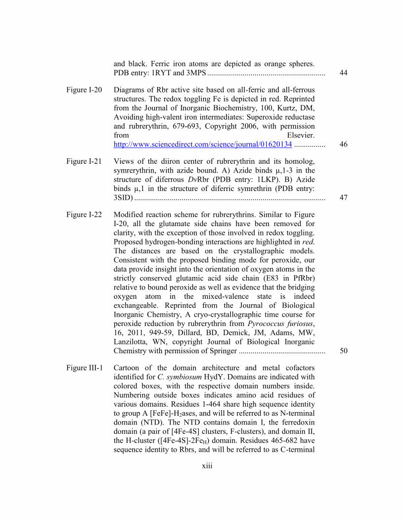
Figure III-1 Cartoon of the domain architecture and metal cofactors
identified for C. symbiosum HydY. Domains are indicated with
colored boxes, with the respective domain numbers inside.
Numbering outside boxes indicates amino acid residues of
various domains. Residues 1-464 share high sequence identity
to group A [FeFe]-H2ases, and will be referred to as N-terminal
domain (NTD). The NTD contains domain I, the ferredoxin
domain (a pair of [4Fe-4S] clusters, F-clusters), and domain II,
the H-cluster ([4Fe-4S]-2FeH) domain. Residues 465-682 have
sequence identity to Rbrs, and will be referred to as C-terminal
xiv
domain (CTD). Domains III and V appear to encode
rubredoxin (Rd) domains; domain IV appears to encode a
diiron domain most similar to Rbrs. Here, Rbr will refer to just
the diiron active site (and not the flanking mononuclear iron Rd
sites) to simplify the nomenclature. Standard nomenclature
refers to Rbr as a diiron site with a C-terminal Rd, whereas
reverse-Rbr refers to a diiron site with a N-terminal Rd. No
previously characterized Rbr contains both N- and C-terminal
Rds ..................................................................................................... 68
Figure III-2 Analytical gel filtration reveals quaternary structure of HydY
constructs. NTD (1 mg/mL, solid line) elutes at its calculated
molecular weight (54 kD). CTD (1 mg/mL, dashed line) elutes
as a tetramer (120 kD; monomer MW = 28 kD). HydYIM
(5
mg/mL, dotted line) elutes at molecular weight of 230 kD
(monomer molecular weight 80 kD), indicating a trimer. Based
on oligomers formed from the synthetic constructs, HydY
likely is an elongated dimeric species, causing it to elute at
slightly larger molecular weight. All runs are the average of at
least two experiments. Inset indicates standards run to estimate
molecular weight of HydY constructs ............................................... 69
Figure III-3 Potential dependence of electrocatalytic reduction of H2O2 by
HydYIM
. The black trace shows the current of the cyclic
voltammogram of HydYIM
in the presence of 0.15 mM H2O2.
The dotted line is the response of a freshly polished electrode
without a protein film. Cyclic voltammograms are obtained at a
potential scan rate of 20 mV s-1
and electrode rotation rate of
2750 rpm in mixed buffer at pH 7. The arrow indicates the
starting point and direction of potential cycling ................................ 70
Figure III-4 HydYIM
shows time-dependent scavenging for H2O2.
Chronoamperometry of HydIM
absorbed onto a rotating PGE
electrode poised at -0.3 V in the presence of 0.2 mM H2O2.
Multiple injections of 0.15 mM H2O2 (marked by arrows)
electrocatalytic response to additional substrate. Other
experimental conditions include: electrode rotation rate 2750
rpm and buffer pH of 7.0. .................................................................. 71
Figure III-5 Spectroscopic evidence that the CTD (HydY residues 465-682)
contains Rbr and Rd iron sites. a) Electronic absorbance of
CTDox (red), CTDox after addition of 1 M NaN3 (blue), and
CTDred (grey). Mössbauer of 0.75 mM 57
Fe-CTD in oxidized
(b) and reduced (c) states. The red lines are simulated spectra

xv
of the total components, while other colors represent
simulations of individual components (see Supplemental Table
1). The oxidized samples were prepared by addition of 4
equivalents of H2O2. The reduced samples were prepared by
addition of 2-fold excess of dithionite in the presence of 50 μM
methyl viologen (as redox mediator) ................................................ 72
Figure III-6 Reductive titration of oxidized CTD by dithionite. Four
reducing equivalents (2 mol dithionite) are required to
completely bleach chromophores associated with oxidized
CTD ................................................................................................... 73
Figure III-7 Mössbauer spectra of 57
Fe-CTD. Spectra of 0.75 mM57
Fe-
CTD (a) oxidized state recorded at 150 K (b) and a partially
oxidized sample recorded at 5 K and 0.05 mT field. The red
lines are simulated spectra of the total components, while other
colors represent simulations of individual components (see
Table III-2) ........................................................................................ 75
Figure III-8 CTD contains multiple iron binding sites. EPR spectrum of as-
isolated CTD (a) or 57
CTD in oxidized (b), partially oxidized
(c) and fully reduced (d) states. Parameters for EPR
measurement were: sample temperature, 10K; microwave
frequency, 9.43 GHz; microwave power, 0.519 mW ........................ 77
Figure III-9 Consumption of H2O2 by CTD is dependent upon Rd and
NROR concentration. a) The rate of NADH oxidation
dependent upon rubredoxin (Rd) concentration, saturating with
50 µM Rd. A Michaelis-Menten fit was applied to give the
following parameters: kcat, 380 ± 40 min-1
; Km, 8 ± 2 µM;
kcat/Km, 8 ± 2 x10⁶ M
-1 s
-1. b) The rate of NADH oxidation
saturated with 5 µM NADH:Rd oxidoreductase (NROR). The
data was fit with the Michaelis-Menten equation to give the
following parameters: kcat, 230 ± 10 min-1
; Km, 0.25 ± 0.07 µM;
kcat/Km, 1.5 ± 0.43 x10⁷ M
-1 s
-1. All assays were performed at
10-12°C, following loss of NADH signal (see Materials and
Methods), with 0.1 mM H2O2 and 0.5 µM NROR (a) or 0.5 µM
Rd (b). One unit is defined as 1 µmol H2O2 reduced per
minute (1 µmol NADH reduced 1 µmol H2O2). Background
consumption of NADH was subtracted from measured rates ........... 78
Figure III-10 The CTD is an efficient H2O2 reductase. Steady-state
reduction of H2O2 was investigated using an in vitro system of
0.2 mM NADH, 5 μM NROR, 50 μM Rd, and 0.1 μM CTD

xvi
The decrease of NADH absorbance (ε340 = 6.22 mM-1
cm-1
)
was used to measure initial rate of H2O2 consumption after
injection of the CTD. Data was fit to the Michaelis-Menten
equation to yield the following kinetic parameters: kcat, 1890 ±
70 min-1; Km, 3.0 ± 0.8 µM; kcat/Km, 1.1 ± 0.3 x10⁷ M-1
s-1
. All
assays were performed at 10-12°C. Background consumption
of NADH was subtracted from measured rates ................................. 79
Figure III-11 Spectroscopic investigation of the NTDIM
. a) Electronic
absorbance of as-isolated NTDIM
(25 µM) reveals broad
absorbance features between 300 and 500 nm, typical of Fe-S
centers. b) EPR spectrum of reduced NTDIM
. Parameters for
EPR measurement were: sample temperature, 10K; microwave
frequency, 9.43 GHz; microwave power, 0.519 mW. Sample
was prepared by adding excess NaDT to as-isolated NTDapo
(150 µM) ........................................................................................... 81
Figure III-12 Inactivation of NTDe
by O2. NTDe (2 mL) in buffer C in a
septum-sealed dram vial (20 mL) was stirred by stir plate
incubated at 22 °C. At zero time, the septum was removed to
expose NTDe to air. At the designated intervals, samples were
analyzed for residual H2 uptake activity (see Materials and
Methods). Data was fit to exponential model, yielding a fit
y=119.7e-0.433x and a t1/2 of 1.44 minutes ..................................... 83
Figure III-13 CsHydY is biased toward H2 oxidation. a) NTDe under
continuous sparging of 1 atm 100% H2 (black trace, 0.78 mM)
and 1 atm N2 (gray trace) with 5 mV s-1
scan rate. The light
gray dotted scan is an electrode under the same conditions that
has not been exposed to enzyme. b) NTDs under 1 atm 100%
H2. HydYs under 1 atm of c) 100% H2 or d) 1% H2. Other
experimental conditions include: electrode rotation rate 2750
rpm and buffer pH 7.0. The horizontal line indicates zero
current and the vertical line indicates the H+/H2 couple at pH
7.0 and 1 atm 100% H2 (-420 mV, a-c) or 1% H2 (-350 mV, d) ....... 84
Figure III-14 Catalytic cyclic voltammograms for NTDs adsorbed to a PGE
electrode over a range of pH values. The pH values shown are
8.0, 7.5, 6.5, 6.0, 5.5, 5.0, 4.5, and 4.0. Other experimental
conditions include: electrode rotation rate 2750 rpm and
potential scan rate of 20 mV s-1
. Catalytic signals were
normalized such that the average activity at 0 mV at pH 7 is 1.
The concentration of dissolved hydrogen is held at 0.78 mM by

xvii
continuously sparging with 1 atm of hydrogen gas. The black
arrow signifies the starting potential and scan direction ................... 85
Figure III-15 Cyclic voltammograms of HydYs adsorbed to a PGE electrode
over a range of pH values. a) Reverse, reductive sweep of
HydYs electrochemical activity at pH values 8.0, 7.5, 6.5, 6.0,
5.5, 5.0, 4.5, and 4.0 (forward, oxidative scan omitted to better
reveal onset potential). b) Potential of H2 oxidation onset as a
function of pH; the grey diagonal line indicates the expected
equilibrium potential for the 2H+/H2 couple pH dependence.
The onset potential (Eonset) is defined as the zero-current
intercept. c) pH dependence of anaerobic, oxidative
inactivation for HydYs. The switch potential (Eswitch) was
obtained from the inflection point of the reductive reactivation
area, determined from the derivative (-di/dE) of the reverse
scan .................................................................................................... 87
Figure III-16 H2-dependent reduction of CTD by NTDe. a) The CTDox (20
µM, blue trace) in H2-saturated exhibits no changes in spectral
features. Upon anaerobic addition of NTDe
(88 nM), spectral
features of oxidized CTD dissipate, replaced with features
consistent with reduced CTD. b) Kinetic analysis of a)
monitored at 490 nm for CTD (blue diamonds) or 456 nm for
E. coli Fdx (green circles). ................................................................ 88
Figure III-17 Phylogenetic grouping of modular HydY homologs. a)
Phylogenetic tree of CsHydY homologs identified by BLAST
(December 2015) calculated using MrBayes (scale bar: 0.2
substitutions per site, 15000000 generations sampled every
1000 generations with an average standard deviation of split
frequencies <0.0002) and visualized with FigTree. Homologs
of HydY were identified in Firmicutes, Fusobacteria and
Proteobacteria. b) HydY homologs are highly modular and
separate into three classes. Class I contains [4Fe-4S] cluster, H-
cluster, Rd and Rbr binding domains. Sub-classes of Class I (a-
c) retain a C-terminal rubrerythrin domain, but vary in putative
electron transfer domains. The class II HydY homolog has a C-
terminal domain with high sequence identity to glutamate
synthase domain. Class III homologs contain only varying
numbers of Rd domains and lack putative C-terminal redox
active sites ......................................................................................... 90
Figure III-18 Representation of the H-cluster in C. pastuerinium HydA1
(CpI; PDB:3C8Y) with important protein interactions.

xviii
Hydrogen bonds are shown in yellow. Protein residues within
van der Wall’s contact are depicted in cyan. Hydrophobic
interactions (A230 to bridging CO) are shown in pink. Note,
the bridgehead of the azadithiolate was modeled as oxygen,
and was colored blue to represent currently accepted
bridgehead nitrogen. .......................................................................... 91
Figure III-19 Sequence alignment reveals conserved substitutions near H-
cluster and [4Fe-4S] cluster motifs. Alignment of N-terminal
domain of CsHydY with structurally characterized [FeFe]-
H2ases C. reinhardtii (Cr), D. desulfuricans (Dd) and C.
pasteurianum I (CpI) or biochemically characterized [FeFe]-
H2ases C. acetobutylicum (Ca) and C. pasteurianum II (CpII).
MUSCLE alignments were visualized by Jalview and residues
are colored by sequence identity, with intensity color increment
of 100%. Conserved motifs for the H-cluster domain are
boxed: P1,TSCCPxW; P2,MPCxxKxxE; P3,
ExMACxxGCxxG; and previously unidentified ‘P4,’ APxVR.
Both CpII and HydY show similar substitutions in P1, P3 and
P4 (yellow boxes) motifs. A ‘positive patch’ of Arg and Lys
residues (red triangles) within the F-cluster domain is also
found in HydY and CpII .................................................................... 92
Figure III-20 Sequence logos of HydY homologs reveal conservation of
hydrophilic substitutions within H-cluster and F-cluster motifs.
Green one letter codes indicate conservation in HydY that are
identical to well-characterized [FeFe]-H2ases. Blue codes
indicate conservation in HydY homologs that are different in
typical [FeFe]-H2ases, and grey indicate variable regions. b) P1
motif is completely conserved. c) P2 motif contains a mostly
conserved Thr substituted for Met, which is within VDW
contact of bridging CO (PDBID: 3C8Y). d) P3 motif contains a
mostly conserved ThrAla substitution, which would be near
the cubane cluster of [4Fe-4S]H. e) HydY homologs contain
completely conserved AlaSer substitution. This Ala is within
3 Å of bridging CO. f) A positive patch of mostly conserved
Arg/Lys/Gln near a [4Fe-4S] motif ................................................... 93
Figure III-21 Model of H2-dependent peroxidase role of HydY. Under
typical, reducing cellular conditions, HydY is inactive. An
oxidative shift (caused by ROS) activates the NTD and triggers
oxidation of excess H2 that was generated during anaerobic
metabolism. Reducing equivalents recycled from H2 are
directly transferred to CTD for H2O2, and maybe O2, reduction ...... 97

xix
Figure IV-1 Crystallographic model of the [FeFe]-H2ase active site taken
from the structure of CpI (PDB 3C8Y). Interactions from
protein to [2Fe]H are depicted as: yellow, polar interactions;
blue, hydrogen bonds; magenta, hydrophobic interactions.
Note, the bridgehead was changed from O to N to reflect
current literature ................................................................................ 100
Figure IV-2 FTIR spectra NTD in as-isolated (a) and oxidized (b)
conditions. All spectra were subtracted from reference sample
with buffer only. Experimental conditions: temperature, 21 °C;
resolution, cm-1
; number of sample scans, 1024; protein
concentration, 700 µM ...................................................................... 102
Figure IV-3 HydY variants exhibit starkly different electrocatalytic profiles.
A new batch of purified wild-type HydY (red) shows activity
similar to that reported previously. NTDS97A (blue) and
NTDS97A/T221M (black) display decreased resistance to
oxidative inactivation and decrease H2 oxidation activity.
Experimental conditions include: electrode rotation rate 2750
rpm, 1 atm 100% H2, 5 mV s-1
scan rate, and phospate buffer
pH 7.0 ................................................................................................ 105
Figure IV-4 Electrocatalytic profiles of NTDS97A (a) and
NTDS97A/T221M (b) vs pH in the presences of 1 atm of
100% H2. The legend indicates the pH of the 0.1 M phosphate
buffer with 0.15 M KCl. Other experimental conditions
include: electrode rotation rate 2750 rpm and a 5 mV s-1
scan
rate ..................................................................................................... 107
Figure IV-5 Transient catalytic current measured for H2 oxidation by wild-
type HydY (a) and NTDS97A/T221M (b), following the
electrochemical cell being flushed with N2. Inset reveals the
residual plot after fitting with equation IV-1. Experimental
conditions: E, 0 mV; rotation rate, 2750 rpm; temperature,
21°C ................................................................................................... 108
Figure IV-6 Optimized structures of Fe(I)Fe(II)[H-] in wild-type (a) and
with water (b) modeling NTDS97. The labels r1-r5 refer to
bond distances in Å, and energies are in kcal/mol ............................ 109
Figure V-1 Depictions of Rbr active site based on diferric and diferrous
structures. The redox toggling Fe (Fe1 is depicted in red. Main
chain atoms are not depicted for clarity. Reprinted from the
Journal of Inorganic Biochemistry, 100, Kurtz, DM, Avoiding
high-valent iron intermediates: Superoxide reductase and

xx
rubrerythrin, 679-693, Copyright 2006, with permission from
Elsevier.
http://www.sciencedirect.com/science/journal/01620134 ................ 116
Figure V-2 Sequence alignment of Rbr homologs and CsHydY-CTD.
Coloring based on a 40% sequence identity threshold. Red box
highlight the 7th
Glu characteristic of Rbr proteins ........................... 118
Figure V-3 Overall fold adopted by CTD. a) Represents the domain-
swapped dimer found in the asymmetric unit. Each subunit is
colored differently for clarity. b) Tetramer of formed from
extensive contacts within the crystal ................................................. 121
Figure V-4 Overlays of RdB domain of CTD (green) with oxidized DvRbr
(a, salmon) and reduced PfRbr (b, purple) ........................................ 123
Figure V-5 2Fo – Fc map reveals the active site of CTD. Clearly from this
structure, the 7th
Glu (E148) is rotated away from the active
site, not coordinating either iron atom. Fe1 is represented as the
leftmost iron ...................................................................................... 124
Figure V-6 Overlay of the CTD diiron site (green, cyan) with oxidized
DvRbr (a, salmon) and reduced PfRbr (b, purple). Main chain
atoms are omitted for clarity. Iron and water atoms are
discolored slightly to specify which structure to which they
belong ................................................................................................ 125
Figure V-7 X-ray fluorescence data of a CTD crystal before (blue) and
after (red) data collection at SSRL beamline 7-1 reveals x-ray
photoreduction ................................................................................... 127
Figure V-8 Surface representation of the CTD dimer reveals a solvent
canyon near the diiron active site. Multiple coordinated waters
are found here, along with a PEG-400. The blue and green
surfaces represent subunit 1 and 2, respectively ............................... 128
Figure VI-1 Sequence similarity network of [FeFe]-H2ase sequences using
a stringency expect value of E-150
...................................................... 135
Figure VI-2 Domain architectures of [FeFe]-H2ase sequences identified
from the sequence similarity network. The green box indicates
an H-cluster binding domain ............................................................. 136
Figure VI-3 Conserved H-cluster binding motifs P1-P4 that provide non-
covalent interactions to the cofactor (a). Conserved motifs

xxi
identified in sub-group 4 of the sequence similarity network of
P1 (b), P2 (c), P3 (d), P4 (e) and SPQ (f). Weblogos are
colored according to chemical identity. A bit score of 4
indicates a completely conserved residue ......................................... 140
Figure VI-4 Depiction of the [4Fe-4S] cluster from CpI (PDB: 3C8Y; a)
and the corresponding positive patch sequence logo from sub-
group 4 from the sequence similarity network. Colors of the
one letter codes indicate chemical identity, and conserved
residues have bit scores of 4 .............................................................. 142
Figure VI-5 Sequence logos proposed H-cluster binding motifs of PAS
domain fusions identified. Conservations of substituted
residues in P1 (a), P2 (b), P3 (c), P4 (d) and SPQ (e) imply that
PAS domains would bind an H-cluster in an usual manner, if at
all ....................................................................................................... 143

xxii
LIST OF TABLES
Page
Table II-1 Strains and plasmids used in this study ............................................. 52
Table III-1 Mössbauer parameters of CTD in comparison with similar iron-binding proteins ......................................................................... 74
Table III-2 Mössbauer parameters of 57
CTD ....................................................... 76
Table III-3 H2ase activity parameters for NTDe and various other [FeFe]-
H2ases ................................................................................................ 82
Table IV-1 Solution activity measurements for NTD and variants ..................... 104
Table V-1 Data collection and refinement statistics for CsCTD ........................ 119

1
CHAPTER I
THE ANAEROBIC WAY OF LIFE
Introduction
Since the discovery of H2 oxidation and production pathways in the early 20th
century,1 the metabolism of H2 has been reported in bacteria, archaea and eukaryotes.
2,3
H2 metabolism is now abundantly clear to be essential to many microbes: from
organotrophs (microbes that catabolize organic compounds) to phototrophs (microbes
that harness solar energy) and lithotrophs (microbes that use inorganic substrates for
reducing equivalents), aerobes to anaerobes alike. H2 has even been postulated to have
been central to primordial evolution of life.4
Recent studies suggest that H2 metabolism is far more widespread (and diverse)
than previously postulated.5,6
Central to H2 metabolism are hydrogenases (H2ases).
These enzymes are named according their active site metals: [FeFe], [NiFe] and [Fe]
(see Diversity of hydrogenases). In general, [FeFe]-H2ases are described to favor H2
production and are utilized in H2 production pathways, [NiFe]-H2ases in H2 oxidation
pathways and [Fe]-H2ases specific to H2 uptake in methanogens. Dependent mostly on
[FeFe]-H2ases, strict anaerobic environments were originally speculated to be the only
niche for H2 production pathways, but the discovery that phototrophs produce H2
challenged that assumption.7 Soil surface bacteria display H2-dependent growth,
presumably scavenging H2 from the lower atmosphere, utilizing [NiFe]-H2ases.8 This
finding of aerobic H2 oxidation disproved the dogma that these metabolisms can exist

2
only in low O2/high H2 environments. Additionally, using deep genome sequencing,
over half the identified microbiota flora in higher eukaryotes5,9,10
contain at least one
H2ase enzyme. For microbes identified in the Human Microbiome Project, [FeFe]-
H2ases are detected in higher numbers than the other two types of H2ases.9 Since H2 does
not accumulate in the gut of humans, interspecies H2 transfer (H2 syntrophy) is proposed,
where microbes import substrates and export metabolic end products to create a
‘community budget’ for resources necessary for survival.11
Here, the focus will be on H2 production pathways, with a particular primary
focus on anaerobic fermentation, and the enzymes utilized for this reaction. A short
vignette on H2 consuming metabolic pathways (and the required enzymes) will also be
described.
H2 evolving microbes: Anaerobic metabolism
Anaerobic fermentation is a process of energy production from substrate-level
phosphorylation using oxidation of organic matter and relievement of excess reducing
equivalents by evolving H2.12
The route of fermentative H2 production begins with
glycolysis. Carbohydrates, specifically glucose, are preferred substrates in this
pathway,13
where these substrates are converted to pyruvate, ATP and NADH in both
strict anaerobes (such as members of the genus Clostridium) and facultative anaerobes
(such as the proteobacteria Escherichia coli, Ec). Most Clostridria convert pyruvate to
acetyl-CoA and CO2 using pyruvate ferredoxin oxidoreductase (PFOR).13-15
PFOR
utilizes a thiamine cofactor to activate pyruvate by two successive, one electron
oxidation reactions that are transferred through Fe-S cluster cofactors to ferredoxins

3
(Fds).16,17
Reduced ferredoxins are used to produce H2 via [FeFe]-H2ases. Hydrogenases
act as terminal electron acceptors, harnessing the low potential electrons from pyruvate
oxidation (E’ = -500 mV) to drive proton reduction (Figure I-1).3,15
A similar pathway
exists for the green algae Chlamdynomous rheinhardtii (Cr) through dark fermentation,18
as it also does in the hyperthermophile Pyrococcus furiousus (Pf), although this archaea
instead uses a [NiFe]-H2ase to accomplish the same chemistry.19
Maximally, 4 moles of
H2 can be produced per mole of glucose from glycolysis. NADH has been speculated to
act as an electron source for H2 production from monomeric H2ases.13
This reaction uses
NADH:ferredoxin oxidoreductase (NFOR) and can only proceed at very low H2 partial
Figure I-1. Highlighting the role of [FeFe]-H2ases in fermentative H2 production. A)
Fermentative H2 production pathway in clostridia. There are two routes for H2
production. One is linked to the action of pyruvate:ferredoxin oxidoreductase and
reducing equivalents can be passed to monomeric or bifurcating H2ases (1). The
second pathway involves NADH:ferredoxin oxidoreductase (2), but only operates
under extremely low H2 partial pressures. B) Stoichiometry of anaerobic
fermentation of glucose.

4
pressures (see Electrochemistry of Hydrogenases). Highlighting the ingenuity of nature,
recent studies of heteromultimeric [FeFe]-H2ases have been found to minimize energy
loss. These enzymes couple the endergonic reaction of NADH oxidation (E’ = -320 mV)
with ferredoxin oxidation (E’ = -450 to -500 mV) to use protons as electron acceptors.20-
23 NAD
+ can be regenerated to form acetate, butyrate or ethanol. Under certain cellular
conditions, NAD+ can be formed directly from pyruvate, forming lactate.
13,14 Most
Clostridium sp also reduce nitrate to ammonia,24
but this is a H2-independent pathway
and lies outside the scope of this review. Acetate fermentation maximally yields four
moles of H2, while butyrate only yields 2 moles.13
Rarely are these yields obtained due
to various other metabolic shunts for reducing equivalents.
In facultative anaerobes, pyruvate is converted to acetyl-CoA and formate by
pyruvate formate lyase (PFL).13
Under acidic conditions and high formate
concentrations, formate is disproportionated to CO2 and H2 by a membrane bound
formate hydrogenlyase (FHL) complex.4 Formate oxidation to electrons, protons and
CO2 occurs at a molybdenum- and selenocysteine-dependent enzyme, and these reducing
equivalents are transferred to a [NiFe]-H2ase (Hyd-3 in Ec) to produce H2. Since two
formate molecules are produced per mole of pyruvate, PFL theoretically produces 2
moles of H2. Competing pathways for formate minimize H2 production from pyruvate.13
The FHL reaction is highly reversible. Indeed, Hyd-3 has received recent attention due
to its intriguing chemistry of ‘fixing’ H2 into a more easily transportable form, formate.25
Outside of anaerobic fermentation, H2 is produced through several other
pathways.4 H2 photoproduction pathways exist in a few green algae, including

5
Chlamdynomous rheinhardtii, Cr, that depend on both photosystem (PS) I and II (direct)
or only PS II (indirect).18,26
The direct pathway requires light-induced oxidation of water
at PS I, electron transfer from PS I to PS II, and then light-dependent excitation of those
electrons to reduce ferredoxin (called PetF). PetF then transfers electrons to an [FeFe]-
H2ase for H2 production. The indirect pathway appears to only require PS I reduced
plastoquinone, and may represent a mechanism to dissipate excess reducing equivalents
under specific cellular conditions.18
Additionally, H2 is also produced as a by-product of
N2 fixation by nitrogenase-containing microbes. Nitrogenase activates the inert N2
molecule using a specialized metallocofactor with the chemical formula MFe7S9,
M=Mo, V or Fe.27-31
Though nitrogenases show significantly slower catalytic rates of H2
production in comparison to hydrogenases, this paradoxical H2 production by
nitrogenase is speculated to be a major source of the H2 oxidized by soil bacteria.4,8
To
further demonstrate the breadth of hydrogenase activity, a fairly new class of bacteria
has been identified that couples CO oxidation to H2 production.4 This new class of
bacteria, called hydrogenogens, can be grown exclusively on CO as a carbon source,
utilizing a [NiFe]-H2ase and membrane-bound carbon monoxide dehydrogenase.32
CO is
a potent inhibitor for all H2ases,7 so these [NiFe]-H2ases must somehow minimize CO
diffusion into the active site.
H2 consuming bacteria
In comparison, the major metabolic activities of H2-consuming bacteria are wide-
ranging.2,3
Some bacteria found at surface waters use H2 as an electron donor and O2 as
the terminal electron acceptor.33,34
Such microbes are called Knallgas (German for

6
‘bang-gas’) bacteria. Ralstonia eutropha (Re) is the best studied Knallgas bacteria. Re
uses a membrane bound hydrogenase [NiFe]-H2ase (MBH) anchored to the periplasmic
side of the cytoplasm.4 This MBH is critical for establishing a H
+ gradient and
transferring electrons to quinone and cytochromes for respiration. ReMBH has attracted
significant attention recently due to its ability to maintain H2 oxidation in O2.33,35,36
Furthermore, sulfate-reducing bacteria couple H2 oxidation to sulfate reduction, with H2
likely a product of syntrophy. Sulfate-reducing bacteria utilize either [FeFe]- or [NiFe]-
H2ases to accomplish H2 oxidation, dictated by either metal availability or H2 partial
pressure. There are also the so-called ‘ancient’ pathways for H2 oxidation:
methanogenesis and acetogenesis.21
Methanogens can convert H2 and CO2 (or methanol
in some cases) to methane and water. The reducing power of H2 is harnessed by multiple
H2ases, including membrane-associated and soluble [NiFe]-H2ases as well as an [Fe]-
only H2ase. The [Fe]-H2ase appears to be up-regulated under nickel-limiting conditions.4
In addition, fumarate respiration is a H2-dependent pathway.3 Oxidation of H2 requires a
periplasmic oriented membrane-bound [NiFe]-H2ase that transfers electrons to a
membrane associated fumarate reductase anchored on the cytoplasmic side of the
membrane. Lastly, additional H2 oxidation dependent pathways exist in bacteria,
including metal reduction (respiration), dehalogenation respiration and anaerobic
photosynthesis, that require [NiFe]-H2ases.4
Diversity of hydrogenases
Hydrogenases are complex catalysts utilized by myriad microorganisms to
reversibly reduce protons to H2. All H2ases share a common theme of first row, low-

7
valent transition metals stabilized by small inorganic ligands (CO and/or CN-), which
were first detected by FTIR spectroscopy. While all H2ases share active site similarity,
they belong to three phylogenetically distinct H2ases classes and are named according to
the transition metal content: [NiFe]- and [FeFe]-H2ases, for which protons, electrons,
and H2 are the only substrates and products, and [Fe]-H2ases that couples H2 uptake to
pterin reduction in methanogens.37
[FeFe]-H2ases are superior hydrogenase catalysts,38
with H2 production turnover frequencies39
of 6,000-9,000 s-1
and H2 oxidation turnovers
estimated up to 50,000 s-1
,40
while [NiFe]-H2ases show catalytic rates at least an order of
magnitude lower.40,41
Ratios of H2 production and consumption point to the metabolic
roles of each respective enzyme. This section will focus primarily on [FeFe]-H2ases with
strong comparisons to [NiFe]-H2ases.
Structure and function of [Fe]-H2ases
[Fe]-H2ases, named because the active site contains only a single iron atom are
also called Hmd H2ases (H2-evolving N5-N10-methlenetetrahydromethanopternin
dehydrogenases).42-44
[Fe]-H2ases catalyze the uptake of H2 to reduce a methenyl-
tetrahydromethanopterin (methylene-H4MPT). These H2ases were originally discovered
in methanogenic Methanothermobacter marburgensis.43
A homolog from
Methanocaldococcus jannaschii was crystalized, revealing the active site of this unusual
H2ase binds two CO ligands at an octahedral iron site (Figure I-2).45
Surprisingly, the
iron appears to remain redox neutral, transferring a hydride to reduce methenyl-H4MPT+
methylene-H4MPT.

8
Structure and function of [NiFe]-H2ases
As deduced from the discussion of H2 metabolism, [NiFe]-H2ases are diffused
across microbial communities. The minimal core of the [NiFe]-H2ase consists of a
catalytic subunit that incorporates a Ni-Fe(CO)(CN)2 cofactor with additional Fe-S
cluster binding motifs for electron transfer (Figure I-3).46
[NiFe]-H2ases reversibly react
with oxygen, leading to considerable interest in [NiFe]-H2ase-based biotechnological
applications. While commonly described in the literature as H2 uptake biased, [NiFe]-
H2ases have diverse catalytic activities, highlighted above.
Phylogenetic analysis of large data sets of [NiFe]-H2ase sequences have led to a
classification of [NiFe]-H2ases, consisting of four different groups. Group 1 H2ases are
membrane-bound and energy transducing, coupling the oxidation of H2 to electron
Figure I-2. Structure of Fe-H2ase from Methanocaldococcus jannaschii
determined to 1.75 Å (PDB entry: 3F47). Inset reveals active site of the [Fe]-
H2ase consisting of a Cys residue, two CO molecules, pyridol-GMP and a
coordinated water from solvent.

9
transport phosphorylation. Group 2 H2ases contain soluble uptake hydrogenases found in
cyanobacteria and specialized sensory H2ases that act as signal transducing components.
Group 3 H2ases are found in archaeal and bacteria species. This group consists of
complex subunits and often called bidirectional; the catalytic direction in vivo is highly
sensitive to the cellular environment. Lastly, group 4 H2ases are multisubunit and
membrane bound. These are typically utilized in vivo for H2 production coupled to
oxidation of another substrate, similar to EcPFL.3
Significant interest in H2ase interactions with O2 have led to a greater
understanding of O2 inactivation and the identification and characterization of H2ases
that retain activity after O2 exposure, so-called O2-tolerant enzymes. Initial reports with
Figure I-3. Structure of [NiFe]-H2ase from Desulfovibrio gigas
(PDB entry: 2FRV). All [NiFe]-H2ases identified to-date are
heterodimeric, with one subunit encoded Fe-S clusters (green) and
the other housing the catalytic subunit (blue). Inset reveals the
catalytic site made up of a Ni atom (green sphere) coordinated by
four Cys residues, two of which bridge to an Fe atom (orange
sphere). The Fe contains one CO and two CN- ligands. A bridging
H2O (red sphere), likely a hydroxide, is modeled.

10
[NiFe]-H2ases indicated O2 exposure caused formation of an inactive species that
required long incubations under H2 before activity returned.47
Two spectroscopically
distinct species are formed. A quickly recovering “ready” Ni-B, and a slowly
reactivating “unready” Ni-A arise from O2 exposure. Structural studies led to models in
which the Ni-B and Ni-A states resulted from hydroxide and peroxide molecules bound
to the Ni ion, respectively.46
Hypotheses to improve O2 tolerant H2ases included limiting
access of O2 to the active site and somehow avoiding the formation of Ni-A. Structural
and modeling studies identified gas channels that connected the active site with the
surface of the protein.46
Remarkably, mutations designed to constrict the hydrophobic
channel did result in increased O2 tolerance.48
Recently, ReMBH was found to oxidize
H2 in O2.49
Reports first circulated that this increased O2 tolerance was due to limited O2
diffusion to the active site;50
however, spectroscopic studies indicated an exclusive
formation of Ni-B and an apparently never-before characterized [Fe-S] species.51
Two structural reports, published in quick succession, elucidated the
determinants for the remarkable O2 tolerance of this class of [NiFe]-H2ases (Figure I-
4).35,52
ReMBH is attached to the cytoplasmic membrane and consists of a large subunit
containing the active site, and a small subunit that contains three Fe-S cluster centers that
function in electron transfer. Traditional [NiFe]-H2ases contain [4Fe-4S] clusters ligated
by four cysteine residues (Figure I-3). In contrast, ReMBH contains a specialized cluster
ligated by six cysteine residues, where one sulfide ion is replaced by two thiols, leading
to [4Fe-3S] cluster (Figure I-4b). This unusual ligation scheme appears to facilitate two
redox transitions within a narrow redox window – transitions unlikely for typical [4Fe-

11
4S]-clusters.53
Upon O2 binding, four electrons are quickly passed directly to [NiFe] site
to completely reduce O2 to water,54
thus avoiding Ni-A formation. Recent evidence
suggests that the [NiFe] site provides one electron, while proximal and medial clusters
donate two and one electrons, respectively.55
Although electron flow is “uphill”
thermodynamically to the [NiFe] site, electron transfer rates are efficient because the
overall reaction (reduction of O2 to water) is much more thermodynamically
Figure I-4. Schematic depiction and structure of O2-tolerant ReMBH. (a)
ReMBH consists of a [NiFe]-H2ase subunit (cyan), an Fe-S cluster
containing subunit (green) and an integral membrane subunit (magenta).
The proximal [4Fe-3S] is circled. (b) The proximal [4Fe-3S] displays
redox-dependent structural changes. The Fe atoms are ligated by all thiol
(sulfides and thiolates), whereas the superoxided the Fe atoms are ligated
by a peptide amide and a water molecule. Adapted and reprinted by
permission from Macmillan Publishers Ltd: Nature, Frielingdorf, S et al,
2014.

12
“downhill.”56
This MBH has been used in a fuel cell, oxidizing H2 at the anode, at
ambient O2 concentrations.57
Structure and function of [FeFe]-H2ases
[FeFe]-H2ases (previously known as Fe-only H2ases) are mostly found in strict
anaerobic eubacteria, such as fermentative bacteria (Clostridium, Thermotoga,
Ruminococcus) and sulfate reducers (Desulfovibrio), and occasionally in eukaryotes,
including some anaerobic protozoa and green algae (Chlamydomonas). [FeFe]-H2ases
have been given the name HydA, with a number to indicate the genomic context.58,59
High-resolution X-ray crystallographic data now exits for representative bacterial H2ases
Clostridium pastueranium HydA-1 (CpI) and Desulfovibrio desulfuricans HydA (Dd) as
well as algal Cr (Figure I-5). For most bacterial HydA enzymes, a variable length N-
terminal domain, called the F-domain, that contains a chain of Fe-S clusters. These
clusters are spaced ~12 Å apart, which is ideal for facile electron transfer, and lead from
the surface to the active site. The H-cluster active site is contained within the C-terminal
core, and is an extended six iron core. The H-cluster consists of a typical [4Fe4S] cluster
with a bridging cysteine ligand coordinating an additional two iron subsite (2FeH). The
2FeH contain two low spin irons (distal and proximal Fe, named according to proximity
to the [4Fe-4S] cluster), each coordinated by biologically toxic CN- and CO ligands.
Along with a bridging CO, the 2FeH is also bridged by dithiolate ligand. This unique
ligand has only recently unequivocally been demonstrated to be an azadiothiolate,60
and
the bridgehead N is positioned to act as a catalytic base for catalysis.7

13
[FeFe]-H2ases are quite modular in domain architecture. Based on the structure
of CrHydA1, the core catalytic domain is called the H-domain. This domain is
comprised of all the cysteines required to coordinate the H-cluster as well as amino acids
that produce second-shell interactions: P1, TSCCPxW; P2, MPCxxKxxE; and P3,
ExMACxxGCxxGGGxP (Figure I-6).3 Canonical H-domains contain a span of ~350
residues with these strictly conserved motifs, represented in the structures mentioned
above. Some identified homologs have extended H-domains up to 800 residues.
Substitutions within these motifs, or cases where a motif is absent, are considered non-
canonical H-domains, as in the case of NAR proteins.61
Many [FeFe]-H2ases contain
additional, accessory domains. A variable length F-domain is the most common domain
found in [FeFe]-H2ase homologs, exemplified in structures of CpI and Dd. A multitude
Figure I-5. Structure of CpI (PDB entry: 3C8Y). A) Overall fold of [FeFe]-H2aes CpI
with minimal H-cluster domain colored green and ferredoxin(F)-domain in grey. B)
Active site of CpI consisting of [4Fe-4S]H and [2Fe]H. The [2Fe]H consists of a two irons
coordinated by CO, CN and a unique azadithiolate. The irons are named according to
proximity to the [4Fe-4S]H, proximal (Fep) and distal (Fed). Fed is modeled with a
terminal water.

14
of other accessory domains have been identified, including many hydrogenase fusion
proteins, and highlight the modular nature of [FeFe]-H2ases in strict anaerobes. Attempts
to separate HydA homologs into distinct phylogenetic groupings have failed, likely due
to variability of additional domains.2,3,6
Monomeric H2ases can be considered the most classical in regards to both in
vitro and in vivo function. For example, the structurally characterized CpI and the well-
studied C. acetobutylicum (Ca) HydA are both monomeric and found in the cytoplasm.7
Both are astonishingly active enzymes, with CpI catalyzing H2 evolution at 5500 U/mg
protein and H2 oxidation at 24,000 U/mg protein (one U is defined as 1 µmol H2
produced or consumed per min)62
and CaHydA exhibits comparable activity. The ratio
Figure I-6. Sequence alignment of classical [FeFe]-H2ases studied in vitro recently.
Residues are shaded according to a 90% sequence identity threshold.

15
of oxidation/evolution rates is 4.36). CpI and CaHydA are also extremely sensitive to
O2, where exposure to atmosphere O2 results in a complete abatement of hydrogenase
activity (T1/2 < 5 min). In vivo, both these monomeric H2ases function in relieving
anaerobic fermenters of reducing equivalents by evolving H2.3 Results indicate these
types of monomeric H2ases accept low potential electrons from ferredoxins (Fdx),
derived from oxidation of pyruvate to acetyl-CoA by pyruvate:ferredoxin oxidoreductase
(E’ -520 mV) – the final step of anaerobic glycolysis. CrHydA is an additional
‘classical’ monomeric H2ase from green algae. In vitro, CrHydA behaves similarly to
the clostridial H2ases above, but its hydrogenase activity is somewhat lower (H2
evolution, 1000 U/mg; H2 oxidation, 1000 U/mg; oxidation/reduction, 10).63,64
Additionally, CrHydA appears confined to chloroplasts in vivo, utilized for redox
balancing during dark fermentation or light driven H2 production, where electrons from
photosystem I and transferred to CrHydA by a ferredoxin called PetF.26,65,66
In summary,
classical [FeFe]-H2ases are monomeric, capable of both fast H2 evolution and oxidation
and function in vivo primarily in evolving H2.
In contrast, Dd is heterodimeric and located in the periplasm. The structure of Dd
revealed a 42 kD catalytic H-domain with two [4Fe-4S] clusters in the F-domain,
appearing quite similar to CpI. However, Dd structure also contained a small subunit
consisting of an 11 kD ‘belt’ peptide surrounding the H-domain.67,68
The exact function
of the small subunit is unknown, but has been speculated to aid in signaling transport to
the periplasm. Dd is suspected to be involved in promoting a H+ gradient in the
periplasm by oxidizing H2. Desulfovibrio desulfuricans, interestingly, also encodes

16
[NiFe]-H2ases.3 Why sulfate reducers employ both hydrogenases also remains unclear.
In vitro, Dd is highly active in H2 production (Vmax, 10,400 U/mg) and H2 consumption
(50,000 U/mg), yielding a ratio of activities similar to monomeric hydrogenases (4.8).40
Additionally, Dd can be purified from the periplasm aerobically, meaning it retains
activity in the presence of air. Aerobically purified Dd has much lower hydrogenase
activity and requires reductive activation for maximal H2 oxidation activity.40
Somewhat
surprisingly, if Dd is reduced with H2, the resulting enzyme is now inactivated by
oxygen at similar rates as monomeric H2ases.
While monomeric and dimeric [FeFe]-H2ases catalyze one of the simplest
chemical reactions, more complicated reactions involving hydrogenases have been
reported.3,6,14
For instance, studies in cell lysates of fermentative anaerobes have been
known to display NAD(P)H-dependent H2 evolution.69
Use of NAD(P)H (E’ = -320 mV)
is thermodynamically unfavorable for H2 production (E’ = -420 mV). Recently,
heterotrimeric and heterotetrameric [FeFe]-H2ases have been found to catalyze this
reaction in Thermotoga maritima, Ruminococcus albus and Acetobacterium woodii,
among others; these complexes are called bifurcating hydrogenases (BF-H).15,22,23,59,70
BF-Hs are complexes of H- and F-domain containing proteins (α-subunit), a ß-subunit
with a FMN cofactor and NAD(P)H binding site, ferredoxin binding γ subunit with an
occasional fourth subunit. BF-Hs catalyze H2 production (Vmax, 7.8 U/mg) in the
presence of reduced ferredoxin and NADH and H2 oxidation in presence of oxidize
ferredoxin and NAD+
(Vmax, 49.4 U/mg).15
Surprisingly in vitro, BF-Hs show no activity
in absence of pyridine nucleotides or ferredoxin. BF-Hs have been ascribed a role in

17
redox balance by conserving energy. In fermentative bacteria, BF-Hs are proposed to
regenerate NAD+ for glycolysis by evolving H2,
15 whereas in acetogens BF-H’s are
speculated to oxidize H2.21
In addition to BF-Hs, acetogenic bacteria, such A. woodii,
also encode for a heterotetrameric H2-dependent carbon dioxide reductase (HDCR),
catalyzing the reduction of CO2 to formate using H2.21,71
HDCRs encode an [FeFe]-
H2ase with a H-domain and two [4Fe-4S]-clusters in the F-domain, a Mo/W-
molybdopterin and two (or sometimes three) electron transfer subunits that bind [4Fe-
4S]. Sequence alignments of both BF-Hs and HDCRs indicate a canonical H-domain.
Hidden within the annals of the scientific literature is C. pasteurianum
hydrogenase II (CpII), an [FeFe]-H2ase with unusual catalytic properties. CpII was first
isolated and characterized from the cytoplasm of native source by Chen and co-workers
in 1978.72
CpII appeared monomeric, was a poor H2 production catalyst and from the
preparation it was unclear of the protein bound Ni or simply Fe. Additional work
demonstrably found the CpII bound only Fe and S, thus it was an [FeFe]-H2ase.73
This
enzyme catalyzed H2 oxidation at rates comparable to CpI and Dd (Vmax, 34,000 U mg-1
),
yet it catalyzed H2 production at rates several orders of magnitude lower (Vmax, 10 U mg-
1). CpII also showed the same rate of O2 inactivation as CpI. Essentially, CpII was a
unidirectional H2ase (oxidation/production ratio: 3,400).74
Potentiometric titrations
monitoring spin active species by EPR revealed the H-cluster had the same midpoint
potential as CpI (Em = -400 mV). However, the two [4Fe-4S] clusters within the F-
domain of CpII had significantly increased (more oxidizing) potentials (Em = -180 and -
300 mV) than similar clusters of CpI (Em = -420 mV).75
Based the large differences in

18
potentials of electron transfer centers and the active site, CpII was proposed to be a
unidirectional [FeFe]-H2ases because it appeared thermodynamically predestined for H2
oxidation only. No sequence information was known at the time of discovering CpII and
no structure was determined. Additionally, no in vivo function was ever assigned for this
unique [FeFe]-H2ase.
As the cost of complete genome sequencing as diminished, new insights have
been gained into the diversity, distribution, and most importantly, the function of
hydrogenases in strict anaerobes.2,3,5,6,10,14,76
Several bioinformatic surveys of
hydrogenases have further implanted the idea that H2 metabolism is quite complex.
Many anaerobes apparently encode multiple putative copies of [FeFe]-H2ases (as many
as seven).6,14
A survey of clostridrial hydrogenases revealed the C-terminal additions
with annotated chemical activity, such as rubrerythrin-like, glutamate synthase-like,
carbon-monoxide dehydrogenase-like, etc (Figure I-7).5,14
Rubrerythrins are diiron
proteins almost exclusively found in anaerobic bacteria believed to be involved in
reducing H2O2, and perhaps O2, in reactive oxygen species defense systems (See
Anaerobes and the problem with oxygen). Glutamate synthases are proteins that use
NADH or reduced ferredoxins with co-substrates glutamine and α-ketogluturate to
produce two glutamates.77
[FeFe]-H2ase fusions to a PAS domain, which often function
to sense some signal,70,78
have also been identified. The H-domain of sequences with a
PAS domain has been mentioned in several studies to be non-canonical.70
These protein
fusions have yet to be investigated in vivo or in vitro. Additionally, hydrogenase-like
sequences have been identified in higher eukaryotes, including humans.6 These genes
19
appear to encode a H-domain like gene product, but lack many cysteines important to H-
cluster binding. This gene product in humans has been demonstrated to localize to the
nucleus and interact with prelamin A, thus the name NarF.79
A similar Nar gene is
speculated to be involved in cytosolic Fe-S biosynthesis in higher eukaryotes.80,81
The diversity of [FeFe]-H2ase domain architectures and large variations of
activity undercuts the general consensus that [FeFe]-H2ases function primarily in H2
production pathways. The large swath of activities highlights the need for a broader
understanding of [FeFe]-H2ase function in vivo and better fundamental understanding of
the protein framework’s role in tuning active site chemistry.

20
Figure I-7. Cartoon depictions of modular domains of clostridial [FeFe]-H2ases. M,
Monomeric; D, dimeric; TR, trimeric; TE, tetrameric; bD, binding domain; Fd,
ferredoxin; CODH, carbon monoxide dehydrogenase. Republished with permission of
Microbiology, from The surprising diversity of clostridial hydrogenases: a comparative
genomic perspective, Calusinka, M, Happe, T, Joris, B, Wilmotte, A,156, 2010;
permission conveyed through Copyright Clearance Center, Inc.

21
Electrochemistry of hydrogenases
Protein film electrochemistry (PFE) has become the nouveau technique in the
study of H2ases. This technique consists of absorption of a protein onto an electrode,
forming a protein film. PFE’s pioneering work was accomplished by Armstrong in 1999
using Allochromatium vinosum [NiFe]-H2ase, where it was shown that pmol of enzyme
dried on an electrode resulted in observable currents dependent upon substrate.82
Since
that initial study, PFE has become the nouveau technique for H2ase research, supplanting
traditional dye-based assays.7,83
Indeed, PFE even showed dye-based assays could be
rate-limiting for H2ase catalysis.82
The power of PFE is beyond simply ‘breaking’ older
assays, because in concert with other techniques such as EPR, IR and X-ray
crystallography, significant progress has been made in understanding the mechanism of
H2 turnover, formation and catalytic rate of intermediate states, as well as the
mechanisms of inhibition for O2, CO2 and even product (H2).7
Basics of protein film electrochemistry
PFE is a dynamic electrochemical technique where an observed electric current
generated by an enzyme on an electrode corresponds to electron transfer and/or catalytic
activity.84
It is well established that redox active enzymes can be absorbed,82,85
or in
some cases covalently attached,86,87
on electrode surfaces and exhibit fast electron
transfer rates from electrode to protein (interfacial). A signal (current) is observed as
electrons exchange between the electrode and the enzyme. The catalytic turnover is
directly controlled by variations in electrode potential.83

22
A simple model for observed currents is given by Figure I-8. Once a protein is
deposited onto an electrode, some amount of the protein will be oriented for direct
interfacial electron transfer; this correctly oriented enzyme is called electroactive
enzyme. It is assumed, then, that fast electron transfer can occur from the electrode to
the protein. Electrons must traverse across the interface, through the protein relay and
ultimately to active site for catalysis. If mass transport is minimized by rotating the
electrode, then the observed current corresponds to electrocatalysis. In this general
scheme, it is assumed that the observed rate of electrocatalysis depends solely on a
chemical step and not electron transfer into the active site.7
A measured current is proportional to the inherent activity of the enzyme. For
H2ases, the potential as which current can be observed is given by the Nernst equation,
Figure I-8. Model for protein film electrochemistry. Reprinted
and adapted with permission from Vincent, KA, Parkin, A,
Armstrong, FA, Chem Rev, 107 (10), 2007. Copyright 2007
American Chemical Society.

23
where T is the absolute temperature, R is the gas constant, n is the number of electrons
transferred (2), and p(H2) is the partial pressure of H2. The standard reduction potential
(E0
= 0 V) is the standard hydrogen electrode (SHE), under 1 bar of H2, pH 0 and 25 °C.
(1)
(2)
Under typical laboratory experimental conditions (pH 7 and 1 atm H2), the reduction
potential of this reaction is -414 mV vs SHE.88
This so-called equilibrium potential
(‘Eeq’) varies considerably by pH and partial pressures of H2 (Figure I-9).89
For instance,
under low H2 partial pressure (0.01%) – a pressure likely encountered by bacteria83
–
Figure I-9. pH dependence of the 2H+/H2 equilibrium
potential as dictated by the Nernst equation.

24
NADH (E’ = -320 mV) can drive H2 evolution (Eeq = -296 mV). Thus, the direction of
electron flow (measured current) is dictated by the Nernst equation.
PFE allows for the possibility of thermodynamically pushing the enzyme by
varying potential. Under cases when the current maximizes (plateaus), the observed
current is equal to kcat as described by (equation 3)
(3)
where kcat is turnover frequency of the enzyme, ilim is the maximum catalytic current
measured, n is the number of electrons transferred, F is Faraday’s constant, A is the area
of the electrode and Γ is the coverage of the electrode by electroactive enzyme.83
Several
important points arise from this equation. First, the magnitude of observed current
depends on the area of the electrode and amount of electroactive enzyme. The amount of
enzyme that orients correctly on the electrode is not controllable, making direct current
comparisons between experiments nearly impossible. Additionally, direct comparison of
the observed current to a turnover number is incorrect, unless Γ is measured. Electrode
coverage can only be measured under nonturnover conditions, i.e. measurement of redox
potentials of bound cofactors. This is often difficult to measure due to weak or broad
currents.83
Though activity per enzyme (kcat) is often not able to be reported, qualitative
comparison of relative electroactive properties of enzymes on electrodes has been
extremely insightful.
In a general sense, the current is a direct measure of the rate of catalytic electron
transfer and reflects the enzyme’s turnover rate. Negative currents indicate cathodic
processes, i.e. reduction of H+, and positive currents indicate anodic processes. The

25
importance of minimizing mass transport by quickly rotating the electrode should not be
overlooked. A stationary electrode with a hydrogenase film will exhibit a negative
current at potentials more negative than Eeq (Figure I-10).90
However, as the scan
continues to more oxidizing conditions, H2 produced from the negative potential regions
builds up at the electrode surface. This H2 is then re-oxidized as the potential crosses Eeq,
leading to a sharp oxidizing current.89
When the electrode is rapidly rotated, this peak is
not observed since the evolved H2 is dissipated to the bulk solution. A comparison of
stationary and rotating CVs confirms that the measured currents are due to H2ase
electrocatalytic activity.
Figure I-10. A comparison of voltammograms of
EcHyd-1 at pH 3.0 recorded under stationary
(blue) and rotating conditions (black).
Reproduced from Ref 101 with permission of
The Royal Society of Chemistry.
dx.doi.org/10.1039/C3EE43652G

26
Protein film electrochemistry with hydrogenases
Cyclic voltammograms (CVs) have provided great insight into functional
differences between [NiFe]- and [FeFe]-H2ases.7,34,91-95
A comparison of electrocatalytic
CVs of two [NiFe]-H2ases (Hyd-1 and Hyd-2) and two [FeFe]-H2ases (CrHydA and
CaHydA) is provided in Figure I-11. Immediately, it is clear that Hyd-1 catalyzes of H+
reduction while Hyd-2 does not at pH 6 and 1 atm 100% H2. The [FeFe]-H2ases display
much faster H+ reduction under the same conditions, highlighting the first difference
between these hydrogenases: [NiFe]-H2ases display a large product (H2) inhibition of H+
reduction ( ) while [FeFe]-H2ases are mostly unaffected by the presence of a H2
Figure I-11. Electrocatalytic CVs of various hydrogenases adsorbed onto a PGE
electrode. A) [NiFe]-H2ases Hyd-1 and Hyd-2 with 10% H2 atm, pH 6 and scan rate = 1
mV s-1
. B) [FeFe]-H2ases Cr and CaHydA with 100% H2 atm, pH 6 and scan rate = 5
mV s-1
. Blue lines indicate Eeq and orange line indicate the onset potential. Red circles
indicate oxidative inactivation. Arrows indicate the scan direction. Redox potentials of
commonly used redox dyes are indicated on the bottom (methyl viologen, MV; benzyl
viologen, BV; methylene blue, MB). Reprinted with permission from Armstrong, FA,
Evans, RM, Hexter, SV, et al. Accounts of Chemical Research 49 (2016).
26

27
atmosphere.96,97
The currents for Hyd-1, Cr and Ca immediately change sign on either
side of Eeq, indicating that electrocatalysis is reversible.90
Hyd-2, though, is not
reversible and requires overpotential (see below). All H2ases exhibit fast H2 oxidation.
Interestingly, [FeFe]-H2ases, in contrast with [NiFe]-H2ases, display sizable losses in
current as the pH2 is lowered. Numerous electrocatalysis studies have shown NiFe]-
H2ases have much lower Km values for H2 than [FeFe]-H2ases.96
Lastly, all H2ases
display loss of current (inactivation) under high (oxidizing) potentials.34,83,85,94
This
inactivation appears mostly reversible. Upon scanning in the reverse direction of the CV,
the fast H2 oxidation current can be recovered. The inflection point of this reactivation is
defined as the switch potential (Eswitch).84
The inactivated species formed by [NiFe]- and
[FeFe]-H2ases are presumed to be analogous to inactivated species formed in
potentiometric experiments, i.e. Ni-B and Hoxinactive
, respectively.34
It must be pointed out,
however, that Eswitch is a model dependent apparent reduction potential that can be
controlled experimentally by kinetics (scan rate) and pH.7,85
Thus, direct comparisons
between electrocatalytic and potentiometric species are difficult. Qualitatively, Eswitch
defines the potential window in which H2ases are active (Figure I-12).83
Standard
[NiFe]- and [FeFe]-H2ases display electrocatalysis from -400 mV to ~ +50 mV, while
the O2-tolerant hydrogenase (Hyd-2) oxidize H2 from -280 mV to +100 mV.7,34
Thus,
despite the overpotential of Hyd-2, it remains active in a larger redox window than
standard H2ases, emphasizing the physiological roles and potential niches of these
enzymes.83

28
Comparison of the potential dependence of electrocatalysis of H2ases
additionally reveals the limitation of redox dyes (Figure I-11). For instance, dithionite-
reduced methyl viologen (MV; E’ = -450 mV) is a commonly utilized reagent to probe
H2ase reactivity. Most H2ases display electrocatalytic H+ reduction that is potential
dependent – more negative potential equates to a faster rate. Oxidized MV has also been
used as electron sink for H2 oxidation. Due to its potential, it is only effective at basic pH
values. So, use of MV does not reveal the ‘true’ or maximal catalytic rate, but instead
can act as a bottleneck for catalysis. Additionally, methylene blue (MB, E’ = +16 mV) is
another dye often used for H2 oxidation. Often, this potential is at or near the inactivation
range observed in PFE.
Electrocatalytic CVs also have exposed the H2ase’s catalytic bias. It is important
Figure I-12. Potential window for H2 oxidation activity for four different H2ases,
defined as the range from Eonset to oxidative inactivation. Vincent, KA, Parkin, A,
Armstrong, FA, Chem Rev, 107 (10), 2007. Copyright 2007 American Chemical
Society.

29
to note here that a true catalyst cannot affect the ratio of products and reactants
(thermodynamics) of a reaction, but instead only affects the rate of catalysis. Bias here
reflects a comparison of observed currents under different electrode conditions. Catalytic
bias is now defined as the ratio of maximally observed electrocatalysis, although the role
of H2 in affecting different types of H2ases must be accounted. As stated above, [FeFe]-
H2ases display marginal effects of H+ reduction in the presence of H2 ( ) and
relatively high ; the opposite is true for [NiFe]-H2ases. Thus, the bias of hydrogenase
appears to be dictated by apparent H2 affinity and inhibition constants.
When the potential of the electron transfer relay of H2ase is near Eeq, then the
H2ase is capable of reversible catalysis and requires minimal overpotential (Echem –
Eeq).98
This is true for all [FeFe]-H2ases studied by electrochemistry so far, as well as the
standard [NiFe]-H2ases. However, if an electron relay center or active site potential lies
significantly far from the Eeq, then the enzyme will preferentially operate in a particular
direction. The catalytic bias depends on the difference between Echem and Eeq.90
This was
shown to be true for the O2-tolerant Hyd-2. Hyd-2 contains the special distal [4Fe-4S]
cluster that is required for O2-tolerance. The distal cluster has an especially high redox
potential (+160 mV), which thermodynamically poises Hyd-2 to oxidize H2.89

30
Biosynthesis of hydrogenases
Biosynthesis of these organometallic catalysts has garnered much attention, and
tremendous progress has been made in understanding the maturation processes. The
[NiFe]-H2ase follows a complex assembly process, utilizing at least six accessory hyp
proteins (HypA-F) and an endopeptidase.47,99
HypA binds and inserts Ni, while HypC
binds Fe and is the scaffold for synthesis of the diatomic ligands. Carbomylphosphate
(CP) is the precursor for CN- biosynthesis, requiring CP transferase (HypF) and HypE,
which ultimately generates a transferrable thiocyanate moiety to HypC. The remaining
hyp genes are involved in scaffolding, proteolysis, and structural rearrangements. In
contrast, H-cluster specific maturation machinery appears quite simple, requiring only
three proteins.100
The genes required to fully mature [FeFe]-H2ases were initially discovered in
green algae by screening for mutants that lacked the ability to produce H2.101
The gene
responsible for the loss of H2 evolution was mapped to the gene hydEF. Upstream of
hydEF was an additional gene, hydG. The hydEF fusion gene contains two distinct
domains that are homologous to two trenchant prokaryotic domains, hydE and hydF.
These gene products were proposed to be the sole maturation factors for [FeFe]-H2ase.
Functional studies in E. coli demonstrated that the homologous three Clostridium
acetobutylicum (Ca) gene products are required to mature [FeFe]-H2ases: HydE, HydF,
and HydG.101,102
HydE and HydF belong to the radical SAM superfamily, that is they
contain a [4Fe-4S] cluster binding domain with an open coordination site where S-

31
adenosyl methionine can bind as a ligand. HydF has clear GTP- and [Fe-S]-binding
domain that are both essential for complete maturation.
H-cluster maturation first requires a preformed [4Fe-4S],103
which is synthesized
by general iron-sulfur cluster biosynthetic machinery (Figure I-13). Based on
recombinant studies where HydF was spectroscopically (FTIR and EPR) demonstrated
to bind a [2Fe]H-like species, HydF is proposed to be the scaffold protein upon which the
nascent [2Fe]H is synthesized.104
HydG uses radical SAM chemistry to cleave tyrosine to
p-cresol and an intermediate, suggested perhaps to be dehydroglycine.105,106
Recent
studies by the Britt group have demonstrated that two turnovers of Tyr lead to the
Figure I-13. Cartoon depiction for biosynthesis of [2Fe]H. a) Two possible pathways for
[FeFe]-hydrogenase maturation. In both pathways, the maturation process involves
synthesis and assembly of the 2Fe subcluster of the H-cluster, followed by insertion of
this 2Fe subcluster to generate the active hydrogenase. JBIC, H-cluster assembly during
maturation of the [FeFe]-hydrogenase, 19 (6), 2014, 747-757, Broderick, JB, with
permission from Springer. b) Formation of the Fe synthon by HydG. Reprinted from
BBA – Molecular Cell Research, 1853 (6), Peters, JW, Schut, GJ, Boyd, ES, Mulder
DW, Shepard, EM, Broderick, JB, King PW, Adams, MWW, [FeFe]- and [NiFe]-
hydrogenase diversity, mechanism, and maturation, 1350-1369, Copyright 2015 with
permission from Elsevier. http://www.sciencedirect.com/science/journal/01674889

32
formation of an Fe(CO)2(CN) synthon that can be transferred to mature the
hydrogenase.107-109
HydE, then, must be involved in generation of the azadithiolate
ligand.110
Little evidence exists as to the substrate or mechanism, but cysteine is believed
to be the substrate for HydE,111-113
and HydE has been shown to form carbon-sulfur
bonds from 1,3-thiazolidines using a carbon-based radical species.114
To complete H-
cluster synthesis, the newly synthesized [2Fe]H on HydF is transferred intact to
HydA.60,115,116
Recently, synthetic mimics have been shown to fully activate immature forms of
[FeFe]-H2ases (H2ases containing the [4Fe-4S] cluster but lacking the [2Fe]H). A seminal
study found HydF could be loaded with a synthetic mimic of the [2Fe]H and activate an
[FeFe]-H2ase to wild-type levels.60
A follow-up study found that HydF was not required
for maturation, the synthetic mimic was capable of spontaneously activating the
hydrogenase.117
Additional studies have demonstrated the active site of [FeFe]-H2ase
Figure I-14. Structures of CpI reveal the structurally rigid H-cluster binding residues. A)
CpI reconstituted semi-synthetically with azadithiolate (PDB entry: 4XDC). B) CpI
expressed in the absence of maturation factors, CpIIM
(PDB entry: 4XDD). Overall
RMSD: 0.33 Å.

33
accommodates various other synthetic mimic derivatives, but these other mimics cannot
achieve full activation. Structures of the immature form of CrHydA, along with CrHydA
reconstituted with a few synthetic mimics illustrate the active site of the protein is rigid,
adopting the same conformation regardless of the presence of a [2Fe]H (Figure I-14).118
These results demonstrate the essential role of the dithiolate central bridgehead nitrogen
and the importance of the protein framework in dictating the conformation of the diiron
site.
Anaerobes and the problem with molecular oxygen
Microbes display disparate abilities to survival in oxygen, a fact that defines the
microbial world. Figure I-15 displays a cartoon depiction of a pond, displaying the types
of niches microbes evolved to proliferate within.8,119
The surface of the water and soil is
saturated with air, giving rise to O2 concentrations, maximally, of ~0.28 mM. Organisms
found here not only survive in oxygen, but also thrive, utilizing the oxidizing power of
O2 to drive their metabolism. Oxygen has limited access to sub-surface environments,
and microbes found here are either facultative anaerobes, meaning their metabolism can
harness the oxidizing power of O2 or can switch to other metabolic routes under O2-
deplete conditions, or microaerophiles.8 Microaerophiles are an interesting set of
microbes because they require O2 for survival, but can only tolerate sub-saturating air
conditions (5-10% O2). Microbes found here are aerobic, requiring oxygen for survival.
Knallgas bacteria, such as Ralstonia eutrophia, which encodes for a O2-tolerant [NiFe]-
H2ase, requires both O2 and H2 for growth.33
Toward the bottom of the pond, anaerobic
microbes such as Clostridia and Desulfovibrio species rarely encounter O2, so they lack

34
metabolic pathways to utilize O2.8 These obligate anaerobes utilize other pathways for
metabolism, such as fermentation (see section above), sulfate reduction, etc. Thus, H2
concentrations can be considered inverted to O2. Additionally, it appears H2 generated
from fermentative anaerobes is ‘recycled’ by organisms in aerobic zones.4,5
In turn,
organic detritus generated from the surface eventually sinks to the bottom of the pond
where it is degraded by various pathways to generate H2 again. The general scheme of
the anaerobic pond seems to be mirrored in other microbial communities, such as the gut
of higher mammals11,70
and soil sediments.8
These anaerobic microbial communities are under constant threat of exposure to
oxygen. Storms may disturb hypoxic waters, exposing air-sensitive microbes. The gut of
higher eukaryotes, such as humans, contains a large ecological community of
commensal, symbiotic and pathogenic microbes. These microbes must be capable of
Figure I-15. Schematic depiction of the distribution of various
microbes in within pond waters. Adapted from ref 49. Copyright
2005 American Chemical Society.

35
surviving O2 exposure long enough to encounter a host organism.120-122
For instance,
strict anaerobes were once thought to completely die upon oxygen exposure, but studies
with the human pathogen Clostridium perfringens123
or the commercially valuable
Clostridium acetobutylicum124
have found these anaerobic bacteria can survive up to
10% O2.122
Clearly, environmental stresses have resulted in mechanisms for increased
survivability of strict anaerobes to oxygen.
To ascertain the mechanisms by which anaerobes tolerate O2, it is necessary to
understand why O2 is inherently toxic to these microbes. Oxygen is a potent two and four
electron oxidant (Figure I-16).125,126
However, these reactions do not occur readily.
Ground state oxygen contains two unpaired electrons in its frontier molecular orbital.
Both electrons have the same spin state, meaning ground state oxygen is triplet (S=1,
2S+1). Triplet oxygen is kinetically stable because most biomolecules are singlet species
(S=1), and reactions between triplet and singlet molecules are spin disallowed. Oxygen
can act as an oxidant to form various reactive oxygen species (ROS), but this requires a
source of low potential electrons.126
Unfortunately for fermentative anaerobes, they are
virtual cornucopias for ROS formation, where their metabolism utilizes PFOR (E’ = -
Figure I-16. Reduction potential diagram for oxygen at pH 7. The units are in V vs
SHE. Adapted from ref 125.

36
450 to -510 mV, see above section) ferredoxins (E’ = -440 mV), reduced flavin
nucleotide cofactors, as well as various reduced metal centers and thiols.14,23,120,127
Oxidation of these reduced species disrupts anaerobic metabolism, can damage protein
cofactors (especially Fe-S clusters) and lead to oxidatively modified biomolecules. H2O2
is also a potent one electron oxidant, forming the hydroxyl radical.125
OH. reacts at near
diffusion controlled rates with virtually all biomolecules, stressing it as the most potent
ROS.126
H2O2 can also form hydroxyl radical from reaction with superoxide, called the
Haber-Weiss reaction (reaction 4), which is iron-mediated and actually the sum of the
Fenton reaction plus reduction of ferric iron with superoxide (reactions 5-6).128
In
summary, exposure of O2 to anaerobes results in the formation of highly reactive ROS
(called ROS stress) that are capable of oxidatively modifying any biomolecule such as
DNA or protein, which decreases growth rates, increase rates of genomic mutagenesis
and, potentially, cellular death.126
(4)
(5)
(6)
A few misconceptions arise that must be taken into account in regards to defense
against ROS stress. First, it was initially proposed, and to some extent still propagated,
that strict anaerobes lack any ability to defend against ROS stress. Many strict anaerobes
are quite capable of surviving limited exposure to oxygen, exemplified above. Lastly,

37
although aerobes and facultative anaerobes employ O2, incomplete reduction of O2 to
water is common. The steady-state concentration of H2O2 in E. coli, in fact, has been
measured in the low micromolar range,129,130
and higher aeration rates increased the
H2O2 concentration.121
This latter effect is also seen in higher eukaryotes, when the
respiratory complex decouples to form ROS,131
either by mutations in the electron
transport chain or upon exposure to increased oxygen concentrations, such as those in
hyperbaric chambers.132
Mechanisms to avoid oxygen stress, then, must be central to
survival of all forms of life, especially anaerobes.
ROS scavenging enzymes across life appear mostly conserved, and enzymes
from aerobes and facultative anaerobes have been well characterized.127,133
These
microbes employ a large suite of ROS detoxification of enzymes including catalases,
superoxide dismutases and peroxidases. Catalase was first identified in 1900 in tobacco
leaf extracts from observations that addition of H2O2 resulted in the formation of trace
amounts of O2.134
Catalase catalyzes reaction 7 at near diffusion controlled rates.133
(7)
These enzymes use heme iron as a cofactor, though non-heme dimanganese proteins are
found in aerobic microbes that lack heme biosynthetic machinery.135
Superoxide
dismutases (reaction 8) are identified by the metal cofactor, containing either a Cu/Zn
(8)
site or Fe, Mn or even Ni (monomeric).136
Lastly, peroxidases are much broader class of
ROS scavenging enzymes (reaction 9).122
These enzymes typically use the reducing

38
(9)
power of NADH (-320 mV) to reduce H2O2. Peroxidase enzymes range from
peroxiredoxins (as known as akyl hydroperoxide reductase, Ahp), thiol peroxidases,
glutathione peroxidase and cytochrome c peroxidase. Peroxiredoxins, in particular,
appear to be efficient H2O2 scavengers (kcat/Km, 107 M
-1 s
-1)137
using two surface exposed
cysteine thiols to catalyze hydrogen peroxide reduction.
In comparison, anaerobes use even more enzymes to protect themselves from the
stresses of ROS (Figure I-17).128,138
Generally, most anaerobes are deemed catalase
negative.139
The evolutionary logic must be that O2 is deleterious to healthy anaerobes,
so there is not much need to regenerate O2. However, genome searches of anaerobes
reveal the presence non-heme, dimaganese catalases (KatBs).135
The role of these non-
heme catalases is currently vague, but it appears some sporulating microbes line their
cell walls with KatB homologs in their vegetative state.140-142
Likely, KatB serves to
minimize excessive formation of mutagenic hydroxyl radicals while the organism is in
the spore state. Anaerobes also encode for SODs.12,128
Along with the above mentioned
peroxidases, anaerobes also employ superoxide reductase (SOR) and rubrerythrins
(reaction 10 and 8, respectively).These two proteins are specific to anaerobes and are
often located on the same operon, indicating they may act in concert.143
(10)

39
SORs and rubrerythrins are hypothesized to act in concert to scavenge for
adventitiously generated superoxide and fully reduce this species to water.144
SOR is a
non-heme, mononuclear enzyme that uses a (His)3Cys ligation scheme at the metal
center. This enzyme has been shown to be effective at scavenging for
superoxide,128,136,145-148
and thus represents a bona fide ROS enzyme. Rbr, on the other
hand, uses a diiron site for NADH-dependent H2O2 reduction, but its catalytic parameters
in vitro call its role as a ROS scavenger into question.122
Rbrs will be enumerated in
further detail below. A recent study in the obligate anaerobe Bacteroides
thetaiotaomicron confirmed this large coalition of ROS scavenging enzymes was
utilized in vivo. Additionally, the authors provided data that Rbr and SOR act as the first
Figure I-17. Reactive oxygen species enzyme response system in anaerobic bacteria.
Reprinted with permission from ref 128.

40
line of defense against ROS. Upon further ROS stress, SOD and various other catalases,
including peroxiredoxins, supplant SOR and Rbr as primary ROS scavengers.121
Rubrerythrins
Rubrerythrins (Rbr) were first isolated from the sulfate-reducing, obligate
anaerobe Desulfovibrio vulgaris strain Hildenborough (Dv) by purification of a dark red
fraction from cell-lysate.149,150
Subsequent spectroscopic characterization identified two
distinct iron sites: a mononuclear iron site ligated by four cysteines most similar to
rubredoxin; and a non-heme diiron site similar to hemerythrin.150,151
Thus, the name
rubrerythrin is a contraction of the two different iron sites. Rubredoxins (Rds) are small,
monomeric proteins characterized by a tetrahedral Cx2Cx30Cx2C iron binding site that is
dark red/ruby in color (hence the name). Rds found as fusions, as in the original Dv Rbr,
contain a shortened Cx2Cx12-20Cx2C motif. Generally, it is believed that Rds function in
electron transfer.128
Conventional Rbr proteins contain an N-terminal diiron center and a
C-terminal Rd. Additional reverse rubrerythrins (revRbr) have been annotated and
characterized, where the Rd found at the N-terminus of the diiron site.152
Anaerobic
bacteria typically encode for ‘classic’ and reverse Rbrs, often with multiple copies,
underscoring the importance of Rbrs in anaerobes. Sequence comparison suggested the
diiron site Dv Rbr was ligated differently than hemerythin, which contains four
histidines and two carboxylates.153
Indeed, Mössbauer spectroscopy of 57
Fe grown Dv
identified the diiron site to be most consistent with four carboxylates and two histidines,
similar to μ-oxo diiron proteins methane monooxygenase (MMO), ribonucleotide
reductase subunit 2 (RNR-R2) and ferritin.151

41
Despite this similarity to other μ-oxo diiron proteins, Rbrs share no ability to
activate O2 via a ferryl intermediate (MMO and RNR-R2) or assemble large oligomers
for iron storage; instead, most studies indicate Rbrs function as ROS scavengers.154-156
Rbrs were first demonstrated in vivo in ROS detoxification in the microaerophilic
Spirillum volutans (Sv). A mutant strain of Sv that was H2O2 resistant exhibited high
levels of NADH-dependent peroxidase activity, and a Rbr homolog was expressed in
surprisingly high levels.157
Complementation experiments, where DvRbr was over-
expressed, increased growth rate and viability following exposure to external H2O2.143
Moreover, Rbr homologs in P. gingivalis158,159
and C. acetobutylicum160-163
were induced
upon aeration, apparently controlled by perR, an ROS response gene in anaerobic
bacteria.124
Lastly, Rbr was shown to be vital for function of the O2-sensitive nitrogenase
in cyanobacteria species Albaena sp. upon ROS stress.164
Initial reports suggested Rbrs catalyzed multiple reactions, including ferroxidase,
O2-activation and pyrophosphatase activity, but only ROS – namely H2O2 and O2 –
reduction have been reproducibly observed in vitro.154-156
Reaction of Rbr with H2O2
causes oxidation of the irons within the diiron center, resulting in reduction of substrate
to H2O. To complete the catalytic cycle, two additional electrons must be transferred
from the reduced rubredoxin domain to the diferric site. The (now) oxidized rubredoxin
can then be reduced by some external reductant pool.
If the function of Rbr truly is in H2O2 scavenging, then anaerobic bacteria must
have a pathway to catalytically regenerate reduced irons sites within Rbr. Toward that
end, rubredoxin has been proposed to act as a reductant for the iron sites of Rbr since Rd

42
genes are adjacent to Rbrs within the genomes of both Pyrococcus furiosus (Pf) and
Dv.165
Incorporation of a separate reduced Rd protein for assays of Rbr-catalyzed H2O2
reduction resulted in kcat estimates of greater than 2000 min-1
(reaction 11).154
A
complete in vitro peroxidase system (reaction 7) has been proposed where electrons are
derived from the NADH pool. A flavin-dependent oxidoreductase is required to transfer
the electrons from NADH (via hydride) to a separate, one electron accepting Rd. Coulter
and coworkers first demonstrated that DvRbr was capable of catalyzing the peroxidase
reduction (3), with an estimated kcat of 300 min-1
.154
The oxidoreductase, BenC, was
shown to influence the catalytic rate, nearly saturating at 5 µM. This steady-state system,
while promising, yields a kcat that is an order of magnitude slower than the rates
measured from a reduced Rd pool only. The authors suggested use of non-physiological
electron donors was responsible for the difference in catalytic rates.154
Additional Rbr
homologs were shown to catalyze reaction 7 using other flavin-containing enzymes,
such as ferredoxin:NADP+ reductase (FNR) from spinach and the now annotated
NADH:Rd oxidoreductase (NROR), have found that other Rbr homologs are capable of
catalyzing the peroxidase reaction.164,166,167
A complete in vitro system has been
developed that allows for measurements of steady-state kinetics using NADH (Figure I-
18). Measured rates for reaction (7) vary between homologs of Rbr, with reported
specific activities from 1 – 50 µmol NADH consumed min-1
mg protein-1
.164,166,167
Reports of a Km for H2O2 are scarce, but two values of 35 µM167
and 2 mM164
have been
reported from Pf and a cyanobacterial species (Albaena), respectively. Additional Rbr
(11)

43
peroxidase rates are only reported at a single H2O2 concentration, making direct
comparison to kcat difficult. Assuming the concentration of H2O2 is in fact saturating the
Rbr active site (a large assumption), these rates are several orders of magnitude slower
than the non-NADH coupled reaction. It should be noted that most NADH peroxidase
assays only utilize a catalytic amount (0.5-1 µM) of Rd or oxidoreductase. These
discrepancies between measured coupled assays and single-turnover experiments
suggest that electron transfer into the diiron site is – or at least contributing to – the rate
limiting step measured in steady-state experiments. Additional experiments where
concentrations of substrate, Rd and flavin-containing proteins are varied will be
necessary to better understand how Rbr functions in H2O2 reduction.
Reduction of O2, in comparison, by Rbr is even less understood. Based on in vivo
results and spectroscopic similarity to O2-activating enzymes, Rbr was proposed to also
function in reduction of O2 completely to water. However, numerous attempts to confirm
an oxidase function for Rbr have undercut this reaction as a possibility.154
For example,
incubation of reduced Rbr with air-saturated buffer show a slow reaction with O2,
Figure I-18. Schematic for steady-state electron delivery to Rbr for
peroxidase function. Reprinted from Journal of Bacteriology, 525, Mishra, S,
Imlay, J, Why do bacteria use so many enzymes to scavenge hydrogen
peroxide?, 145-160, Copyright 2012, with permission from Elsevier.
http://www.sciencedirect.com/science/journal/00039861

44
requiring several minutes for complete oxidation of Rbr.154
Similar aerobic conditions
with H2O2 included only required seconds. These results led to the suggestion that the
proposed oxidase function of Rbr is artefactual.128
However, a recent report of a revRbr
from C. acetobutylicum indicated an increased oxidase rate in comparison to a classical
Rbr called RubY. Additionally, over-expression of revRbr in C. acetobutylicum led to
significantly increased growth tolerance to both O2 and H2O2. Interestingly, revRbr, like
conventional Rbrs, was shown to preferentially use H2O2 over O2 as a substrate, despite
the increased reactivity with O2. The authors speculated that this rearranged domain
architecture might improve electron transfer rates from the Rd domain to the diiron
site.152
All Rbrs studied to-date adopt a homodimeric quaternary fold, which has now
Figure I-19. X-ray crystal structures of DvRbr (A) and PfRbr (B) homodimer structures.
The two subunits are colored as grey and black. Ferric iron atoms are depicted as orange
spheres. PDB entry: 1RYT and 3MPS.

45
become an additional characteristic of Rbrs, and is reflected in various crystal structures
of classical Rbrs from DvH and Pf (Figure I-19).168-174
These structures contain an N-
terminal four-helix domain containing the diiron site, and a C-terminal domain
containing the Rd-like Fe(Cys)4 site. In addition, the diiron and Fe(Cys)4 sites are
orientated to achieve a distance of 12-13 Å for efficient interdomain electron transfer
(Figure 1-19).128
Electron transfer between sites is presumably through-bonds since
conserved aromatic residues are found between the two iron sites. While the distance
between the Fe sites is maintained, the separation is achieved differently in the two
structures. DvRbr adopts a ‘head-to-tail’ dimer that places the iron sites across the dimer
interface within 12 Å (Figure I-19a).174
A Rbr homolog that lacks Rd domains, called
symerythrin, has this same core four-helix bundle fold.168,175
In the PfRbr structure, the
four-helix bundles containing the diiron site are domain swapped, meaning the bundle
consists of two helical regions from both subunits.169,170
The intersite distance for PfRbr
is within a single subunit (Figure I-19b). Sulerythrin, a rubrerythrin-like homolog found
in strict aerobes that only encodes for the four-helix bundle domain, also adopts this
domain swapped dimer.176
From the first structure of oxidized Rbr, it was clear that the active site of Rbr
differed significantly from other diiron proteins. As expected from numerous
spectroscopic studies, an oxo-species was observed between the diferric site with two
bridging carboxylates.149,151,177-179
However, the structure also revealed an extra terminal
glutamate ligand (Figure I-20; numbering according to PDB entry 3RYT) bound to
Fe1.174
This extra Glu ligand, called the seventh Glu, has no counterpart in O2-activating

46
enzymes and is now considered a hallmark motif for Rbr homologs.128
The expected
histidine (H56) is 4.2 Å away from the iron, too far to be considered a ligand.
Subsequent structures of reduced (all ferrous) Rbr indicated that the seventh Glu was no
longer a ligand to Fe1, and instead ‘toggled’ 1.8 Å to now be ligated by His56.180
The
protein backbone and His56 do not appear to indicate any redox-dependent movement.
Only a bidentate glutamate (Glu20, Figure I-20, right) appears to move appreciably,
essentially paralleling the movement of Fe1. The distance between Fe1 and Fe2 also
increases considerably upon this redox toggle, from 3.3 Å to 4.0 Å, respectively, and the
oxo-bridge is replaced by solvent molecules upon reduction. The redox-dependent
Figure I-20. Diagrams of Rbr active site based on all-ferric and all-ferrous
structures. The redox toggling Fe is depicted in red. Reprinted from the Journal of
Inorganic Biochemistry, 100, Kurtz, DM, Avoiding high-valent iron
intermediates: Superoxide reductase and rubrerythrin, 679-693, Copyright 2006,
with permission from Elsevier.
http://www.sciencedirect.com/science/journal/01620134

47
movement was confirmed by advanced EPR techniques, and appears to occur at a
mixed-valent state (FeII/Fe
III).
177 Co-crystallization of reduced Rbr with azide found this
ligand to bind µ-1,3, replacing solvent molecules at the active site (Figure I-21a).169,180
Another feature of Rbr is that the active site is highly solvent accessible, with two
helices from the bundle creating essentially a coordinated solvent canyon. All of these
features, including the redox toggling of Fe1 and solvent accessibility of the active site,
are characteristic of Rbrs, and have been confirmed in other homologous structures.
Additional crystallographic work with oxidized Rbr even indicated that synchrotron
radiation was capable of photoreduction of the diiron site, and Fe1 stilled showed
movement at 100K.172
Interestingly, the structure of oxidized symerythrin in complex
with azide finds the ligand binds µ-1 to the diferric site, replacing the oxo bridge (Figure
I-21b).168
The seventh Glu has two positions in the structure; either partial coordination
Figure I-21. Views of the diiron center of rubrerythrin and its homolog, symrerythrin,
with azide bound. A) Azide binds µ,1-3 in the structure of diferrous DvRbr (PDB entry:
1LKP). B) Azide binds µ,1 in the structure of diferric symrethrin (PDB entry: 3SID).

48
to Fe1 or rotated to interact with solvent. Combined, these results indicate that the Rbr
active site is quite plastic, capable of redox and ligand dependent movements.
From these structures, one can reasonably assume that the Rd site is responsible
for electron transfer to the diiron site, where H2O2 reduction occurs. Through-bond
electron transfer has been inferred based on placement of aromatic residues between the
two iron sites. Substitution of one CysSer (presumably forming a Fe(Cys)3Ser site)
caused a considerable decrease in the rate of (7). Additionally, Rbr substituted with Zn
resulted in a complete abatement of reaction (7).155
These experimental results support
external reducing equivalents travel through the Rd iron site to the diiron center. The
solvated canyon near the solvent exposed diiron site may contribute to the relatively
sluggish reactivity with O2 by essentially selecting for polar substrates. The surprisingly
high reduction potential (E’ > + 200 mV) can, at least, be rationalized by this highly
solvent exposed active site and redox-dependent toggling of Fe1. The diferrous state
reacts with H2O2 quickly (millisecond time scale), with no formation of higher oxidation
states of iron, i.e. ferryl intermediates seen in MMO and RNR-R2, when diferric Rbr is
incubated with additional substrate. Furthermore, E97A or E97F substitutions lead to
significantly decreased reactivity of reduced Rbr and H2O2 (requiring minutes for
complete oxidation of Rbr), highlighting the important role Glu97 plays in coordinating
substrate.155
Based on these results, a mechanism has been proposed for Rbr (Figure I-22),
starting at the reduced (diferrous state) due to the reduction potential of irons.128,169
Substrate binds, displacing coordinating waters, and the seventh Glu has a stronger

49
interaction with one of the oxygen atoms of peroxide. Next, two electrons are transferred
to peroxide (successive one electron transfers from the diiron center), concomitant with
the Fe1 toggle to Glu97 and water release. The seventh Glu likely becomes protonated at
this step, due to its strong interaction upon substrate binding. One electron is then
transferred from the Fe(Cys)4 site to the diiron site , likely forming a mixed-valent Fe1III
-
Fe2II
species. In the presence of external reducing equivalents, another electron transfer
event from the Rd site can occur. Fe1 toggles back to His ligation, the bridging hydroxo
is protonated and an additional water molecule binds in the now open diferrous active
site to complete the catalytic cycle.169

50
Figure I-22. Modified reaction scheme for rubrerythrins. Similar to Figure I-20, all the
glutamate side chains have been removed for clarity, with the exception of those
involved in redox toggling. Proposed hydrogen-bonding interactions are highlighted in
red. The distances are based on the crystallographic models. Consistent with the
proposed binding mode for peroxide, our data provide insight into the orientation of
oxygen atoms in the strictly conserved glutamic acid side chain (E83 in PfRbr) relative
to bound peroxide as well as evidence that the bridging oxygen atom in the mixed-
valence state is indeed exchangeable. Reprinted from the Journal of Biological
Inorganic Chemistry, A cryo-crystallographic time course for peroxide reduction by
rubrerythrin from Pyrococcus furiosus, 16, 2011, 949-59, Dillard, BD, Demick, JM,
Adams, MW, Lanzilotta, WN, copyright Journal of Biological Inorganic Chemistry
with permission of Springer.

51
CHAPTER II
EXPERIMENTAL SECTION FOR SUBSEQUENT CHAPTERS
Experimental methods for Chapter III
Reagents and general procedures
Reagents and buffers were of highest commercial purity. DNA constructs were
confirmed by sequencing (Gene Technologies Lab, TAMU). Cell cultures were grown in
a 15 L bioreactor (ChemGlass Life Sciences) equipped with a pH controller that
maintained pH 7.0 by titrating with 1 M KOH. Growth temperatures were sustained in a
water bath by a water circulator (VWR).
All proteins were purified using an ÄKTAFPLC (GE LifeSciences) located
inside a refrigerated anaerobic glovebox (mBraun, 10-14 C, < 0.5 ppm O2) and were
manipulated in this anaerobic glovebox, unless otherwise stated. Soluble protein
fractions were isolated by centrifugation at 20,000xg in anaerobic centrifuge tubes.
Proteins were concentrated on preparative scale by a 50 mL pressurized Amicon (EMD
Millipore) using Ar gas passed through an O2 trap (Supelco, < 0.5 ppm O2). Spin
concentrators (30 kD MWCO, GE Lifesciences) were to concentrate proteins for
spectroscopic analysis. All pH values are temperature-adjusted.
Cloning and protein expression
All cell lines and plasmids used are represented in Table II-1. The gene encoding
C. symbiosum hydY (WP_044902140.1, GenBank ID: EGA94013.1) was codon
optimized for Escherichia coli and synthesized (Life Technologies) with a TEV protease
recognition site. The synthesized gene was cloned into pET28a (Novagen) using

52
MEGAWHOP mutagenesis181
between the 5’ His6-tag and XhoI sites, and the resulting
plasmid (pHydY) was transformed into E. coli BL21(DE3) pLemo21 cells182
for protein
expression.
E. coli BL21(DE3) pLemo21 cells containing pHydY were grown aerobically
(10L/min air) in LB supplemented with 30 μg/mL chloramphenical, 30 μg/mL
kanamycin, 50 μM L-rhamnose, and 0.002% anti-foam B at 30°C. At OD600 of 0.6, 0.5
mM L-Cys, 0.5 mM (NH4)2Fe(SO4)2 and 0.4 mM IPTG was added to the culture. Cells
were grown for an additional 16 hours at 20°C with an air flow of 2 L/min.
Table II-1. Strains and plasmids used in this study.
Strains and plasmids Relevant characteristics Source or reference
Strains
Lemo21(DE3) pLemo21(DE3) NEB
BL21(DE3) BL21(DE3) Invitrogen
Rosetta™2(DE3)pLysS Express rare codons for E. coli Novagen
Plasmids
pHydY pET28a/His6-Tev-HydY This study
pCTD pET28a/His6-CTD This study
pNTD pET28a/His6-NTD This study
pCaI-hydE pETDuet-1/CaI-Strep 102
phydF-hydG pCDFDuet-1 102
pNTD-hydE pETDuet-1/ His6-NTD-Strep This study

53
The sequence encoding the C-terminal domain of CshydY (CTD, codons
corresponding to residues 465-682), was cloned into the cloning site of pHydY using
MEGAWHOP mutagenesis. The resulting plasmid (pCTD) was transformed in E. coli
BL21(DE3) cells and grown aerobically (10 L/min air flow rate) at 37 °C in LBP
supplemented with 30 μg/mL kanamycin and 0.002% anti-foam B (Sigma) until
reaching an OD600 of 1.0 - 1.2. Cultures were induced with 0.5 mM IPTG and media was
supplemented with 0.5 mM (NH4)2Fe(SO4)2, 0.5% (w/v) glucose and 0.5% (v/v)
glycerol. The gas supply was switched to 2 L/min N2 and temperature was adjusted to 20
°C during the additional 16-18 hours of protein expression.
The sequence encoding the N-terminal domain of Cs hydY (NTD, codons
corresponding to residues 1-464) was cloned into two distinct plasmids. First, NTD was
cloned in the same manner as CTD, generating pNTD. pNTD was transformed into E.
coli BL21(DE3) cells, allowing for production of NTD without an H-cluster (NTDIM
).
Additionally, NTD (with 5’ His6-tag from pNTD) was cloned into the first cloning site
of pCaI-hydE using NcoI and BamHI restriction sites. pNTD-hydE was transformed into
E. coli Rosetta2(DE3)pLysS cells harboring phydF-hydG (pCaI-hydE and phydF-hydG
were gifts from Dr. Paul King)102
allowing for production and insertion of H-cluster.
Cells were grown aerobically (>10 L/min air) at 37 °C in LBP (LB plus 50 mM
Na2HPO4, 50 mM KH2PO4, 25 mM (NH4)2SO4 and 2 mM MgSO4)183
with 0.002% anti-
foam B. Cultures of pNTD were supplemented with 30 µg/mL kanamycin, while
cultures of pNTD-hydE/hydF-hydG was supplemented with 100 μg/mL ampicillin, 50
μg/mL spectinomycin and 30 μg/mL chloramphenicol. When the OD600 reached 0.6, the

54
cultures were supplemented with 1 mM L-cys and 1 mM ferric ammonium citrate. At
OD600 of 1.2-1.5, 25 mM sodium fumurate, 0.5% (w/v) glucose and 0.5% (v/v) glycerol
were added, the temperature was reduced to 20 °C, and the supply gas switched to 5
L/min N2. After a 20 min purge with N2, the cells were induced with 1.5 mM IPTG and
grown for an additional 16-18 hours under 2 L/ min N2.
Following protein expression, cultures were transferred to a glovebox by a
peristaltic pump and centrifuged in anaerobic centrifuge bottles. Cell pellets were frozen
in N2 (l) and stored at -80°C until further use.
Protein purification
Cell pellets of E. coli Lemo21(DE3) with phydY were resuspended and lysed in
Buffer A (50 mM MOPS, 250 mM NaCl, 10 mM imidiazole, 5% glycerol, pH 7.0) with
0.5 mM (NH4)2Fe(SO4)2, 0.5 mM L-Cys, 1 mM DTT and 2 mM sodium dithionite
(NaDT, ε315=8 mM-1
cm-1
),184
by sonication (Branson sonifier 450). The soluble fraction
was isolated by centrifugation and loaded onto a 5 mL Ni-NTA column (GE
LifeSciences). Column-bound protein as eluted with Buffer B (A plus 0.5 M imidiazole).
HydY were incubated with 2 mM DTT and 1 mM (NH4)2Fe(SO4)2, concentrated by
Amicon and loaded onto an analytical S200 gel filtration column equilibrated in Buffer
SE (50 mM Mops, 250 mM NaCl, 5% glycerol, temperature adjusted pH=7.0). The
dimeric fractions of HydY were combined and incubated with 1 mM (NH4)2Fe(SO4)2 for
2 hours. Excess iron was removed by 5 mL Hi-trap desalting columns (GE
LifeSciences). Typical yields were <1 mg HydY/15L culture.

55
The CTD was purified from cell pellets of E. coli BL21(DE3) in a similar
manner as HydY, except NaDT was omitted and a preparative-scale S300 gel filtration
column (GE Life Sciences) was used. Dimeric fractions of CTD were incubated with 1
mM (NH4)2Fe(SO4)2 overnight and typical yields were 20-30 mg CTD/L of culture.
NTDIM
cell pellets were resuspended in buffer A supplemented with 20 mg
lysozyme, 30 mg protease inhibitor A (Sigma), 1 mM (NH4)2Fe(SO4)2, 1 mM L-Cys, 2
mM DTT and 10 mM NaDT and lysed by sonication. NTDIM
was purified by Ni-NTA
using a linear gradient buffer B. NTDIM
was concentrated in the presence of 1 mM DTT
and was loaded onto an analytical S200 gel filtration column equilibrated in Buffer SE.
Dark-brown monomeric fractions were collected with typical yields of 2-3 mg/L of E.
coli culture.
Cell pellets with over-expressed NTD, HydE, HydF and HydG were thawed,
resuspended in Buffer A2 (50 mM Tris-base, 250 mM NaCl, 5% glycerol, pH=8.0)
supplemented with 20 mg lysozyme, 30 mg protease inhibitor A (Sigma), 1 mM
(NH4)2Fe(SO4)2, 1 mM L-Cys, 2 mM DTT and 10 mM NaDT and lysed by sonication.
The soluble protein fraction was loaded onto a 5 mL Strep-Tactin column (GE
LifeSciences). NTD fractions were eluted using a linear gradient of Buffer B2 (Buffer
A2 plus 5 mM desthiobiotin). NTD was concentrated in the presence of 1 mM DTT and
10 mM NaDT. The concentrated protein was loaded onto analytical S200 gel filtration
column equilibrated in Buffer SE. Dark-brown monomeric fractions were collected with
typical yields of 1-2 mg/L of E. coli culture.

56
T. maritima NADH/rubredoxin oxidoreductase (TmNROR)185
and D. vulgaris
rubredoxin (DvRd),186
as well as E. coli ferredoxin (EcFdx)187
were purified as described
previously.
Protein purity was >95% as judged by SDS-PAGE. Protein concentrations were
estimated by Bradford, using BSA as a standard, unless otherwise stated. Protein iron
and sulfur content was determined using assays developed by Fish188
or Heibert,
respectively.189
EPR spectroscopy
An as-isolated NTD sample (final concentration 120 μM) was oxidized with 20-
fold molar excess of thionine and subsequently frozen in N2(l). A similar NTD EPR
sample (final concentration of 90 μM) was prepared by addition of 1 mM NaDT. X-band
continuous wave EPR spectra were recorded using a Bruker ELEXSYS II E 500
spectrometer equipped with a liquid helium cryostat.
Mössbauer spectroscopy
Isotopically labeled 57
Fe-CTD was prepared for Mössbauer experiments by using
57FeCl3 throughout the expression and purification protocols.
57Fe-CTD was
concentrated to 750 μM and 600 μL was frozen in Mössbauer cups at -80°C. Reduced
CTD was prepared by addition of 2 equiv of NaDT in the presence of 50 μM methyl
viologen (as an electron mediator). Oxidized CTD was prepared by addition of 4 equiv
of H2O2. EPR spectra were also collected on the oxidized and reduced CTD samples.
Low-field Mössbauer spectra were recorded using a model MS4 WRC spectrometer
(SEE Co., Edina, MN) at 5 K and 0.05 T (field applied parallel to the γ radiation), and

57
analyzed using WMOSS software (SEE Co.). Parameters are quoted relative to those of
α-Fe foil at 298 K.
H2 oxidation/H2 production assays
H2 oxidation activities were assayed in 1 mL septum-capped (SubaSeal, Sigma)
quartz cuvettes in H2-saturated (~0.78 mM H2) Buffer C (50 mM Mops, 150 mM NaCl,
pH 7.0). Reduction of methyl viologen (MV; one mol of H2 reduces 2 mol of MV2+
) was
monitored by the increase in absorbance at 604 nm (ε604 = 13.6 mM-1
cm-1
)102
using an
Agilent 8453 spectrophotometer. Alternatively, activity was assayed via reduction of
0.15 mM methylene blue (MB; ε590 = 10.7 mM-1
cm-1
in buffer C).74
Assays were
initiated by addition of purified enzymes via gas-tight syringe.
H2 production assays were performed in 20 mL septum-capped glass vials in 2
mL of Buffer C with 10 mM MV2+
and 25 mM NaDT. NTD solutions were placed in
double-septa sealed glass vials and removed from the anaerobic glovebox. Assays were
initiated via addition of an aliquot of NTD by gas-tight syringe. At 5.5 min intervals, 100
μL headspace samples were removed and injected into a GC-TCD (Thermo) equipped
with carbosieve S-II 100/120 column (Sigma) using Ar as carrier gas. Headspace H2 was
quantified by comparison to H2 standard curve. All measurements were conducted at
room temperature.
Oxygen inactivation assay
Oxygen inactivation was assayed as described previously.74
Briefly, the NTD
(8.8 μM) in 2 mL buffer C was placed in a septum-sealed 20 mL glass vial. The vial was
brought of the glovebox and at time zero the septum was removed, exposing the protein

58
solution to air. At intervals of 0, 1, 2.5, 5 and 10 min, samples were removed and
residual activity was measured using methylene blue H2 uptake assays.
H2O2/O2 reduction steady-state measurements
Reduction of H2O2 and O2 were determined by using an adaptation of a NADH
reduction assay156
with TmNROR and DvRd as electron mediators. Oxidation of NADH
was monitored by the decrease in absorbance at 340 nm (ε = 6.22 mM-1
cm-1
) upon
addition of CTD in Buffer C. Initial NADH peroxidase rates were measured in an
anaerobic (10-14 °C) glovebox with an Ocean Optics USB 2000+ spectrophotometer.
Optimized conditions were determined by varying concentrations of TmNROR and
DvRd190
using H2O2 as substrate. Final assay conditions were 0.2 mM NADH, 0.1 mM
EDTA, 5-750 μM H2O2 (ε240 = 43.6 M-1
cm-1
),191
5 μM TmNROR, 50 μM DvRd and 0.1
or 0.05 μM CTD listed in the order of injection. O2 reduction assays were measured in 1
mL rubber septum-capped (SubaSeal, Sigma) quartz cuvettes using air-saturated Buffer
C at 12°C (~280 μM O2) with an Agilent 8453 spectrophotometer. Assay conditions
were the same as H2O2 reduction assays with the following exceptions: 5-280 μM O2 and
initiated by injecting CTD (final concentration 0.1 μM) using gas-tight syringe.
Background H2O2 and O2-dependent NADH consumptions were subtracted from assays
with CTD.
Reconstitution of hydrogenases with synthetic H-cluster
[(Et4N)2-{Fe2[(SCH2)2NH](CO)4(CN)2] (1) was synthesized under a nitrogen
atmosphere on a Schlenk vacuum as described previously.192-195
Hydrogenase constructs
were reconstituted by incubating 50 μL of immature protein solution under strictly
59
anaerobic conditions in 300 μl of 0.1 M potassium phosphate buffer (pH 6.7) with 2 mM
sodium dithionite (NaDT) and a thousand fold molar excess of 1 at 25 °C. Unbound
inorganic precursor was removed and reconstituted holo-protein simultaneously isolated
by size-exclusion chromatography using a NAP-5 column (GE Healthcare).
Electrochemistry
Electrochemical experiments were performed in an anaerobic glovebox under a
nitrogen atmosphere (Vacuum Atmospheres) using an autolab electrochemical analyzer
employing GPES software. A single compartment, temperature-controlled
electrochemical cell was used with platinum wire as the counter electrode, a Ag/AgCl
reference electrode, and a rotating pyrolytic graphite edge (PGE) electrode (area 36π or
1π cm2). Reported potentials are corrected to the standard hydrogen electrode (SHE)
using 88
The electrode was polished with 1.0 μm Buehler
deagglomerated alpha alumina, sonicated, rinsed with water and dried in preparation for
protein adsorption. To immobilize enzyme on the electrode surface, 1.0 μL of enzyme
solution (0.5-5.5 μM) was spotted directly onto the electrode surface and allowed to
incubate for five minutes before commencement of electrochemical experiments. The
working electrode was rotating at a rate of 2750 rpm in all experiments. To account for
irreversible loss of immobilized enzyme activity from denaturation or dissociation from
the electrode surface, currents are, in some cases, normalized to the current of the same
film measured at pH 7.0. In short, activity was evaluated at pH 7.0 both before and after
the desired experiment, and data are normalized such that the average activity at 0 V at
pH 7 is 1.

60
H2-dependent reduction of CTD
Assays were prepared in 1 mL septum-capped (SubaSeal, Sigma) quartz cuvettes
in H2-saturated buffer C. Purified CTD or E. coli Fdx (25 μM) were added via gas-tight
syringe and spectra were acquired for 5 minutes to establish baseline absorbance
(Agilent 8453). NTDe (88 nM) was then added via gas-tight syringe and spectra were
acquired for an additional 5 minutes.
Sequence alignments
Homologous sequences were identified by BLAST genome search using
CsHydY as bait, with the requirement of at least one rubredoxin-like domain fused to an
[FeFe]-H2ase-like domain. Identified sequences, as of August 2014, were aligned with
ClustalW196,197
using gap open and extension penalties of 10 and 0.2, respectively.
Mesquite198
was used to generate a MrBayes199,200
Nexus file, and MrBayes was used to
calculate a phylogenetic tree using default values. Calculations were run until the split
frequency standard deviation was <0.001. FigTree201
was used to visualize and
manipulate the resulting rooted phylogenetic tree.

61
Experimental section for Chapter IV
Protein preparation
The QuikChange method (Agilent) was used to introduce point mutants (S97A
and T221M) into pNTD-CahydE, and the mutagenesis was confirmed by DNA
sequencing. Plasmids containing the NTD variants were individually transformed in E.
coli Rosetta2 BL21(DE3) pLysS competent cells harboring pCahydF-hydG. Proteins
were expressed and purified as described previously (see Chapter II – Experimental
Methods for Chapter III).
Solution activity measurements
H2 oxidation and H2 production measurements were conducted as described
previously (see Chapter II – Experimental Methods for Chapter III).
FTIR
FTIR spectra were collected with a Nicolet 6700 FTIR Spectrometer (Thermo
Fisher Scientific) equipped with a liquid nitrogen cooled HgCdTe (MCT) detector, a
KBr beam splitter and OMNIC software. The light path, MCT detector and sample
chamber were purged with dry nitrogen during the measurements. The IR spectra were
collected with a total of 1024 scans of both the sample and the buffer reference at 2 cm-1
resolution. The anaerobic sample cell consisted of CaF2 windows with an approximately
50 µM Teflon spacer (Pike Technologies), with a sample volume of approximately 50
µL. All spectra were recorded at room temperature. Absorbance baselines were adjusted
using a manual baseline spline within the OMNIC software package.

62
Protein-film electrochemistry
Electrochemical experiments were conducted as previously described (see
Chapter II – Experimental Methods for Chapter III) using a rotating pyrolytic graphite
edge (PGE) electrode with an area of 36π. The electrochemical measurement for Km as
described previously,202
with slight modifications. 1 atm 100% H2 gas was switched to 1
atm N2 at indicated time points. The electrode potential was poised at 0 mV vs SHE to
maximize the H2 oxidation current while minimizing anaerobic inactivation. All studies
were conducting in 0.1 M K2PO4, 0.15 mM KCl and pH 7.0, unless otherwise stated. The
electrode was rotated at 2750 rpm in order to minimize mass transport effects.
Computational modeling
DFT calculations were performed using a diffuse hybrid functional [M06/BS1,
BS1=6-31+G(d)(Fe) and 6-31-G] implanted in Gaussian 09.203
1-fluorooctane (ε=3.89)
was used to solvate models with M06/B21 and SMD solvent corrections. Geometry
optimizations were conducted with the above basis set using atomic coordinates of
Clostridium pastueranium [FeFe]-H2ase I (3C8Y).204
Methanethiol was used as a
surrogate for [4Fe-4S]H. A water molecule was modeled into the active site assuming
approximately the same position as Ala230.
Experimental methods for Chapter V
Sequence alignments
Rbr homologs were aligned by MUSCLE205,206
using default parameters and
visualized using the JalView2 package.207
Residues were colored with a 40% sequence
63
identity cut-off. Determination of secondary structure was predicted by JNET and
compared to known structures.
Protein purification
CTD was purified as described previously in Chapter II. After thawing, a slight
excess of H2O2 was added to CTD. Fully oxidized CTD (1 mL) was loaded onto an
analytical S200 gel filtration column (GE LifeSciences) pre-equilibrated in 10 mM
MOPS pH 6.8 at room temperature (pH 7 at 10°C). The tetrameric fraction was then
pooled and concentrated in spin concentrators (30 kD MWCO, GE LifeSciences) to a
final concentration of 21 mg/ml.
Crystallization and data collection
Initial sparse matrix screen were performed in 96-well sitting drop plates
(Hampton) at 22°C. Thin plate crystals grew after six-months in 0.1 M Bis-Tris pH 6.5,
0.225 M NaCl and 18% PEG-3350. A round of additive screens (Hampton) using
hanging-drap method at room temperature resulted in larger crystals. CTD (21 or 32
mg/mL) in 10 mM MOPS (pH 7), was mixed with optimized well solution [16-19%
PEG-3350, 0.225 M NaCl, 0.1 M Bis-Tris (pH 6.5) and 1.5-4% xylitol] in a 1:1 ratio to
make 4 µL hanging drops. Crystals large enough for data collection required two to three
weeks for growth. Large, thin plate-like crystals grew in space group P22121, but with
slightly varying cells constants. Before being cryo-cooled, the crystals were
cryoprotected in solution mother liquor and 20% PEG-400. After a 30s soak, the crystals
were looped and flash-frozen in liquid N2.

64
Data collection, processing, structure determination
Data was collected first at an in-house data-collection facility using a Cu-Kα
rotating anode generator (Rigaku) and Rigaku R-AXIS IV++
detector. Higher resolution
data was collected at SSRL beamline 7-1 equipped with a Visex microscope (Jan
Scientific) and an ADSC Q315R CCD detector. Images were integrated and scaled with
iMosfilm208
and Aimless of the CCP4 suite209
was used to process all the data sets.
Phases were determined by molecular replacement with Phaser210
using 1LKO180
as search model, and then those coordinates were used as molecular replacement for the
SSRL 7-1 data set. Difference electron density and omit maps were manually fit using
COOT211
package and refined using PHENIX,212
except 5% was used for Rfree
calculations. After several rounds of refinement, the active site was further built using a
simulated annealing omit map of the active site to minimize model bias.
Experimental methods for Chapter VI
Sequence similarity network generation
All [FeFe]-H2ase sequence were obtained from the National Center for
Biotechnology Information (https://www.ncbi.nlm.nih.gov/). Input sequences were
obtained by standard protein-protein BLAST searches (blastp) using Clostridium
symbiosum HydY (CsHydY, GI:769118840) from the non-redundant (nr) protein
database. Redundant sequences (99%) were removed using CD-HIT,213
totaling nearly
8000 sequences. No additional manipulation of the sequences was done; no manually
truncated sequences were used. The manually curated sequence database, trimmed but
not aligned, of [FeFe]-H2ase sequences were used as nodes (with respective GI numbers)

65
and edges were generated using the all-by-all BLAST function (expect value E-150
) from
a local install of the NCBI BLAST suite (v. 2.2.28+) in FASTA format. Initial networks
were generated using the Blast2Sim214
plug-in within Cytoscape (v. 2.8.3)215
with a
BLAST threshold of 0.01. Properties of interest (mainly domain architecture and
functional annotation) were identified using Batch Conserved Domain (CD) search
tool216
from NCBI or a MOTIF search (http://www.genome.jp/tools/motif/) using the
Pfam database were tabulated manually and overlaid onto the network using the
Cytoscape VizMapper plug-in.215
Sequence alignments
Identified sub-group sequences were retrieved using Batch Entrez
(http://www.ncbi.nlm.nih.gov/sites/batchentrez) and aligned by MUSCLE205,206
(for
<1000 sequences) within JalView2207
or by Clustal Omega217
(for >1000 sequences)
using the EMBLE-EBI web service.197
Sequence logos were generated using WebLogo
3218
(http://weblogo.berkeley.edu/) using default values.

66
CHAPTER III
A CLASS OF ITS OWN: HydY, FROM CLOSTRIDIUM SYMBIOSUM, IS A
H2-DEPENDENT PEROXIDASE
Introduction
Hydrogenases (H2ases) reversibly catalyze production or oxidation of
hydrogen3,7,219
and are classified according to their active site metals (NiFe or FeFe).
[FeFe]-H2ases exhibit the highest H2 evolution rates,220
and are a target for biological
hydrogen production.83
The active site of the [FeFe]-H2ase, termed the H-cluster,
consists of a standard cubane [4Fe-4S] cluster ([4Fe-4S]H) and a diiron subcluster (2FeH)
bridged by a cysteine. The irons of 2FeH are coordinated by CO and CN- ligands and a
bridging azadithiolate.7 Biosynthesis of the 2FeH requires three auxiliary maturases,
HydE, HydF and HydG.221,222
Remarkably, insertion of inactive, chemically synthesized
2FeH complexes in vitro generates fully-functional [FeFe]-H2ases.60,117
The protein
environment clearly influences catalysis, yet studies to elucidate the roles of second-
sphere ligands in tuning active site chemistry have only recently been initiated.118,223,224
All studies to date7,34,94,225
have found that [FeFe]-H2ases are catalytically biased
toward H2 production, pointing to their typical in vivo function. For example, the [FeFe]-
H2ases from Chlamydomonas reinhardtii (Cr) and Desulfovibrio desulfuricans (Dd) are
capable of maintaining H2-evolution in the presence of an atmosphere of H2, contrasting
with [NiFe]-H2ases that exhibit strong H2 product inhibition.34,225
Recent studies of
heteromultimeric, bifurcating H2ases15,22,226
have found this class also functions
similarly, but couples NADH and ferredoxin oxidation to drive H2 production.

67
Additionally, multi-domain [FeFe]-H2ases have been identified via bioinformatics,5,14
including fusion proteins with apparent N-terminal H2ases and C-terminal rubrerythrin
domains, but have not been investigated for catalytic activity.
Rubrerythrins (Rbrs) contain rubredoxin-type (Fe(Cys)4) and non-heme, His/Glu
ligated diiron sites. In vivo, these enzymes appear to function in the reactive oxygen
species (ROS) response system in air-sensitive microbes128
as primary scavengers under
anaerobic conditions.121
Their diiron sites are ligated by pairs of D/Ex29-37Ex2H motifs,
similar to the diiron sites in methane monooxygenase (MMOH) and ribonucleotide
reductase (RNR); however, oxygen activation has not been reported for Rbrs.128
Instead,
Rbrs rapidly reduce H2O2 (kcat > 2000 min-1
)156
to water using C-terminal or N-terminal
rubredoxin (Rd) sites as electron transfer agents. Additionally, Rbrs that contain N-
terminal Rds exhibit a much faster O2 reduction (O2R) rate.152
The mechanism for
increased O2R remains uncertain, and to-date, no di-rubredoxin Rbr has been studied.
Herein, we report on the first investigation of an [FeFe]-H2ase-Rbr fusion
protein, named HydY, from the obligate anaerobe Clostridium symbiosum. Our study
indicates the N-terminal domain (NTD) of HydY contains [4Fe-4S] and H-cluster sites
and the C-terminal domain (CTD) is a Rbr flanked by two Rd sites (Figure III-1). We
show that the CTD reduces H2O2 at rates similar to typical Rbrs, whereas the NTD
exhibits atypical [FeFe]-H2ases catalysis: H+ reduction inhibition by H2 and a catalytic
bias toward H2 oxidation. These catalytic properties contrast starkly with well-
characterized [FeFe]-H2ases and suggest specific protein-tuning features for HydY.
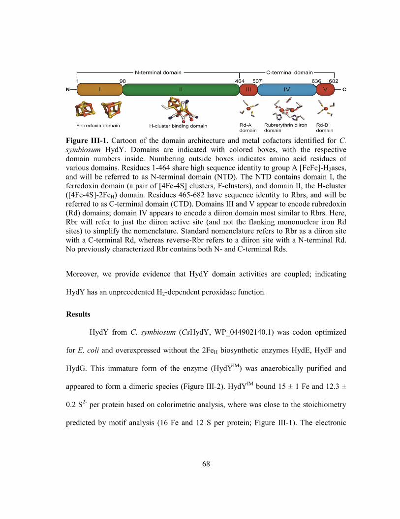
68
Moreover, we provide evidence that HydY domain activities are coupled; indicating
HydY has an unprecedented H2-dependent peroxidase function.
Results
HydY from C. symbiosum (CsHydY, WP_044902140.1) was codon optimized
for E. coli and overexpressed without the 2FeH biosynthetic enzymes HydE, HydF and
HydG. This immature form of the enzyme (HydYIM
) was anaerobically purified and
appeared to form a dimeric species (Figure III-2). HydYIM
bound 15 ± 1 Fe and 12.3 ±
0.2 S2-
per protein based on colorimetric analysis, where was close to the stoichiometry
predicted by motif analysis (16 Fe and 12 S per protein; Figure III-1). The electronic
Figure III-1. Cartoon of the domain architecture and metal cofactors identified for C.
symbiosum HydY. Domains are indicated with colored boxes, with the respective
domain numbers inside. Numbering outside boxes indicates amino acid residues of
various domains. Residues 1-464 share high sequence identity to group A [FeFe]-H2ases,
and will be referred to as N-terminal domain (NTD). The NTD contains domain I, the
ferredoxin domain (a pair of [4Fe-4S] clusters, F-clusters), and domain II, the H-cluster
([4Fe-4S]-2FeH) domain. Residues 465-682 have sequence identity to Rbrs, and will be
referred to as C-terminal domain (CTD). Domains III and V appear to encode rubredoxin
(Rd) domains; domain IV appears to encode a diiron domain most similar to Rbrs. Here,
Rbr will refer to just the diiron active site (and not the flanking mononuclear iron Rd
sites) to simplify the nomenclature. Standard nomenclature refers to Rbr as a diiron site
with a C-terminal Rd, whereas reverse-Rbr refers to a diiron site with a N-terminal Rd.
No previously characterized Rbr contains both N- and C-terminal Rds.

69
absorbance spectrum of HydYIM
(data not shown) revealed broad features from 300 to
500 nm, consistent with charge transfer bands for [4Fe-4S] clusters.
To evaluate the catalytic activity of HydYIM
, we immobilized it on a pyrolytic
graphite edge electrode. When an enzyme is adsorbed to an electrode, the current
response is proportional to turnover frequency. Figure III-3 shows a cyclic
voltammogram from a HydYIM
-coated electrode in the presence of 0.15 mM H2O2. At
reducing potentials below approximately +0.1 V (all potentials are reported relative to
SHE), a catalytic current is observed, indicating reduction of H2O2 by HydYIM
. The
response on a freshly polished electrode under the same experimental conditions does
Figure III-2. Analytical gel filtration reveals quaternary
structure of HydY constructs. NTD (1 mg/mL, solid line)
elutes at its calculated molecular weight (54 kD). CTD (1
mg/mL, dashed line) elutes as a tetramer (120 kD;
monomer MW = 28 kD). HydYIM
(5 mg/mL, dotted line)
elutes at molecular weight of 230 kD (monomer molecular
weight 80 kD), indicating a trimer. Based on oligomers
formed from the synthetic constructs, HydY likely is an
elongated dimeric species, causing it to elute at slightly
larger molecular weight. All runs are the average of at least
two experiments. Inset indicates standards run to estimate
molecular weight of HydY constructs.

70
not show catalysis, demonstrating that enzyme is absolutely required for catalytic
activity. Furthermore, the catalytic activity depends on [H2O2] at low concentrations
(Figure III-4). Additionally, the potential at which a catalytic current was obtained (Eonset
= +100 mV) designated the generation of a species capable of either peroxidase activity
or electron transfer, and this potential was similar to midpoint potentials measured for
Rbrs.150
Together, these results support the ability of HydYIM
to catalytically reduce
H2O2 (peroxidase activity). We hypothesized that this peroxidase activity was initiated at
the CTD, which contains apparent Rbr and Rd centers.
Figure III-3. Potential dependence of electrocatalytic
reduction of H2O2 by HydYIM
. The black trace shows
the current of the cyclic voltammogram of HydYIM
in
the presence of 0.15 mM H2O2. The dotted line is the
response of a freshly polished electrode without a
protein film. Cyclic voltammograms are obtained at a
potential scan rate of 20 mV s-1
and electrode rotation
rate of 2750 rpm in mixed buffer at pH 7. The arrow
indicates the starting point and direction of potential
cycling.

71
Spectroscopic and functional characterization of the CTD
Yields of HydYIM
were exceedingly low (< 1 mg/15 L of E. coli culture),
preventing efforts to spectroscopically characterize this protein. CsHydY is annotated to
contain multiple [4Fe-4S] clusters as well as three other non-heme iron centers, thus
efforts to characterize the iron centers would likely result in overlapping spectroscopic
signals, limiting potential insights. In an effort to aid characterization of HydY and
increase yields of protein, a synthetic construct of the C-terminal domain (CTD) of
CsHydY (residues 465-682, Figure III-1) was cloned. The CTD has high sequence
identity to Rbrs with C-terminal Rds (D. vulgaris Rbr, 41%) as well as Rbrs with N-
Figure III-4. HydYIM
shows time-dependent
scavenging for H2O2. Chronoamperometry of HydIM
absorbed onto a rotating PGE electrode poised at -0.3
V in the presence of 0.2 mM H2O2. Multiple
injections of 0.15 mM H2O2 (marked by arrows)
electrocatalytic response to additional substrate.
Other experimental conditions include: electrode
rotation rate 2750 rpm and buffer pH of 7.0.

72
terminal Rds (C. acetobutylicum reverse-Rbr, 32%). Thus, we hypothesized that the
CTD could be expressed as an independent domain. In solution, the CTD appeared
mostly tetrameric, with a propensity to oligomerize, especially if purified in air (Figure
III-2). Incubation of the higher order oligomer of CTD with DTT, a reductant, resulted in
reformation of the tetrameric state, consistent with reduction of disulfides.
Quantification of protein-bound iron by colorimetric analysis revealed 4.1 ± 0.3
Fe/protein (expected 4 Fe/protein) from n = 3 preparations of this protein construct.
Spectroscopic properties of the CTD are reminiscent of both Rd and Rbr centers
(Figure III-5). UV-vis spectra from oxidized CTD (CTDox) include characteristic
features of Rbrs at 340, 492 and 575 nm, with a shoulder at 378 nm (Figure III-5a, red
Figure III-5. Spectroscopic evidence that the CTD (HydY residues 465-682) contains
Rbr and Rd iron sites. a) Electronic absorbance of CTDox (red), CTDox after addition of 1
M NaN3 (blue), and CTDred (grey). Mössbauer of 0.75 mM 57
Fe-CTD in oxidized (b)
and reduced (c) states. The red lines are simulated spectra of the total components, while
other colors represent simulations of individual components (see Supplemental Table 1).
The oxidized samples were prepared by addition of 4 equivalents of H2O2. The reduced
samples were prepared by addition of 2-fold excess of dithionite in the presence of 50
μM methyl viologen (as redox mediator).

73
trace).150,227
Sodium azide is often used as a probe for diiron site; coordination of azide
to the diiron site results in an increase in the high-energy features. Addition of 1 M
buffered sodium azide resulted in increased ε at 340 and 490 nm (Figure III-5, blue
trace). The UV-vis spectrum of reduced CTD (CTDred) is largely featureless in the
visible region with a weak absorbance band at 320 nm, which is consistent with other
reduced Rd sites (Figure III-5, gray trace).156
A reductive titration revealed that four
electrons were sufficient to fully reduce CTDox (Figure III-6).
Low temperature and low field Mössbauer spectrum of 57
Fe-enriched CTDox
(Figure III-5b) consisted of a three species in a 2:1:1 ratio. The central doublet had
parameters typical of similar to other oxygen-bridged diiron centers, such as Rbr, MMO
and RNR (Table III-1).150,228-230
The magnetic features were simulated at two S = 5/2
sites, with mostly equal intensity (Figure III-5b, blue and green traces). Simulated
parameters of these sites fit with one tetrahedral ferric site with four cysteine sulfur
Figure III-6. Reductive titration of oxidized CTD by
dithionite. Four reducing equivalents (2 mol dithionite)
are required to completely bleach chromophores
associated with oxidized CTD.

74
donors. The other site is best simulated with ferric iron coordinated primarily by 5 or 6
O/N donors (Table III-1). A low-temperature low-field Mossbauer spectrum of the fully
reduced protein exhibited two quadrupole doublets of unequal intensities (Figure III-5c).
The dominant double had parameters typical of diferrous clusters and the other double
was best simulated to parameters of reduced Rd centers ((Table III-1). Unexpectedly, the
diferrous cluster accounted for 75% of the Fe absorption and Rd site only 25%. This
difference in Fe adsorption indicates that one Fe site, likely a Rd site, has mostly O/N
Table III-1. Mössbauer parameters of CTD in comparison with similar iron-binding
proteins.
Oxidized Reduced
Protein
δ
(mm/s)
ΔEQ
(mm/s)
Area
(%)
δ
(mm/s)
ΔEQ
(mm/s)
Area
(%)
ref
HydY-CTD
0.55 1.66 57 1.3 3.02 75
this
work
0.23 0.72 20 0.74 3.2 25
0.51 0.58 23 1.3 3.02
Rbr
0.52 1.38 -
1.3 3.14
- 150,228
0.53 1.74 -
Rd 0.25 0.75 - 0.70 3.15 - 150
MMOH 0.5 1.07 - 1.30 3.14 - 229
RNR-R2
0.55 1.62 -
1.26 3.13
- 230
0.45 2.44 - -

75
ligation that has similar parameters to diferrous clusters. These observations suggest one
of the putative Rd-like was destabilized in some manner during 57
Fe reconstitution, or
one Rd-like site ligates iron in a non-classical fashion, i.e. ligation to metal with non-
sulfur ligands.
To resolve incongruities in the metal centers, further Mössbauer studies were
undertaken. First, a Mössbauer spectrum of 57
Fe-CTDox at 150K was acquired to
delineate the two magnetic features observed at low temperature (Figure III-7a). The
dominant doublet had parameters consistent, but slightly shifted, from a typical oxo-
bridged diferric cluster (Table III-2). The less intense doublet had parameters consistent
Figure III-7. Mössbauer spectra of 57
Fe-CTD.
Spectra of 0.75 mM 57
Fe-CTD (a) oxidized
state recorded at 150 K (b) and a partially
oxidized sample recorded at 5 K and 0.05 mT
field. The red lines are simulated spectra of the
total components, while other colors represent
simulations of individual components (see
Table III-2).

76
with ferric iron coordinated by 5-6 O/N ligands. The ferric iron site for the Rd-centers
appears hidden within the spectral baseline. We conclude that at 150 K, the O/N ligated
iron species collapses in the fast-relaxation limit, while the Rd center is transitioning
between fast and slow relaxation limits. Additionally, we prepared a partially oxidized
sample by adding 1.5 equivalents of H2O2 to 57
CTDred. Incubation of substrate resulted
in the color change from colorless (reduced CTD) to a light pink color (partially
oxidized CTD). No attempts were made to purify excess reductant from 57
CTDred, and
the partially oxidized sample was not purified after incubation with substrate. The
Table III-2. Mössbauer parameters of 57
CTD.
Site (mm/s)
EQ
(mm/s)
(mm/s)
%
(spectrum) Ax,y,z/gnn
(kG)
D
(cm-1
) E/D
[FeIII
-O-
FeIII
]
0.53; ox
100 K
0.51; ox
150 K
1.60; ox
100 K
1.58; ox
150 K
0.35; ox
100 K
0.35; ox
150 K
50; ox
100K
50; ox
150K
2%; semi
--- --- --- ---
[FeII-O-
FeII]
1.29;
semi
3.03;
semi
0.53;
semi
52 of 77%
semi --- --- --- ---
FeIII
(Scys)4
0.23; ox
5K
0.23; ox
100K
0.23; ox
150 K
0.72; ox
5K
0.72; ox
100K
0.72; ox
150K
0.34; ox
5K
1.0; ox
100K
1.0; ox
150K
20; ox
100K
20; ox 150
K
13; semi
-60, -88,
-148
ox 5K;
ox 100
K; ox
150 K;
semi
0.12 0.24 3.6
FeII(Scys)4
0.68;
semi
3.26;
semi
0.26;
semi 9; semi
--- --- --- ---
FeIII
(O/N)4
0.47; ox
100 K
0.45; ox
150K
0.76; ox
100K
0.75; ox
150 K
0.33; ox
100 K
25; ox
100K
25; ox 150
K
-300, -
156, -183
ox 5 K
0.12 0.24 10
FeII(O/N)4
1.29;
semi
3.03;
semi
0.53;
semi
25 of 77%
semi --- --- --- ---

77
resulting Mössbauer spectrum at low temperature and low field exhibited three partially
overlapping quadrupole doublets and some magnetic (S = 5/2) species (Figure III-7b).
The major doublet had parameter typical of diferrous iron clusters, implying that all the
iron centers were re-reduced by the Rd-like sites upon substrate addition, especially
evidenced by small (~4%) absorption typical of oxo-bridged diferric iron clusters (Table
III-2). The other observed doublet arose from the classical diferrous Rd site. Magnetic
features were also simulated, but due to the weak features observed, it is unclear of these
sites represent the sulfur-ligated or O/N ligated mononuclear iron site.
To further characterize the iron sites of CTD, EPR spectra were collected. The
as-isolated CTD yields a weak, rhombic S = ½ EPR signal with g-values of 1.95, 1.76
and ~1.56 (Figure III-7a). This gave < 2 signal arises from antiferromagnetically coupled
Figure III-8. CTD contains multiple iron binding sites. EPR spectrum of as-isolated
CTD (a) or 57
CTD in oxidized (b), partially oxidized (c) and fully reduced (d) states.
Parameters for EPR measurement were: sample temperature, 10K; microwave
frequency, 9.43 GHz; microwave power, 0.519 mW.

78
pair of high-spin FeIII
(S = 5/2) and FeII
(S = 2) ions, and is a hallmark feature of diiron
proteins with O/N ligation, such as Rbr. High-spin iron(III) accounts for g-values of 4.3
and 9.4, attributable to Fe3+
(Cys)4 sites in Rds. However, the as-isolated CTD appears to
contain two types of overlapping g = 4.3 signals, implying there may be two slightly
distinct Rd sites, or Rd sites close enough to perturb individual signals. Indeed, EPR of
57CTDred contains only the ‘sharp’ g=4.3 signal (Figure III-8b), while the fully oxidized
contains both g = 4.3 signals (Figure III-8a). The EPR and Mössbauer results support a
model that includes four irons per CTD, including a diiron oxo site, a classical FeIII
rubredoxin site, and a mononuclear high-spin FeIII
(O/N)5-6 site.
Figure III-9. Consumption of H2O2 by CTD is dependent upon Rd and NROR
concentration. a) The rate of NADH oxidation dependent upon rubredoxin (Rd)
concentration, saturating with 50 µM Rd. A Michaelis-Menten fit was applied to give
the following parameters: kcat, 380 ± 40 min-1
; Km, 8 ± 2 µM; kcat/Km, 8 ± 2 x10⁶ M
-1 s
-1.
b) The rate of NADH oxidation saturated with 5 µM NADH:Rd oxidoreductase
(NROR). The data was fit with the Michaelis-Menten equation to give the following
parameters: kcat, 230 ± 10 min-1
; Km, 0.25 ± 0.07 µM; kcat/Km, 1.5 ± 0.43 x10⁷ M
-1 s
-1. All
assays were performed at 10-12°C, following loss of NADH signal (see Materials and
Methods), with 0.1 mM H2O2 and 0.5 µM NROR (a) or 0.5 µM Rd (b). One unit is
defined as 1 µmol H2O2 reduced per minute (1 µmol NADH reduced 1 µmol H2O2).
Background consumption of NADH was subtracted from measured rates.

79
Taken together, high sequence identity, metal content and results from multiple
spectroscopies provide clear evidence that the CTD contains two types of metal centers:
a Rbr-like diiron site and two mononuclear iron sites, one similar to Rd-like and one
with mostly O/N ligation. Further studies are required to resolve the identity of this third
iron site.
Figure III-10. The CTD is an efficient H2O2
reductase. Steady-state reduction of H2O2
was investigated using an in vitro system of
0.2 mM NADH, 5 μM NROR, 50 μM Rd,
and 0.1 μM CTD The decrease of NADH
absorbance (ε340 = 6.22 mM-1
cm-1
) was
used to measure initial rate of H2O2
consumption after injection of the CTD.
Data was fit to the Michaelis-Menten
equation to yield the following kinetic
parameters: kcat, 1890 ± 70 min-1; Km, 3.0 ±
0.8 µM; kcat/Km, 1.1 ± 0.3 x10⁷ M-1
s-1
. All
assays were performed at 10-12°C.
Background consumption of NADH was
subtracted from measured rates.
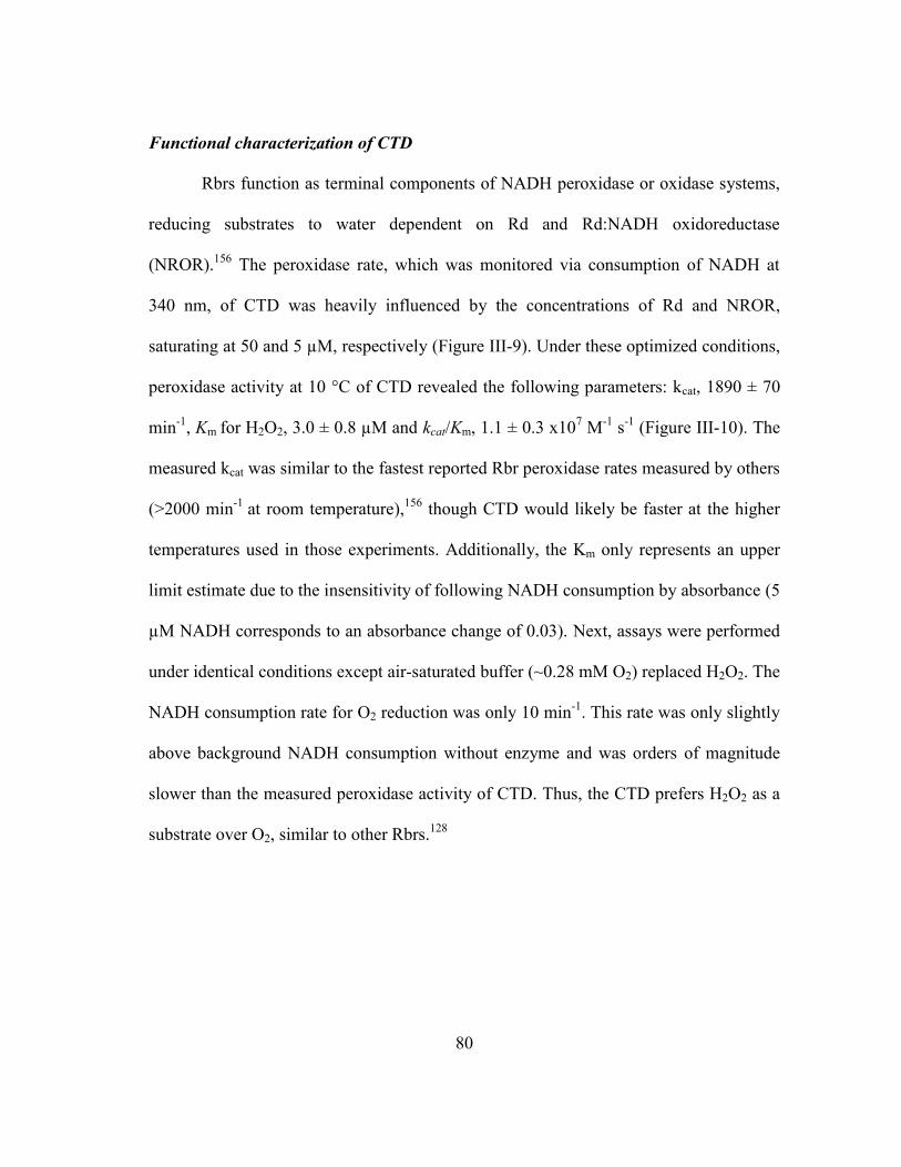
80
Functional characterization of CTD
Rbrs function as terminal components of NADH peroxidase or oxidase systems,
reducing substrates to water dependent on Rd and Rd:NADH oxidoreductase
(NROR).156
The peroxidase rate, which was monitored via consumption of NADH at
340 nm, of CTD was heavily influenced by the concentrations of Rd and NROR,
saturating at 50 and 5 µM, respectively (Figure III-9). Under these optimized conditions,
peroxidase activity at 10 °C of CTD revealed the following parameters: kcat, 1890 ± 70
min-1
, Km for H2O2, 3.0 ± 0.8 µM and kcat/Km, 1.1 ± 0.3 x10⁷ M
-1 s
-1 (Figure III-10). The
measured kcat was similar to the fastest reported Rbr peroxidase rates measured by others
(>2000 min-1
at room temperature),156
though CTD would likely be faster at the higher
temperatures used in those experiments. Additionally, the Km only represents an upper
limit estimate due to the insensitivity of following NADH consumption by absorbance (5
µM NADH corresponds to an absorbance change of 0.03). Next, assays were performed
under identical conditions except air-saturated buffer (~0.28 mM O2) replaced H2O2. The
NADH consumption rate for O2 reduction was only 10 min-1
. This rate was only slightly
above background NADH consumption without enzyme and was orders of magnitude
slower than the measured peroxidase activity of CTD. Thus, the CTD prefers H2O2 as a
substrate over O2, similar to other Rbrs.128

81
Spectroscopic and functional characterization of the NTD
The NTD of CsHydY (residues 1-464, Figure III-1), which has high sequence
identity to monomeric [FeFe]-H2ases (Dd, 44%; Cr, 38%), was also expressed as an
independent construct. NTD was prepared in the absence (NTDIM
) or the presence
(NTDe) of hydrogenase maturases.
102 Proteins derived from both NTD-expressing
constructs are monomeric (Figure III-2), and contain 10.5 Fe and 11 S2-
per protein. The
electronic absorbance spectrum of NTD exhibited features consistent with Fe-S clusters
(Figure III-11). Dithionite-reduced NTDIM
also has an S = ½ EPR signal with
temperature and power saturation properties consistent with [4Fe-4S] clusters (Figure
III-10). Taken together, the NTD appears to bind three [4Fe-4S] clusters.
Figure III-11. Spectroscopic investigation of the NTDIM
. a) Electronic absorbance of as-
isolated NTDIM
(25 µM) reveals broad absorbance features between 300 and 500 nm,
typical of Fe-S centers. b) EPR spectrum of reduced NTDIM
. Parameters for EPR
measurement were: sample temperature, 10K; microwave frequency, 9.43 GHz;
microwave power, 0.519 mW. Sample was prepared by adding excess NaDT to as-
isolated NTDapo
(150 µM).
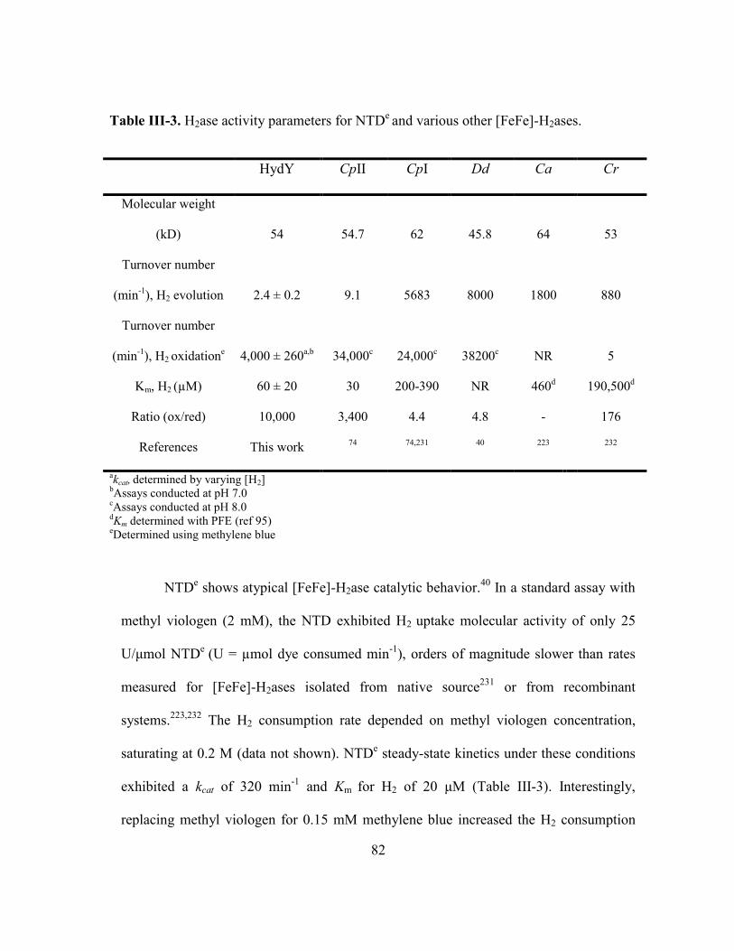
82
NTDe shows atypical [FeFe]-H2ase catalytic behavior.
40 In a standard assay with
methyl viologen (2 mM), the NTD exhibited H2 uptake molecular activity of only 25
U/μmol NTDe
(U = µmol dye consumed min-1
), orders of magnitude slower than rates
measured for [FeFe]-H2ases isolated from native source231
or from recombinant
systems.223,232
The H2 consumption rate depended on methyl viologen concentration,
saturating at 0.2 M (data not shown). NTDe steady-state kinetics under these conditions
exhibited a kcat of 320 min-1
and Km for H2 of 20 μM (Table III-3). Interestingly,
replacing methyl viologen for 0.15 mM methylene blue increased the H2 consumption
Table III-3. H2ase activity parameters for NTDe and various other [FeFe]-H2ases.
HydY CpII CpI Dd Ca Cr
Molecular weight
(kD) 54 54.7 62 45.8 64 53
Turnover number
(min-1
), H2 evolution 2.4 ± 0.2 9.1 5683 8000 1800 880
Turnover number
(min-1
), H2 oxidatione
4,000 ± 260a,b
34,000c
24,000c
38200c
NR 5
Km, H2 (µM) 60 ± 20 30 200-390 NR 460d
190,500d
Ratio (ox/red) 10,000 3,400 4.4 4.8 - 176
References This work 74 74,231 40 223 232
akcat,
determined by varying [H2]
bAssays conducted at pH 7.0
cAssays conducted at pH 8.0
dKm determined with PFE (ref 95)
eDetermined using methylene blue

83
rate 12.5 fold (4,000 min-1
) and only marginally affected the Km (60 μM). Additionally,
NTDe
displayed marginal H2 production, with a specific activity of 130 U/mg protein.
Similar catalysis has been described for another [FeFe]-H2ase, the H2-uptake C.
pasteurianum H2ase II (CpII, 58% identity to HydY).74
The lowered H2 production
activity of NTDe had no effect on O2 inactivation rates (Figure III-12), similar to
inactivation rates observed for numerous other [FeFe]-H2ases.74,233
Electrochemical investigation of HydYs and NTD
e/s
Figure III-13 shows that NTDe electrocatalytically reduces protons and, in the
presence of hydrogen, oxidizes hydrogen. In the absence of hydrogen, negative current
attributable to proton reduction is observed at potentials below that of the H+/H2 couple
(Figure III-13a, gray). Upon introduction of an atmosphere of H2, several changes in the
Figure III-12. Inactivation of NTDe
by O2. NTDe (2 mL) in
buffer C in a septum-sealed dram vial (20 mL) was stirred by
stir plate incubated at 22 °C. At zero time, the septum was
removed to expose NTDe to air. At the designated intervals,
samples were analyzed for residual H2 uptake activity (see
Materials and Methods). Data was fit to exponential model,
yielding a fit y=119.7e-0.433x and a t1/2 of 1.44 minutes.

84
voltammetric response are noticeable (Figure III-13a, black). First, proton reduction
current is significantly decreased, suggesting that H2 is a strong product inhibitor for
proton reduction. Such activity is well known for [NiFe]-H2ases but has not been
previously reported for an [FeFe]-H2ases.96
Second, at oxidizing potentials, a large
Figure III-13. CsHydY is biased toward H2 oxidation. a) NTDe under continuous
sparging of 1 atm 100% H2 (black trace, 0.78 mM) and 1 atm N2 (gray trace) with 5 mV
s-1
scan rate. The light gray dotted scan is an electrode under the same conditions that has
not been exposed to enzyme. b) NTDs under 1 atm 100% H2. HydY
s under 1 atm of c)
100% H2 or d) 1% H2. Other experimental conditions include: electrode rotation rate
2750 rpm and buffer pH 7.0. The horizontal line indicates zero current and the vertical
line indicates the H+/H2 couple at pH 7.0 and 1 atm 100% H2 (-420 mV, a-c) or 1% H2 (-
350 mV, d).

85
positive current arises from electrocatalytic oxidation of hydrogen. The hydrogen
oxidation current is rotation rate dependent at low rotation rates, indicating that catalysis
is limited by mass transfer of hydrogen to the electrode surface. Thus, all subsequent
experiments were undertaken at a high electrode rotation rate (2750 rpm).
Additionally, oxidative catalysis is not observed until the experimental potential
is approximately 100 mV more oxidizing than the H+/H2 couple. This excess potential is
referred to as overpotential. Although a requirement for considerable overpotential for
hydrogen oxidation has been reported for some oxygen-tolerant [NiFe]-H2ases,
overpotential has not been previously described as essential for catalysis by an [FeFe]-
Figure III-14. Catalytic cyclic voltammograms
for NTDs adsorbed to a PGE electrode over a
range of pH values. The pH values shown are
8.0, 7.5, 6.5, 6.0, 5.5, 5.0, 4.5, and 4.0. Other
experimental conditions include: electrode
rotation rate 2750 rpm and potential scan rate of
20 mV s-1
. Catalytic signals were normalized
such that the average activity at 0 mV at pH 7 is
1. The concentration of dissolved hydrogen is
held at 0.78 mM by continuously sparging with 1
atm of hydrogen gas. The black arrow signifies
the starting potential and scan direction.

86
H2ase.34
As suggested by solution assays of NTDe, the voltammetric experiments
demonstrate considerably higher rates for hydrogen oxidation than H2 production. At the
most extreme potentials assayed, H2 is oxidized 5-25 times faster than it is produced
under reducing conditions (depending on whether reduction is assayed in the presence or
absence of hydrogen). Such an extreme electrocatalytic bias towards H2 oxidation has
not been reported previously for an [FeFe]-H2ases. It is worth noting that electrocatalytic
activity depends on pH (Figure III-14), and thus the magnitude of the bias is also pH
dependent.
To ensure this atypical electrocatalytic profile was due only to HydY-specific
tuning, NTDIM
and HydYIM
were reconstituted with [(Et4N)2{Fe2-
[(SCH2)2NH](CO)4(CN)2] (1, see Methods)117
to yield synthetically matured enzyme
(NTDs
and HydYs, respectively). By extending experiments to more extreme oxidizing
potentials and slower potential scan rates, both constructs undergo anaerobic, oxidative
inactivation at high potentials (Figure III-13b,c). The hydrogen oxidation current peaks
at approximately +100 mV and then decreases at higher driving forces, i.e. more
oxidizing potentials. This suggests that oxidation of the active site produces a
catalytically incompetent state, as has been described previously other for [FeFe]-
H2ases.34
Figure III-15 shows Eswitch, an apparent thermodynamic feature that defines this
process in terms of the reactivation observed when potentials are returned to more
reducing values. Eswitch, the point at which -di/dE attains a local maximum, is not a
reduction potential for the active site but instead represents a convolution of active site

87
thermodynamics and kinetics of reductive reactivation. Oxidative inactivation/reductive
reactivation of HydY/NTD is pH dependent with more inactivation observed as lower
pH values. This is the opposite of the trend usually observed for hydrogenases and
suggests the active site chemistry of HydY may be unique. Interestingly, the rate of
Figure III-15. Cyclic voltammograms of HydYs adsorbed to a PGE electrode over a
range of pH values. a) Reverse, reductive sweep of HydYs electrochemical activity at pH
values 8.0, 7.5, 6.5, 6.0, 5.5, 5.0, 4.5, and 4.0 (forward, oxidative scan omitted to better
reveal onset potential). b) Potential of H2 oxidation onset as a function of pH; the grey
diagonal line indicates the expected equilibrium potential for the 2H+/H2 couple pH
dependence. The onset potential (Eonset) is defined as the zero-current intercept. c) pH
dependence of anaerobic, oxidative inactivation for HydYs. The switch potential (Eswitch)
was obtained from the inflection point of the reductive reactivation area, determined
from the derivative (-di/dE) of the reverse scan.

88
anaerobic inactivation had a local minimum at pH 7.0, suggesting that the inactivation
process was primarily a kinetic process. Furthermore, Eonset for H2 oxidation does not
follow a Nerstian decay (Figure III-14b).
In a 1% H2 environment (Figure III-13d), HydYs
showed similar magnitudes of
H+ reduction and H2 oxidation currents, indicating that the apparent H2 inhibition
constant ( ) for HydY must be below a 1% H2 atm. The oxidative current is
significantly decreased as well, suggesting that the Km for H2 is above 1% H2. The
concentration of dissolved H2 under these conditions (~8 µM) agrees with the Km
measured by dye-coupled assays (20-60 µM) These results indicate HydY displays
inhibition like [NiFe]-H2ase, and Km for H2 higher that [NiFe]-H2ase but lower than
[FeFe]-H2ase. Additionally, HydYs requires minimal overpotential under 1% H2 atm.
This result suggests that the rate limiting step for catalysis is governed, at least, by active
Figure III-16. H2-dependent reduction of CTD by NTDe. a) The CTDox (20 µM, blue
trace) in H2-saturated exhibits no changes in spectral features. Upon anaerobic addition
of NTDe
(88 nM), spectral features of oxidized CTD dissipate, replaced with features
consistent with reduced CTD. b) Kinetic analysis of a) monitored at 490 nm for CTD
(blue diamonds) or 456 nm for E. coli Fdx (green circles).

89
site chemistry. However, a residual slope near the H+/H2 couple (-360 mV) suggests
electron transfer also contributes to the rate-limiting step.98,234
H2-dependent reduction of the CTD by NTDe
We tested whether the activities of the N- and C-terminal domains of HydY can
be coupled by combining the independently expressed and purified domains in situ
(Figure III-16). Anaerobic incubation of CTDox (25 µM) in a H2-saturated buffer (~0.78
mM) does not cause a change in the absorbance spectrum (blue trace). On the other
hand, an additional anaerobic injection of NTDe (88 nM) results in the loss of CTDox
absorbance peaks and formation CTDred features characteristic of reduced Rd centers
(red trace). The change in absorbance at 490 nm indicates that injection of NTDe resulted
in near immediate reduction of CTDox, suggesting NTDe directly reduces CTD (Figure
III-15b). To evaluate whether NTDe could reduce additional protein substrates, it was
also incubated with E. coli Fdx, a [2Fe-2S] cluster-binding protein that functions in one-
electron transfer processes. No spectral changes of Fdx were detected. This implies that
reduction of CTD by NTD is: 1) dependent upon H2 and not mediated by spurious
dithionite from NTD purification; and 2) not a general process since Fdx was unable to
be reduced by NTDe. Overall, the rapid reduction of a >250-fold molar excess of CTDox
is consistent with reducing equivalents originating from dissolved H2.
Bioinformatic analysis of HydY homologs
We next used BLAST to identify HydY homologs. We filtered the BLAST hits
by requiring that the hits be fusion protein with the conserved H-cluster motifs and at
least one Rd domain. In all, 40 HydY homologs were identified (Figure III-17).

90
Homologs of CsHydY are scattered throughout Firmicutes, Fusobacteria and δ-
Proteobacteria, with the Clostridia order (35 homologs) mostly represented. Distinct
classes (I, II and III) of HydY homologs were identified. Class I is the most common,
with motifs similar to that of CsHydY (Figure III-I). Deviations from this architecture,
such as missing the second Rd motif but retaining the Rbr domain, were classified as I-
A, B or C (Figure III-17). Class II HydY sequences contained alternate C-terminal
domain fusions other than Rbr, such as a glutamate synthase (GS) domain in HydY from
Robinsellia sp. RH5. GSs synthesize glutamate from glutamine and α-ketoglutarate,
requiring external electrons from either ferredoxin or NAD(P)H.77
Lastly, Class III
Figure III-17. Phylogenetic grouping of modular HydY homologs. a) Phylogenetic tree
of CsHydY homologs identified by BLAST (December 2015) calculated using MrBayes
(scale bar: 0.2 substitutions per site, 15000000 generations sampled every 1000
generations with an average standard deviation of split frequencies <0.0002) and
visualized with FigTree. Homologs of HydY were identified in Firmicutes, Fusobacteria
and Proteobacteria. b) HydY homologs are highly modular and separate into three
classes. Class I contains [4Fe-4S] cluster, H-cluster, Rd and Rbr binding domains. Sub-
classes of Class I (a-c) retain a C-terminal rubrerythrin domain, but vary in putative
electron transfer domains. The class II HydY homolog has a C-terminal domain with high
sequence identity to glutamate synthase domain. Class III homologs contain only varying
numbers of Rd domains and lack putative C-terminal redox active sites.

91
HydY sequences lacked additional redox active sites and variable numbers of Rd
domains, thus the electron donor/acceptor is ambiguous.5,14
Sequence analysis of all identified HydY homologs and CpII revealed marked
differences within second-shell motifs for binding H-cluster and [4Fe-4S] clusters. Three
conserved motifs provide second-shell interactions for the H-cluster: P1, TSCCPxW; P2,
MPCx2Kx2E; and P3, ExMACx2GCx2GGGxP (Figure III-18). HydY homologs contain
conserved substitutions within canonical H-cluster binding motifs and a third in a
previously unidentified region (Figure III-19). All HydYs and
CpII have completely conserved P1 sequences. Within the P2 motif, a conserved
Figure III-18. Representation of the H-cluster in C.
pastuerinium HydA1 (CpI; PDB:3C8Y) with
important protein interactions. Hydrogen bonds are
shown in yellow. Protein residues within van der
Wall’s contact are depicted in cyan. Hydrophobic
interactions (A230 to bridging CO) are shown in
pink. Note, the bridgehead of the azadithiolate was
modeled as oxygen, and was colored blue to
represent currently accepted bridgehead nitrogen.

92
substitution of Met to Thr was found in all identified HydY homologs (Figure III-20b).
This methionine residue (M353, all numbering according to PDB:3C8Y204
), is
positioned beneath the μ-CO, and is within van der Walls contact (Figure III-18). A
mostly conserved Ala to Thr substitution was also identified in the P3 motif, which is
Figure III-19. Sequence alignment reveals conserved substitutions near H-cluster
and [4Fe-4S] cluster motifs. Alignment of N-terminal domain of CsHydY with
structurally characterized [FeFe]-H2ases C. reinhardtii (Cr), D. desulfuricans
(Dd) and C. pasteurianum I (CpI) or biochemically characterized [FeFe]-H2ases
C. acetobutylicum (Ca) and C. pasteurianum II (CpII). MUSCLE alignments
were visualized by Jalview and residues are colored by sequence identity, with
intensity color increment of 100%. Conserved motifs for the H-cluster domain are
boxed: P1,TSCCPxW; P2,MPCxxKxxE; P3, ExMACxxGCxxG; and previously
unidentified ‘P4,’ APxVR. Both CpII and HydY show similar substitutions in P1,
P3 and P4 (yellow boxes) motifs. A ‘positive patch’ of Arg and Lys residues (red
triangles) within the F-cluster domain is also found in HydY and CpII.
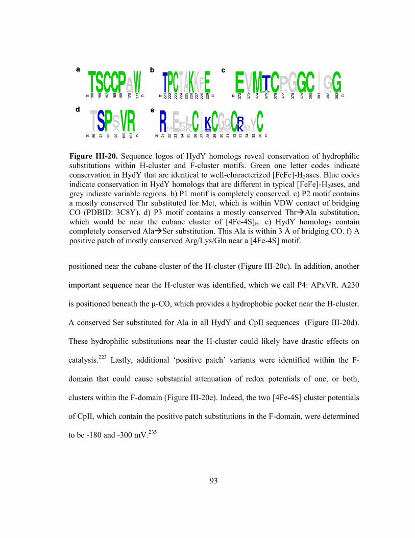
93
positioned near the cubane cluster of the H-cluster (Figure III-20c). In addition, another
important sequence near the H-cluster was identified, which we call P4: APxVR. A230
is positioned beneath the μ-CO, which provides a hydrophobic pocket near the H-cluster.
A conserved Ser substituted for Ala in all HydY and CpII sequences (Figure III-20d).
These hydrophilic substitutions near the H-cluster could likely have drastic effects on
catalysis.223
Lastly, additional ‘positive patch’ variants were identified within the F-
domain that could cause substantial attenuation of redox potentials of one, or both,
clusters within the F-domain (Figure III-20e). Indeed, the two [4Fe-4S] cluster potentials
of CpII, which contain the positive patch substitutions in the F-domain, were determined
to be -180 and -300 mV.235
Figure III-20. Sequence logos of HydY homologs reveal conservation of hydrophilic
substitutions within H-cluster and F-cluster motifs. Green one letter codes indicate
conservation in HydY that are identical to well-characterized [FeFe]-H2ases. Blue codes
indicate conservation in HydY homologs that are different in typical [FeFe]-H2ases, and
grey indicate variable regions. b) P1 motif is completely conserved. c) P2 motif contains
a mostly conserved Thr substituted for Met, which is within VDW contact of bridging
CO (PDBID: 3C8Y). d) P3 motif contains a mostly conserved ThrAla substitution,
which would be near the cubane cluster of [4Fe-4S]H. e) HydY homologs contain
completely conserved AlaSer substitution. This Ala is within 3 Å of bridging CO. f) A
positive patch of mostly conserved Arg/Lys/Gln near a [4Fe-4S] motif.

94
Discussion
We have reported on the first study on a homodimeric [FeFe]-H2ase rubrerythrin
fusion from the anaerobe C. symbiosum, called HydY, which performs two seemingly
dichotomous reactions: H+ reduction/H2 oxidation and H2O2 reduction. The CTD of
HydY is the site for H2O2 reduction, and is a highly efficient peroxidase (kcat/Km 1.1x10⁷
M-1
s-1
). Multiple spectroscopic tools indicate that the CTD binds a diiron site, with
Mössbauer assigning it as a rubrerythrin-like. The NTD of HydY has high sequence
identity (38-44%) to the well-studied [FeFe]-H2ase, such as Cr or Dd, and is the site for
hydrogenase activity.
Using a coupled biochemical and electrochemical approach, HydY (via the CTD)
was found to catalytically reduce H2O2. Rbrs preferentially react with H2O2 and
sluggishly reduce O2.128
A recent report with a reverse domain orientated Rbr, i.e. Rd-
Rbr, was found to have an increased reactivity with O2, but still maintained substrate
preference for H2O2.152
No di-rubredoxin, to our knowledge has been reported. Cyclic
voltammograms with HydYIM
, in the presence of H2O2, show catalytic, reductive
currents at potentials ≤ +100 mV (Figure III-3). The Eonset measured for HydYIM
is much
more negative than those measured for typical Rbrs. Reductive titrations with DvRbr
have determined mid-point potentials +150 to +230 mV for either the diiron or FeS4 Rd-
like sites.150,227
Rd proteins, i.e. those not found as chimeras with Rbrs, have mid-point
potentials of ~ 0 mV.150
This hints at a lowered reduction potential for the CTD. Further
work must be done to understand this apparent negative redox potential shift.

95
Our results indicate HydY functions differently than standard [FeFe]-H2ases: H+
reduction is inhibited by H2 and it readily consumes H2. Solution assays with methyl
viologen (-450 mV) present the case that HydY is a poor catalyst for both H+ reduction
and H2 uptake. Replacing the electron donor with methylene blue (+12 mV) makes
HydY a more active enzyme for H2 oxidation. Reports by Adams and Mortenson show
CpII also show this same ‘bias’ toward H2 oxidation, where CpII was speculated to be a
poor H+ reduction catalyst because H2 removal was rate limiting.
74 Cyclic
voltammograms of HydY further enhance the understanding of this reactivity. Under 1
atm of H2 and near the H+/H2 couple (-420 mV), the maturated HydY constructs show
minimal catalytic currents. At +20 mV, all the maturated HydY constructs show near
maximal oxidative currents. These results illustrate that HydY requires an overpotential
for H2ase activity indicating that electron transfer is rate-limiting,98
contrasting with
standard [FeFe]-H2ases.7
The electrochemical results presented for HydY contrast with those of typical
[FeFe]-H2ases. For example, HydY displays a bias for H2 oxidation (25) that is likely
due product (H2) inhibition. Under similar conditions, standard [FeFe]-H2ases have
reported biases of 0.4 (hydrogen oxidation/protein reduction activities) for Ca and 2 for
Cr, whereas standard a [NiFe]-H2ase displayed a bias of ~12 and O2-tolerant varieties
were even more biased toward H2 oxidation.98
The mechanism for bias is complex,98,234
but appears to not be influenced by active site chemistry. HydYs and NTD
s display
anaerobic reactivation (Eswitch)85
values of +120 mV. Eswitch is a qualitative
parameter34,236
used to describe anaerobic oxidative inactivation observed for H2ases. In

96
comparison, under similar conditions Eswitch values for Dd[FeFe]-H2ase were +10 mV,
while O2-tolerant [NiFe]-H2ases were +100 mV.34
Eswitch was originally hypothesized to
be over oxidation for [FeFe]-H2ases, similar to Hoxinactive
,94
but a recent study suggests
anaerobic inactivation involves a more complex mechanism.237
Regardless of the species
formed under oxidizing conditions, HydY and standard [FeFe]-H2ases display clearly
different electrocatalytic profiles.
Tuning activity of [FeFe]-H2ases is mediated by active site residues.223
In fact,
recent studies of apo and maturated CpI with various synthetic 2FeH complexes118,238
indicate the protein matrix induces the H-cluster into the active, ‘rotated’ isomer.239
Interestingly, substitutions of Leu for Met in P2 or P3 resulted in a drastic decrease in
activity.223
Additionally, recent spectroscopic240
and electrochemical237
investigations
support a model for H-cluster isomerization. Mechanisms for these isomerizations
remain unclear, but highlight the importance of second-shell interactions for H-cluster
stability and catalytic activity. HydY homologs contain conserved substitutions with H-
cluster and [4Fe-4S] cluster binding motifs (Figures III-19&20). We hypothesize that
variants identified herein are the determinants for marked catalytic differences of HydY,
and likely CpII, with positive patch substitutions largely affecting the catalytic bias.98
We propose that CsHydY acts in recycling H25
generated by prototypical [FeFe]-
H2ases3 and transfers reducing equivalents directly to the CTD active site for H2O2
reduction (Figure III-21). Typical anaerobic bacteria operate under reducing redox
potentials, allowing for multiple copies (4 in C. symbiosum; 6 in human pathogen C.
perfringens) of [FeFe]-H2ases to presumably be active in H2 production; HydY would be

97
inactive under these typical cellular conditions. ROS stress, however, would disrupt
anaerobic metabolism and shift cellular potentials more positive, signaling for oxidative
stress response via perR and similar genes.124
This oxidative shift would trigger HydY
for H2 oxidation, consequently allowing for efficient H2O2 scavenging by the CTD.
Upon removal of ROS, the anaerobe would return to reducing, homeostatic conditions
and HydY would be inactive due to the inhibition of H2. The conservation of
substitutions within H-cluster binding motifs suggests that all HydYs function similarly.
We hypothesize that HydY enzymes catalyze H2-dependent reduction of H2O2 (Class I)
or an unknown substrate (Class III). The class II HydY from Robinsellia sp. RH5
contains a glutamate synthase (GS) domain would use H2 as a reductant, in lieu of
NAD(P)H or reduced ferredoxin.
Figure III-21. Model of H2-dependent peroxidase role of HydY. Under typical,
reducing cellular conditions, HydY is inactive. An oxidative shift (caused by ROS)
activates the NTD and triggers oxidation of excess H2 that was generated during
anaerobic metabolism. Reducing equivalents recycled from H2 are directly transferred
to CTD for H2O2, and maybe O2, reduction.

98
Overall, we have characterized a novel [FeFe]-H2ase rubrerythrin fusion, called
HydY, that is biased toward H2 oxidation. Our evidence suggests that HydY is capable
of transferring reducing equivalents generated from H2 oxidation directly to the
rubrerythrin domain for H2O2 reduction, i.e. H2-dependent H2O2 reduction. We have
identified possible determinants for the bias and apparent overpotential requirement of
HydY. Elucidating and understanding the factors that tune H2ase chemistry may lead to
the better design of small molecule compounds that could have greater implications in
energy production.
99
CHAPTER IV
INVESTIGATION OF SECOND-SHELL CONTRIBUTIONS
FOR HYDY CATALYSIS
Introduction
Hydrogenases reversibly catalyze conversion of dihydrogen into protons and
electrons, with many that exhibit turnover frequencies exceeding 1,000 s-1
.219,220
The
three classes of hydrogenases (H2ases) are named by the metals employed at the active
site, nominally [FeFe]-, [NiFe]- and [Fe]-H2ases Though phylogenetically distinct,2,3
H2ase active sites are characterized by their low oxidation and spin states stabilized by
unique CO and CN- ligands. The [FeFe]-H2ase is the superior catalyst.
38
The active site of [FeFe]-H2ases, as originally revealed by the Peters, et al241
and
Nicolet, et al,67,242
utilizes a complex organometallic cofactor called the H-cluster,
comprised of a cubane [4Fe-4S] cluster ([4Fe-4S]H) bridged by a cysteine thiolate to a
diiron subcluster ([2Fe]H). These irons are referred to as proximal (Fep) or distal (Fed),
relative to their position to the cubane cluster. Each iron of the [2Fe]H coordinates a
terminal CO and CN-, along with a bridging CO, and a bridging dithiolate ligand (Figure
1). This pendant ligand has been modeled as a propandithiolate241,243
,
dithiomethylether244
, and dithiomethylamine. Theoretical245
, spectroscopic246
, and, most
recently, biomimetic60
studies have unequivocally proven the bridgehead atom to be
nitrogen. Spectroscopic results have found that exogenous CO inhibits [FeFe]-H2ases by
binding to the open coordination site of Fed, indicating this is the active site metal. The
CN- ligands are likely held in place by H-bonds from the protein backbone, while the

100
CO ligands are free within relatively hydrophobic pockets (Figure IV-1).238,241,247
Four
highly conserved motifs within this protein pocket – TSCCPxW (P1), MPCx2Kx2E (P2),
ExMACx2GCx2G3xP (P3) and APx2R (P4) – contribute second-shell interactions to the
[2Fe]H. Mutation of Cys within P1 has revealed it as the likely proton donor,223,233
whereas substitutions of Leu for Met in P2 or P3 or Arg for Lys in P2 resulted in
drastically decreased H-cluster incorporation.223
The mechanism of [FeFe]-H2ases stem mostly from contributions using FTIR
and EPR spectroscopy. Both [4Fe-4S]H and [2Fe]H are redox active. The [4Fe-4S]H
cycles between the oxidized [4Fe-4S]2+
and EPR-active reduced [4Fe-4S]+ states,
whereas the [2Fe]H can either be: overoxidized, Fe(II)Fe(II); oxidized Fe(I)Fe(II); or
reduced, Fe(I)Fe(I). Only the oxidized [2Fe]H state is EPR active. Spectroscopic studies
Figure IV-1. Crystallographic model of the [FeFe]-H2ase
active site taken from the structure of CpI (PDB 3C8Y).
Interactions from protein to [2Fe]H are depicted as: yellow,
polar interactions; blue, hydrogen bonds; magenta,
hydrophobic interactions. Note, the bridgehead was
changed from O to N to reflect current literature.

101
indicate the catalytically active, resting state forms of [FeFe]-H2ases are the Hox ([4Fe-
4S]2+
Fe(I)Fe(II)) and Hred ([4Fe-4S]2+
Fe(I)Fe(I)) states,7 each that bear characteristic
FTIR bands. An additional state, the so-called Hsred,248
consists of [4Fe-4S]+Fe(I)Fe(I)
state. Hsred accumulates in some [FeFe]-H2ases248,249
and is likely transiently formed
during catalysis. A simple proton-coupled mechanism, then, can be assumed to form a
hydride intermediate [Fe(I)Fe(II)(H-)] followed by the formation of H2, forming a
transient (Fe(I)Fe(II)H2) species.245
We recently reported on [FeFe]-H2ase fusion protein, called HydY, which had
near uni-directional H2 uptake activity, contrasting with typical [FeFe]-H2ases. All
HydY homologs contain conserved substitutions in conserved H-cluster domain binding
residues. We speculated that the conserved Ala to Ser mutation in P4 (S97, numbering
according to HydY residues) and Thr to Met in P2 (T221) significantly contributed to
the unique properties observed for HydY. We found that these conserved residues within
the second-shell sphere of [2Fe]H appear to tune catalysis of the H-cluster by H-bonding
directly to CO and CN- ligands. We speculate that the µ-CO is the ‘hotspot’ for [FeFe]-
H2ase catalysis.
Results
FTIR Spectroscopy
The N-terminal domain (NTD) of HydY was purified and exhibited a similar
catalytic bias as previous batches (turnover number for H2 oxidation of 4,500 min-1
and
2.4 ± 0.2 min-1
for H2 production). To examine the potential molecular determinants for
this surprising lack of H+ reduction for HydY, Fourier transform infrared (FTIR)
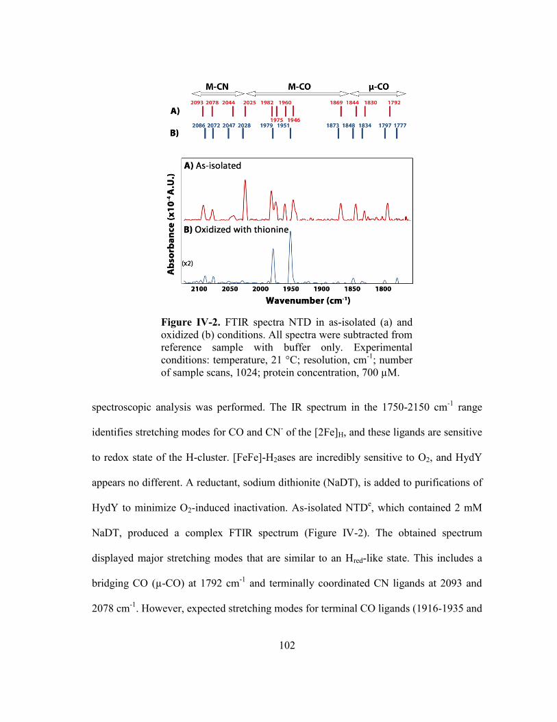
102
spectroscopic analysis was performed. The IR spectrum in the 1750-2150 cm-1
range
identifies stretching modes for CO and CN- of the [2Fe]H, and these ligands are sensitive
to redox state of the H-cluster. [FeFe]-H2ases are incredibly sensitive to O2, and HydY
appears no different. A reductant, sodium dithionite (NaDT), is added to purifications of
HydY to minimize O2-induced inactivation. As-isolated NTDe, which contained 2 mM
NaDT, produced a complex FTIR spectrum (Figure IV-2). The obtained spectrum
displayed major stretching modes that are similar to an Hred-like state. This includes a
bridging CO (µ-CO) at 1792 cm-1
and terminally coordinated CN ligands at 2093 and
2078 cm-1
. However, expected stretching modes for terminal CO ligands (1916-1935 and
Figure IV-2. FTIR spectra NTD in as-isolated (a) and
oxidized (b) conditions. All spectra were subtracted from
reference sample with buffer only. Experimental
conditions: temperature, 21 °C; resolution, cm-1
; number
of sample scans, 1024; protein concentration, 700 µM.

103
1891-1894 cm-1
)7,250-252
are not present. Instead, multiple lower energy stretching modes
(1830-1869 cm-1
) and higher energy modes (1960-2025 cm-1
) are observed. These
stretches are most similar to those detected for inactivated states of Dd,253
but have not
been reported for clostridial H2ases.254
This spectrum suggests as-isolated NTDe, in the
presence of NaDT, exists primarily either in Hred or Hinact. The concentration of NaDT
used was only in slight excess to protein (800 µM), and O2 induced inactivation cannot
be ruled out since the FTIR spectrometer used was outside of the glovebox.
To deconvolute the multiple states observed for the as-isolated sample, NTDe
was incubated with a ten-fold excess of thionine (E’ = 16 mV vs SHE) (Figure V-2b).
Previous studies have found addition of thionine results in the formation of Hox.250,253,255-
258Consistent with these studies, the obtained spectrum of oxidized NTD
e displays
stretching modes for terminal CN- ligands
251 at 2072 and 2086 cm
-1 and major stretching
modes for terminally ligated CO at 1951 and 1979 (Hox terminal CO stretching modes
typically 1940 and 1965 cm-1
)7,251,253
. The stretching mode for the µ-CO was detected at
1797 cm-1
, several wavenumbers blue-shifted compared to previously reported
values.7,253
Weaker stretching modes were also detected from 1834, 1848, 1873, 2028
and 2047 cm-1
that are difficult to assign. It is currently unclear if these peaks correspond
to authentic stretching modes or represent poor data analysis. Taken together, these
initial FTIR studies on NTDe suggest that second-shell contributions near the [2Fe]H
influence stretching modes for CO and CN- in manners not observed for classical
[FeFe]-H2ases.

104
Solution characterization of NTD substitutions
The NTD of CsHydY, and all HydY homologs, contains multiple hydrophilic
substitutions within conserved motifs of [FeFe]-H2ases. The Met residue within P2
(MPCx2Kx2E) resides beneath the µ-CO in structures of [FeFe]-H2ases, and the Ala
residue within the so-called P4 motif appears to create a hydrophobic pocket at the µ-CO
as well. HydY homologs have a Met to Thr substitution in P2 and an Ala to Ser
substitution in P4. In order to investigate the role of these conserved substitutions in
catalysis, we attempted to ‘evolve’ CsHydY back to a typical [FeFe]-H2ase by creating
single (S97A) and double (S97A/T221M) NTD mutants. The purified variants both
exhibited the expected amounts of Fe and S atoms (11 and 12, respectively) and
appeared to migrate as monomers on gel filtration columns, similar to native NTD.
Solution activity measurements of the NTD variants revealed dramatic
differences compared to native NTD (Table IV-1). In the standard methylene blue assay
with H2-saturated buffer, the S97A variant exhibited a H2 oxidation turnover number of
Table IV-1. Solution activity measurements for NTD and variants.
H2 oxidation H+ reduction
Variant
Turnover
number
(min-1
)
%
of WT
Turnover
number
(min-1
)
%
of WT
NTD -WT 4500 100 2.4 ± 0.2 100
NTDS97A 600 ± 50 13 5.8 ± 0.1 240
NTD97A/T221M 400 ± 100 8.9 NDa -
aND, not determined.

105
600 ± 50 min-1
. This is 13% of the wild-type activity. The double NTD variant,
S97A/T221M, exhibited even slower H2 oxidation turnover number (400 ± 100 min-1
),
or 8.9% of the wild-type activity. Interestingly, the H2 evolution activity of S97A
increased 2.4 fold (5.8 ± 0.1 min-1
). We were unable to consistently measure H2
evolution for the double mutant, however. The changes in activity for these variants can
be explained by substitutions in the active site resulting in poor [2Fe]H incorporation or
directly impacting catalysis.
Figure IV-3. HydY variants exhibit starkly different
electrocatalytic profiles. A new batch of purified wild-type
HydY (red) shows activity similar to that reported
previously. NTDS97A (blue) and NTDS97A/T221M
(black) display decreased resistance to oxidative
inactivation and decrease H2 oxidation activity.
Experimental conditions include: electrode rotation rate
2750 rpm, 1 atm 100% H2, 5 mV s-1
scan rate, and phospate
buffer pH 7.0.

106
Interrogation of NTD constructs by PFE
PFE was used to further investigate the roles of active site substitutions. In the
presence of H2, positive currents representing H2 oxidation were not observed until the
potential was ~+100 mV more positive than the 2H+/H2 couple (Eeq = -420 mV vs SHE
at pH 7.0), as described previously for CsNTD (Figure IV-3). No H+ reduction is
observed due to the small redox window (-300 to + 150 mV vs SHE). At more oxidizing
potentials, all NTD variants undergo anaerobic inactivation and then reductive
reactivation as the potential is swept to more reducing conditions. This rate of anaerobic
inactivation occurs most significantly for NTDS97A/T221M (black), then NTDS97A,
and finally wild-type NTD (red). This suggests that all NTD constructs form an
inactivated species, but evolving NTD back to a traditional [FeFe]-H2ase results in a
protein that is more susceptible to anaerobic inactivation. Oxidative
inactivation/reductive reactivation is also pH dependent for the active site substitution
constructs, with more inactivation occurring at lower pH, as observed for wild-type NTD
(Figure IV-4). Surprisingly, NTDS97A/T221M begins to show fairly significant H+
reduction currents in these studies (Figure V-4b). This indicates that the conserved active
site substitutions favor the enzymes toward H2 oxidation.

107
Previous studies of HydY suggested that the conserved second-shell substitutions
resulted in an [FeFe]-H2ase that had a lower H+ reduction inhibition constant for H2 and
a significantly higher Km for H2 than typical [FeFe]-H2ases. We used time-dependent
PFE to measure the apparent Km for H2 for the NTD constructs.83,96,202
In a typical PFE
experimental set-up, an H2ase is absorbed onto a rotating electrode that is submerged in
a buffer with H2 directly bubbling into the apparatus. Under these conditions, if the
potential is set to favor oxidation of substrate, and H2 gas is switched to N2 or Ar gas,
then the measured current can be described by equation 1: where i(t)
is the current at any given time, Km is the Michaelis-Menten complex, CH2(0) is the
concentration of H2 at time point zero (~0.78 mM at 1 atm 100% H2), and τ is the time
Figure IV-4. Electrocatalytic profiles of NTDS97A (a) and NTDS97A/T221M (b) vs pH
in the presences of 1 atm of 100% H2. The legend indicates the pH of the 0.1 M
phosphate buffer with 0.15 M KCl. Other experimental conditions include: electrode
rotation rate 2750 rpm and a 5 mV s-1
scan rate.

108
constant for H2 to dissipate from the electrode surface and diffuse out of the
electrochemical cell.96,202
Indeed, a sigmoidal response is observed for both HydY and
NTDS97A/T221M (Figure V-5). For wild-type HydY, an average Km of 13 ± 2 µM was
measured, whereas NTDS97A/T221M was found to increase four-fold to 43 ± 8. As can
be seen from the residual plots (Figure V-5, inset), the fits for this data show systematic
errors, suggesting this model does is not accurate in depicting this data. No Km was
reproducibly fit for NTDS97A. However, this data further suggests that active site
substitutions effect chemistry at the [2Fe]H.
Computational investigation for hydrophilic active site substitutions
Figure V-6 displays our computational model of the H-cluster before H2
formation. Using the coordinates from PDB 3C8Y, the geometries were optimized
Figure IV-5. Transient catalytic current measured for H2 oxidation by wild-type HydY
(a) and NTDS97A/T221M (b), following the electrochemical cell being flushed with N2.
Inset reveals the residual plot after fitting with equation IV-1. Experimental conditions:
E, 0 mV; rotation rate, 2750 rpm; temperature, 21°C.

109
Figure IV-6. Optimized structures of Fe(I)Fe(II)[H-] in wild-type (a) and with water (b)
modeling NTDS97. The labels r1-r5 refer to bond distances in Å, and energies are in
kcal/mol.
assuming [Fe(I)Fe(II)(H-)] state in wild-type conditions (1, Figure V-6a). HydY contains
an Ala to Ser substitution (S97), located near the µ-CO X-ray structures (Figure IV-I).
This Ser substitution was modeled into the active site as a water molecule, and then
geometry optimized (1H2O, Figure V-6b). The bond distances for this transient hydride
complex are remarkably similar, including the distances for Fep---µ-CO (2.8 Å) and Fed-
--µ-CO (1.8 Å). The relative energies of the protonated adduct, analogous to
[Fe(I)Fe(II)(H2)] (2 and 2H2O, respectively), and the transition state are similar.
Additionally, there is relatively little difference in the energy between 2 and 2H2O.

110
Surprisingly, the transition state energy for 2H2O (-0.4 kcal/mol) is significantly
decreased compared to 2. This result suggests that H-bonds near the [2Fe]H affect active
site chemistry by making H2 cleavage more favorable.
Discussion
The active site of [FeFe]-H2ases appear finely tuned for its activity: reversibly
producing and consuming H2. Using only iron, this metallocofactor amazingly tunes the
acidity of iron through a complex process of π-accepting and σ-donating abiotic CO and
CN- ligands for thousands of turnovers per second.
7,259 The CN
- ligands are likely held in
place by H-bonds from the protein backbone, while the CO ligands are free within
relatively hydrophobic pockets.238,241,247
The unique pendant amine7,60
of the [2Fe]H is
positioned to act as a general base during catalysis.245
Relatively little information exists
for second-shell interaction of the [2Fe]H, though. Mutational studies indicate a cysteine
near the pendant amine functions as a general base.223,233
Substitutions of conserved
methionines for leucines in the [2Fe]H resulted in drastically decreased solution activity,
but no substantive effect in electrochemistry, suggesting decreased H-cluster
incorporation. Additionally, loss of a conserved lysine in the P2 motif resulted in no
incorporation of H-cluster whatsoever.223
Clearly, the environment around the H-cluster
facilitates this enzymes’ catalysis, but mutagenesis studies have only revealed minor
contributions58
of the second-shell interactions.
Analysis NTD, and several engineered variants, indicates that second-shell
contributions to the CO and CN-
ligands of the [2Fe]H result in large perturbations in
spectroscopic and catalytic properties. H-bonding residues in HydY cause a shift in

111
FTIR stretching modes, and loss of H-bonds causes a substantive change in observed
catalytic rates. Neither wild-type HydY nor NTDS97A display much H+ reduction or
reversible catalysis;98
however, NTD97A/T221M begins to display near reversible
catalysis. Interestingly, PFE of the NTD variants also resulted in an increased
susceptibility to oxidative inactivation, with NTD97A/T221M inactivating like
traditional [FeFe]-H2ases.34,83
This indicates second-shell contributions from the protein
influence catalytic properties of the cofactor.
FTIR of NTD in reduced and oxidized reveals unsuspected stretching modes.
Bands in the as-isolated state were attributed to the Hred state [Fe(I)Fe(I)], however
additional stretching modes were observed from 1830-1869 cm-1
, as well as modes from
2025-2044. These stretches lie between a terminal CO and µ-CO and terminal CN/CO
stretching frequencies (Figure IV-2). We tentatively assign these peaks to effects of H-
bonding to the [2Fe]H. No FTIR data was collected on mutants of NTD, so further work
is needed to better understand this interaction.
Computational modeling of H-bonding interactions near the [2Fe]H indicate a
significant effect upon catalysis. Modeling of a single water molecule near the µ-CO
resulted in apparent H-bonds to both CN- ligated to Fep (2.1 Å) and µ-CO (2.3 Å). Such
an interaction aids in the interpretation of shifted IR vibrational signatures for both CO
and CN-. Additionally, the transition state for H2 formation decreased from 3.5 kcal/mol
to -0.4 kcal/mol (Figure IV-6). This results suggest that a hydrogen bond near the µ-CO
of the [2Fe]H causes the [FeFe]-H2ase to be a uni-directional enzyme by significantly
decreasing the activation energy for cleave of the dihydrogen bond. While potentially

112
aligning with both the solution and electrochemical results, further studies are needed to
fully understand the mechanistic implications of the conserved substitutions found in
HydY homologs.
We have undertaken additional studies to elaborate the mechanistic determinants
for the catalytic bias of HydY homologs. Suspected H-bonding residues conserved in
HydY were found to affect not only IR vibrational signatures of CO and CN- ligands, but
also catalytic properties that can be explained by withdrawing electron density of the H-
cluster, therefore favoring H2 oxidation. Since HydY contains a µ-CO, additional studies
mimicking this interaction with small molecules may lead to the development of more
stable rotated [2Fe]H mimics239
that may have implications for hydrogen-based fuel cells.

113
CHAPTER V
STRUCTURAL INVESTIGATION OF CTD
Introduction
Life emerged on Earth some 3.5 billion years ago, when the early biosphere was
highly reducing, containing mostly methane, dihydrogen and carbon dioxide and lacked
significant O2 concentrations, very different than it is today.260
Following the
transformative ‘Great Oxidation Event’ roughly 2.4 billion ago,261
the biosphere shifted
to a much increased oxygen atmosphere, driven mostly by oxygenic photosynthesis from
cyanobacteria. This increased presence of O2 significantly shifted the metabolic
pathways of existing bacteria, and likely gave rise to entirely new metabolic pathways.
Concomitant with the rise of oxygen was also the rise of deleterious reactive oxygen
species (ROS), i.e. superoxide, hydrogen peroxide and hydroxyl radical.136
Life then
required antioxidant systems and enzymes to protect against the side effects of now
living in oxygen.
Aerobic bacteria and higher organisms harness the oxidizing potential of O2, but
production of ROS is still a strong possibility.120-122
A large number of enzymes are
utilized by these organisms to avoid ROS-induced death. These systems mainly include
superoxide dismutases and catalases, but also a large class of various peroxidases that
reduce H2O2 using external reducing equivalents.127
For anaerobic bacteria, that is
bacteria that lack oxidative phosphorylation metabolic pathways, encountering O2 arrests
growth and often leads to cellular death.120,127,262
Anaerobic bacteria, then, appear to use
an ‘all hands on deck’ approach to survive O2 exposure. Along with the above

114
mentioned antioxidant enzymes, strict anaerobes also employ two enzymes specific to
anaerobes, namely superoxide reductase (SOR) and rubrerythrin (Rbr).127,128,263
Superoxide reductase reduces superoxide, instead of regenerating the dangerous O2
molecule, whereas Rbr functions as a peroxidase.128,136,148
Recent observations reveal
these two proteins act in vivo in concert, where the H2O2 produced from SOR is reduced
directly by Rbr. A bevy of in vivo121,124,143,152,158,160,161,164,264
and in vitro152,156,164,167
results affirm that Rbr functions as an NADH-dependent peroxidase, in the presence of
NADH:rubredoxin oxidoreductase and a rubredoxin (Rd) (equation 12).
(12)
Rubrerythrins are characterized by their combination of domains, both of which
contain a metal center. The smaller metal-binding domain, classically located at the C-
terminus, contains a Cx2Cx10Cx2C that binds a single, tetrahedral iron atom. A recently
identified N-terminal Rd Rbr protein (called reverse Rbr or RevRbr) was identified that
is up-regulated upon O2 stress. The larger domain, classically located at the N-terminus,
is comprised of four helix bundle domain with two pairs of Ex29-37Ex2H motifs that
ligate a non-heme, oxo-bridged diiron site similar to methane monooxygenase (MMOH)
and ribonucleotide reductase (RNR). However, no homologous O2 activation chemistry
has been reported for Rbr. Instead, numerous studies present evidence that the diiron site
reacts specifically with H2O2 (millisecond to second time scale) and to a much lesser
extent O2 (minutes time scale).154,155
The RevRbr was shown to have an increased
reactivity to O2 in comparison to classical Rbr, and the molecular basis for this increased

115
reactivity is currently unknown. The function of the Rd domains, either N- or C-
terminal, is presumed to transfer electrons to the diiron site during catalysis.
The mechanism of rubrerythrins is deduced mostly from the crystal
structures.128,169
The iron sites of Rbr are surprisingly high (E’ > +200 mV vs
SHE)150,228
, so the resting state of Rbrs is diferrous. In the diferrous state, the
coordination geometry is pseudo-octahedral, with two bridging Glu residues, axial
histidines and two axial solvent molecules (Figure V-1). Upon oxidation, Fe1 is shown
to ‘toggle’ 2.2 Å, replacing a histidine (His off) with the 7th
Glu (Glu on). This redox-
dependent toggling must occur at least as fast as the turnover number (up 2,000 min-1
)
and has been observed at cryogenic temperatures.179,265
This redox toggle has only been
observed in Rbr homologs and can be considered a defining characteristic of this protein
family.128
Additionally, a water molecule bridges the diiron site, which was confirmed
by ENDOR to be an oxo-bridge in the diferric state.266
This extra glutamate, E97
(numbering according to 1RYT267
), appears vital to stabilization of the diferrous-
dihydroperoxo intermediate.128,169
Substitution of the E97 to alanine resulted in a
complete loss of peroxidase activity, with an increased autooxidation with oxygen.155
Also, no high-valent ferryl species have been reported for Rbr, and oxidized Rbr appears
to show no additional reactivity to oxygen or hydrogen peroxide.128,154,155
Overall, the
mechanism of hydrogen peroxide reactivity with substrate involves oxidation of the
diferrous center to a diferric iron center, ensuant with Fe1 toggling to Glu-on and His-off
ligation geometry. The diferric center then contains a bridging oxo-species, that, after

116
two, one electron transfers from the Rd domain, returns to diferrous and the oxo is
protonate to make water.
Recently, we reported a fusion protein from Clostridium symbiosum called
HydY, which contained a C-terminal four-helix bundle domain, flanked by two Rd-like
domains. The CTD of HydY was spectroscopically confirmed to contain an oxo-bridged
diferric center, most similar to Rbr-type proteins. However, Mössbauer parameters of the
other iron centers were most consistent with the assignment of one classical Rd-like
center, and one non-classical. The non-classical Rd center was best fit with mostly O/N
ligation, and perhaps one sulfur ligation.
Figure V-1. Depictions of Rbr active site based on diferric
and diferrous structures. The redox toggling Fe (Fe1 is
depicted in red. Main chain atoms are not depicted for
clarity. Reprinted from the Journal of Inorganic
Biochemistry, 100, Kurtz, DM, Avoiding high-valent iron
intermediates: Superoxide reductase and rubrerythrin, 679-
693, Copyright 2006, with permission from Elsevier.
http://www.sciencedirect.com/science/journal/01620134

117
To further understand of the iron centers of CTD, we determined the crystal
structure to 1.77 Å. Surprisingly, the structure of CTD revealed the first reported case of
bacterial Rbr with three dimensionally domain swapped dimer. Additionally, one of Rd
domains was hydrolyzed during crystallization. The structure confirms the diiron center
of CTD is most similar to Rbr, and the C-terminal Rd is likely the classical Rd observed
by Mössbauer.
Results
Sequence alignment of rubrerythrins
A MUSCLE alignment of all Rbr homologs characterized in vitro with CsCTD
reveals relatively little conservation amongst Rbr homologs (Figure V-2). At a threshold
of 40% sequence identity, only the metal-binding residues appear conserved. Rbr
proteins contain two types of iron binding sites: a non-heme, diiron site and a four
cysteine rubredoxin-like site. CTD contains two Rd-like Cx2Cx21-12Cx2C motifs at both
the N- and C-terminus of the Rbr protein, unlike previously characterized Rbr homologs.
Previous studies of CTD of CsHydY identified the diiron to be similar to oxo-bridged
diiron centers, and functional assays highlighted the CTD to be an efficient peroxidase.
The non-heme, diiron site of CTD contains a pair of Ex30-35Ex2H motifs sandwiching a
conserved 7th
Glu found within helix 3 of the core four-helix bundle. Classical Rbrs also
contain a large linker between helices 2 and 3. CTD also has this 7th
Glu in helix 3, but
contains a shortened linker between these two helices. The linker between helices 2 and
3 of PfRbr is also comparatively short, and its crystal structure revealed a domain-
swapped dimer.169,170
Interestingly, Clostridium acetobutylicum (Ca) encodes for at least

118
two Rbrs: a classical one, called RubY, and non-classical RevRbr.152
RevRbr appears to
be missing the extra, 7th
Glu in helix 3. This alignment also reveals conservation of a few
non-metal binding residues, namely Tyr near the active site, which are thought to be
important in H-bonding to active site residues, stabilizing their orientation.267,268
Based
on this the multiple sequence alignment, the shortened linker between helices 2 and 3
will likely influence the fold of CTD, but no clear active site substitutions appear likely
candidates to explain the catalytic efficiency observed for HydY.
Figure V-2. Sequence alignment of Rbr homologs and CsHydY-CTD. Coloring
based on a 40% sequence identity threshold. Red box highlight the 7th
Glu
characteristic of Rbr proteins.

119
Table V-1. Data collection and refinement statistics for CsCTD.
CTD Cu-Kα CTD SSRL BL7-1
Data collection and processing
Space group P22121 P22121
Cell dimensions 53.99, 63.08, 113.38 54.49, 64.06, 113.31
Wavelength (Å) 1.54180 1.12708
Resolution 2.50 1.77
Total unique reflections 13,192 39,548
Completeness (%) 95.1 (94.1) 100 (100)
Redundancy 6.4 (6.4) 7.7 (7.7)
<I/σI> 9.3 (2.3) 14.2 (3.9)
Rmerge (%) 15.9 (77.2) 9.1 (44.2)
CC1/2 (%) 99.1 (88.2) 99.8 (94.4)
Model refinement
R-work (%) 17.4 14.02
R-free (%) 24.2 17.20
Fe atoms 6 6
Water molecules 100 363
PEG-400 - 2
RMS (bonds) (Å) .007 .009
RMS (angles) (°) 0.972 1.13
Ramachandran favored (%) 97.1 98
Ramachandran
Outliers (%)
0.0 0.0
Average B-factor (Å2) 32.48 19.96
Protein 18.11
H2O 31.41
Fe 12.84
PEG-400 40.03

120
Overall fold
Crystals of CTD were first obtained after 6 months from sparse matrix screens.
Optimized crystallization conditions (0.1 mM Bis-Tris pH 6.5, 0.225 M NaCl, 18%
PEG-3350 and 3% xylitol) requiring only weeks for crystal growth.. The structure of
CTD was determined by molecular replacement using oxidized DvRbr (PDB entry:
1LKO; sequence identity, 41%) as a search model. The structure was refined to 2.50 Å
resolution with a Rwork/Rfree of 17.4/24.2 and reasonable geometry (Table V-1).
Additional, higher diffracting crystals were identified by increasing the protein
concentration (21 mg/ml to 32 mg/mL). Phases were determined for a 1.77 Å CTD data
set using the previously refined CTD structure as a search model for molecular
replacement, and the structure was refined to a Rwork/Rfree 14.0/17.2, again with
reasonable geometry.
CTD adopts a four-helix bundle fold characteristic of the Rbr-family, with two
metal atoms bound in the center of the helix (Figure V-3). Helix 3 contains a 20° kink
near the dimetal center (called π-helix); this distortion is typical of Rbrs and contains the
so-called 7th
Glu. The crystal structure also revealed a C-terminal fold that contains a
mononuclear metal center. This fold is typical of Rd-like domains. The metal ions were
assigned as iron based on the bound metal content (3.8 Fe/monomer CTD) and the
occupancies were individually refined. The iron atoms bound at the Rd sites occupancies
were 100%, while the diiron sites occupancies were 65 and 70% for site 1, and 67 and
75% for diiron site 2.

121
CTD adopts a domain-swapped dimer in the crystal structure (Figure V-3a).
Helices 3 and 4 from subunit one creates a four-helix bundle with helices 1 and 2 from
subunit two, and vice versa for the other half of the dimer. This domain-swapping is a
ramification of the shortened linker between helices two and 4. The diiron site, then, is
ligated across the dimer interface. Residues from both subunits (E145, E179, H182/ E84,
E117 and H120) contribute to the active site. This is the first structure of a domain-
swapped Rbr from bacteria. Domain swapping has been characterized for two Rbr
homologs from thermophilic archaea,169,170,269
and was speculated to be important for
increased thermostability. Domain-swapping occurring in a mesophile such as
Clostridum symbiosum implies that this architecture is a more general phenomenon than
previously suspected.
Figure V-3. Overall fold adopted by CTD. a) Represents the domain-swapped dimer
found in the asymmetric unit. Each subunit is colored differently for clarity. b) Tetramer
of formed from extensive contacts within the crystal.

122
Unexpectedly, the structure of CTD did not reveal the N-terminal Rd (RdA).
There is no residual density for any additional domains. Moreover, crystal packing
analysis reveals that there is insufficient room for this domain in the unit cell. We
suspect that the RdA site was hydrolyzed (or proteolytically cleaved) during the
crystallization trials. Crystals of CTD first grew over a period of months from a film of
red/brown precipitate. Rd sites with irons are ruby red in color, so it is possible that the
precipitate within the crystal drop is mostly the hydrolyzed RdA domain. Furthermore,
SDS-PAGE of crystallized CTD ran at a much smaller molecular weight (22 kD) than
solutions of CTD (28 kD). How, and, to a larger extent, why RdA was cleaved during
crystallization is unclear, though, it must be stressed that CTD is a synthetic construct of
a much larger protein. Perhaps the designed construct was destabilized in a manner that
destabilized N-terminal Rd domain, making it more susceptible to hydrolysis.
Subunit interactions
Though the asymmetric unit contains dimeric CTD, there are extensive contacts
within the crystal that forms tetramer (Figure V-3b). The tetramer is best described as a
dimer of dimers, with individual dimers adopting a twisted, head-to-head fold mediated
by helices one and two from subunit one and helices three and four from subunit two. In
this conformation, the Rd domains are separated by too great an intersite distance (>30
Å) for efficient electron transfer (Figure V-3a). The diiron centers in this conformation
are surprisingly close (22 Å). The tetramer interface is best described as a head-to-tail
conformation, mediated mostly by Rd domains (Figure V-3b). The shortest intersite iron
distance comes from this tetramer, placing the Rd domains close enough (10-12 Å) for

123
efficient electron transfer pathways. The relevance of this tetramer is in agreement of the
calculated molecular weight judged by analytical gel filtration (110 kD). However, the
functional relevance of the tetrameric Rd placement is difficult to judge without the
presence of the RdA domain
Comparison of the RdB of CTD suggests conformational mobility of the
rubredoxin sites. An overlay of the classical DvRbr (PDB entry 1RYT) with CTD (C-α
r.m.s.d = 1.089 Å) reveals both Rd domains are positioned like caps at the bottom of
four-helix bundle (Figure V-4). In 1RYT, a dimer is formed through a head-to-tail type
conformation. However, for the domain swapped dimer found in thermophilic archaeal
PfRbr (PDB entry 3MPS), the random coil extending from the four-helix bundle to the
Rd domain is extended, allowing for an intersite distance from Rd to diiron site of 11 Å.
CTD and PfRbr structures are quite similar (C-α r.m.s.d = 0.9 Å). This large movement
Figure V-4. Overlays of RdB domain of CTD (green) with oxidized DvRbr (a, salmon)
and reduced PfRbr (b, purple).

124
of domains between various homologs suggests the Rd domains conformation is fluid,
and crystallization could trap a particular conformation. However, the Rd domains have
not shown redox dependent conformational changes.
The diiron site
The diiron sites of CTD display a significantly different structure than classical
Rbrs. As seen from the 2Fo-Fc map, the two iron atoms have slightly distinct
coordination geometries (Figure V-5). Fe1 is best described as pseudo-square pyramidal,
while Fe2 is approximately octahedral. The diiron site is comprised of residues spanning
the dimer interface. Fe1 is coordinated by a terminal, bidendate E145, bridged by E179
Figure V-5. 2Fo – Fc map reveals the active site of CTD. Clearly from this structure, the
7th
Glu (E148) is rotated away from the active site, not coordinating either iron atom.
Fe1 is represented as the leftmost iron.

125
and an axial H182 from subunit 1. Fe1 also shares a bridging E117. Along with E117,
Fe2 is ligated by a terminal, bidentate E84, an axial H120, and a water. Unexpectedly,
E148, the 7th
Glu, is rotated away from the active site, interacting with solvent. There is
no residual density to place this mechanistically important glutamate residue near either
iron within the active site. The terminal, bidendate Glu residues are hydrogen-bonded to
conserved Tyr residues (Y153 from subunit 1 and Y91 from subunit 2). These tyrosine
residues are speculated to be important for positioning the active site. The iron centers
do not appear to be fully occupied as the sites refined to occupancies of 65% and 70%,
respectively. Lastly, the bridging E179 was modeled in two different conformations: a
bridging and monodentate to Fe2. The relative occupancies of E179 were not individual
refined, however. Both diiron active sites appear quite similar in the crystal structure.
Since CTD was oxidized with slightly excess H2O2 before being brought out of
an anaerobic atmosphere and crystalized in air, CTD was expected to be in an oxidized
Figure V-6. Overlay of the CTD diiron site (green, cyan) with oxidized DvRbr (a,
salmon) and reduced PfRbr (b, purple). Main chain atoms are omitted for clarity. Iron
and water atoms are discolored slightly to specify which structure to which they belong

126
state. No electron density is present between the diiron centers and the distance between
Fe1 and Fe2 is 3.8 Å, which are inconsistent with the presence of bridging oxo
species.267
This is surprising as the 57
Fe-reconstituted CTDox had Mössbauer parameters
consistent with an assignment of a bridging-oxo species.
To further understand this discrepancy between the CTD diiron site, this active
site was overlaid with Rbr in both oxidized and reduced diiron states. First, a comparison
of CTD with oxidized DvRbr (Figure V-6a). The diiron site of CTD (Figure V-6a, green
and cyan) is in great agreement of backbone Cα of oxidized DvRbr (Figure V-6a,
salmon) with an RMSD of 0.615 Å. However, the geometries of irons are completely
different. In the oxidized DvRbr structure, Fe1 is ligated by the 7th
Glu (Glu-on) and the
histidine is not coordinating a metal (His-off). Additionally, water is modeled as
bridging the irons. The Fe1-----Fe2 distance is ~3.2 Å and is consistent with a diiron
center bridged by an oxo species.267
In comparison, the Fe1 of CTD has moved 2.2 Å to
adopt a His-on/Glu-off conformation, and as a result, the diiron distance is 3.8 Å. To
compensate for this movement, a terminal Glu rotates ~70°, with both oxygen atoms in
equatorial geometry positions. Furthermore, the bridging glutamate ligands are in
different conformations as well, with the 7th
ligating Fe1 in the DvRbr structure, whereas
the corresponding Glu in the CTD structure adopts a conformation away from the active
site. There are no appreciable differences at Fe2.
In comparison, an overlay of reduced PfRbr and CTD are in great agreement
(Figure V-6b). Both iron sites have an Fe1----Fe2 distance of ~3.8 Å and the absence of
a bridging oxo species.128
While the sites are similar, Fe1----F1 distance are moved by

127
~0.3 Å. This Fe1 movement is likely dictated by a slight movement of the iron ligating
residues. The 7th
Glu of PfRbr, despite reduction, is still rotated toward the iron centers,
interacting with two solvent molecules. These similarities of the CTD active with
reduced Rbr imply that CTD was reduced from the synchrotron radiation. Indeed, X-ray
absorbance on another CTD crystal displays a shift in the Fe-edge from 7138 eV before
data collection, to 7136 eV with pre-edge features at 7128 eV (Figure V-7). This X-ray
absorption data gives further credence that CTD was phootoreduced. No absorbance data
was collected during data collection, however.
Solvent canyon
All diiron sites of Rbr are highly solvent accessible, and the diiron site of CTD is
no different. Two water molecules were modeled near the diiron active site. A channel,
Figure V-7. X-ray fluorescence data of a CTD
crystal before (blue) and after (red) data
collection at SSRL beamline 7-1 reveals x-ray
photoreduction.

128
only 7 Å long, leads directly from these waters at the active site to the surface.
Immediately outside is a solvent canyon, filled with many ordered waters (Figure V-8).
Additionally, PEG-400 molecules were modeled near both active site that interact with
surface exposed lysine residues. A 30 second soak of mother liquor plus 20% PEG-400
was used as a cryogenic protectant. This indicates the solvent canyon exchanges readily,
and may aid in explaining the high catalytic efficiency observed for CTD.
Discussion
HydY was found to consist of two domains: an N-terminal [FeFe]-H2ase and a C-
terminal rubrerythrin. The CTD of HydY appears to be comprised of two Rd-like sites
Figure V-8. Surface representation of the CTD dimer reveals a solvent canyon near the
diiron active site. Multiple coordinated waters are found here, along with a PEG-400.
The blue and green surfaces represent subunit 1 and 2, respectively.

129
flanking a diiron site, most similar to Rbrs. This CTD construct was found to have the
highest catalytic efficiency (1.7x10⁷ M
-1 s
-1) and a kcat at least as fast (1870 min
-1) as
those reported for DvRbr (estimated to be 2000 min-1
). As an attempt to understand the
molecular details of this increased reactivity with H2O2, a structure of CTD was refined
at 1.77 Å resolution to a Rwork/Rfree 14.0/17.2. Unlike classical Rbr proteins, CTD adopts
a domain-swapped dimer and is the first Rbr from a bacterial species that uses this fold.
This domain swapping is a consequence of a shortened linker between helices 2 and 3.
Domain swapping in Rbr may be more pronounced than previously suspected,169
especially since CaRevRbr contains the same shortened linker. The implications for
domain swapping remain unclear, though Dillard and coworkers169
suggested this fold
might have increased Rbr to tolerance to stresses, such as increased thermostability. The
thermostability of the CTD construct has not yet been determined. Certainly, this
explanation fits the original finding of domain-swapped Rbr homologs in thermophilic
archaeal species.169,269
CaRevRbr was found to be induced by both O2 and increased
temperature, causing some to speculate it was a general heat-shock protein.124,162,270
Perhaps the domain swapping observed in CTD, and speculated in CaRevRbr (Figure 1),
allows these proteins to function during various cellular stresses, including ROS
stress.152
The observed three dimensional domain swapping observed for the CTD may
also explain why full-length HydY is difficult to prepare in significant quantities. Surely,
expressing an 80 kD protein in E. coli is arduous, but also requiring two 80 kD proteins
to domain-swap for a stable fold is even more demanding.

130
We were initially intrigued by the di-Rd domains of CTD and suspected the
additional RdA domain was one determinant for the high catalytic efficiency of CTD. No
di-Rd Rbr has been structurally characterized to-date, and unfortunately this study was
unable to provide that information. It appears the RdA domain was hydrolyzed in situ
before crystallization. It is tempting to speculate that RdA would occupy the opposite
side of the four-helix bundle, allowing for electron transfer to the diiron site from either
face. This is given credence by the position of the Rd domain in the tetramer and the
position of the remaining N-terminal peptide. However, the role of RdA remains unclear.
Additional studies where other CTD constructs are generated, such as the one observed
in the crystal structure, RdA and the diiron domain or simply the diiron domain only, and
characterized biochemically and biophysically would greatly enhance the understanding
of the roles of Rd domains.
Comparison of the diiron site of CTD with other Rbr indicates photoreduction at
the iron sites. Crystals of CTD were prepared in the oxidized state, and Mossbauer
parameters collected previously were consistent with the assignment of µ-oxo bridge
species in the diferric state. The structure of CTD determined to 1.77 Å resolution,
however, clearly indicates no µ-oxo species (Figure #). Instead, a single water molecule
was modeled as ligating Fe2, with an open coordination site on Fe1. Additionally, Fe1 is
ligated by His182, instead of the expect His off/7th
Glu on identified in many oxidized
Rbr homologs.168-170,173,179,180,267,269,271
The location and ligation of Fe1 intimates all the
irons of the active site are reduced. Indeed, this suspicion of X-ray photoreduction
(XPR) from synchrotron radiation was confirmed on an additional CTD crystal (Figure

131
#). XPR has been documented for DvRbr179
and appears to be a general issue with X-ray
structures of metalloproteins.272-274
Reduction of the iron centers during the course of
data collection at 100 K explains the His182-on ligation as well as the lack of an oxo-
bridging species. Additionally, Rbr active sites are quite plastic in regard to redox state,
thus the structure of CTD likely represents some average of oxidize, partially-reduced
and fully-reduced diiron active sites. The heterogeneity of the active site is reflected in
the modeled dual-conformations of ligating residues and compounds our interpretation
of the CTD active site.
Despite the observed XPR, the position of E148, the 7th
Glu, in CTD is
intriguing. E148 is rotated to interact with solvent, instead of ligation to or near the
diiron center.169,180
The 7th
Glu is mechanistically important, where it is positioned for
interactions with the diferrous-dihydroperoxo intermediate.128,155
The 7th
Glu has been
speculated to be important for imparting substrate specificity, essentially screening
against O2 binding to the diiron site.128
Why Glu148 adopts this rotation away from the
active site remains unclear because no specific interactions with other protein residues
are readily discernable. Mutation of the ‘extra’ Glu in DvRbr to phenylalanine or alanine
resulted in a complete abatement peroxidase activity, and a slow (minute time scale)
oxidation of the diferrous center with H2O2.155
A structure of DvRbrE97A indicated the
loss of the extra ligating Glu resulted in rotation of the bridging glutamate residues to
bidentate and terminal.275
It should be noted that the coordinates to this structure were
never deposited into the RCSB. Also, DvRbrE97A protein preparations bound sub-
stoichiometric amounts of iron. Interestingly, CaRevRbr contains a Glu to Gly

132
substitution at the 7th
Glu (Figure V-1). CaRevRbr shows increased reactivity to O2 in
comparison to typical Rbr homologs. A follow-up study where CTD was re-engineered
in a manner similar to CaRevRbr would be warranted to determine if those substitutions
increased O2 reduction chemistry.

133
CHAPTER VI
INVESTIGATION OF THE DIVERSITY OF HYDROGENASES THROUGH
PROTEIN SIMILARITY NETWORKS
Introduction
Hydrogenases (H2ases) reversibly catalyze the reduction of protons to
dihydrogen,7 and are essential to metabolism in a variety of microbial communities.
5,9-
11,276,277 The [FeFe]-H2ase, which employs a unique organometallic diiron cofactor called
the H-cluster, is considered the superior H2ases catalyst38
due to its high rates of catalytic
H2 production (turnovers > 1000 s-1
)219,220
and H2 consumption (turnovers estimated >
10000 s-1
),40
and this fact was lead to usage in biological fuel cells applications.219,278-280
High rates of catalysis by [FeFe]-H2ases depends on influence of the protein
framework223
to modulate active site geometry239,281,282
and tune chemical properties of
the cofactor.224,283-286
Therefore, understanding the mechanistic implications of the
protein framework is paramount to evolving increased H2 production rates and possible
designing more efficient H2 producing catalysts.
Examination of X-ray crystal structures of the simplest [FeFe]-H2ase
Chlamydomonas reinhardtii (CrHydA) or Clostridium pastueranium hydrogenase I
(CpI, PDB:3C8Y), the largest [FeFe]-H2ase crystalized to-date, reveal the H-cluster is
buried within the protein environment, where the protein provides second-shell
interactions with the cofactor. Alignments of the sequences of crystalized proteins and
other [FeFe]-H2ase homologs reveal the conserved binding sites for the H-cluster: P1,
TSCCPxW; P2, MPCx2KKxE; P3, M(A)Cx2xGCx2GGGQ.2,3,6
Four cysteine residues

134
provide ligands to the cubane [4Fe-4S] cluster, and the first cysteine is positioned to act
as a general base during catalysis.7,223,233
Other residues, such as the Met in P2 and the
first Lys in P2, provide non-covalent interactions to CO and CN- ligands,
respectively.223,238
Mutagenesis studies targeting either of these residues led to decreased
H-cluster incorporation, and possible affected catalysis.223
These residues appear to be
strictly conserved in all [FeFe]-H2ases studied to-date.
Recently, a novel [FeFe]-H2ase-rubrerythrin protein from Clostridium
symbiosum (CsHydY) was found to display near uni-directional H2-uptake catalysis and
was remarkably tolerant to anaerobic, oxidative inactivation. CsHydY, in this sense,
appeared to have catalytic properties reminiscent of O2-tolerant [NiFe]-H2ases. We
speculated that the protein matrix of CsHydY interacted with the bound H-cluster in a
non-traditional fashion. We speculated that conserved substitutions in P2 (Met to Thr)
and in another H-cluster binding motif called P4 (typically, APAxVR; first Ala to Ser in
CsHydY) resulted in the displayed catalytic bias (see Chapter III).
In this manuscript, the conservation of this unique H2-uptake biased [FeFe]-
H2ase was surveyed using bioinformatics. Generation of a sequence similarity network
of nearly 4000 [FeFe]-H2ase sequences found a large functional diversity in domain
architectures, hinting a possible diversity in chemistry of these sequence homologs. H2-
uptake biased [FeFe]-H2ase, i.e. CsHydY-like, sequences clustered into a sub-group.
Analysis of the second-shell interactions from the protein framework suggested that the
CsHydY-like H-cluster interactions may be conserved in most members of this sub-
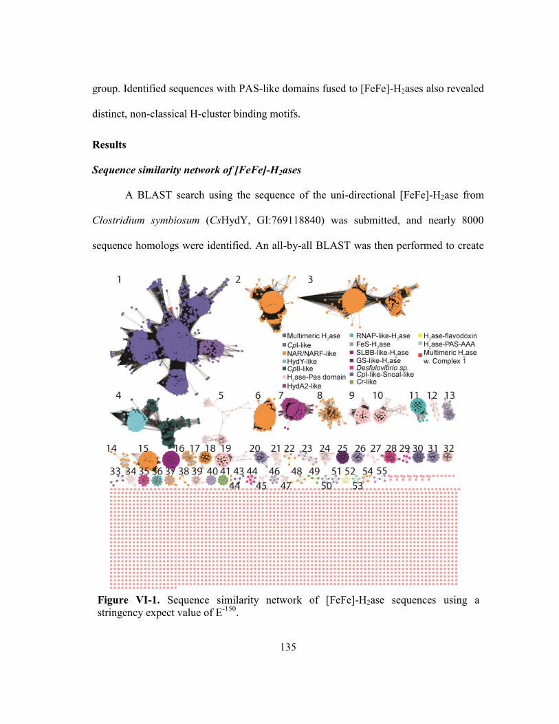
135
Figure VI-1. Sequence similarity network of [FeFe]-H2ase sequences using a
stringency expect value of E-150
.
group. Identified sequences with PAS-like domains fused to [FeFe]-H2ases also revealed
distinct, non-classical H-cluster binding motifs.
Results
Sequence similarity network of [FeFe]-H2ases
A BLAST search using the sequence of the uni-directional [FeFe]-H2ase from
Clostridium symbiosum (CsHydY, GI:769118840) was submitted, and nearly 8000
sequence homologs were identified. An all-by-all BLAST was then performed to create

136
a sequence similarity network (SSN) at a strict E-value cutoff of 10-150
. With this
stringency for sequence homology, a total of 55 subgroups could be identified as
depicted in Figure VI-I, totaling nearly 4000 sequences. [FeFe]-H2ase homologs are
distributed across bacterial classes (Clostridia, Thermotogae and δ-Proteobacteria),
anaerobic protists, green algae and even higher eukaryotes.
Identified H2ase homologs display a wide-range of modularity for catalysis, as
previously described.3,6,14
The most abundantly identified homologs grouped within
Figure VI-2. Domain architectures of [FeFe]-H2ase
sequences identified from the sequence similarity network.
The green box indicates an H-cluster binding domain.

137
cluster 1, encompassing over half (~1800 sequences) of homologs that grouped within
the SSN. The overwhelming majority of these homologs appear to be multimeric H2ases,
confirming a speculation that bifurcating H2ases are the most representative H2ase in
bacteria (Figure IV-2).287
The N-terminal ferredoxin-domain (F-domain) appears quite
variable, where [2Fe-2S], NuoG-like [4Fe-4S] and typical [4Fe-4S] clusters binding
domains are exchangeable. Other predicted domains range from H2ases with thioredoxin
and Complex I-like domains (found in anaerobic protists), glutamate synthase binding
domains, rubredoxin/rubrerythrin domains and flavodoxin-like domains. The
thioredoxin/Complex I-like domains are hypothesized to take part in metabolism in
hydrogenosomes by regenerating NAD+, suggesting these microbes share a common
ancestry with mitochondria.288
Rubrerythrins are diiron proteins involved in H2O2
reduction during ROS stress, and the Rd domain efficiently transfers electrons into the
diiron site using external reducing equivalents.128
CsHydY, which contains these
domains, was recently characterized in vitro and displayed near uni-directional H2
uptake catalysis, contrasting with classical H2ases. It was speculated that HydY catalyzes
a H2-dependent peroxidase reaction. Glutamate synthases (GS) are proteins that use
NADH or reduced ferredoxins with co-substrates glutamine and α-ketogluturate to
produce two glutamates.77
Two GS/[FeFe]-H2ase homologs were identified, one within
the HydY cluster (Figure IV-1), which also appears to contain rubredoxin-like (Rd)
domain linking the H2ase to the and another sub-group with no Rd. Based on the need
for external reducing equivalents, this fusion protein likely functions similarly to
CsHydY, consuming H2 to produce glutamate. These GS-H2ase fusions could even

138
operate in the opposite direction, i.e. H2 production from glutamate, depending on
cellular resources. Flavodoxins are electron transfer proteins that bind flavin
mononucleotides. Why these H2ases homologs contain a C-terminal flavodoxin domain
is unclear (Figure IV-2).
Additionally, [FeFe]-H2ase homologs with soluble ligand binding domains
(SLBB) and archaeal RNA polymerase domains were identified, that group separately
(Figures IV-1 and 2). SLBB domains can be secreted or periplasmic, where they bind
some ligand, in some cases even cobalamin, and this ligand is moved into the cell in a
ABC-type transporter-dependent fashion.289
The inclusion of RNAP-like domain with an
[FeFe]-H2ase is perplexing, but further supports the notion of that nature repurposes
tools for increased fitness.
[FeFe]-H2ase homologs also emphasize an evolutionary repurposing of the H-
cluster domain. Numerous identified homologs contain PAS-like domains and, in
eukaryotes, [FeFe]-H2ase like domains called nuclear architecture related (NAR) and
nuclear prelamin A recognition factor (NARF). PAS domains function as signal sensors,
binding some substrate transducing it as a signal for some cellular response. In archaea
and bacteria, these domains appear to function in sensing oxygen and redox
environments.290,291
Given the idea of syntrophy for H2 in microbial environments, this
[FeFe]-H2ase-PAS protein could sense H2 to turn on H2-consuming metabolic pathways.
Or, alternatively, it could sense ROS stress by binding O2/H2O2 or sense oxidative
conditions. It is unclear if this fusion would bind an H-cluster, although non-classical H-
cluster motifs were speculated for Ruminococcus albus.70
NAR-like homologs are

139
thought to be involved in cytosolic Fe-S cluster biosynthesis,81
and homologs were
identified in all higher eukaryotes. NAR/NARF proteins are still poorly understood, and
the cluster content of these [FeFe]-H2ase homologs remains a mystery.
Despite the clear domain modularity of [FeFe]-H2ase homologs, additional
domains do not dictate the sub-grouping of these homologs (Figure VI-1). For instance,
both the Desulfovibrio sp. and green algae [FeFe]-H2ases clustered into subgroups, as
did the CsHydY homologs. This observation suggests that there are additional
determinants for the groupings of H2ases.
Analysis of sub-group 4
To further understand the reasons for this diversity in [FeFe]-H2ase homologs,
sub-group 4 was analyzed in further detail by examining conserved cofactor binding
motifs. Cluster 4 was selected because it contains CpII and HydY homologs, which were
recently shown to be uni-directional H2 uptake [FeFe]-H2ases. This sub-group contains
234 sequences, which should avoid over-interpretation due to the relative size. Second-
shell contributions near the H-cluster were speculated to be the determinants for this
change in catalysis. Previous studies have attempted similar forays into understanding
protein-specific tuning, but only utilized a small sample size, through the lens of
characterized [FeFe]-H2ases CpI and CrHydA1, to understand the influence of the
protein matrix.

140
The active site of the [FeFe]-H2ase is found buried within the protein, formed
between several loops and helices. Residues that non-covalently interact with the H-
cluster exhibit a high degree of conservation, with these residues specified in a number
of studies and reviews (Figure VI-3).3,6,58,223,233,292
These bioinformatics studies
identified three motifs that coordinate the H-cluster: P1, TSCCPxW; P2, MPCx2KKxE;
P3, M(A)Cx2xGCx2GGGQ. Additional motifs coordinate the H-cluster near the
Figure VI-3. Conserved H-cluster binding motifs P1-P4 that provide non-covalent
interactions to the cofactor (a). Conserved motifs identified in sub-group 4 of the
sequence similarity network of P1 (b), P2 (c), P3 (d), P4 (e) and SPQ (f). Weblogos
are colored according to chemical identity. A bit score of 4 indicates a completely
conserved residue.

141
diatomic ligands, with ‘P4’ (APAxR) interacting with the bridging CO and proximal CN-
and a SPQ motif interacting with the distal CN- and H-bonding with other conserved
residues.
A MUSCLE alignment of the 234 sub-group 4 homologs agree with this
proposed active site conservation; however, there are a few key differences. Within the
P2 motif, the conserved M353 (all numbering according to PDB: 3C8Y) is substituted,
with 87% Thr, 6% Asn and 5% Ala. Substitutions of this Met residue appears to be
specific only to this sub-cluster. Substitution of hydrophilic residues, or small
hydrophobic residues, would likely largely affect catalysis, as hypothesized previously
(see Chapters III and V). The P3 motif provides Met and Cys residues that interact with
the azadithiolate and the cubane cluster, between these residues, Ala is substituted to Thr
57% in sub-group 4. The so-called P4 motif (described previously, Chapter III), occurs
before the P1 motif in sequence but described after introduction of P1-P3 motif
nomenclature, contains three interactions to the H-cluster. The first Ala creates a
hydrophobic pocket for the bridging-CO, and this residue is substituted to Ser 66% of
the identified homologs. This Ser was suggested to a determinant for catalytic bias of
CsHydY, but the lack of conservation (as was seen in P2) argues otherwise. However, all
HydY homologs, annotated based on containing at least a C-terminal Rd, contain this
Ser substitution. Other homologs within this group that contain the Ala are all from the
class Negativicutes, and these homologs lack Rd domains. Thus, it appears the Ser
substitution also is concomitant with a Rd domain. Perhaps the Ser near the H-cluster is
important not for catalytic bias, but instead minimizes anaerobic, oxidative inactivation

142
as monitored by protein film electrochemistry. Furthermore, an additional SPQ motif,
which provides interactions with the proximal CN- through electrostatics of the Pro and
main-chain Gln, has a substitution of an isoleucine (97% occurrence) at the Gln residue
and Cys (10% occurrence) at the Pro. The Ile likely does not preclude interactions with
the CN-, but the hydrophobic side chain would be inserted into a hydrophilic pocket.
This isoleucine substitution would result in the movement of the coil housing the SPQ
motif, or hydrophilic residues would require conformational changes. Either scenario
would result in a modified H-cluster binding pocket that might affect catalysis.
While the above mentioned active site substitutions might affect catalysis, these
substitutions do not explain the overpotential requirement observed in electrochemistry
studies. The overpotential was hypothesized to be a result of ‘positive-patch’
substitutions within the F-domain (Chapter III). As depicted in Figure VI-4, multiple
positively charged residues occupy positions near a [4Fe-4S] cluster. Lysine, arginine
Figure VI-4. Depiction of the [4Fe-4S] cluster from CpI (PDB: 3C8Y; a) and the
corresponding positive patch sequence logo from sub-group 4 from the sequence
similarity network. Colors of the one letter codes indicate chemical identity, and
conserved residues have bit scores of 4.

143
and asparagine residues flank all of the conserved Cys residues that are annotated to bind
a [4Fe-4S] cluster. These positive charges near the [4Fe-4S] cluster would likely
attenuate the potential of the cluster to significantly more positive potentials. Indeed, the
redox potential of clusters within CpII were found to be increased to nearly -180 mV,
hundreds of mV more positive than typical [4Fe-4S] clusters.235
Thus, analysis sub-
cluster 4 of this SSN suggests the defining motifs of these hydrogenases are likely to H-
bonding substitutions within H-cluster binding motifs and positively charged residue
substitutions within a [4Fe-4S] cluster, which likely dictates catalytic properties, as
suggested previously (see Chapter III).
Analysis of PAS domain proteins
We next analyzed all PAS domains fused to [FeFe]-H2ases. We hypothesized
Figure VI-5. Sequence logos proposed H-cluster binding motifs of PAS domain fusions
identified. Conservations of substituted residues in P1 (a), P2 (b), P3 (c), P4 (d) and
SPQ (e) imply that PAS domains would bind an H-cluster in an usual manner, if at all.

144
that since PAS domains bind soluble substrates or respond to cellular redox conditions,
these homologs would bind the H-cluster in a non-canonical manner in order to facilitate
signal transduction.70
A total of 282 [FeFe]-H2ase-PAS domain fusions were analyzed.
All motifs appear to have significant variability within the H-cluster binding motifs
(Figure VI-5). The cysteine residues required to bind the cubane [4Fe-4S] cluster appear
to be the only conserved residues, while residues important for coordinating proximal
and distal CN- ligands (K in P2, 94% occurrence and P in P4, 97.5% occurrence) is
nearly absolutely conserved. The first cysteine in P1, identified as the general base
during the catalytic cycle, only occurs 48.2% in the PAS domain homologs, with Ala
(33%) the next most encountered residue, likely indicating PAS domains would lack the
ability to catalyze H+
reduction or H2 oxidation. Methionine residues in P2 and P3 that
stack below and above the H-cluster (Figure VI-5) are also lacking. The P2 Met residue
is substituted with Gly (91.8% occurrence), while the P3 Met residues is equally
substituted with N/S/L residues.
Discussion
The rapidly increasing NCBI non-redundant database has led to the discovery
and annotation of a myriad of hydrogenases, many of them orphans, lacking any
information other than sequence identity and similarity of various domains. We have
attempted begin the monumental task of assigning functions for these enzymes. Nature
has clearly evolved numerous repurposed functions for this old enzyme, and, in a sense,
taught ‘old dogs new tricks.’ [FeFe]-H2ases, as has been described by numerous others,
are highly modular in nature, and this theme appears to underscore the chemical
145
diversity [FeFe]-H2ases are capable of undertaking. The newest members of this pack
are the uni-directional H2-uptake [FeFe]-H2ases. Using SSNs for [FeFe]-H2ases, it is
clear that this sub-group shares some conservation in active site mutants, but also some
substitutions specific to groups within the larger sub-group.
A total of 55 sub-groups were identified from this SSN of [FeFe]-H2ases,
consisting nearly 4000 sequences with enough similarity to cluster together under strict
conditions (expect values of E-150
). The overwhelming majority of these homologs were
monomeric or multimeric [FeFe]-H2ases, but additional domains including SLBB-,
RNAP-, GS- and apparent DNA-binding-like domains were identified (Figure VI-2).
SLBB domains are believed to excreted or periplasmic, where they recruit a required
extracellular molecule, such as cobalamin. While studies are the SLBB fusion protein
are needed, it is tempting to speculate that perhaps the evolutionary logic of this fusion is
to use a H+
gradient to transport the soluble ligand. The inclusion of PAS and apparent
DNA-binding domains suggests that [FeFe]-H2ase-like homologs were evolutionarily
repurposed to recognize and transduce specific cellular conditions. These domain fusion
functions become especially clear due to the recent observation of syntrophic H2 usage
within microbial communities,5,10,11,70,76,293
highlighting the broader scope of H2
metabolism within anaerobic and microaerobic organisms.
We focused on active site substitutions within both HydY-like H2ases and PAS-
domain fusions. Conservation of the H-cluster of HydY homologs was clearly evident.
Substitutions within the active site and auxiliary cluster motifs suggest these evolved
changes may be characteristic of the sub-groups, but further work is needed to further

146
elucidate the sub-grouping of these [FeFe]-H2ase homologs. For instance, green algal,
Desulfovibrio sp and HydA2 homologs segregate into various respective groups. All
these homologs appear to bind the H-cluster in a classical manner. Differences within F-
domains do not preclude this as a major discriminating factor, but additional factors may
also influence the sub-grouping. Putative gas channels, proton transfer relays and
electron transfer pathways may be subtly different within the sub-groups, which
warrants further inspection using more advanced studies. Inspection of conserved
surface residues may also indicate likely electron transfer partners in vivo, as would
inspection of possible cellular location signals (periplasmic targeting in prokaryotes,
organelle-specific sequence in eukaryotes) may also point to the in vivo function.
[FeFe]-H2ase-PAS domain fusions are widespread throughout the SSN. PAS
domain fusions, based on the sequence identity within supposedly conserved H-cluster
binding motifs, likely do not bind an H-cluster in a classical manner, or if at all. How a
signal is recognized, what the signal is, and how the signal is transduced is not clear
from sequence analysis, and will require a brave soul to purify and characterize in vitro,
likely from native source to fully understand how these proteins function.70
The identification of a multitude of [FeFe]-H2ase sub-groups, with the variability
of apparent H-cluster motifs, implicates the influence of the protein matrix in dictating
catalysis of the H-cluster. The individual influence of protein residues at second- or even
third-shell contributions could have unplumbed effects on the chemical nature of the H-
cluster. Furthermore, the sub-grouping of the 4000 sequences highlights that there are
determinants of [FeFe]-H2ases that may lead to future phylogenetic grouping and

147
elucidate the influence of the protein framework on catalysis. Understanding the
influence of various evolved [FeFe]-H2ase homologs may lead to improved small
molecule H-cluster mimics that have increased catalytic rates or minimized O2-induced
damage.

148
CHAPTER VII
CONCLUDING REMARKS
The function-discovery project for the CsHydY, the [FeFe]-H2ase-Rbr fusion,
started out with simple, relatively straightforward questions: 1) Is CsHydY the first O2-
tolerant [FeFe]-H2ase? 2) If so, then how? And finally, 3) Why does this fusion protein
exist? At the time (~2010), recombinant studies with [FeFe]-H2ases were exploding onto
the scientific scene. Exploratory studies had, at the time, recently identified recombinant
systems for expressing [FeFe]-H2ases with the required maturation machinery, HydE, F
and G.102
And now, unraveling the mechanism of [FeFe]-H2ase biosynthesis appears to
be the nouveau research avenue. Before the seminal discovery of the minimal
maturation machinery, [FeFe]-H2ase research relied on purifying material from native
sources, limiting diversity of research groups capable of probing this enzyme.
Recombinant protein production allowed for increased yield of protein, enhancing the
scientific community’s understanding of [FeFe]-H2ase mechanism and function. Sample
intensive techniques, such as EPR, FT-IR, protein crystallography and X-ray absorption
spectroscopy were now ripe for study with these increased protein yields. Now,
researchers were capable of – and this is truly the amazing part to me – choosing to
expose their precious preparations of [FeFe]-H2ases to O2 in order to elaborate the
mechanism of O2 induced destruction of the H-cluster. At the time I started this project,
irreversible damage by O2 was speculated to result from O2 diffusion into the active site,
where it ultimately led to loss of the cubane [4Fe-4S].93,294,295
The current model of O2-
based degradation of the [FeFe]-H2ase active site is now thought to first result in the loss

149
of the [2Fe]H.296
We reasoned that if ROS were the source of [FeFe]-H2ase inactivation,
then [FeFe]-H2ases with the inherent ability to remove such species (a la a rubrerythrin
module) might result in a more O2 tolerant enzyme. In retrospect, these all were
somewhat naïve speculations. Rubrerythrins are poor O2 reduction catalysts, and require
external reducing equivalents, in the form of one electron transfers, for catalysis. Any
such rubrerythrin based system would eventually suffer the same fate as an [FeFe]-
H2ase. Regardless, a story of CsHydY function eventually came together. Using insights
gleaned from giants in the field of H2ase research, from the pioneers and prolific
publishers,1,7,46,60,67,83,90,117,219,220,248,253
we improved our hypothesis for the function of
this truly novel fusion protein. It is on the shoulders of giants in which we can now
annotate the function of CsHydY.
Using a coupled biochemical and electrochemical approach, we found that
CsHydY is a poor H+
reduction catalyst and readily consumes H2, making it the first
electrochemical study (initial work with CpII72,74,235
) of an [FeFe]-H2ase that was
catalytically biased toward H2 oxidation. In fact, our careful biochemical work agreed
with our electrochemical measurements. This catalytic bias discovery was met by
significant barriers, namely the (incorrect) assumption that [FeFe]-H2ases are biased
toward H+ reduction. In fact, [FeFe]-H2ases are the superior H2ase catalysts,
38 producing
and consuming H2 at least an order of magnitude faster than their [NiFe]-H2ase
counterparts.40
Additionally, we found that H+ reduction CsHydY was inhibited by H2
and it was much less susceptible to anaerobic oxidative inactivation. This observation
did not escape our collaborators, who then reconstituted unmaturated HydY constructs

150
with synthetic mimics to convince themselves of the result. The fact that enzymes can be
matured with biomimetic compounds still astounds me.60,117
Thus, CsHydY appears to
have evolved to function in manner somewhere between that of both [NiFe]- and [FeFe]-
H2ases. Not only this, but we also showed that electrons from H2 can be transferred from
the H2ase domain to Rbr domain. Because our Rbr construct showed surprisingly high
catalytic efficiency when reduced from NADH, we speculated that HydY likely
functions as a H2-dependent peroxidase.
Though we have assigned a putative function for CsHydY, it is by no means an
open-and-closed case. The lack of conclusive H2 dependency on the peroxidase catalysis
is an open wound. We speculate that reduction of Rbr domain from dissolved H2 could
be used to reduce ROS, but do not directly, experimental ‘prove’ the function. We have
now designed, and plan to attempt forays with as soon as possible, an assay to test our
hypothesis. Using a coupled horse-radish peroxidase assay, we will incubate the HydY
constructs with H2O2 and various concentrations of dissolved H2 and measure, indirectly,
the amount of H2O2 consumed. This will, in theory, allow for direct counting of
electrons required to consume H2. We speculate that HydY will function in a
stoichiometric manner, i.e. no non-productive turnover of H2. While these studies might
include a possible function in vitro, the true function in vivo is an enigma wrapped in a
mystery. Unraveling the function in a host organism would be the cherry on the
proverbial sundae. We have a potential collaborator here at Texas A&M that is willing to
help use grow Clostridium scindens, which encodes a homolog of HydY. We have
envisioned quantitative RT-PCR and hydY gene knock-out studies to elucidate the role

151
this protein plays in anaerobic bacteria. I believe these studies are paramount to
assigning H2-dependent peroxidase chemistry to HydY.
Using mutagenesis, bioinformatics and computational studies, we have also
identified conserved residues that provide second-shell interactions with the H-cluster
that are the likely molecular determinants for HydY’s catalytic bias. The initial idea of
hydrogen bonding donor interacting with CO ligands was difficult comprehend, but it is
now clear that these residues act like Lewis acids near the H-cluster. Understanding the
fundamental chemistry here and potential implications for biomimetic complexes are
natural segues. Clearly, the next frontier for biomimetic compounds will be the rational
design and incorporation of controlled second-sphere interactions with catalysts. I
believe that the interactions identified in CsHydY will result in incorporation of Lewis
acid-like moieties near the bridging CO to stabilize this important ligand. Sequence
similarity networks of nearly 4000 [FeFe]-H2ase sequences lead to the development of
55 sub-group sequences. While there are clear modularity in the domain architectures of
these sequences, differences in the domains cannot completely account the sub-
groupings of the sequences. Further, more in-depth analysis is warranted to investigate
potential surface exposed domains, which might be important for protein-protein
interactions, putative gas-channels, H+ relays or even other nearby active site
substitutions that are I mistakenly overlooked. Of all the various domain architectures
identified in the [FeFe]-H2ase SSN, the fusions with SLBB and ATPase AA domains
caught my attention. How these enzymes might function continually occupy my
thoughts. My hope is that this SSN will spark the curiosity of at least one brave scientist

152
to investigate their roles. That, or I will just have to do it eventually. The broad swath of
potential chemical diversity that are central to [FeFe]-H2ases astounds me, and
underscores the extents to which evolution pushes the repurposing of these old enzymes
for new functions.

153
REFERENCES
1 Stephenson, M. & Stickland, L. H. Hydrogenase: a bacterial enzyme activating
molecular hydrogen: The properties of the enzyme. Biochem J 25, 205-214,
(1931).
2 Vignais, P. M., Billoud, B. & Meyer, J. Classification and phylogeny of
hydrogenases. Fems Microbiology Reviews 25, 455-501, (2001).
3 Vignais, P. M. & Billoud, B. Occurrence, classification, and biological function
of hydrogenases: an overview. Chem Rev 107, 4206-4272, (2007).
4 Schwartz E, F. J., Friedrich B. in The Prokaryotes - Prokaryotic Physiology and
Biochemistry 545-577 (2013).
5 Greening, C. et al. Genomic and metagenomic surveys of hydrogenase
distribution indicate H2 is a widely utilised energy source for microbial growth
and survival. ISME J, (2015).
6 Meyer, J. [FeFe] hydrogenases and their evolution: a genomic perspective. Cell
Mol Life Sci 64, 1063-1084, (2007).
7 Lubitz, W., Ogata, H., Rüdiger, O. & Reijerse, E. Hydrogenases. Chem Rev 114,
4081-4148, (2014).
8 Conrad, R. Soil microorganisms as controllers of atmospheric trace gases (H2,
CO, CH4, OCS, N2O, and NO). Microbiol Rev 60, 609-640, (1996).
9 Wolf, P. G., Biswas, A., Morales, S. E., Greening, C. & Gaskins, H. R. H
metabolism is widespread and diverse among human colonic microbes. Gut
Microbes, 0, (2016).
10 Ballor, N. R., Paulsen, I. & Leadbetter, J. R. Genomic analysis reveals multiple
[FeFe] hydrogenases and hydrogen sensors encoded by treponemes from the
H(2)-rich termite gut. Microb Ecol 63, 282-294, (2012).

154
11 Fischbach, M. A. & Sonnenburg, J. L. Eating for two: how metabolism
establishes interspecies interactions in the gut. Cell Host Microbe 10, 336-347,
(2011).
12 Schmitz, R. A., Daniel, R., Deppenmeier, U. & Gottschalk, G. The Anaerobic
Way of Life. Prokaryotes: A Handbook on the Biology of Bacteria, Vol 2, Third
Edition, 86-101, (2006).
13 Vardar-Schara, G., Maeda, T. & Wood, T. K. Metabolically engineered bacteria
for producing hydrogen via fermentation. Microb Biotechnol 1, 107-125, (2008).
14 Calusinska, M., Happe, T., Joris, B. & Wilmotte, A. The surprising diversity of
clostridial hydrogenases: a comparative genomic perspective. Microbiology 156,
1575-1588, (2010).
15 Schut, G. J. & Adams, M. W. The iron-hydrogenase of Thermotoga maritima
utilizes ferredoxin and NADH synergistically: a new perspective on anaerobic
hydrogen production. J Bacteriol 191, 4451-4457, (2009).
16 Ragsdale, S. W. Pyruvate ferredoxin oxidoreductase and its radical intermediate.
Chem Rev 103, 2333-2346, (2003).
17 Furdui, C. & Ragsdale, S. W. The role of pyruvate ferredoxin oxidoreductase in
pyruvate synthesis during autotrophic growth by the Wood-Ljungdahl pathway. J
Biol Chem 275, 28494-28499, (2000).
18 Posewitz, M. C., Dubini, A., Meuser, J. E., Seibert, M. & Ghirardi, M. L.
Hydrogenases, Hydrogen Production, and Anoxia. The Chlamydomonas
Sourcebook 3-Vol set 2, 217-255, (2009).
19 Chou, C. J., Jenney, F. E., Jr., Adams, M. W. & Kelly, R. M. Hydrogenesis in
hyperthermophilic microorganisms: implications for biofuels. Metab Eng 10,
394-404, (2008).
20 Wang, S., Huang, H., Kahnt, J. & Thauer, R. K. A reversible electron-bifurcating
ferredoxin- and NAD-Dependent [FeFe]-Hydrogenase (HydABC) in Moorella
thermoacetica. Journal of Bacteriology 195, 1267-1275, (2013).

155
21 Poehlein, A. et al. An ancient pathway combining carbon dioxide fixation with
the generation and utilization of a sodium ion gradient for ATP synthesis. PLoS
One 7, e33439, (2012).
22 Schuchmann, K. & Muller, V. A bacterial electron-bifurcating hydrogenase. J
Biol Chem 287, 31165-31171, (2012).
23 Schut, G. J. & Adams, M. W. W. The iron-hydrogenase of Thermotoga maritima
utilizes ferredoxin and NADH synergistically: a new perspective on anaerobic
hydrogen production. Journal of Bacteriology 191, 4451-4457, (2009).
24 Zhou, Z. et al. Ammonia fermentation, a novel anoxic metabolism of nitrate by
fungi. J Biol Chem 277, 1892-1896, (2002).
25 McDowall, J. S. et al. Bacterial formate hydrogenlyase complex. P Natl Acad Sci
USA 111, E3948-3956, (2014).
26 Ghirardi, M. L., Dubini, A., Yu, J. & Maness, P. C. Photobiological hydrogen-
producing systems. Chem Soc Rev 38, 52-61, (2009).
27 Hu, Y. & Ribbe, M. W. Nitrogenase and homologs. J Biol Inorg Chem 20, 435-
445, (2015).
28 Hu, Y. & Ribbe, M. W. A journey into the active center of nitrogenase. J Biol
Inorg Chem 19, 731-736, (2014).
29 Ribbe, M. W., Hu, Y., Hodgson, K. O. & Hedman, B. Biosynthesis of
nitrogenase metalloclusters. Chem Rev 114, 4063-4080, (2014).
30 Bothe, H., Schmitz, O., Yates, M. G. & Newton, W. E. Nitrogen fixation and
hydrogen metabolism in cyanobacteria. Microbiol Mol Biol Rev 74, 529-551,
(2010).
31 Seefeldt, L. C., Hoffman, B. M. & Dean, D. R. Mechanism of Mo-dependent
nitrogenase. Annu Rev Biochem 78, 701-722, (2009).
156
32 Wu, M. et al. Life in hot carbon monoxide: the complete genome sequence of
Carboxydothermus hydrogenoformans Z-2901. PLoS Genet 1, e65, (2005).
33 Cracknell, J. A., Wait, A. F., Lenz, O., Friedrich, B. & Armstrong, F. A. A
kinetic and thermodynamic understanding of O2 tolerance in [NiFe]-
hydrogenases. Proc Natl Acad Sci U S A 106, 20681-20686, (2009).
34 Vincent, K. A. et al. Electrochemical definitions of O2 sensitivity and oxidative
inactivation in hydrogenases. J Am Chem Soc 127, 18179-18189, (2005).
35 Shomura, Y., Yoon, K. S., Nishihara, H. & Higuchi, Y. Structural basis for a
[4Fe-3S] cluster in the oxygen-tolerant membrane-bound [NiFe]-hydrogenase.
Nature 479, 253-256, (2011).
36 Liang, Y. et al. Increased biological hydrogen production by deletion of
hydrogen-uptake system in photosynthetic bacteria. Microbiol Res 164, 674-679,
(2009).
37 Shima, S. et al. The crystal structure of [Fe]-hydrogenase reveals the geometry of
the active site. Science (New York, N.Y.) 321, 572-575, (2008).
38 Frey, M. Hydrogenases: hydrogen-activating enzymes. ChemBioChem 3, 153-
160, (2002).
39 Cammack, R. Redox enzymes. Splitting molecular hydrogen. Nature 373,
(1995).
40 Adams, M. W. The structure and mechanism of iron-hydrogenases. Biochim
Biophys Acta 1020, 115-145, (1990).
41 Cammack, R. Hydrogenase sophistication. Nature 397, 214-215, (1999).
42 Pilak, O. et al. The crystal structure of the apoenzyme of the iron-sulphur cluster-
free hydrogenase. J Mol Biol 358, 798-809, (2006).

157
43 Korbas, M. et al. The iron-sulfur cluster-free hydrogenase (Hmd) is a
metalloenzyme with a novel iron binding motif. J Biol Chem 281, 30804-30813,
(2006).
44 Shima, S. & Thauer, R. K. A third type of hydrogenase catalyzing H2 activation.
Chem Rec 7, 37-46, (2007).
45 Hiromoto, T. et al. The crystal structure of C176A mutated [Fe]-hydrogenase
suggests an acyl-iron ligation in the active site iron complex. FEBS Lett 583,
585-590, (2009).
46 Fontecilla-Camps, J. C., Volbeda, A., Cavazza, C. & Nicolet, Y.
Structure/function relationships of [NiFe]- and [FeFe]-hydrogenases. Chem Rev
107, 4273-4303, (2007).
47 Lubitz, W., Reijerse, E. & van Gastel, M. [NiFe] and [FeFe] hydrogenases
studied by advanced magnetic resonance techniques. Chem Rev 107, 4331-4365,
(2007).
48 Dementin, S. et al. Introduction of methionines in the gas channel makes [NiFe]
hydrogenase aero-tolerant. Journal of the American Chemical Society 131,
10156-10164, (2009).
49 Cracknell, J. a., Wait, A. F., Lenz, O., Friedrich, B. & Armstrong, F. a. A kinetic
and thermodynamic understanding of O2 tolerance in [NiFe]-hydrogenases.
Proceedings of the National Academy of Sciences of the United States of America
106, 20681-20686, (2009).
50 Buhrke, T., Lenz, O., Krauss, N. & Friedrich, B. Oxygen tolerance of the H2-
sensing [NiFe] hydrogenase from Ralstonia eutropha H16 is based on limited
access of oxygen to the active site. J Biol Chem 280, 23791-23796, (2005).
51 Pandelia, M. E. et al. Characterization of a unique [FeS] cluster in the electron
transfer chain of the oxygen tolerant [NiFe] hydrogenase from Aquifex aeolicus.
Proc Natl Acad Sci U S A 108, 6097-6102, (2011).

158
52 Fritsch, J. et al. The crystal structure of an oxygen-tolerant hydrogenase uncovers
a novel iron-sulphur centre. Nature, 3-7, (2011).
53 Fritsch, J., Lenz, O. & Friedrich, B. Structure, function and biosynthesis of O(2)-
tolerant hydrogenases. Nat Rev Microbiol 11, 106-114, (2013).
54 Lauterbach, L. & Lenz, O. Catalytic production of hydrogen peroxide and water
by oxygen-tolerant [NiFe]-hydrogenase during H2 cycling in the presence of O2.
J Am Chem Soc 135, 17897-17905, (2013).
55 Evans, R. M. et al. Principles of sustained enzymatic hydrogen oxidation in the
presence of oxygen--the crucial influence of high potential Fe-S clusters in the
electron relay of [NiFe]-hydrogenases. J Am Chem Soc 135, 2694-2707, (2013).
56 Parkin, A. & Sargent, F. The hows and whys of aerobic H2 metabolism. Curr
Opin Chem Biol 16, 26-34, (2012).
57 Vincent, K. a. et al. Electricity from low-level H2 in still air--an ultimate test for
an oxygen tolerant hydrogenase. Chemical communications (Cambridge,
England), 5033-5035, (2006).
58 Winkler, M., Esselborn, J. & Happe, T. Molecular basis of [FeFe]-hydrogenase
function: an insight into the complex interplay between protein and catalytic
cofactor. Biochim Biophys Acta 1827, 974-985, (2013).
59 Poehlein, A. et al. An ancient pathway combining carbon dioxide fixation with
the generation and utilization of a sodium ion gradient for ATP synthesis. PloS
One 7, e33439, (2012).
60 Berggren, G. et al. Biomimetic assembly and activation of [FeFe]-hydrogenases.
Nature 499, 66-69, (2013).
61 Peters, J. W. & Broderick, J. B. Emerging paradigms for complex iron-sulfur
cofactor assembly and insertion. Annu Rev Biochem 81, 429-450, (2012).

159
62 Adams, M. W. W. & Mortenson, L. E. The physical and catalytic properties of
hydrogenase II of Clostridium pasteurianum. A comparison with hydrogenase I.
Journal of Biological Chemistry 259, 7045-7055, (1984).
63 Forestier, M. et al. Expression of two [Fe] -hydrogenases in Chlamydomonas
reinhardtii under anaerobic conditions. Eur J Biochem 2758, 2750-2758, (2003).
64 Girbal, L. et al. Homologous and heterologous overexpression in Clostridium
acetobutylicum and characterization of purified clostridial and algal Fe-only
hydrogenases with high specific activities. Appl Environ Microbiol 71, 2777-
2781, (2005).
65 Ghirardi, M. L. et al. Microalgae: a green source of renewable H(2). Trends in
Biotechnology 18, 506-511, (2000).
66 Ghirardi, M. L. et al. Hydrogenases and hydrogen photoproduction in oxygenic
photosynthetic organisms. Annu Rev Plant Biol 58, 71-91, (2007).
67 Nicolet, Y., Piras, C., Legrand, P., Hatchikian, C. E. & Fontecilla-Camps, J. C.
Desulfovibrio desulfuricans iron hydrogenase: the structure shows unusual
coordination to an active site Fe binuclear center. Struct Fold Des 7, 13-23,
(1999).
68 Nicolet, Y. et al. Crystallographic and FTIR spectroscopic evidence of changes
in Fe coordination upon reduction of the active site of the Fe-only hydrogenase
from Desulfovibrio desulfuricans. J Am Chem Soc 123, 1596-1601, (2001).
69 Thauer, R. K., Jungermann, K. & Decker, K. Energy conservation in
chemotrophic anaerobic bacteria. Bacteriol Rev 41, 100-180, (1977).
70 Zheng, Y., Kahnt, J., Kwon, I. H., Mackie, R. I. & Thauer, R. K. Hydrogen
Formation and its Regulation in Ruminococcus albus: Involvement of an
Electron-Bifurcating [FeFe]-Hydrogenase, of a Non Electron-bifurcating [FeFe]-
hydrogenase and of a Putative Hydrogen-Sensing [FeFe]-Hydrogenase. J
Bacteriol 196, 3840-3852, (2014).

160
71 Schuchmann, K. & Muller, V. Direct and reversible hydrogenation of Co2 to
formate by a bacterial carbon dioxide reductase. Science 342, 1382-1386, (2013).
72 Chen, J. S. & Blanchard, D. K. Isolation and properties of a unidirectional H2-
oxidizing hydrogenase from the strictly anaerobic N2-fixing bacterium
Clostridium pasteurianum W5. Biochem Biophys Res Commun 84, 1144-1150,
(1978).
73 Adams, M. & Mortenson, L. The purification of hydrogenase II (uptake
hydrogenase) from the anaerobic N2-fixing bacterium Clostridium pasteurianum.
Biochimica et Biophysica Acta (BBA) - Bioenergetics 766, 51-61, (1984).
74 Adams, M. W. & Mortenson, L. E. The physical and catalytic properties of
hydrogenase II of Clostridium pasteurianum. J Biol Chem 259, 7045-7055,
(1984).
75 Adams, M. W. W. The mechanisms of hydrogen activation and carbon monoxide
binding by hydrogenase I and hydrogenase II of Clostridium pasteurianum.
Journal of Biological Chemistry 262, 15054-15061, (1987).
76 Schmidt, O., Drake, H. L. & Horn, M. A. Hitherto unknown [Fe-Fe]-
hydrogenase gene diversity in anaerobes and anoxic enrichments from a
moderately acidic fen. Appl Environ Microbiol 76, 2027-2031, (2010).
77 Suzuki, A. & Knaff, D. B. Glutamate synthase: structural, mechanistic and
regulatory properties, and role in the amino acid metabolism. Photosynth Res 83,
191-217, (2005).
78 Ballor, N. R. & Leadbetter, J. R. Analysis of extensive [FeFe] hydrogenase gene
diversity within the gut microbiota of insects representing five families of
Dictyoptera. Microb Ecol 63, 586-595, (2012).
79 Hackstein, J. H. P. Eukaryotic Fe-hydrogenases -- old eukaryotic heritage or
adaptive acquisitions? Biochemical Society transactions 33, 47-50, (2005).

161
80 Lill, R., Srinivasan, V. & Muhlenhoff, U. The role of mitochondria in cytosolic-
nuclear iron-sulfur protein biogenesis and in cellular iron regulation. Curr Opin
Microbiol 22, 111-119, (2014).
81 Lill, R. & Muhlenhoff, U. Maturation of iron-sulfur proteins in eukaryotes:
mechanisms, connected processes, and diseases. Annu Rev Biochem 77, 669-700,
(2008).
82 Pershad, H. R. et al. Catalytic electron transport in Chromatium vinosum [NiFe]-
hydrogenase: application of voltammetry in detecting redox-active centers and
establishing that hydrogen oxidation is very fast even at potentials close to the
reversible H+/H2 value. Biochemistry 38, 8992-8999, (1999).
83 Vincent, K. A., Parkin, A. & Armstrong, F. A. Investigating and exploiting the
electrocatalytic properties of hydrogenases. Chem Rev 107, 4366-4413, (2007).
84 Leger, C. et al. Enzyme electrokinetics: using protein film voltammetry to
investigate redox enzymes and their mechanisms. Biochemistry 42, 8653-8662,
(2003).
85 Jones, A. K. et al. Enzyme electrokinetics: electrochemical studies of the
anaerobic interconversions between active and inactive states of Allochromatium
vinosum [NiFe]-hydrogenase. J Am Chem Soc 125, 8505-8514, (2003).
86 Baffert, C. et al. Covalent attachment of FeFe hydrogenases to carbon electrodes
for direct electron transfer. Anal Chem 84, 7999-8005, (2012).
87 Le Goff, A. et al. From hydrogenases to noble metal-free catalytic nanomaterials
for H2 production and uptake. Science 326, 1384-1387, (2009).
88 Bard, A. J. & Faulkner, L. R. Electrochemical Methods : Fundamentals and
Applications. 2nd edn, (Wiley, 2001).
89 Murphy, B. J., Sargent, F. & Armstrong, F. a. Transforming an oxygen-tolerant
[NiFe] uptake hydrogenase into a proficient, reversible hydrogen producer.
Energy & Environmental Science 7, 1426-1433, (2014).

162
90 Armstrong, F. A. et al. Guiding Principles of Hydrogenase Catalysis Instigated
and Clarified by Protein Film Electrochemistry. Accounts of Chemical Research,
acs.accounts.6b00027, (2016).
91 Hexter, S. V., Grey, F., Happe, T., Climent, V. & Armstrong, F. A. Correction
for Hexter et al., Electrocatalytic mechanism of reversible hydrogen cycling by
enzymes and distinctions between the major classes of hydrogenases.
Proceedings of the National Academy of Sciences 109, 18232-18233, (2012).
92 Lukey, M. J. et al. How Escherichia coli is equipped to oxidize hydrogen under
different redox conditions. J Biol Chem 285, 3928-3938, (2010).
93 Goldet, G. et al. Electrochemical kinetic investigations of the reactions of
[FeFe]-hydrogenases with carbon monoxide and oxygen: comparing the
importance of gas tunnels and active-site electronic/redox effects. Journal of the
American Chemical Society 131, 14979-14989, (2009).
94 Parkin, A., Cavazza, C., Fontecilla-Camps, J. C. & Armstrong, F. A.
Electrochemical investigations of the interconversions between catalytic and
inhibited states of the [FeFe]-hydrogenase from Desulfovibrio desulfuricans. J
Am Chem Soc 128, 16808-16815, (2006).
95 Armstrong, F. A. Hydrogenases: active site puzzles and progress. Curr Opin
Chem Biol 8, 133-140, (2004).
96 Fourmond, V. et al. The mechanism of inhibition by H2 of H2-evolution by
hydrogenases. Chem Commun 49, 6840-6842, (2013).
97 Vincent, K. a. et al. Electrochemical definitions of O2 sensitivity and oxidative
inactivation in hydrogenases. Journal of the American Chemical Society 127,
18179-18189, (2005).
98 Hexter, S. V., Grey, F., Happe, T., Climent, V. & Armstrong, F. A.
Electrocatalytic mechanism of reversible hydrogen cycling by enzymes and
distinctions between the major classes of hydrogenases. Proc Natl Acad Sci U S
A 109, 11516-11521, (2012).

163
99 Forzi, L. & Sawers, R. G. Maturation of [NiFe]-hydrogenases in Escherichia coli.
Biometals 20, 565-578, (2007).
100 Mulder, D. W. et al. Insights into [FeFe]-Hydrogenase Structure, Mechanism,
and Maturation. Structure (London, England : 1993) 19, 1038-1052, (2011).
101 Posewitz, M. C. et al. Discovery of two novel radical S-adenosylmethionine
proteins required for the assembly of an active [Fe] hydrogenase. J Biol Chem
279, 25711-25720, (2004).
102 King, P. W., Posewitz, M. C., Ghirardi, M. L. & Seibert, M. Functional studies of
[FeFe] hydrogenase maturation in an Escherichia coli biosynthetic system. J
Bacteriol 188, 2163-2172, (2006).
103 Mulder, D. W. et al. Activation of HydA(DeltaEFG) requires a preformed [4Fe-
4S] cluster. Biochemistry 48, 6240-6248, (2009).
104 Czech, I., Silakov, A., Lubitz, W. & Happe, T. The [FeFe]-hydrogenase maturase
HydF from Clostridium acetobutylicum contains a CO and CN- ligated iron
cofactor. FEBS Lett 584, 638-642, (2010).
105 Shepard, E. M. et al. [FeFe]-hydrogenase maturation: HydG-catalyzed synthesis
of carbon monoxide. Journal of the American Chemical Society 132, 9247-9249,
(2010).
106 Driesener, R. C. et al. [FeFe]-hydrogenase cyanide ligands derived from S-
adenosylmethionine-dependent cleavage of tyrosine. Angewandte Chemie
(International ed. in English) 49, 1687-1690, (2010).
107 Suess, D. L. M., Kuchenreuther, J. M., De La Paz, L., Swartz, J. R. & Britt, R. D.
Biosynthesis of the [FeFe] Hydrogenase H Cluster: A Central Role for the
Radical SAM Enzyme HydG. Inorganic Chemistry 55, 478-487, (2016).
108 Suess, D. L. et al. The Radical SAM Enzyme HydG Requires Cysteine and a
Dangler Iron for Generating an Organometallic Precursor to the [FeFe]-
Hydrogenase H-Cluster. J Am Chem Soc 138, 1146-1149, (2016).

164
109 Kuchenreuther, J. M. et al. The HydG enzyme generates an Fe(CO)2(CN)
synthon in assembly of the FeFe hydrogenase H-cluster. Science 343, 424-427,
(2014).
110 Peters, J. W. et al. [FeFe]- and [NiFe]-hydrogenase diversity, mechanism, and
maturation. Biochim Biophys Acta 1853, 1350-1369, (2015).
111 Boswell, N. Biochemical Characterization of the -hydrogenase Maturation
Protein HydE and Identification of the Substrate Master of Science thesis,
Montana State University, (2011).
112 Betz, J. N. et al. [FeFe]-hydrogenase maturation: insights into the role HydE
plays in dithiomethylamine biosynthesis. Biochemistry 54, 1807-1818, (2015).
113 Kuchenreuther, J. M., Stapleton, J. A. & Swartz, J. R. Tyrosine, cysteine, and S-
adenosyl methionine stimulate in vitro [FeFe] hydrogenase activation. PLoS One
4, e7565, (2009).
114 Rohac, R. et al. Carbon-sulfur bond-forming reaction catalysed by the radical
SAM enzyme HydE. Nat Chem 8, 491-500, (2016).
115 Mulder, D. W. et al. Stepwise [FeFe]-hydrogenase H-cluster assembly revealed
in the structure of HydA(DeltaEFG). Nature 465, 248-251, (2010).
116 Cendron, L. et al. Crystal structure of HydF scaffold protein provides insights
into [FeFe]-hydrogenase maturation. J Biol Chem 286, 43944-43950, (2011).
117 Esselborn, J. et al. Spontaneous activation of [FeFe]-hydrogenases by an
inorganic [2Fe] active site mimic. Nat Chem Biol 9, 607-609, (2013).
118 Esselborn, J. et al. A structural view of synthetic cofactor integration into [FeFe]-
hydrogenases. Chem. Sci., (2016).
119 Vincent, K. A. et al. Electrocatalytic hydrogen oxidation by an enzyme at high
carbon monoxide or oxygen levels. Proc Natl Acad Sci U S A 102, 16951-16954,
(2005).

165
120 Imlay, J. A. Iron-sulphur clusters and the problem with oxygen. Mol Microbiol
59, 1073-1082, (2006).
121 Mishra, S. & Imlay, J. A. An anaerobic bacterium, Bacteroides thetaiotaomicron,
uses a consortium of enzymes to scavenge hydrogen peroxide. Mol Microbiol 90,
1356-1371, (2013).
122 Mishra, S. & Imlay, J. Why do bacteria use so many enzymes to scavenge
hydrogen peroxide? Arch Biochem Biophys 525, 145-160, (2012).
123 Kawasaki, S. et al. Adaptive responses to oxygen stress in obligatory anaerobes
Clostridium acetobutylicum and Clostridium aminovalericum. Appl Environ
Microbiol 71, 8442-8450, (2005).
124 Hillmann, F., Fischer, R. J., Saint-Prix, F., Girbal, L. & Bahl, H. PerR acts as a
switch for oxygen tolerance in the strict anaerobe Clostridium acetobutylicum.
Mol Microbiol 68, 848-860, (2008).
125 Wood, P. M. The potential diagram for oxygen at pH 7. Biochem J 253, 287-289,
(1988).
126 Fridovich, I. Fundamental aspects of reactive oxygen species, or what's the
matter with oxygen? Ann N Y Acad Sci 893, 13-18, (1999).
127 Imlay, J. A. The molecular mechanisms and physiological consequences of
oxidative stress: lessons from a model bacterium. Nat Rev Microbiol 11, 443-
454, (2013).
128 Kurtz, D. M., Jr. Avoiding high-valent iron intermediates: superoxide reductase
and rubrerythrin. J Inorg Biochem 100, 679-693, (2006).
129 Seaver, L. C. & Imlay, J. A. Are respiratory enzymes the primary sources of
intracellular hydrogen peroxide? J Biol Chem 279, 48742-48750, (2004).
130 Seaver, L. C. & Imlay, J. A. Hydrogen peroxide fluxes and compartmentalization
inside growing Escherichia coli. J Bacteriol 183, 7182-7189, (2001).

166
131 Weinberg, S. E. & Chandel, N. S. Targeting mitochondria metabolism for cancer
therapy. Nat Chem Biol 11, 9-15, (2015).
132 Thom, S. R. Hyperbaric Oxygen: Its Mechanisms and Efficacy. Plast Reconstr
Surg 127, 131s-141s, (2011).
133 Apel, K. & Hirt, H. Reactive oxygen species: metabolism, oxidative stress, and
signal transduction. Annu Rev Plant Biol 55, 373-399, (2004).
134 Loew, O. A New Enzyme of General Occurrence in Organismis. Science 11,
701-702, (1900).
135 Whittaker, J. W. Non-heme manganese catalase--the 'other' catalase. Arch
Biochem Biophys 525, 111-120, (2012).
136 Sheng, Y. et al. Superoxide dismutases and superoxide reductases. Chem Rev
114, 3854-3918, (2014).
137 Parsonage, D., Karplus, P. A. & Poole, L. B. Substrate specificity and redox
potential of AhpC, a bacterial peroxiredoxin. Proc Natl Acad Sci U S A 105,
8209-8214, (2008).
138 Lumppio, H. L., Shenvi, N. V., Summers, A. O., Voordouw, G. & Kurtz D.M., J.
Rubrerythrin and rubredoxin oxidoreductase in Desulfovibrio vulgaris: A novel
oxidative stress protection system. Journal of Bacteriology 183, 101-108, (2001).
139 Hentges, D. J. in Medical Microbiology (ed S. Baron) (1996).
140 Abhyankar, W. et al. In pursuit of protein targets: proteomic characterization of
bacterial spore outer layers. J Proteome Res 12, 4507-4521, (2013).
141 Permpoonpattana, P. et al. Functional characterization of Clostridium difficile
spore coat proteins. J Bacteriol 195, 1492-1503, (2013).

167
142 Checinska, A., Burbank, M. & Paszczynski, A. J. Protection of Bacillus pumilus
Spores by Catalases. Applied and Environmental Microbiology 78, 6413-6422,
(2012).
143 Lumppio, H. L., Shenvi, N. V., Summers, A. O., Voordouw, G. & Kurtz, D. M.,
Jr. Rubrerythrin and rubredoxin oxidoreductase in Desulfovibrio vulgaris: a
novel oxidative stress protection system. J Bacteriol 183, 101-108, (2001).
144 Thorgersen, M. P., Stirrett, K., Scott, R. A. & Adams, M. W. Mechanism of
oxygen detoxification by the surprisingly oxygen-tolerant hyperthermophilic
archaeon, Pyrococcus furiosus. Proc Natl Acad Sci U S A 109, 18547-18552,
(2012).
145 Riebe, O., Fischer, R. J. & Bahl, H. Desulfoferrodoxin of Clostridium
acetobutylicum functions as a superoxide reductase. FEBS Letters 581, 5605-
5610, (2007).
146 Grunden, A. M. et al. In vitro reconstitution of an NADPH-dependent superoxide
reduction pathway from Pyrococcus furiosus. Appl Environ Microbiol 71, 1522-
1530, (2005).
147 Fournier, M., Dermoun, Z., Durand, M. C. & Dolla, A. A new function of the
Desulfovibrio vulgaris Hildenborough [Fe] hydrogenase in the protection against
oxidative stress. J Biol Chem 279, 1787-1793, (2004).
148 Adams, M. W., Jenney, F. E., Jr., Clay, M. D. & Johnson, M. K. Superoxide
reductase: fact or fiction? J Biol Inorg Chem 7, 647-652, (2002).
149 Huynh, B. H. et al. Desulfovibrio vulgaris hydrogenase: a nonheme iron enzyme
lacking nickel that exhibits anomalous EPR and Mössbauer spectra. P Natl Acad
Sci USA 81, 3728-3732, (1984).
150 LeGall, J. et al. Isolation and characterization of rubrerythrin, a non-heme iron
protein from Desulfovibrio vulgaris that contains rubredoxin centers and a
hemerythrin-like binuclear iron cluster. Biochemistry 27, 1636-1642, (1988).

168
151 Ravi, N., Prickril, B. C., Kurtz, D. M. & Huynh, B. H. Spectroscopic
characterization of 57Fe-reconstituted rubrerythrin, a non-heme iron protein with
structural analogies to ribonucleotide reductase. Biochemistry 32, 8487-8491,
(1993).
152 Riebe, O., Fischer, R. J., Wampler, D. A., Kurtz, D. M., Jr. & Bahl, H. Pathway
for H2O2 and O2 detoxification in Clostridium acetobutylicum. Microbiology 155,
16-24, (2009).
153 Prickril, B. C., Kurtz, D. M., LeGall, J. & Voordouw, G. Cloning and sequencing
of the gene for rubrerythrin from Desulfovibrio vulgaris (Hildenborough).
Biochemistry 30, 11118-11123, (1991).
154 Coulter, E. D., Shenvi, N. V. & Kurtz, D. M., Jr. NADH peroxidase activity of
rubrerythrin. Biochem Biophys Res Commun 255, 317-323, (1999).
155 Coulter, E. D. et al. Rubrerythrin-catalyzed substrate oxidation by dioxygen and
hydrogen peroxide. Inorganica Chimica Acta 297, 231-241, (2000).
156 Coulter, E. D. & Kurtz, D. M., Jr. A role for rubredoxin in oxidative stress
protection in Desulfovibrio vulgaris: catalytic electron transfer to rubrerythrin
and two-iron superoxide reductase. Arch Biochem Biophys 394, 76-86, (2001).
157 Alban, P. S., Popham, D. L., Rippere, K. E. & Krieg, N. R. Identification of a
gene for a rubrerythrin/nigerythrin-like protein in Spirillum volutans by using
amino acid sequence data from mass spectrometry and NH2-terminal
sequencing. J Appl Microbiol 85, 875-882, (1998).
158 Sztukowska, M., Bugno, M., Potempa, J., Travis, J. & Kurtz, D. M., Jr. Role of
rubrerythrin in the oxidative stress response of Porphyromonas gingivalis. Mol
Microbiol 44, 479-488, (2002).
159 Mydel, P. et al. Roles of the host oxidative immune response and bacterial
antioxidant rubrerythrin during Porphyromonas gingivalis infection. PLoS
Pathog 2, e76, (2006).

169
160 Kawasaki, S., Sakai, Y., Takahashi, T., Suzuki, I. & Niimura, Y. O2 and reactive
oxygen species detoxification complex, composed of O2-responsive
NADH:rubredoxin oxidoreductase-flavoprotein A2-desulfoferrodoxin operon
enzymes, rubperoxin, and rubredoxin, in Clostridium acetobutylicum. Appl
Environ Microbiol 75, 1021-1029, (2009).
161 Kawasaki, S. et al. An O2-inducible rubrerythrin-like protein, rubperoxin, is
functional as a H2O2 reductase in an obligatory anaerobe Clostridium
acetobutylicum. FEBS Lett 581, 2460-2464, (2007).
162 Hillmann, F., Fischer, R. J. & Bahl, H. The rubrerythrin-like protein Hsp21 of
Clostridium acetobutylicum is a general stress protein. Arch Microbiol 185, 270-
276, (2006).
163 Kawasaki, S., Ishikura, J., Watamura, Y. & Niimura, Y. Identification of O2-
induced peptides in an obligatory anaerobe, Clostridium acetobutylicum. FEBS
Lett 571, 21-25, (2004).
164 Zhao, W., Ye, Z. & Zhao, J. RbrA, a cyanobacterial rubrerythrin, functions as a
FNR-dependent peroxidase in heterocysts in protection of nitrogenase from
damage by hydrogen peroxide in Anabaena sp. PCC 7120. Mol Microbiol 66,
1219-1230, (2007).
165 Das, A., Coulter, E. D., Kurtz, D. M., Jr. & Ljungdahl, L. G. Five-gene cluster in
Clostridium thermoaceticum consisting of two divergent operons encoding
rubredoxin oxidoreductase- rubredoxin and rubrerythrin-type A flavoprotein-
high-molecular-weight rubredoxin. J Bacteriol 183, 1560-1567, (2001).
166 Riebe, O., Fischer, R.-J., Wampler, D. a., Kurtz, D. M. & Bahl, H. Pathway for
H2O2 and O2 detoxification in Clostridium acetobutylicum. Microbiology 155,
16-24, (2009).
167 Weinberg, M. V., Jenney, F. E., Jr., Cui, X. & Adams, M. W. Rubrerythrin from
the hyperthermophilic archaeon Pyrococcus furiosus is a rubredoxin-dependent,
iron-containing peroxidase. J Bacteriol 186, 7888-7895, (2004).

170
168 Cooley, R. B., Arp, D. J. & Karplus, P. A. Symerythrin structures at atomic
resolution and the origins of rubrerythrins and the ferritin-like superfamily. J Mol
Biol 413, 177-194, (2011).
169 Dillard, B. D., Demick, J. M., Adams, M. W. & Lanzilotta, W. N. A cryo-
crystallographic time course for peroxide reduction by rubrerythrin from
Pyrococcus furiosus. J Biol Inorg Chem 16, 949-959, (2011).
170 Tempel, W. et al. Structural genomics of Pyrococcus furiosus: X-ray
crystallography reveals 3D domain swapping in rubrerythrin. Proteins 57, 878-
882, (2004).
171 Jin, S., Kurtz, D. M., Liu, Z.-J., Rose, J. & Wang, B.-C. Displacement of iron by
zinc at the diiron site of Desulfovibrio vulgaris rubrerythrin: X-ray crystal
structure and anomalous scattering analysis. Journal of inorganic biochemistry
98, 786-796, (2004).
172 Jin, S., Kurtz, D. M., Liu, Z.-J., Rose, J. & Wang, B.-C. X-ray crystal structure of
Desulfovibrio vulgaris rubrerythrin with zinc substituted into the [Fe(SCys)4]
site and alternative diiron site structures. Biochemistry 43, 3204-3213, (2004).
173 Li, M. et al. Crystal structure studies on rubrerythrin: enzymatic activity in
relation to the zinc movement. J Biol Inorg Chem 8, 149-155, (2003).
174 Kurtz, D. & Nordlund, P. The structure of Desulfovibrio vulgaris rubrerythrin
reveals a unique combination of rubredoxin-like FeS4 and ferritin-like diiron
domains. Nature Structural & Molecular Biology, (1996).
175 Cooley, R. B., Arp, D. J. & Karplus, P. A. Evolutionary origin of a secondary
structure: pi-helices as cryptic but widespread insertional variations of alpha-
helices that enhance protein functionality. J Mol Biol 404, 232-246, (2010).
176 Wakagi, T. Sulerythrin, the smallest member of the rubrerythrin family, from a
strictly aerobic and thermoacidophilic archaeon, Sulfolobus tokodaii strain 7.
FEMS Microbiology Letters 222, 33-37, (2003).

171
177 Smoukov, S., Davydov, R. & Doan, P. EPR and ENDOR evidence for a 1-His,
hydroxo-bridged mixed-valent diiron site in Desulfovibrio vulgaris rubrerythrin.
Biochemistry, 6201-6208, (2003).
178 LeGall, J. et al. Isolation and characterization of rubrerythrin, a non-heme iron
protein from Desulfovibrio vulgaris that contains rubredoxin centers and a
hemerythrin-like binuclear iron cluster. Biochemistry 27, 1636-1642, (1988).
179 Jin, S., Kurtz, D. M., Jr., Liu, Z. J., Rose, J. & Wang, B. C. Displacement of iron
by zinc at the diiron site of Desulfovibrio vulgaris rubrerythrin: X-ray crystal
structure and anomalous scattering analysis. J Inorg Biochem 98, 786-796,
(2004).
180 Jin, S., Kurtz, D. M., Jr., Liu, Z. J., Rose, J. & Wang, B. C. X-ray crystal
structures of reduced rubrerythrin and its azide adduct: a structure-based
mechanism for a non-heme diiron peroxidase. J Am Chem Soc 124, 9845-9855,
(2002).
181 Miyazaki, K. MEGAWHOP cloning: a method of creating random mutagenesis
libraries via megaprimer PCR of whole plasmids. Methods Enzymol 498, 399-
406, (2011).
182 Wagner, S. et al. Tuning Escherichia coli for membrane protein overexpression.
Proc Natl Acad Sci U S A 105, 14371-14376, (2008).
183 Studier, F. W. Protein production by auto-induction in high-density shaking
cultures. Protein Expr Purif 41, 207-234, (2005).
184 McKenna, C. E., Gutheil, W. G. & Song, W. A method for preparing analytically
pure sodium dithionite. Dithionite quality and observed nitrogenase-specific
activities. BBA-Gen Subjects 1075, 109-117, (1991).
185 Hillmann, F. et al. Reductive dioxygen scavenging by flavo-diiron proteins of
Clostridium acetobutylicum. FEBS Lett 583, 241-245, (2009).

172
186 Eidsness, M. K., Richie, K. A., Burden, A. E., Kurtz, D. M. & Scott, R. A.
Dissecting contributions to the thermostability of Pyrococcus furiosus
rubredoxin: β-sheet chimeras. Biochemistry 36, 10406-10413, (1997).
187 Vranish, J. N. et al. Fluorescent probes for tracking the transfer of iron-sulfur
cluster and other metal cofactors in biosynthetic reaction pathways. J Am Chem
Soc 137, 390-398, (2015).
188 Fish, W. W. in Methods in Enzymology Vol. 158 Ch. 27, 357-364 (1988).
189 Beinert, H. Semi-micro methods for analysis of labile sulfide and of labile sulfide
plus sulfane sulfur in unusually stable iron-sulfur proteins. Analytical
Biochemistry 131, 373-378, (1983).
190 Fang, H. et al. Histidine ligand variants of a flavo-diiron protein: effects on
structure and activities. J Biol Inorg Chem 17, 1231-1239, (2012).
191 Noble, R. W. & Gibson, Q. H. The reaction of ferrous horseradish peroxidase
with hydrogen peroxide. J Biol Chem 245, 2409-2413, (1970).
192 Stanley, J. L., Rauchfuss, T. B. & Wilson, S. R. Studies on the condensation
pathway to and properties of diiron azadithiolate carbonyls. Organometallics 26,
1907-1911, (2007).
193 Li, H. & Rauchfuss, T. B. Iron carbonyl sulfides, formaldehyde, and amines
condense to give the proposed azadithiolate cofactor of the Fe-only
hydrogenases. J Am Chem Soc 124, 726-727, (2002).
194 Schmidt, M., Contakes, S. M. & Rauchfuss, T. B. First generation analogues of
the binuclear site in the Fe-only hydrogenases: Fe-2(mu-
SR)(2)(CO)(4)(CN)(2)(2-). J Am Chem Soc 121, 9736-9737, (1999).
195 Lawrence, J. D., Li, H. X., Rauchfuss, T. B., Benard, M. & Rohmer, M. M.
Diiron azadithiolates as models for the iron-only hydrogenase active site:
Synthesis, structure, and stereoelectronics. Angew Chem Int Edit 40, 1768-1771,
(2001).

173
196 Larkin, M. A. et al. Clustal W and Clustal X version 2.0. Bioinformatics 23,
2947-2948, (2007).
197 McWilliam, H. et al. Analysis Tool Web Services from the EMBL-EBI. Nucleic
Acids Research 41, W597-W600, (2013).
198 Maddison, W. & Maddison, D. Mesquite: a modular system for evolutionary
analysis. Version 3.10 http://mesquiteproject.org. (2016).
199 Huelsenbeck, J. P. & Ronquist, F. MRBAYES: Bayesian inference of
phylogenetic trees. Bioinformatics 17, 754-755, (2001).
200 Ronquist, F. & Huelsenbeck, J. P. MrBayes 3: Bayesian phylogenetic inference
under mixed models. Bioinformatics 19, 1572-1574, (2003).
201 Morariu, V. I., Srinivasan, B. V., Raykar, V. C., Duraiswami, R. & Davis, L. S.
Automatic online tuning for fast Gaussian summation. Advances in Neural
Information Processing Systems (NIPS), (2008).
202 Léger, C., Dementin, S., Bertrand, P., Rousset, M. & Guigliarelli, B. Inhibition
and aerobic inactivation kinetics of Desulfovibrio fructosovorans NiFe
hydrogenase studied by protein film voltammetry. Journal of the American
Chemical Society 126, 12162-12172, (2004).
203 Gaussian 09 (Gaussian, Inc., Wallingford, CT, USA, 2009).
204 Pandey, A. S., Harris, T. V., Giles, L. J., Peters, J. W. & Szilagyi, R. K.
Dithiomethylether as a ligand in the hydrogenase H-cluster. J Am Chem Soc 130,
4533-4540, (2008).
205 Edgar, R. C. MUSCLE: a multiple sequence alignment method with reduced
time and space complexity. Bmc Bioinformatics 5, 1-19, (2004).
206 Edgar, R. C. MUSCLE: multiple sequence alignment with high accuracy and
high throughput. Nucleic Acids Research 32, 1792-1797, (2004).

174
207 Waterhouse, A. M., Procter, J. B., Martin, D. M. A., Clamp, M. & Barton, G. J.
Jalview Version 2-a multiple sequence alignment editor and analysis workbench.
Bioinformatics 25, 1189-1191, (2009).
208 Battye, T. G. G., Kontogiannis, L., Johnson, O., Powell, H. R. & Leslie, A. G. W.
iMOSFLM: a new graphical interface for diffraction-image processing with
MOSFLM. Acta Crystallogr D 67, 271-281, (2011).
209 Bailey, S. The Ccp4 Suite - Programs for Protein Crystallography. Acta
Crystallogr D 50, 760-763, (1994).
210 Mccoy, A. J. et al. Phaser crystallographic software. J Appl Crystallogr 40, 658-
674, (2007).
211 Emsley, P., Lohkamp, B., Scott, W. G. & Cowtan, K. Features and development
of Coot. Acta Crystallogr D 66, 486-501, (2010).
212 Adams, P. D. et al. PHENIX: a comprehensive Python-based system for
macromolecular structure solution. Acta Crystallogr D 66, 213-221, (2010).
213 Li, W. Z. & Godzik, A. Cd-hit: a fast program for clustering and comparing large
sets of protein or nucleotide sequences. Bioinformatics 22, 1658-1659, (2006).
214 Wittkop, T. et al. Comprehensive cluster analysis with Transitivity Clustering.
Nature Protocols 6, (2011).
215 Cline, M. S. et al. Integration of biological networks and gene expression data
using Cytoscape. Nature Protocols 2, 2366-2382, (2007).
216 Marchler-Bauer, A. & Bryant, S. H. CD-Search: protein domain annotations on
the fly. Nucleic Acids Research 32, W327-W331, (2004).
217 Sievers, F. et al. Fast, scalable generation of high-quality protein multiple
sequence alignments using Clustal Omega. Molecular Systems Biology 7, (2011).

175
218 Crooks, G. E., Hon, G., Chandonia, J. M. & Brenner, S. E. WebLogo: A
sequence logo generator. Genome Res 14, 1188-1190, (2004).
219 Cammack, R., Frey, M. & Robson, R. Hydrogen as a fuel: learning from nature.
(Taylor & Francis, 2001).
220 Cammack, R. Redox Enzymes - Splitting Molecular-Hydrogen. Nature 373, 556-
557, (1995).
221 Mulder, D. W. et al. Activation of HydA(ΔEFG) requires a preformed [4Fe-4S]
cluster. Biochemistry 48, 6240-6248, (2009).
222 Mulder, D. W. et al. Stepwise [FeFe]-hydrogenase H-cluster assembly revealed
in the structure of HydA(ΔEFG). Nature 465, 248-251, (2010).
223 Knörzer, P. et al. Importance of the protein framework for catalytic activity of
[FeFe]-hydrogenases. J Biol Chem 287, 1489-1499, (2012).
224 Miyake, T. et al. Does the environment around the H-cluster allow coordination
of the pendant amine to the catalytic iron center in [FeFe] hydrogenases?
Answers from theory. J Biol Inorg Chem 18, 693-700, (2013).
225 Baffert, C. et al. Hydrogen-activating enzymes: activity does not correlate with
oxygen sensitivity. Angew Chem Int Ed Engl 47, 2052-2054, (2008).
226 Huang, H., Wang, S., Moll, J. & Thauer, R. K. Electron bifurcation involved in
the energy metabolism of the acetogenic bacterium Moorella thermoacetica
growing on glucose or H2 plus CO2. J Bacteriol 194, 3689-3699, (2012).
227 Gupta, N. et al. Recombinant Desulfovibrio vulgaris rubrerythrin. Isolation and
characterization of the diiron domain. Biochemistry 34, 3310-3318, (1995).
228 Ravi, N., Prickril, B. C., Kurtz, D. M., Jr. & Huynh, B. H. Spectroscopic
characterization of 57Fe-reconstituted rubrerythrin, a non-heme iron protein with
structural analogies to ribonucleotide reductase. Biochemistry 32, 8487-8491,
(1993).

176
229 Fox, B. G., Surerus, K. K., Munck, E. & Lipscomb, J. D. Evidence for a Mu-
Oxo-Bridged Binuclear Iron Cluster in the Hydroxylase Component of Methane
Monooxygenase - Mossbauer and Electron-Paramagnetic-Res Studies. Journal of
Biological Chemistry 263, 10553-10556, (1988).
230 Lynch, J. B., Juarez-Garcia, C., Munck, E. & Que, L., Jr. Mossbauer and EPR
studies of the binuclear iron center in ribonucleotide reductase from Escherichia
coli. A new iron-to-protein stoichiometry. J Biol Chem 264, 8091-8096, (1989).
231 Erbes, D. L. & Burris, R. H. The kinetics of methyl viologen oxidation and
reduction by the hydrogenase from Clostridium pasteurianum. Biochim Biophys
Acta 525, 45-54, (1978).
232 Vonabendroth, G. et al. Optimized over-expression of [FeFe] hydrogenases with
high specific activity in Clostridium acetobutylicum. International Journal of
Hydrogen Energy 33, 6076-6081, (2008).
233 Morra, S. et al. Site saturation mutagenesis demonstrates a central role for
cysteine 298 as proton donor to the catalytic site in CaHydA [FeFe]-
hydrogenase. PLoS One 7, e48400, (2012).
234 Fourmond, V. et al. Steady-state catalytic wave-shapes for 2-electron reversible
electrocatalysts and enzymes. J Am Chem Soc 135, 3926-3938, (2013).
235 Adams, M. W. The mechanisms of H2 activation and CO binding by
hydrogenase I and hydrogenase II of Clostridium pasteurianum. J Biol Chem
262, 15054-15061, (1987).
236 Liebgott, P. P. et al. Relating diffusion along the substrate tunnel and oxygen
sensitivity in hydrogenase. Nat Chem Biol 6, 63-70, (2010).
237 Fourmond, V. et al. The oxidative inactivation of FeFe hydrogenase reveals the
flexibility of the H-cluster. Nat Chem 6, 336-342, (2014).
238 Siebel, J. F. et al. Hybrid [FeFe]-hydrogenases with modified active sites show
remarkable residual enzymatic activity. Biochemistry 54, 1474-1483, (2015).

177
239 Hsieh, C. H. et al. Structural and spectroscopic features of mixed valent
Fe(II)Fe(I) complexes and factors related to the rotated configuration of diiron
hydrogenase. J Am Chem Soc 134, 13089-13102, (2012).
240 Chernev, P. et al. Hydride binding to the active site of [FeFe]-hydrogenase. Inorg
Chem 53, 12164-12177, (2014).
241 Peters, J. W. X-ray Crystal Structure of the Fe-Only Hydrogenase (CpI) from
Clostridium pasteurianum to 1.8 Angstrom Resolution. Science 282, 1853-1858,
(1998).
242 Nicolet, Y. et al. Crystallographic and FTIR spectroscopic evidence of changes
in Fe coordination upon reduction of the active site of the Fe-only hydrogenase
from Desulfovibrio desulfuricans. Journal of the American Chemical Society
123, 1596-1601, (2001).
243 Nicolet, Y., Piras, C., Legrand, P., Hatchikian, C. E. & Fontecilla-Camps, J. C.
Desulfovibrio desulfuricans iron hydrogenase: the structure shows unusual
coordination to an active site Fe binuclear center. Structure (London, England :
1993) 7, 13-23, (1999).
244 Pandey, A. S., Harris, T. V., Giles, L. J., Peters, J. W. & Szilagyi, R. K.
Dithiomethylether as a ligand in the hydrogenase h-cluster. Journal of the
American Chemical Society 130, 4533-4540, (2008).
245 Siegbahn, P. E. M., Tye, J. W. & Hall, M. B. Computational studies of [NiFe]
and [FeFe] hydrogenases. Chem Rev 107, 4414-4435, (2007).
246 Silakov, A., Wenk, B., Reijerse, E. & Lubitz, W. (14)N HYSCORE investigation
of the H-cluster of [FeFe] hydrogenase: evidence for a nitrogen in the dithiol
bridge. Phys Chem Chem Phys 11, 6592-6599, (2009).
247 Hatchikian, E. C., Magro, V., Forget, N., Nicolet, Y. & Fontecilla-Camps, J. C.
Carboxy-terminal processing of the large subunit of [Fe] hydrogenase from
Desulfovibrio desulfuricans ATCC 7757. J Bacteriol 181, 2947-2952, (1999).

178
248 Adamska, A. et al. Identification and characterization of the "super-reduced"
state of the H-cluster in [FeFe] hydrogenase: a new building block for the
catalytic cycle? Angew Chem Int Ed Engl 51, 11458-11462, (2012).
249 Mulder, D. W. et al. EPR and FTIR analysis of the mechanism of H2 activation
by [FeFe]-hydrogenase HydA1 from Chlamydomonas reinhardtii. J Am Chem
Soc 135, 6921-6929, (2013).
250 Roseboom, W., De Lacey, A. L., Fernandez, V. M., Hatchikian, E. C. &
Albracht, S. P. The active site of the [FeFe]-hydrogenase from Desulfovibrio
desulfuricans. II. Redox properties, light sensitivity and CO-ligand exchange as
observed by infrared spectroscopy. J Biol Inorg Chem 11, 102-118, (2006).
251 Silakov, A., Kamp, C., Reijerse, E., Happe, T. & Lubitz, W.
Spectroelectrochemical characterization of the active site of the [FeFe]
hydrogenase HydA1 from Chlamydomonas reinhardtii. Biochemistry 48, 7780-
7786, (2009).
252 Adamska-Venkatesh, A. et al. New redox states observed in [FeFe] hydrogenases
reveal redox coupling within the H-cluster. J Am Chem Soc 136, 11339-11346,
(2014).
253 De Lacey, A. L., Fernandez, V. M., Rousset, M. & Cammack, R. Activation and
inactivation of hydrogenase function and the catalytic cycle:
spectroelectrochemical studies. Chem Rev 107, 4304-4330, (2007).
254 Chen, Z. et al. Infrared studies of the CO-inhibited form of the Fe-only
hydrogenase from Clostridium pasteurianum I: examination of its light sensitivity
at cryogenic temperatures. Biochemistry 41, 2036-2043, (2002).
255 Gilbert-Wilson, R. et al. Spectroscopic Investigations of [FeFe] Hydrogenase
Maturated with [(57)Fe2(adt)(CN)2(CO)4](2.). J Am Chem Soc 137, 8998-9005,
(2015).
256 Adamska, A. et al. Identification and Characterization of the “Super-Reduced”
State of the H-Cluster in [FeFe] Hydrogenase: A New Building Block for the
Catalytic Cycle? Angewandte Chemie (International ed. in English) 51, 11458-
11462, (2012).

179
257 Kamp, C. et al. Isolation and first EPR characterization of the [FeFe]-
hydrogenases from green algae. Biochim Biophys Acta 1777, 410-416, (2008).
258 De Lacey, A. L. et al. IR spectroelectrochemical study of the binding of carbon
monoxide to the active site of Desulfovibrio fructosovorans Ni-Fe hydrogenase. J
Biol Inorg Chem 7, 318-326, (2002).
259 Kubas, G. J. Fundamentals of H2 binding and reactivity on transition metals
underlying hydrogenase function and H2 production and storage. CHem Rev 107,
4152-4205, (2007).
260 Canfield, D. E. THE EARLY HISTORY OF ATMOSPHERIC OXYGEN:
Homage to Robert M. Garrels. Annual Review of Earth and Planetary Sciences
33, 1-36, (2005).
261 Sessions, A. L., Doughty, D. M., Welander, P. V., Summons, R. E. & Newman,
D. K. The continuing puzzle of the great oxidation event. Curr Biol 19, R567-
574, (2009).
262 Jang, S. & Imlay, J. A. Micromolar intracellular hydrogen peroxide disrupts
metabolism by damaging iron-sulfur enzymes. J Biol Chem 282, 929-937,
(2007).
263 Strand, K. R. et al. Oxidative stress protection and the repair response to
hydrogen peroxide in the hyperthermophilic archaeon Pyrococcus furiosus and in
related species. Arch Microbiol 192, 447-459, (2010).
264 Lakhal, R. et al. Oxygen uptake rates in the hyperthermophilic anaerobe
Thermotoga maritima grown in a bioreactor under controlled oxygen exposure:
clues to its defence strategy against oxidative stress. Arch Microbiol 193, 429-
438, (2011).
265 Iyer, R. B., Silaghi-Dumitrescu, R., Kurtz, D. M., Jr. & Lanzilotta, W. N. High-
resolution crystal structures of Desulfovibrio vulgaris (Hildenborough)
nigerythrin: facile, redox-dependent iron movement, domain interface variability,
and peroxidase activity in the rubrerythrins. J Biol Inorg Chem 10, 407-416,
(2005).

180
266 Smoukov, S. K. et al. EPR and ENDOR evidence for a 1-His, hydroxo-bridged
mixed-valent diiron site in Desulfovibrio vulgaris rubrerythrin. Biochemistry 42,
6201-6208, (2003).
267 deMare, F., Kurtz, D. M., Jr. & Nordlund, P. The structure of Desulfovibrio
vulgaris rubrerythrin reveals a unique combination of rubredoxin-like FeS4 and
ferritin-like diiron domains. Nat Struct Biol 3, 539-546, (1996).
268 Jin, S., Kurtz, D. M., Jr., Liu, Z. J., Rose, J. & Wang, B. C. X-ray crystal
structure of Desulfovibrio vulgaris rubrerythrin with zinc substituted into the
[Fe(SCys)4] site and alternative diiron site structures. Biochemistry 43, 3204-
3213, (2004).
269 Fushinobu, S., Shoun, H. & Wakagi, T. Crystal structure of sulerythrin, a
rubrerythrin-like protein from a strictly aerobic archaeon, Sulfolobus tokodaii
strain 7, shows unexpected domain swapping. Biochemistry 42, 11707-11715,
(2003).
270 May, A., Hillmann, F., Riebe, O., Fischer, R. J. & Bahl, H. A rubrerythrin-like
oxidative stress protein of Clostridium acetobutylicum is encoded by a duplicated
gene and identical to the heat shock protein Hsp21. FEMS Microbiol Lett 238,
249-254, (2004).
271 Sieker, L. C. et al. The 1.9 A crystal structure of the "as isolated" rubrerythrin
from Desulfovibrio vulgaris: some surprising results. J Biol Inorg Chem 5, 505-
513, (2000).
272 Griese, J. J. & Hogbom, M. X-ray reduction correlates with soaking accessibility
as judged from four non-crystallographically related diiron sites. Metallomics 4,
894-898, (2012).
273 Sigfridsson, K. G. et al. Rapid X-ray photoreduction of dimetal-oxygen cofactors
in ribonucleotide reductase. J Biol Chem 288, 9648-9661, (2013).
274 Leidel, N. et al. High-valent [MnFe] and [FeFe] cofactors in ribonucleotide
reductases. Biochim Biophys Acta 1817, 430-444, (2012).

181
275 DeMaré, F. et al. Re-engineering the diiron site in rubrerythrin towards that in
ribonucleotide reductase. Inorganica Chimica Acta 263, 255-262, (1997).
276 Molloy, S. Environmental microbiology: disentangling syntrophy. Nat Rev
Microbiol 12, 7, (2014).
277 Morris, B. E., Henneberger, R., Huber, H. & Moissl-Eichinger, C. Microbial
syntrophy: interaction for the common good. FEMS Microbiol Rev 37, 384-406,
(2013).
278 Biswas, R., Zheng, T., Olson, D. G., Lynd, L. R. & Guss, A. M. Elimination of
hydrogenase active site assembly blocks H2 production and increases ethanol
yield in Clostridium thermocellum. Biotechnol Biofuels 8, 20, (2015).
279 Lubner, C. E., Grimme, R., Bryant, D. A. & Golbeck, J. H. Wiring photosystem I
for direct solar hydrogen production. Biochemistry 49, 404-414, (2010).
280 Hambourger, M. et al. [FeFe]-hydrogenase-catalyzed H2 production in a
photoelectrochemical biofuel cell. Journal of the American Chemical Society
130, 2015-2022, (2008).
281 Justice, A. K., Zampella, G., Gioia, L. D. & Rauchfuss, T. B. Lewis vs. Br?nsted-
basicities of diiron dithiolates: spectroscopic detection of the ?rotated structure?
and remarkable effects of ethane- vs. propanedithiolate. Chemical
Communications, 2019, (2007).
282 Thomas, C. M., Darensbourg, M. Y. & Hall, M. B. Computational definition of a
mixed valent Fe(II)Fe(I) model of the [FeFe]hydrogenase active site resting state.
J Inorg Biochem 101, 1752-1757, (2007).
283 Liu, Z. P. & Hu, P. A density functional theory study on the active center of Fe-
only hydrogenase: characterization and electronic structure of the redox states. J
Am Chem Soc 124, 5175-5182, (2002).
284 Bruschi, M. et al. Functionally relevant interplay between the Fe(4)S(4) cluster
and CN(-) ligands in the active site of [FeFe]-hydrogenases. J Am Chem Soc 132,
4992-4993, (2010).

182
285 Bruschi, M. et al. Influence of the [2Fe]H subcluster environment on the
properties of key intermediates in the catalytic cycle of [FeFe] hydrogenases:
hints for the rational design of synthetic catalysts. Angew Chem Int Ed Engl 48,
3503-3506, (2009).
286 Bruschi, M. et al. in Encyclopedia of Inorganic and Bioinorganic Chemistry.
(2011).
287 Boyd, E. S. et al. [FeFe]-hydrogenase abundance and diversity along a vertical
redox gradient in Great Salt Lake, USA. Int J Mol Sci 15, 21947-21966, (2014).
288 Hrdy, I. et al. Trichomonas hydrogenosomes contain the NADH dehydrogenase
module of mitochondrial complex I. Nature 432, 618-622, (2004).
289 Burroughs, A. M., Balaji, S., Iyer, L. M. & Aravind, L. A novel superfamily
containing the beta-grasp fold involved in binding diverse soluble ligands. Biol
Direct 2, (2007).
290 Zhulin, I. B., Taylor, B. L. & Dixon, R. PAS domain S-boxes in Archaea,
Bacteria and sensors for oxygen and redox. Trends Biochem Sci 22, 331-333,
(1997).
291 Taylor, B. L. & Zhulin, I. B. PAS domains: internal sensors of oxygen, redox
potential, and light. Microbiol Mol Biol Rev 63, 479-506, (1999).
292 Vignais, P. M., Billoud, B. & Meyer, J. in FEMS Microbiology Reviews Vol. 25
455-501 (2001).
293 Boyd, E. S., Hamilton, T. L., Spear, J. R., Lavin, M. & Peters, J. W. [FeFe]-
hydrogenase in Yellowstone National Park: evidence for dispersal limitation and
phylogenetic niche conservatism. ISME J 4, 1485-1495, (2010).
294 Stripp, S. T. et al. How oxygen attacks [FeFe] hydrogenases from photosynthetic
organisms. Proc Natl Acad Sci U S A 106, 17331-17336, (2009).

183
295 Lambertz, C. et al. O2 reactions at the six-iron active site (H-cluster) in [FeFe]-
hydrogenase. J Biol Chem 286, 40614-40623, (2011).
296 Swanson, K. D. et al. [FeFe]-hydrogenase oxygen inactivation is initiated at the
H cluster 2Fe subcluster. J Am Chem Soc 137, 1809-1816, (2015).