A. Chapter 10 Muscular Tissue Lecture slides prepared by Curtis DeFriez, Weber State University.
26
a
-
Upload
dwight-parrish -
Category
Documents
-
view
221 -
download
2
Transcript of A. Chapter 10 Muscular Tissue Lecture slides prepared by Curtis DeFriez, Weber State University.

a

Chapter 10
Muscular
Tissue
Lecture slides prepared by Curtis DeFriez, Weber State University
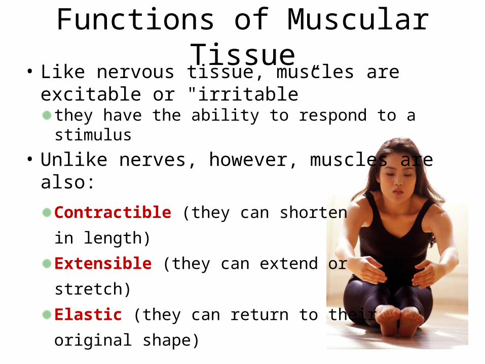
• Like nervous tissue, muscles are excitable or "irritable”
they have the ability to respond to a stimulus• Unlike nerves, however, muscles are also:
Contractible (they can shorten
in length)
Extensible (they can extend or
stretch)
Elastic (they can return to their
original shape)
Functions of Muscular Tissue
• Muscle makes up a large percentage of the body’s
weight
• Their main functions are to:
Create motion – muscles work with nerves, bones, and
joints to produce body movements
Stabilize body positions and maintain posture
Store substances within the body using sphincters
Move substances by peristaltic contractions
Generate heat through thermogenesis
Functions of Muscular Tissue
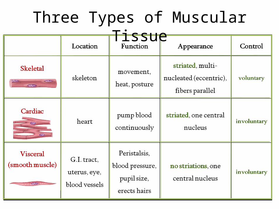
Three Types of Muscular Tissue
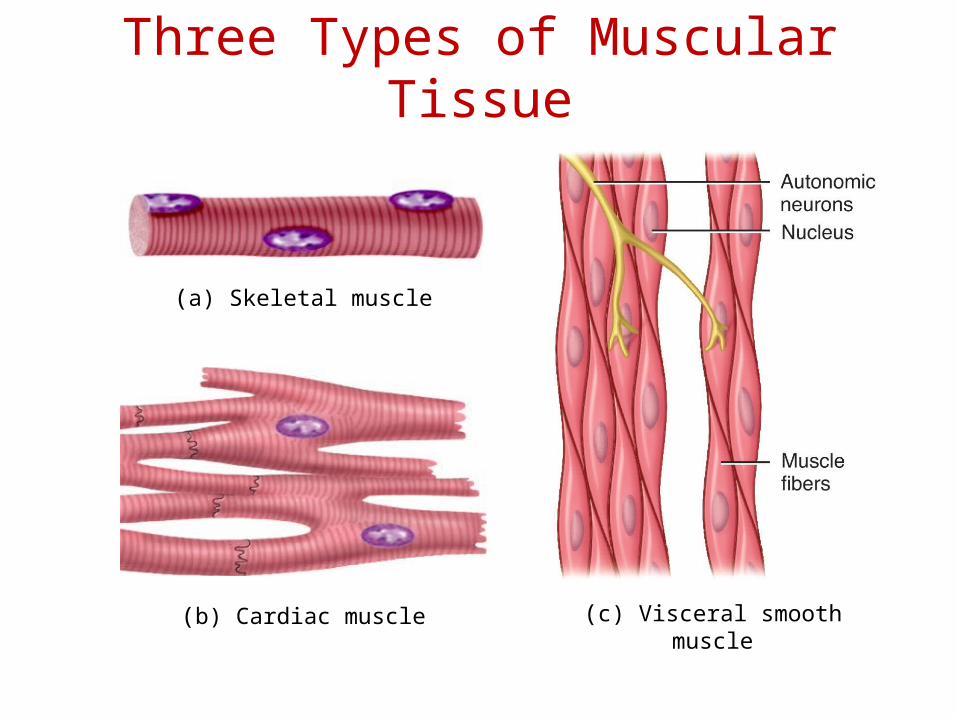
(b) Cardiac muscle (c) Visceral smooth muscle
(a) Skeletal muscle
Three Types of Muscular Tissue

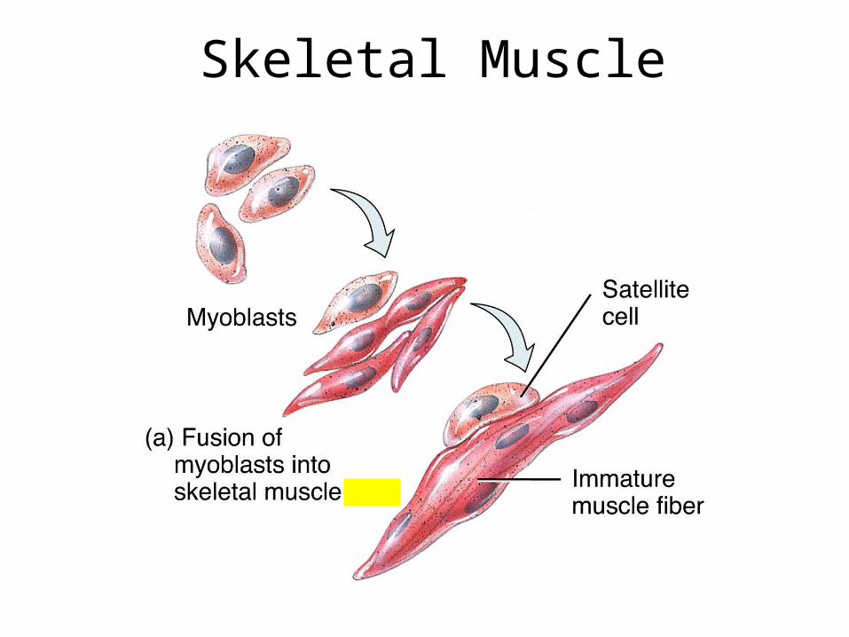
Skeletal Muscle
Skeletal Muscle
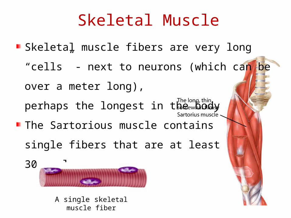
Skeletal muscle fibers are very long “cells” -
next to neurons (which can be over a meter
long),
perhaps the longest in the body
The Sartorious muscle contains
single fibers that are at least
30 cm long
A single skeletal muscle fiber
Skeletal Muscle
Sarcolemma
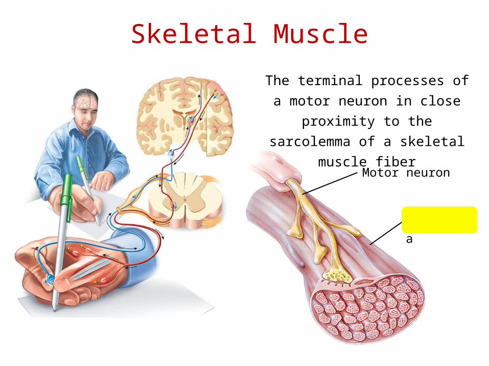
Motor neuron
Skeletal Muscle
The terminal processes of a
motor neuron in close proximity
to the sarcolemma of a skeletal
muscle fiber
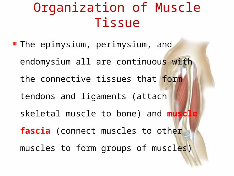
The epimysium, perimysium, and
endomysium all are continuous with
the connective tissues that form
tendons and ligaments (attach
skeletal muscle to bone) and muscle
fascia (connect muscles to other
muscles to form groups of muscles)
Organization of Muscle Tissue

Organization of Muscle Tissue
Organization of a single muscle
belly
Epimysium
Perimysium

Organization of a fasciculus
Organization of Muscle Tissue
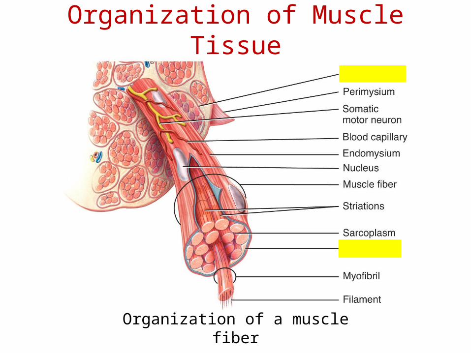
Organization of a muscle fiber
Organization of Muscle Tissue

A muscle, a fasciculus, and a fiber all visualized
Organization of Muscle Tissue

• In groups of muscles the
epimysium continues to
become thicker, forming
fascia which covers many
muscles• This graphic shows the
fascia lata enveloping the
entire group of quadriceps
and hamstring muscles in
the thing
Organization of Muscle Tissue

Organization of Muscle
Tissue• Many large muscle
groups are encased
in both a superficial
and a deep fascia
Real Anatomy, John Wiley and Sons
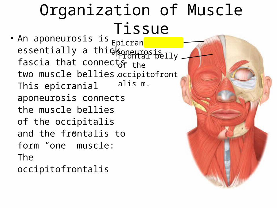
Organization of Muscle Tissue• An aponeurosis is
essentially a thick fascia that connects two muscle bellies. This epicranial aponeurosis connects the muscle bellies of the occipitalis and the frontalis to form “one” muscle: The occipitofrontalis
Epicranial aponeurosisFrontal belly of
the occipitofrontalis m.
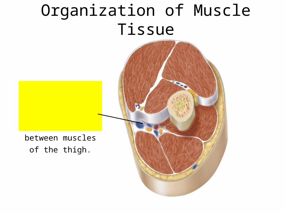
Veins, arteries, and nerves are located in the
deep fascia between muscles of the thigh.
Organization of Muscle Tissue

Beneath the connective tissue endomysium
is found the plasma membrane (called the
sarcolemma) of an individual skeletal
muscle fiber
The cytoplasm (sarcoplasm) of skeletal
muscle fibers is chocked full of
contractile proteins
arranged in myofibrils
The Skeletal Muscle Fiber
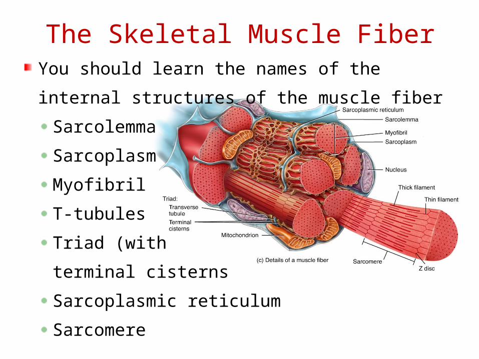
You should learn the names of the internal
structures of the muscle fiber
Sarcolemma
Sarcoplasm
Myofibril
T-tubules
Triad (with
terminal cisterns
Sarcoplasmic reticulum
Sarcomere
The Skeletal Muscle Fiber

The Skeletal Muscle Fiber
Increasing the level of magnification, the
myofibrils are seen to be composed
of filaments
Thick filaments
Thing filaments
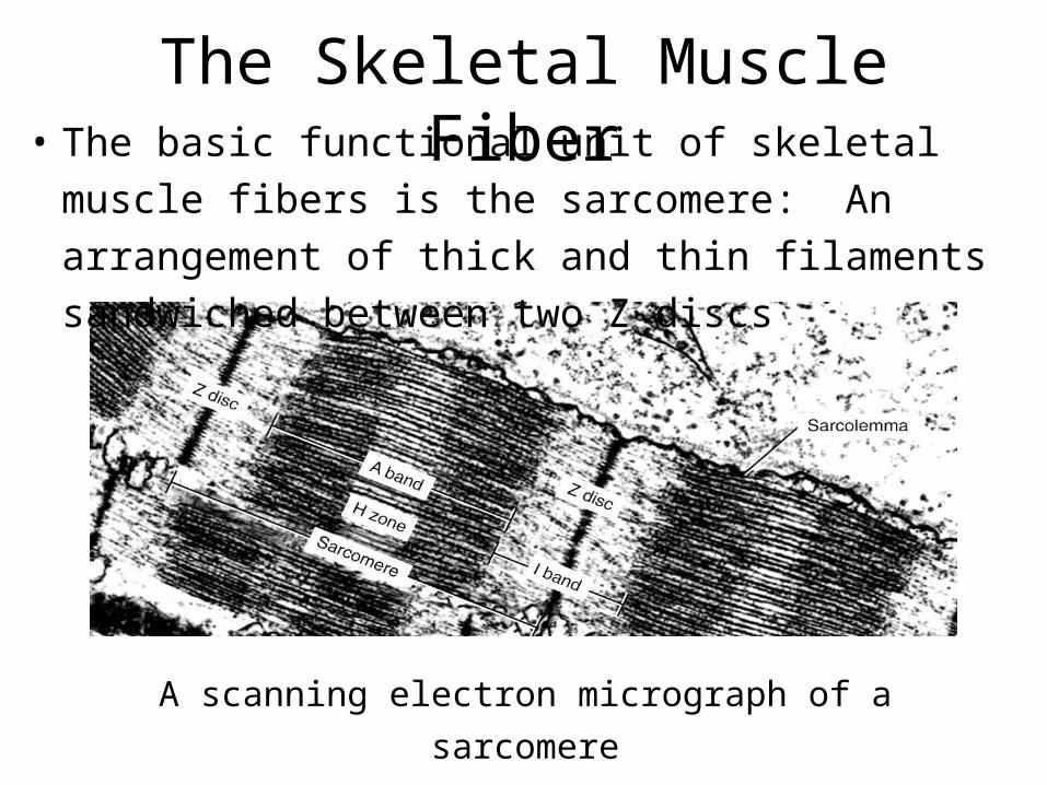
A scanning electron micrograph of a
sarcomere
• The basic functional unit of skeletal muscle fibers is the sarcomere: An arrangement of thick and thin filaments sandwiched between two Z discs
The Skeletal Muscle Fiber
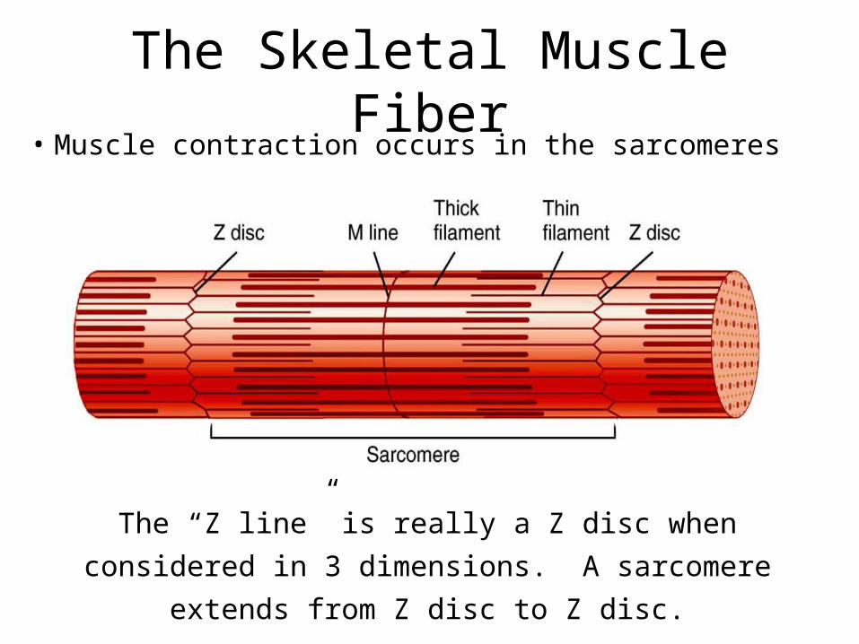
The “Z line” is really a Z disc when considered in
3 dimensions. A sarcomere extends from Z disc
to Z disc.
• Muscle contraction occurs in the sarcomeres
The Skeletal Muscle Fiber

• Myofibrils are built from three groups of proteins
Contractile proteins generate force during contraction
Regulatory proteins help switch the contraction process
on and off
Structural proteins keep the thick and thin filaments in
proper alignment and link the myofibrils to the
sarcolemma and extracellular matrix
Muscle Proteins
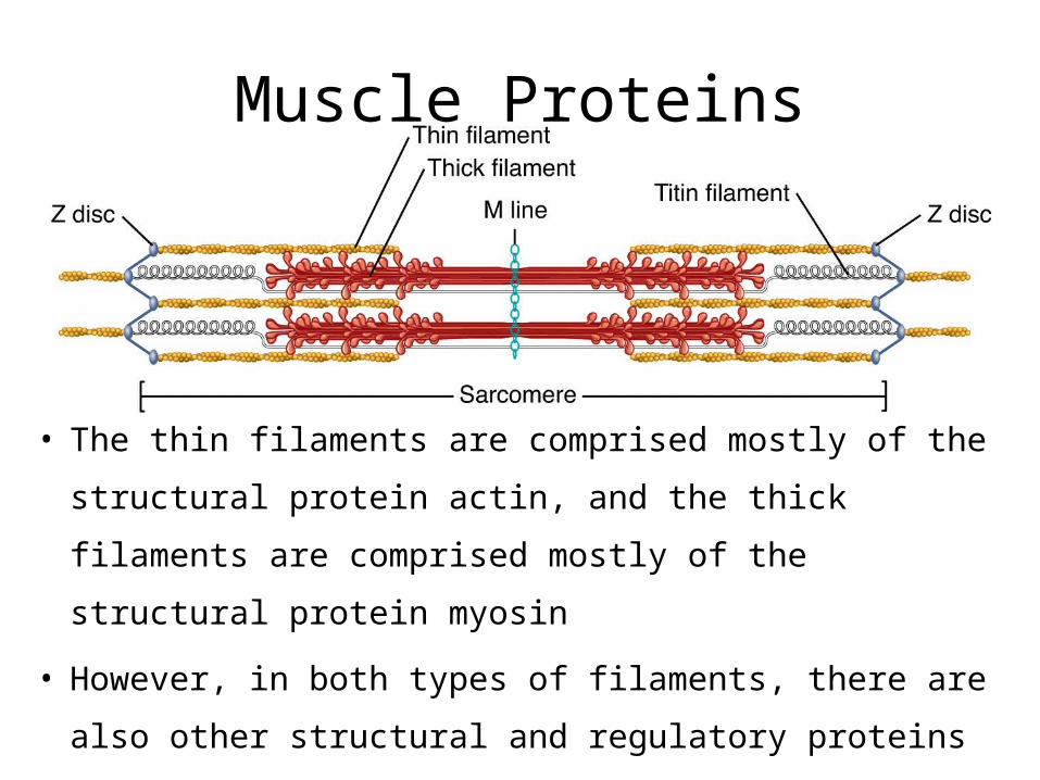
• The thin filaments are comprised mostly of the structural
protein actin, and the thick filaments are comprised
mostly of the structural protein myosin
• However, in both types of filaments, there are also other
structural and regulatory proteins
Muscle Proteins


![[PPT]Chapter 5 - Dr. Gerry Cronindrgerrycronin.weebly.com/uploads/5/9/7/4/5974564/5a.pptx · Web viewChapter 5 The Integumentary System Lecture slides prepared by Curtis DeFriez,](https://static.fdocuments.net/doc/165x107/5aec44127f8b9ac361903e25/pptchapter-5-dr-gerry-viewchapter-5-the-integumentary-system-lecture-slides.jpg)