A CASE STUDY ON ECTOPIC PREGNANCY
13
www.wjpr.net Vol 9, Issue 3, 2020. 1422 A CASE STUDY ON ECTOPIC PREGNANCY Dr. Nilakshi Pravin Deore* 1 , Dr. Deepali Suresh Rajput 2 , Dr. Manda Sanjog Ghorpade 3 1 PG Scholar, Department of Prasutitantra and Streeroga, Sane Guruji Aarogya Kendra Malwadi Hadapsar, Pune 411028. 2 Guide - MD Prasutitantra Streeroga, Department of Prasutitantra and Streeroga, Sane Guruji Aarogya Kendra Malwadi Hadapsar, Pune 411028. 3 HOD - MS Prasutitantra Streeroga, Department of Prasutitantra and Streeroga, Sane Guruji Aarogya Kendra Hadapsar, Pune 411028. ABSTRACT Introduction- Ectopic pregnancy described for the first time in the11th Century and later on it was described as pregnancy complication. Ectopic pregnancy is potentially life-threatening and remains the leading cause of maternal death. The incidence of ectopic pregnancy is increased during last years all over the world. Case presentation- Here we present a case of recurrent ectopic pregnancy with left adnexal mass and stable general condition was treated with medical management by Methotrexate but finally posted for laparotomy. Management and outcome- Though Laparoscopic surgery is still the cornerstone of treatment in the majority of women, medical management is an alternative treatment option. If the diagnosis of ectopic pregnancy can be made earlier non-invasively, medical treatment with systemic intramuscular Methotrexate (MTX) aimed at reducing mortality, morbidity and reducing costs, minimal intervention/non-intervention on comparing with outcomes of surgical treatment. Fertility can be preserved. Discussion- discussion of the case according to the ayurvedic etiopathology is discussed by using nidan panchak. KEYWORDS: Medical management, Ectopic pregnancy. 1) INTRODUCTION An ectopic pregnancy is one in which fertilized ovum is implanted and develops outside the normal endometrial cavity. It refers to the pregnancy occurring outside the uterine World Journal of Pharmaceutical Research SJIF Impact Factor 8.084 Volume 9, Issue 3, 1422-1434. Case Study ISSN 2277– 7105 Article Received on 18 Jan. 2020, Revised on 07 Feb. 2020, Accepted on 28 Feb. 2020, DOI: 10.20959/wjpr20203-16878 *Corresponding Author Dr. Nilakshi Pravin Deore PG Scholar, Department of Prasutitantra and Streeroga, Sane Guruji Aarogya Kendra Malwadi Hadapsar, Pune 411028.
Transcript of A CASE STUDY ON ECTOPIC PREGNANCY

www.wjpr.net Vol 9, Issue 3, 2020.
Deore et al. World Journal of Pharmaceutical Research
1422
A CASE STUDY ON ECTOPIC PREGNANCY
Dr. Nilakshi Pravin Deore*1, Dr. Deepali Suresh Rajput
2, Dr. Manda Sanjog Ghorpade
3
1PG Scholar, Department of Prasutitantra and Streeroga, Sane Guruji Aarogya Kendra
Malwadi Hadapsar, Pune 411028.
2Guide - MD Prasutitantra Streeroga, Department of Prasutitantra and Streeroga, Sane
Guruji Aarogya Kendra Malwadi Hadapsar, Pune 411028.
3HOD - MS Prasutitantra Streeroga, Department of Prasutitantra and Streeroga, Sane Guruji
Aarogya Kendra Hadapsar, Pune 411028.
ABSTRACT
Introduction- Ectopic pregnancy described for the first time in
the11th Century and later on it was described as pregnancy
complication. Ectopic pregnancy is potentially life-threatening and
remains the leading cause of maternal death. The incidence of
ectopic pregnancy is increased during last years all over the world.
Case presentation- Here we present a case of recurrent ectopic
pregnancy with left adnexal mass and stable general condition was
treated with medical management by Methotrexate but finally posted
for laparotomy. Management and outcome- Though Laparoscopic
surgery is still the cornerstone of treatment in the majority of
women, medical management is an alternative treatment option. If
the diagnosis of ectopic pregnancy can be made earlier non-invasively, medical treatment
with systemic intramuscular Methotrexate (MTX) aimed at reducing mortality, morbidity
and reducing costs, minimal intervention/non-intervention on comparing with outcomes of
surgical treatment. Fertility can be preserved. Discussion- discussion of the case according
to the ayurvedic etiopathology is discussed by using nidan panchak.
KEYWORDS: Medical management, Ectopic pregnancy.
1) INTRODUCTION
An ectopic pregnancy is one in which fertilized ovum is implanted and develops outside
the normal endometrial cavity. It refers to the pregnancy occurring outside the uterine
World Journal of Pharmaceutical Research SJIF Impact Factor 8.084
Volume 9, Issue 3, 1422-1434. Case Study ISSN 2277– 7105
Article Received on
18 Jan. 2020,
Revised on 07 Feb. 2020,
Accepted on 28 Feb. 2020,
DOI: 10.20959/wjpr20203-16878
*Corresponding Author
Dr. Nilakshi Pravin Deore
PG Scholar, Department of
Prasutitantra and Streeroga,
Sane Guruji Aarogya
Kendra Malwadi Hadapsar,
Pune 411028.

www.wjpr.net Vol 9, Issue 3, 2020.
Deore et al. World Journal of Pharmaceutical Research
1423
cavity usually in the fallopian tube. In the past ectopic pregnancies were diagnosed at the
time of laparoscopy or surgery, often in women who were unaware that they were
pregnant. Now with the advent of modern diagnostic aids, a pregnancy can easily be
diagnosed even before a menstrual period has been missed. Ectopic pregnancy still
contributes significantly to the cause of maternal morbidity and mortality.
INCIDENCE: The incidence of ectopic pregnancy among all pregnancies is about 0.25-
2.0% worldwide and can occur in any sexually active woman of reproductive age.
Ectopic pregnancy was reported in 0.91% of pregnant women (with no maternal deaths) in
a study done at tertiary care centre in South India.
While there has been about fourfold increase in the incidence over the couple of decades,
but the mortality has been slashed down to 80%.
AETIOLOGY: Aetiology includes tubal damage from different reasons like
inflammation, infections and surgical interventions.
1. Salpingitis and pelvic inflammatory disease
2. Iatrogenic - contraceptive failure, tubal surgery, intrapelvic adhesions, ART,
3. Others - previous ectopic pregnancy (10-15%), prior induced abortion, developmental
defects of tube, transperitoneal migration of ovum.
CLINICAL FEATURES
A) SYMPTOMS
ACUTE UNRUPTURED SUBACUTE (CHRONIC)
Amenorrhoea Presence of delayed period
with features of pregnancy Amenorrhoea
Abdominal pain
Uneasiness on one side of
the flank which continuous
and colicky in nature.
Lower abdominal pain
Vaginal bleeding Vaginal bleeding
Vomiting Bladder irritation
Fainting attack Rectus tenesmus
Shock

www.wjpr.net Vol 9, Issue 3, 2020.
Deore et al. World Journal of Pharmaceutical Research
1424
B) SIGNS
ACUTE UNRUPTURED CHRONIC
Patient perspires conscious
look blanched
P/V - uterus usually soft
showing evidence of
pregnancy. A pulsatile small
circumscribed tender mass
felt through fornix separated
from uterus.
Patient looks ill
Severe pallor Pallor of varying degree
Shock present - pulse rapid
and feeble, hypotension,
extremities cold and
clammy.
Pulse persistently high
P/A - tense , tumid , tender Features of shock absent
P/S - 1. Vaginal mucosa -
blanched white
2. uterus normal in size or
slightly bulky
3. extreme tenderness in
fornix palpation/ on
cervical movement
4. no mass felt , uterus
floats as if in water.
P/A - 1. tenderness,
2. mass may be felt in lower
abdomen-irregular tender,
3. Cullen sign may be present
P/S - 1. Vaginal mucosa- pale
2. uterus bulky or normal in size
3. cervical movement
tenderness
4. Ill-defined mass boggy felt in
posterior fornix.
DIAGNOSIS
(a) History.
(b) Speculum and bimanual examination.
(c) Pregnancy test.
(d) Ultrasonography.
(e) Test to detect serum β-human chorionic gonadotrophin.
(f) Laparoscopy.
MANAGEMENT- Scheme of management of tubal ectopic pregnancy
Detailed history, evaluation of high-risk factors and examination.
Urine- beta hCG.
Ultrasound scan (transvaginal preferred).

www.wjpr.net Vol 9, Issue 3, 2020.
Deore et al. World Journal of Pharmaceutical Research
1425
CASE PRESENTATION
A 32 yrs old female patient presented to OPD of streeroga prasutitantra department of
SGAK with the referral note for further management of left ectopic pregnancy with
complaints of sudden abdominal pain on 09/09/19.
Married life - 12yrs.
LMP- 28/07/19 with bad obstetric history

www.wjpr.net Vol 9, Issue 3, 2020.
Deore et al. World Journal of Pharmaceutical Research
1426
OBSTETRIC HISTORY
1. G1- PTVD of 6 months baby died after 3 days due to RDS,
2. G2- right ectopic tubal pregnancy was operated and salpingectomy was done in 2013.
PAST HISTORY - no any history of major illness.
FAMILY HISTORY - no any history of major illness.
DRUG ALLERGY - no any drug allergy or food allergy. Now she came with left tubal
ectopic pregnancy.
UPT Done - positive and
USG on 09/09/19 suggestive of ET thick 13mm left adnexa - 17 mm thick walled
echogenic lesion adjacent to left ovary vascularity present minimal free fluid seen in pelvis,
she was hemodynamically stable
CLINICAL EXAMINATION
Pulse- 96/min,
BP- 130/80 mm of hg
P/A -abdominal examination revealed lower abdominal tenderness in left iliac region rigidity
and guarding present.
P/V- PV examination revealed tenderness in left fornices, tenderness during cervical
movement with uterus normal in size.
Patient was admitted and all investigations were done on 09/09/19 beta Hcg - 3557
mIU/ml. patient was stable hemodynamically and in order to preserve tube inj
methotrexate was given on 10/09/19. All vitals were monitored on 4th
day beta Hcg was
repeated on 4th
day which was 9753 mIU/ml and on 7th
day which was 679.3 mIU/ml which
shown reducing values hence patient was discharged and asked for follow-up after a week .
Patient came for follow-up after a week with beta hCG report which was (24/09/19) 6302
mIU/ml which was raised, patient gave history of p/v bleeding hence after her menses USG
(TVS) and beta hCG was repeated which suggested of 28*27 mm lesion in left adnexa
patient was admitted beta hCG 4320 mIU/ml (01/10/19) and all investigation were done
second dose of inj methotrexate was given on 02/10/19. Patient was discharged and asked
for regular follow-up after a week patient gave follow-up with beta hCG report (09/10/19)
2526 mIU/ml which was decreased. Later patient’s follow-up’s were taken weekly beta hCG
reports showed declining values, on 15/10/19 - 1404mIU/ml.

www.wjpr.net Vol 9, Issue 3, 2020.
Deore et al. World Journal of Pharmaceutical Research
1427
On 23/10/19 USG and beta hCG were repeated beta hCG - 900.2 and USG Suggestive of
28*28 mm mixed echogenic lesion with no large peritoneum as compared to previous USG
no significant change in this lesion. Patient was asked to give follow-up after 15 days patient
gave follow-up on 08/11/19 with beta hCG- 268.7 and asked for further follow-up after 15
days.
Unfortunately Patient came on 19/11/19 complaining of lower abdominal pain since 2 days
increased suddenly on 19/11/19 USG was advised which suggested of increased in size of
lesion 48*51*41 mm mild echogenicity seen adherent to pelvis is significant hematoma free
fluid was significant .hence emergency laparotomy was performed on 19/11/19 and left
salpingectomy was performed left adnexal hematoma and hemoperitoneum was noted.
Patient was stable and discharged in good condition.
DATE Beta
Hcg Hgm Complaints pulse
Blood
pressure USG
Inj
methotrexate
10/09/2019 3557 9.7 Abdominal
pain 96 130/80
Echogenic lesion
17mm left tubal
ectopic pregnancy
Single dose-
100mg deep IM
14/09/2019 9753 - Mild abdominal
pain 98 110/70 - -
17/09/2019 679.3 10.1 Mild abdominal
pain 96 100/60 - -
24/09/2019 6302 - No any
complaints 88 110/70 - -
02/10/2019 4320 11.1 Mild abdominal
pain 80 110/70
Increased in
Echogenic lesion
28*27 mm
Inj methotrexate
100mg deep
IM 2nd
dose
10/10/2019 2526 - Mild abdominal
pain 88 110/70 - -
16/10/2019 1404 - Mild abdominal
pain 84 110/70 - -
23/10/2019 900.2 - - 88 110/70
Echogenic lesion
28*28 mm, no
significant change
in this lesion
-
07/11/2019 268.7 11.4 Mild abdominal
pain 80 120/70 - -
19/11/2019 11.8 Pain in abdomen
increased 92 130/80
Lesion size
Increased 48*51*41
mm S/O left
adnexal hematoma
significant free
fluid in pelvis.
-
www.wjpr.net Vol 9, Issue 3, 2020.
Deore et al. World Journal of Pharmaceutical Research
1428
2) MANAGEMENT AND OUTCOME
Proper evaluation of pregnancy with associated risk factors and early diagnosis help
preserving tube and in turns her fertility and thus help in decreasing morbidity and
mortality. Hence medical management was done which showed declining result in serum
beta Hcg levels but subsequently there was rise in the size of lesion which further needed
the surgical intervention.
Ruptured ectopic pregnancy is the leading cause of maternal mortality in the first trimester
and accounts for 10-15% of all maternal deaths. The early diagnosis and treatment of this
condition over the past two decades have allowed a definitive medical management of
unruptured ectopic pregnancy even before there were clinical symptoms in these high risk
women. The reason for increasing incidence has not been fully elucidated, but the possible
contribution of pelvic inflammatory disease (PID), ovulation inducing drugs, previous
abdominal-pelvic surgeries and intra-uterine contraceptive device use has been cited as
contributing factors. The diagnosis of ectopic pregnancy has become more frequent during
the last decades, but the incidence of ectopic pregnancy rupture has declined. This declined
is due to quantitative human chorionic gonadotropin measurements, minimally invasive
surgeries, and transvaginal ultrasonography (USG). Early diagnosis reduces the risk of
tubal rupture and allows more conservative medical treatments to be employed.
HENCE TO CONCLUDE WITH
Early diagnosis, proper assessment of principal risk factors and timely intervention in the
form of conventional or surgical treatment will help in reducing the morbidity and mortality
associated with ectopic pregnancy.
3) DISCUSSION
Acharya Charaka has also laid emphasis on the role of Vata in conception. Most of the
activities of central, vegetative and peripheral nervous system including the autonomous
can be identified with the functions ascribed to Vata. So proper function of Vata here refers
to the integrity of neuroendocrine system especially H-P-O-U axis (Hypothalamus-Pituitary-
Ovarian-Uterine axis) which is of prime value as far as concept of fertility is concerned.
The four factors essential for conception are Ritu, Kshetra, Ambu and Beeja. Here,
Kshetra can be taken as the entire female reproductive tract from vagina up to the ovaries
and in close association with Hypothalamus and Pituitary gland.

www.wjpr.net Vol 9, Issue 3, 2020.
Deore et al. World Journal of Pharmaceutical Research
1429
Modern science is based on the Pratyaksha only while Ayurveda does consider the
Anumana as well as Aaptopadesha along with the Pratyaksha. Anatomy in both the sciences
is totally different from each other and one cannot find an equivocal reference in Ayurvedic
texts for various anatomical structures like fallopian tubes. However, some of the Ayurvedic
terms which can be equated with Fallopian tubes are Kshetra, Artavavaha Srotas,
Shukravaha Srotas in female and Ashaya.
Kshetra
Fallopian tubes can be considered a part of the Kshetra mentioned by Acharya Sushruta as
one of the four elements necessary for conception. Kshetra is a broad term and includes all
the structures of the female reproductive tract whose structural and functional integrity is
indispensable for conception. But as it clear that female physiology is entirely under the
neuroendocrinal control, so it must be concluded that Hypothalamus Pituitary Ovarian axis
can also be included in the Kshetra.
Artavavaha Srotas
Understanding of Srotas has always remained the seat of conflicts but its importance and
applicability is of enormous importance. If we do not go through the various conflicts and
try to understand the Srotas in a simpler way, we will find them as the structural as well as
functional units of body. This discussion clarifies that every organ or bodily structure can be
included under the umbrella of any of the Srotasas. Keeping this view in mind, an attempt
has been made to understand the fallopian tubes with the name of Artavavaha Srotas.
Artavavaha Srotas is described by Acharya Sushruta, where he says that there are two
Artavavaha Srotasas, having roots in Garbhashaya and Artavavahi Dhamanis, injury to
which causes Vandhyatva (infertility), Maithunasahishnuttva (dyspareunia) and
Artavanasha (anovulation or amenorrhoea). Some scholars compare Artavavaha Srotas with
uterine arteries, specially their capillary bed, because these arteries are responsible to carry
menstrual blood, which is compared to Artava at several places and injuries to these vessels
may cause infertility too. This correlation of Artavavaha Srotas with uterine arteries does
not seem to be fully acceptable for some reasons.
Ashaya
One more indirect description of fallopian tube can be taken as the extra Ashaya in females
told by Acharya Sharngadhara, where he mentions the Garbhashaya as Dhara and the extra
Ashaya in females. Here Acharya has considered Garbhashaya as a whole to be the site of

www.wjpr.net Vol 9, Issue 3, 2020.
Deore et al. World Journal of Pharmaceutical Research
1430
conception by calling it Dhara. But now, it is a well known fact that the actual fertilization
takes place in the ampullary region of fallopian tube. So, fallopian tubes can also be
accepted as Ashaya. The above discussion tries to bring out the Ayurvedic entities which
should be thought of, when we hear the term fallopian tubes. But for the purpose of
explaining and interpreting ectopic pregnancy, accepting fallopian tubes as the Artavavaha
Srotas appears to be the most appropriate choice.
The way to define the disease or the pathogenesis in Ayurveda is entirely different from
western medical science, and is based more upon the first vitiation of Doshas, i.e. the
pathogenesis of disease from commence. Taking this Ayurvedic view into consideration, an
approach can be developed by finding out the ectoic pregnancy on the basis of Nidanas &
Samprapti. Acharya Charaka has himself given space to Ayurvedic scholars to understand
the newly diagnosed diseases on the basis of Prakriti (Doshas; root cause), Adhishthana
(Dushya; seat), Linga (Lakshanas; features) & Ayatana (Ahara Viharadi Nidanas).
PRAKRITI (SANNIKRISHTA KARANA OR ROOT CAUSE)
According to Ayurvedic point of view, the root or ultimate cause of any disease is the
vitiation of either one or more of the three Doshas by one or more of their Gunas.
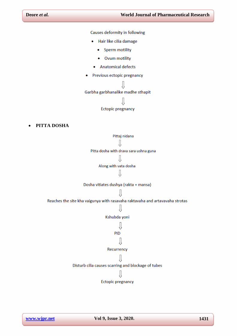
All the three Doshas can be assumed responsible for ectopic pregnancy by causing structural
or functional abnormalities in Artava Beeja Vaha Srotas i.e. fallopian tube. Vitiation of Vata
can be considered the most important factor.
VATA
www.wjpr.net Vol 9, Issue 3, 2020.
Deore et al. World Journal of Pharmaceutical Research
1431
PITTA DOSHA
www.wjpr.net Vol 9, Issue 3, 2020.
Deore et al. World Journal of Pharmaceutical Research
1432
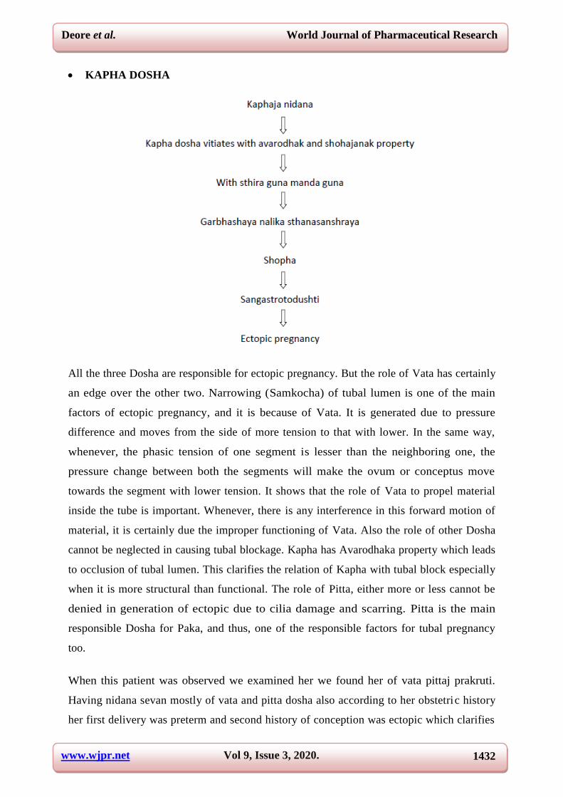
KAPHA DOSHA
All the three Dosha are responsible for ectopic pregnancy. But the role of Vata has certainly
an edge over the other two. Narrowing (Samkocha) of tubal lumen is one of the main
factors of ectopic pregnancy, and it is because of Vata. It is generated due to pressure
difference and moves from the side of more tension to that with lower. In the same way,
whenever, the phasic tension of one segment is lesser than the neighboring one, the
pressure change between both the segments will make the ovum or conceptus move
towards the segment with lower tension. It shows that the role of Vata to propel material
inside the tube is important. Whenever, there is any interference in this forward motion of
material, it is certainly due the improper functioning of Vata. Also the role of other Dosha
cannot be neglected in causing tubal blockage. Kapha has Avarodhaka property which leads
to occlusion of tubal lumen. This clarifies the relation of Kapha with tubal block especially
when it is more structural than functional. The role of Pitta, either more or less cannot be
denied in generation of ectopic due to cilia damage and scarring. Pitta is the main
responsible Dosha for Paka, and thus, one of the responsible factors for tubal pregnancy
too.
When this patient was observed we examined her we found her of vata pittaj prakruti.
Having nidana sevan mostly of vata and pitta dosha also according to her obstetric history
her first delivery was preterm and second history of conception was ectopic which clarifies

www.wjpr.net Vol 9, Issue 3, 2020.
Deore et al. World Journal of Pharmaceutical Research
1433
that there was vitiation of vata dosha when history was taken she mention recurrent lower
abdominal pain which can be correalated to PID. Hence by following etiopathogenesis of
generation of ectopic pregnancy took place.
SAMPRAPTI (PATHOGENESIS)
Samprapti Ghataka
Dosha : Vatapradhana Tridosha
Dushya : Rasa, Rakta, Artava
Agni : Dhatvagni
Srotas : Artavavaha (Artava-Beeja-vaha)
Udbhavasthana :Pakvashaya
Srotodushti : Sanga
Vyaktisthana : Garbhashaya
Avayava : Garbhashaya-nalika

www.wjpr.net Vol 9, Issue 3, 2020.
Deore et al. World Journal of Pharmaceutical Research
1434
Roga Vinishchaya : Garbhashayettar garbha dharana
Sadhyasadhyata : Krichchhrasadhya
4) REFERENCES
1. F. Garry Cunningham, Kenneth j. leveno, Steven L. Bloom, Catherine Y. Spong, Jodi S.
Dashe, Barbara L. Hoffman, Brian M. Casey, Jeanne S. Sheffield. Williams obstetrics.
24th edition. United states. Mc graw hill education. 2014.
2. Pt. K. N. Shastry & G. N. Chaturvedi, Vidyotini Vyakhya, Charaka Samhita,
Chaukhamba Bharati Academy, 2005, Ch. Su. 20/3.
3. Chakrapani commentary on Charaka samhita, Chaukhamba samskrit Samsthana,
Varanasi, 1984, Ch.Su. 20/3.
4. Ibid, Vidyotini Vyakhya, Ch. Su. 20/9.
5. Ayurveda-Tattva Samdipika Vyakhya, Sushruta Samhita, 24/9.
6. Pt. Hemraja Sharma, Vidyotini Hindi commentary, Kashyapa Samhita, Chaukhamba
Sanskrit Sansthan, Varanasi (2009), Kash. Su. 27/29.
7. Dr. K. H. Krishnamurthy, Bhel Samhita English commentary, Chaukhamba Vishvabharti,
Varanasi, Sharir Sthana 2000,12/7.
8. Ibid, Vidyotini Vyakhya, Ch. Su. 12/4.
9. Ibid, Chak. Commentary on Ch. Su. 12/4.
10. Ibid, Vidyotini Vyakhya, Ch. Su. 1/61.
11. Ibid, Ayurveda-Tattva-Samdipika vyakhya, Su. Su. 17/12.
12. Ibid, Ayurvedic concepts of gynaecology, pg. 99.