A Case of Sporadic Intestinal Cryptosporidiosis Diagnosed ... · A Case of Sporadic Intestinal...
5
A Case of Sporadic Intestinal Cryptosporidiosis Diagnosed by Endoscopic Biopsy Sho Ogata a,b* , Toshiyuki Suganuma c , Chizuko Okada c , Kimitoshi Inoue c , Akio Kinoshita d , and Kimiya Sato d a Division of Pathology, Japan Self Defense Forces Hospital Yokosuka, Yokosuka, Kanagawa 237-0071, Japan, b Department of Pathology and Laboratory Medicine, National Defense Medical College, Tokorozawa, Saitama 359-8513, Japan, c Division of Medicine, Japan Self Defense Forces Hospital Kure, Kure, Hiroshima 737-0027, Japan, and d Department of Pathology, Japan Self Defense Forces Central Hospital, Setagaya-ku, Tokyo 154-8532, Japan Sporadic intestinal cryptosporidiosis is not easily diagnosed and might be overlooked. We present here a case of this disease in a 23- year- old Japanese military man with 3 days of abdominal pain, watery diarrhea, and nausea. The frequency of his diarrhea was more than 10 times per day. After his diar - rheal bowel symptoms subsided, a colonoscopy was performed because inflammatory bowel disease was suspected. Although the endoscopic findings indicated non- specific ileitis, intestinal cryptosporidi- osis was suspected from the histology of ileal biopsy specimens, and this was confirmed ultrastructur- ally. At that time, however, the patient was on active duty, and thus it was not possible to confirm this as a definitive diagnosis by an adequate stool examination for cryptosporidium. Routine practi - tioners should be encouraged to carefully inspect patients for this disease, supported by detailed knowledge of it and its diagnosis. If stool - examination results are negative or are not obtained at first, histological diagnosis by endoscopic biopsy could be a useful way to screen for intestinal cryptospori- diosis. Furthermore, stool or histological examination should be performed in recovered patients because the oocysts may continue to be shed for 1 to 4 weeks after the symptoms disappear. Therefore, endoscopic and histological examinations may be useful tools for the early diagnosis of intestinal cryptosporidiosis, although admittedly they are invasive procedures. Key words: intestinal cryptosporidiosis, histology ntestinal cryptosporidiosis is an infectious dis- ease caused by the coccidian parasite crypto- sporidium [1]. Previously considered a zoonotic dis- ease, it can be caused in humans by Cryptosporidium hominis, parvus, and other species [2]. The clinical manifestations are similar among Cryptosporidium spe- cies [3]: namely, after 2 to 10 daysʼ incubation, the patientʼ s chief symptom is severe, sometimes watery, diarrhea [1]. The symptoms usually disappear within several days to 2 weeks, although in immunodeficient patients they are more severe and prolonged, and can be lethal [1]. These agents are transmitted by the fecal - oral route. Besides direct contact with infected animals or humans, contaminated water is considered an impor- tant source of human infection. The first human case was reported in 1976 [4], and since then water- borne diseases and outbreaks (WBDOs) by Cryptosporidium I Acta Med. Okayama, 2009 Vol. 63, No. 5, pp. 287ン291 CopyrightⒸ 2009 by Okayama University Medical School. Case Report http: // escholarship.lib.okayama-u.ac.jp / amo/ Received February 12, 2009 ; accepted May 1, 2009. * Corresponding author. Phone: + 81ン4ン2995ン1505; Fax: + 81ン4ン2996ン5192 E- mail:[email protected] (S. Ogata)
Transcript of A Case of Sporadic Intestinal Cryptosporidiosis Diagnosed ... · A Case of Sporadic Intestinal...

A Case of Sporadic Intestinal Cryptosporidiosis Diagnosed by Endoscopic Biopsy
Sho Ogataa,b*, Toshiyuki Suganumac, Chizuko Okadac, Kimitoshi Inouec, Akio Kinoshitad, and Kimiya Satod
aDivision of Pathology, Japan Self Defense Forces Hospital Yokosuka, Yokosuka, Kanagawa 237-0071, Japan, bDepartment of Pathology and Laboratory Medicine, National Defense Medical College, Tokorozawa, Saitama 359-8513, Japan,
cDivision of Medicine, Japan Self Defense Forces Hospital Kure, Kure, Hiroshima 737-0027, Japan, and dDepartment of Pathology, Japan Self Defense Forces Central Hospital, Setagaya-ku, Tokyo 154-8532, Japan
Sporadic intestinal cryptosporidiosis is not easily diagnosed and might be overlooked. We present here a case of this disease in a 23-year-old Japanese military man with 3 days of abdominal pain, watery diarrhea, and nausea. The frequency of his diarrhea was more than 10 times per day. After his diar-rheal bowel symptoms subsided, a colonoscopy was performed because inflammatory bowel disease was suspected. Although the endoscopic findings indicated non-specific ileitis, intestinal cryptosporidi-osis was suspected from the histology of ileal biopsy specimens, and this was confirmed ultrastructur-ally. At that time, however, the patient was on active duty, and thus it was not possible to confirm this as a definitive diagnosis by an adequate stool examination for cryptosporidium. Routine practi-tioners should be encouraged to carefully inspect patients for this disease, supported by detailed knowledge of it and its diagnosis. If stool-examination results are negative or are not obtained at first, histological diagnosis by endoscopic biopsy could be a useful way to screen for intestinal cryptospori-diosis. Furthermore, stool or histological examination should be performed in recovered patients because the oocysts may continue to be shed for 1 to 4 weeks after the symptoms disappear. Therefore, endoscopic and histological examinations may be useful tools for the early diagnosis of intestinal cryptosporidiosis, although admittedly they are invasive procedures.
Key words: intestinal cryptosporidiosis, histology
ntestinal cryptosporidiosis is an infectious dis-ease caused by the coccidian parasite crypto-
sporidium [1]. Previously considered a zoonotic dis-ease, it can be caused in humans by Cryptosporidium hominis, parvus, and other species [2]. The clinical manifestations are similar among Cryptosporidium spe-cies [3]: namely, after 2 to 10 daysʼ incubation, the
patientʼs chief symptom is severe, sometimes watery, diarrhea [1]. The symptoms usually disappear within several days to 2 weeks, although in immunodeficient patients they are more severe and prolonged, and can be lethal [1]. These agents are transmitted by the fecal-oral route. Besides direct contact with infected animals or humans, contaminated water is considered an impor-tant source of human infection. The first human case was reported in 1976 [4], and since then water-borne diseases and outbreaks (WBDOs) by Cryptosporidium
I
Acta Med. Okayama, 2009Vol. 63, No. 5, pp. 287ン291CopyrightⒸ 2009 by Okayama University Medical School.
Case Report http ://escholarship.lib.okayama-u.ac.jp/amo/
Received February 12, 2009 ; accepted May 1, 2009. *Corresponding author. Phone : +81ン4ン2995ン1505; Fax : +81ン4ン2996ン5192E-mail : [email protected] (S. Ogata)

species have occasionally occurred throughout the world, even in developed countries [5]. The WBDOs reported to be caused by cryptosporidium infection have been associated with either drinking water (com-munity or individual water supply systems) or recrea-tional water (swimming pools or spas) [6]. Oocysts, which show strong infectivity, are resistant to disin-fectants and chlorination, and thus are often found at low levels in treated, “safe” drinking water [7-9]. At present, cryptosporidium is one of the leading causes of reported recreational water-associated outbreaks of gastroenteritis [10], and the incidence of cryptospo-ridiosis is about 1 to 2 per 100,000 population in the United States [11]. Although intestinal cryptospo-ridiosis is considered an emerging infection, sporadic cases of intestinal cryptosporidiosis are often clini-cally and histopathologically overlooked. We report here a sporadic case of intestinal cryptosporidiosis in Japan, which has water supply systems similar to those found in the United States, and we discuss some potential diagnostic pitfalls.
Case Report
A 23-year-old Japanese man, a member of the Japan Self Defense Forces (JSDF), visited an out-patient clinic at the JSDF Hospital Kure (Hiroshima, Japan) complaining of abdominal pain, watery diar-rhea, and nausea for the past 3 days. The diarrheal fluid was non-bloody and translucent yellow, and the frequency of diarrhea was more than 10 times per day. He had been otherwise healthy, but had experienced abdominal pain events once every 4 months or so in recent years. His family history was not remarkable, and he did not travel abroad. Moreover, he had no obvious recent history either of contact with animals or of drinking untreated water. On his first visit, he exhibited low-grade fever, left abdominal tenderness, and increased bowel sounds. Laboratory data revealed a slight elevation in the serum C-reactive protein value. He showed no signs of being an immunocompromised host, and a serological test for human immunodeficiency virus proved nega-tive. Without antibiotics, his symptoms subsided within a few days and his serum C-reactive protein value returned to within normal limits. Since an inflammatory bowel disease such as ulcerative colitis or Crohnʼs disease was suspected, the first total
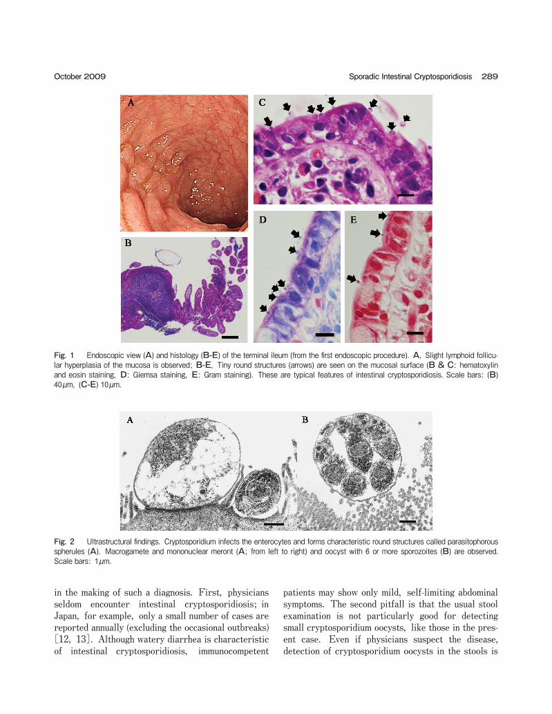
colonoscopy was performed on the 8th day after his first visit. At the time of this total colonoscopy, how-ever, inspection for parasitic oocysts in the stools was not performed because his diarrheal bowel symptoms had already subsided. Endoscopically, the terminal ileal mucosa showed evidence of non-specific ileitis, including slight lymphoid follicular hyperplasia and focal congestion (Fig. 1). In the histological examination performed at the JSDF Hospital Yokosuka (Kanagawa, Japan), the terminal ileum displayed active ileitis (Fig. 1). Char-acteristic tiny round structures were observed on the luminal surface of the mucosal surface epithelium. They were stained purple with hematoxylin-eosin, deep purple with Giemsa, and red with Gram staining methods, compatible with the staining pattern exhib-ited by cryptosporidium [1]. These findings led to a suspicion of intestinal cryptosporidiosis. To confirm or rule out the diagnosis, an electron microscopic examination was performed using paraffin-embedded block samples in which intestinal cryptosporidiosis was suspected. Transmission electron microscopy (Hitachi H-7500, acceleration voltage 80kV; Hitachi, Tokyo, Japan) revealed parasites about 5 micrometers in diameter on the mucosal surface. These represented various stages in the life cycle of the parasite, with features typical of Cryptosporidium species (Fig. 2). From these findings, we confirmed the diagnosis as intestinal cryptosporidiosis, although genetic confir-mation was unfortunately not performed. Following his next visit (which was not until 5 weeks after the first colonoscopy because he was on active duty), a stool examination for the oocysts of parasites, not specifically for cryptosporidium oocysts, was performed by a commercial laboratory. However, none was found. Re-biopsy specimens taken during follow-up colonoscopy revealed no parasites. The immunochromatographic test for cryptosporidium was not performed. The patient made a full recovery and finished by visiting a follow-up outpatient clinic. We found no definite source of infection and no evi-dence of other diarrheal patients in his vicinity.
Discussion
In routine practice, the diagnosis of sporadic intestinal cryptosporidiosis is difficult. On the basis of the present case, three major pitfalls can be identified
288 Acta Med. Okayama Vol. 63, No. 5Ogata et al.
in the making of such a diagnosis. First, physicians seldom encounter intestinal cryptosporidiosis; in Japan, for example, only a small number of cases are reported annually (excluding the occasional outbreaks) [12, 13]. Although watery diarrhea is characteristic of intestinal cryptosporidiosis, immunocompetent
patients may show only mild, self-limiting abdominal symptoms. The second pitfall is that the usual stool examination is not particularly good for detecting small cryptosporidium oocysts, like those in the pres-ent case. Even if physicians suspect the disease, detection of cryptosporidium oocysts in the stools is
289Sporadic Intestinal CryptosporidiosisOctober 2009
Fig. 1 Endoscopic view (A) and histology (B-E) of the terminal ileum (from the first endoscopic procedure). A, Slight lymphoid follicu-lar hyperplasia of the mucosa is observed; B-E, Tiny round structures (arrows) are seen on the mucosal surface (B & C: hematoxylin and eosin staining, D: Giemsa staining, E: Gram staining). These are typical features of intestinal cryptosporidiosis. Scale bars: (B) 40µm, (C-E) 10µm.
Fig. 2 Ultrastructural findings. Cryptosporidium infects the enterocytes and forms characteristic round structures called parasitophorous spherules (A). Macrogamete and mononuclear meront (A; from left to right) and oocyst with 6 or more sporozoites (B) are observed. Scale bars: 1µm.

not easy for inexperienced technicians. Therefore, special oocyst-collecting methods, such as the sucrose-gradient flotation method, may be necessary [14]. The third pitfall is that a total colonoscopy for histological examination is not necessarily performed for patients whose only symptom is diarrhea. Even if adequate biopsy samples are obtained, diagnostic pathologists may not notice the characteristic morpho-logical features of intestinal cryptosporidiosis in materials stained with hematoxylin and eosin, as the oocysts are easily mistaken for stain droplets, con-creted mucus, or epithelial fragments. This problem is related to the first pitfall (viz. the rarity of this infection). When cryptosporidiosis is included within a differential diagnosis histologically, special stains, such as Giemsa, should be added. Furthermore, immunohistochemical or ultrastructural studies are also useful for confirming the diagnosis [1], as in the present case. This discussion indicates that careful inspection, supported by an awareness and detailed knowledge of this disease and how to diagnose it, is a necessity for routine practitioners. However, further species-discrimination for cryptosporidial subtypes is difficult from morphology alone. Indeed, it requires genetic analysis [15], which was unfortunately not performed in the present case. Detection of parasitic oocysts by stool inspection is the gold standard for the diagnosis of diarrheal dis-eases caused by protozoan organisms. Adequate oocyst collection and immunofluorescent detection of cryptosporidium oocysts are required for the diagno-sis of cryptosporidiosis by stool examination. This is a non-invasive test and should be recommended as the first approach. Without such a test, establishing the diagnosis of sporadic cases is especially difficult, because typical manifestations are easily masked by other symptoms. Therefore, many cryptosporidial cases with mild symptoms may be missed as non-spe-cific diarrheal illness. The scenario is different when a patient is suspected of having an inflammatory bowel disease (e.g., ulcerative colitis or Crohnʼs disease), because then an invasive examination such as colonos-copy would be performed as the first approach. Fortunately, this would provide us with a second way of finding cryptosporidiosis, as in the present case. In cases with negative or no stool-examination results, endoscopic and histological examinations may be useful tools for the early diagnosis of intestinal cryptospori-
diosis. In immunocompromised patients, including patients with acquired immunodeficiency syndrome, intestinal cryptosporidiosis may sometimes be lethal, and its diagnosis will therefore be critically important [1]. In such cases, endoscopy should be considered as a second-line diagnostic tool if the patientʼs general condition allows. Pathological examination of biopsy materials may detect not only cryptosporidium, but also isospora, cyclospora, or other gastrointestinal pathogens responsible for opportunistic infections. Isospora or cyclospora, either of which causes similar watery diarrhea, can be discriminated from crypto-sporidiosis because these pathogens are found deeply located within the cytoplasm of the mucosal surface epithelium [1, 16, 17]. In Japan, cryptosporidium oocysts are actually found in rivers and recreational waters [18-19]. From the viewpoint of public health and preventive medicine, it is important that even rare diseases are included in a differential diagnosis of diarrheal illness. Although the source of infection could not be eluci-dated in the present case, hidden sources, such as contaminated water or infected animals or humans, may exist. Recovered patients may still be shedding oocysts, as in the present case (in which active infec-tion was confirmed even after the diarrheal bowel symptoms had subsided). In general, oocysts are thought to continue shedding for up to 1 week, or even 4 weeks, after the symptoms have subsided [20]. Even if the symptoms have subsided, any patient giving cause for suspicion should undergo a stool examination (inspection for parasitic oocysts) or histological examination. Furthermore, suspect patients should avoid recreational waters, not only during the period for which they have a diarrheal bowel symptoms, but also for a few months after recovery (because recovered patients are a potential source of WBDOs). In conclusion, knowledge of the methods necessary to establish a diagnosis of intestinal cryptosporidiosis, together with an awareness of its clinical manifesta-tions, should be mandatory for routine practitioners. If stool-examination results are negative or not obtained, histological diagnosis by endoscopic biopsy is a useful alternative for detecting intestinal crypto-sporidiosis. Furthermore, stool examination or histo-logical examination should be performed in apparently recovered, but still possibly infected, patients to
290 Acta Med. Okayama Vol. 63, No. 5Ogata et al.

determine if they are a potential source of future outbreaks.
Acknowledgments. The authors thank Mrs. Naoko Niihara and Mr. Hitoshi Kaga (JSDF Hospital Yokosuka) for their technical assistance with the histological examination. This work was supported in part by a grant from the Japan Ministry of Defense.
References
1. Orenstein JM: Cryptosporidiosis; in Pathology of infectious dis-eases, Connor DH ed, Appleton & Lange, Stamford, CT (1997) pp 1147-1158.
2. Xiao L, Fayer R, Ryan U and Upton SJ: Cryptosporidium taxonomy: recent advances and implications for public health. Clin Microbiol Rev (2004) 17: 72-97.
3. Xiao L, Bern C, Limor J, Sulaiman I, Roberts J, Checkley W, Cabrera L, Gilman RH and Lal AA: Identification of 5 types of Cryptosporidium parasites in children in Lima, Peru. J Infect Dis (2001) 183: 492-497.
4. Nime FA, Burek JD, Page DL, Holscher MA and Yardley JH: Acute enterocolitis in a human being infected with the proto-zoan Cryptosporidium. Gastroenterology (1976) 70: 592-598.
5. DʼAntonio RG, Winn RE, Taylor JP, Gustafson TL, Current WL, Rhodes MM, Gary GW and Zajac RA: A waterborne outbreak of cryptosporidiosis in normal hosts. Ann Intern Med (1985) 103: 886-888.
6. Kramer MH, Herwaldt BL, Calderon RL and Juranek DD: Surveillance for waterborne-diseases outbreaks―United States, 1993-1994, in Surveillance Summaries, April 12, 1996. MMWR CDC Surveil Summ (1996) 45 (No. SS-1): 1-33.
7. DuPont HL, Chappell CL, Sterling CR, Okhuysen PC, Rose JB and Jakubowski W: The infectivity of cryptosporidium parvum in healthy volunteers. N Engl J Med (1995) 332: 855-859.
8. Korich DG, Mead JR, Madore MS, Sinclair NA and Sterling CR: Effects of ozone, chlorine dioxide, chlorine, and mono-chloramine on Cryptosporidium parvum oocysts viability. Appl Environ Microbiol (1990) 56: 1423-1428.
9. LeChevallier MW, Norton WD and Lee RG: Giardia and Crypto-sporidium spp. in filtered drinking water supplies. Appl Environ Microbiol (1991) 57: 2617-2621.
10. Dziuban EJ, Liang JL, Craun GF, Hill V, Yu PA, Painter J, Moore MR, Calderon RL, Roy SL, Beach MJ; Centers for Disease
Control and Prevention (CDC): Surveillance for waterborne dis-ease and outbreaks associated with recreational water―United States. 2003-2004, in Surveillance Summaries, October 22, 2006. MMWR Surveil Summ (2006) 55 (No. SS-12): 1-30.
11. Yoder JS and Beach MJ; Centers for Disease Control and Prevention (CDC): Cryptosporidiosis surveillance―United States, 2003-2005, in Surveillance Summaries, September 7, 2007. MMWR Surveil Summ (2007) 56 (No. SS-7): 1-10.
12. Yamamoto N, Urabe K, Takaoka M, Nakazawa K, Gotoh A, Haga M, Fuchigami H, Kimata I and Iseki M: Outbreak of crypto-sporidiosis after contamination of the public water supply in Saitama Prefecture, Japan, in 1996. Kansenshogaku Zasshi (2000) 74: 518-526.
13. Nakamura-Uchiyama F, Hiromatsu K, Ishiwata K, Sakamoto Y and Nawa Y: The current status of parasitic diseases in Japan. Intern Med (2003) 42: 222-236.
14. Bukhari Z and Smith HV: Effect of three concentration techniques on viability of Cryptosporidium parvum oocysts recovered from bovine feces. J Clin Microbiol (1995) 33: 2592-2595.
15. Morgan-Ryan UM, Fall A, Ward LA, Hijjawi N, Sulaiman I, Fayer R, Thompson RC, Olson M, Lal A and Xiao L: Cryptosporidium homi-nis n. sp. (Apicomplexa: Cryptosporidiidae) from Homo sapiens. J Eukaryot Microbiol (2002) 49: 433-440.
16. Sun T: Cyclospora infection; in Pathology of infectious diseases, Connor DH ed, Appleton & Lange, Stamford, CT (1997) pp 1159-1162.
17. Orenstein JM: Isosporiasis; in Pathology of infectious diseases, Connor DH ed, Appleton & Lange, Stamford, CT (1997) pp 1185-1190.
18. Ono K, Tsuji H, Rai SK, Yamamoto A, Masuda K, Endo T, Hotta H, Kawamura T and Uga S: Contamination of river water by Cryptosporidium parvum oocysts in Western Japan. Appl Environ Microbiol (2001) 67: 3832-3836.
19. Yokoi H, Tsuruta M, Tanaka T, Tsutake M, Akiba Y, Kimura T, Tokita Y, Akimoto T, Mitsui Y, Ogasawara Y and Ikegami H: Cryptosporidium outbreak in a sport center. Jpn J Infect Dis (2005) 58: 331-332.
20. Ichinohe S, Fukushima T, Kishida K, Sanbe K, Saika S and Ogura M: Secondary transmission of cryptosporidiosis associated with swimming pool use. Jpn J Infect Dis (2005) 58: 400-401.
21. Meinhardt PL, Casemore DP and Miller KB: Epidemiologic aspects of human cryptosporidiosis and the role of waterborne transmission. Epidemiol Rev (1996) 18: 118-136.
291Sporadic Intestinal CryptosporidiosisOctober 2009