A case of nodular posterior scleritis mimicking choroidal mass · Nodular posterior scleritis is a...
4
Case Report A case of nodular posterior scleritis mimicking choroidal mass Abdullah Ozkaya a,⇑ , Cengiz Alagoz a , Alperen Koc a , Hande Mefkure Ozkaya b , Ahmet Taylan Yazıcı a Abstract The aim of this study is to report clinical and imaging findings, and treatment outcomes of a patient with nodular posterior scleritis. A 41-year-old woman was diagnosed as nodular posterior scleritis in the light of clinical and imaging findings. At first admission best corrected visual acuity was 20/50 in her right eye. Fundus examination revealed an amelanotic subretinal mass under the superior temporal arcade associated with subretinal fluid surrounding it. B-scan ultrasonography, optical coherence tomography, fluorescein angiography, and indocyanine green angiography findings confirmed the diagnosis. As treatment, nepafenac eye drops 3 times a day, and flurbiprofen tablet 100 mg twice a day were prescribed. After 4 weeks of treatment, the ocular pain was relieved, BCVA improved to 20/20, and subretinal mass totally regressed. Although the diagnosis of nodular posterior scleritis may be confusing, it has to be kept in mind in patients with a subretinal/choroidal mass. Multimodal fundus imaging may be helpful in differential diagnosis. The condition is usually curable with non-steroidal anti-inflammatory drugs and/or systemic steroids. Keywords: Scleritis, Choroidal mass, Subretinal fluid Ó 2014 Saudi Ophthalmological Society, King Saud University. Production and hosting by Elsevier B.V. All rights reserved. http://dx.doi.org/10.1016/j.sjopt.2014.06.012 Introduction Posterior scleritis is a rare and potentially blinding condi- tion which is usually misdiagnosed due to its varied presenta- tion and low incidence. 1,2 The inflammation may affect the whole sclera (diffuse posterior scleritis) or only a part of it (nodular posterior scleritis). 1–4 Nodular posterior scleritis is more complex than the diffuse form, because it usually simu- lates a choroidal mass which may lead the physician to misdi- agnosis and unwarranted surgeries. 3–6 In this case report we aimed to present a patient with nodular posterior scleritis mimicking a choroidal mass, and who was treated success- fully with topical and systemic non-steroidal anti-inflamma- tory drugs. Case report A 41-year-old woman admitted with a complaint of visual loss associated with ocular pain since 3 days in the right eye. Systemic evaluation was not notable. Her family history was not significant. On ocular examination, best corrected visual acuity (BCVA) was 20/50 in the right eye and 20/20 in the left eye. Slit lamp examination of the anterior segment did not show any abnormality. Intraocular pressure was within normal limits in both eyes. Fundus examination revealed an amela- notic subretinal mass under the superior temporal arcade associated with subretinal fluid surrounding it in the right eye (Fig. 1a). B-scan ultrasonography showed a hyperechoic dome-shaped mass (Fig. 2). Optical coherence tomography Peer review under responsibility of Saudi Ophthalmological Society, King Saud University Production and hosting by Elsevier Access this article online: www.saudiophthaljournal.com www.sciencedirect.com Received 18 May 2014; received in revised form 16 June 2014; accepted 25 June 2014; available online 2 July 2014. a Beyog ˘ lu Eye Research and Training Hospital, Beyog ˘ lu, _ Istanbul, Turkey b Istanbul University, School of Medicine, Department of Endocrinology, _ Istanbul, Turkey ⇑ Corresponding author. Address: Beyoglu Eye Resarch and Education Hospital, Bereketzade Cami Sok. No:2 Beyoglu, _ Istanbul, Turkey. Tel.: +90 2125215900; fax: +90 2122526109. e-mail addresses: [email protected] (A. Ozkaya), [email protected] (C. Alagoz), [email protected] (A. Koc), hndebektas@gmail. com (H.M. Ozkaya), [email protected] (A.T. Yazıcı). Saudi Journal of Ophthalmology (2015) 29, 165–168
Transcript of A case of nodular posterior scleritis mimicking choroidal mass · Nodular posterior scleritis is a...
Saudi Journal of Ophthalmology (2015) 29, 165–168
Case Report
A case of nodular posterior scleritis mimicking choroidal mass
Peer review under responsibilityof Saudi Ophthalmological Society,King Saud University Production and hosting by Elsevier
Access this article onlinwww.saudiophthaljournwww.sciencedirect.com
Received 18 May 2014; received in revised form 16 June 2014; accepted 25 June 2014; available online 2 July 2014.
a Beyoglu Eye Research and Training Hospital, Beyoglu, _Istanbul, Turkeyb Istanbul University, School of Medicine, Department of Endocrinology, _Istanbul, Turkey
⇑ Corresponding author. Address: Beyoglu Eye Resarch and Education Hospital, Bereketzade Cami Sok. No:2 Beyoglu, _Istanbul, Turkey. T2125215900; fax: +90 2122526109.e-mail addresses: [email protected] (A. Ozkaya), [email protected] (C. Alagoz), [email protected] (A. Koc), hndebektascom (H.M. Ozkaya), [email protected] (A.T. Yazıcı).
Abdullah Ozkaya a,⇑, Cengiz Alagoz a, Alperen Koc a, Hande Mefkure Ozkaya b, Ahmet Taylan Yazıcı a
Abstract
The aim of this study is to report clinical and imaging findings, and treatment outcomes of a patient with nodular posterior scleritis.A 41-year-old woman was diagnosed as nodular posterior scleritis in the light of clinical and imaging findings. At first admissionbest corrected visual acuity was 20/50 in her right eye. Fundus examination revealed an amelanotic subretinal mass under thesuperior temporal arcade associated with subretinal fluid surrounding it. B-scan ultrasonography, optical coherence tomography,fluorescein angiography, and indocyanine green angiography findings confirmed the diagnosis. As treatment, nepafenac eyedrops 3 times a day, and flurbiprofen tablet 100 mg twice a day were prescribed. After 4 weeks of treatment, the ocular painwas relieved, BCVA improved to 20/20, and subretinal mass totally regressed. Although the diagnosis of nodular posterior scleritismay be confusing, it has to be kept in mind in patients with a subretinal/choroidal mass. Multimodal fundus imaging may be helpfulin differential diagnosis. The condition is usually curable with non-steroidal anti-inflammatory drugs and/or systemic steroids.
Keywords: Scleritis, Choroidal mass, Subretinal fluid
� 2014 Saudi Ophthalmological Society, King Saud University. Production and hosting by Elsevier B.V. All rights reserved.http://dx.doi.org/10.1016/j.sjopt.2014.06.012
Introduction
Posterior scleritis is a rare and potentially blinding condi-tion which is usually misdiagnosed due to its varied presenta-tion and low incidence.1,2 The inflammation may affect thewhole sclera (diffuse posterior scleritis) or only a part of it(nodular posterior scleritis).1–4 Nodular posterior scleritis ismore complex than the diffuse form, because it usually simu-lates a choroidal mass which may lead the physician to misdi-agnosis and unwarranted surgeries.3–6 In this case report weaimed to present a patient with nodular posterior scleritismimicking a choroidal mass, and who was treated success-fully with topical and systemic non-steroidal anti-inflamma-tory drugs.
Case report
A 41-year-old woman admitted with a complaint of visualloss associated with ocular pain since 3 days in the right eye.Systemic evaluation was not notable. Her family history wasnot significant. On ocular examination, best corrected visualacuity (BCVA) was 20/50 in the right eye and 20/20 in the lefteye. Slit lamp examination of the anterior segment did notshow any abnormality. Intraocular pressure was within normallimits in both eyes. Fundus examination revealed an amela-notic subretinal mass under the superior temporal arcadeassociated with subretinal fluid surrounding it in the righteye (Fig. 1a). B-scan ultrasonography showed a hyperechoicdome-shaped mass (Fig. 2). Optical coherence tomography
e:al.com
el.: +90
@gmail.
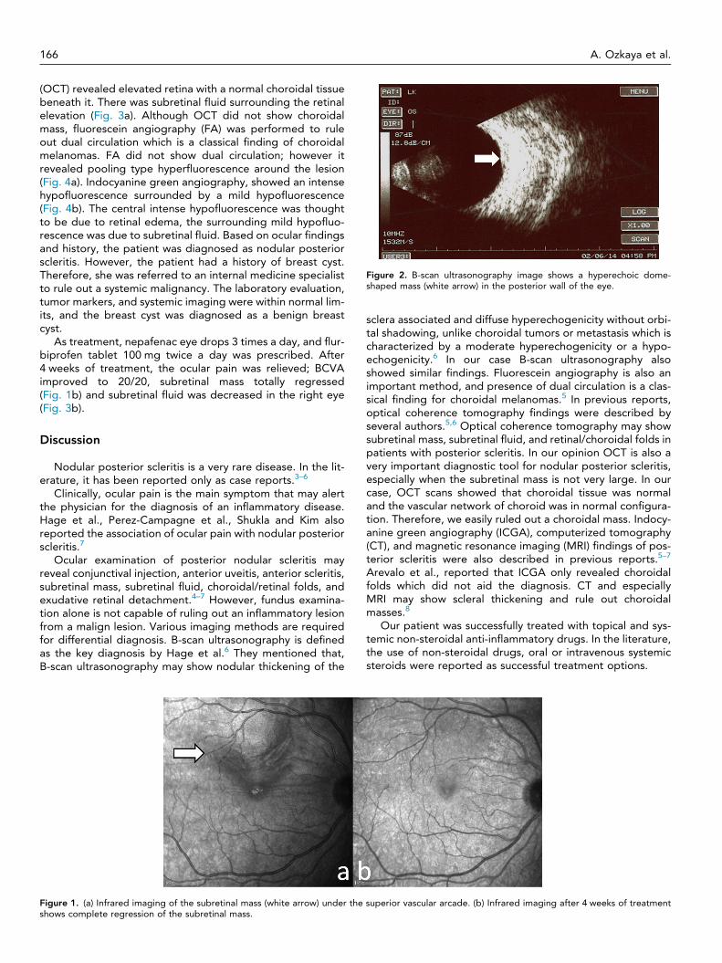
Figure 2. B-scan ultrasonography image shows a hyperechoic dome-shaped mass (white arrow) in the posterior wall of the eye.
166 A. Ozkaya et al.
(OCT) revealed elevated retina with a normal choroidal tissuebeneath it. There was subretinal fluid surrounding the retinalelevation (Fig. 3a). Although OCT did not show choroidalmass, fluorescein angiography (FA) was performed to ruleout dual circulation which is a classical finding of choroidalmelanomas. FA did not show dual circulation; however itrevealed pooling type hyperfluorescence around the lesion(Fig. 4a). Indocyanine green angiography, showed an intensehypofluorescence surrounded by a mild hypofluorescence(Fig. 4b). The central intense hypofluorescence was thoughtto be due to retinal edema, the surrounding mild hypofluo-rescence was due to subretinal fluid. Based on ocular findingsand history, the patient was diagnosed as nodular posteriorscleritis. However, the patient had a history of breast cyst.Therefore, she was referred to an internal medicine specialistto rule out a systemic malignancy. The laboratory evaluation,tumor markers, and systemic imaging were within normal lim-its, and the breast cyst was diagnosed as a benign breastcyst.
As treatment, nepafenac eye drops 3 times a day, and flur-biprofen tablet 100 mg twice a day was prescribed. After4 weeks of treatment, the ocular pain was relieved; BCVAimproved to 20/20, subretinal mass totally regressed(Fig. 1b) and subretinal fluid was decreased in the right eye(Fig. 3b).
Discussion
Nodular posterior scleritis is a very rare disease. In the lit-erature, it has been reported only as case reports.3–6
Clinically, ocular pain is the main symptom that may alertthe physician for the diagnosis of an inflammatory disease.Hage et al., Perez-Campagne et al., Shukla and Kim alsoreported the association of ocular pain with nodular posteriorscleritis.7
Ocular examination of posterior nodular scleritis mayreveal conjunctival injection, anterior uveitis, anterior scleritis,subretinal mass, subretinal fluid, choroidal/retinal folds, andexudative retinal detachment.4–7 However, fundus examina-tion alone is not capable of ruling out an inflammatory lesionfrom a malign lesion. Various imaging methods are requiredfor differential diagnosis. B-scan ultrasonography is definedas the key diagnosis by Hage et al.6 They mentioned that,B-scan ultrasonography may show nodular thickening of the
Figure 1. (a) Infrared imaging of the subretinal mass (white arrow) under theshows complete regression of the subretinal mass.
sclera associated and diffuse hyperechogenicity without orbi-tal shadowing, unlike choroidal tumors or metastasis which ischaracterized by a moderate hyperechogenicity or a hypo-echogenicity.6 In our case B-scan ultrasonography alsoshowed similar findings. Fluorescein angiography is also animportant method, and presence of dual circulation is a clas-sical finding for choroidal melanomas.5 In previous reports,optical coherence tomography findings were described byseveral authors.5,6 Optical coherence tomography may showsubretinal mass, subretinal fluid, and retinal/choroidal folds inpatients with posterior scleritis. In our opinion OCT is also avery important diagnostic tool for nodular posterior scleritis,especially when the subretinal mass is not very large. In ourcase, OCT scans showed that choroidal tissue was normaland the vascular network of choroid was in normal configura-tion. Therefore, we easily ruled out a choroidal mass. Indocy-anine green angiography (ICGA), computerized tomography(CT), and magnetic resonance imaging (MRI) findings of pos-terior scleritis were also described in previous reports.5–7
Arevalo et al., reported that ICGA only revealed choroidalfolds which did not aid the diagnosis. CT and especiallyMRI may show scleral thickening and rule out choroidalmasses.8
Our patient was successfully treated with topical and sys-temic non-steroidal anti-inflammatory drugs. In the literature,the use of non-steroidal drugs, oral or intravenous systemicsteroids were reported as successful treatment options.
superior vascular arcade. (b) Infrared imaging after 4 weeks of treatment
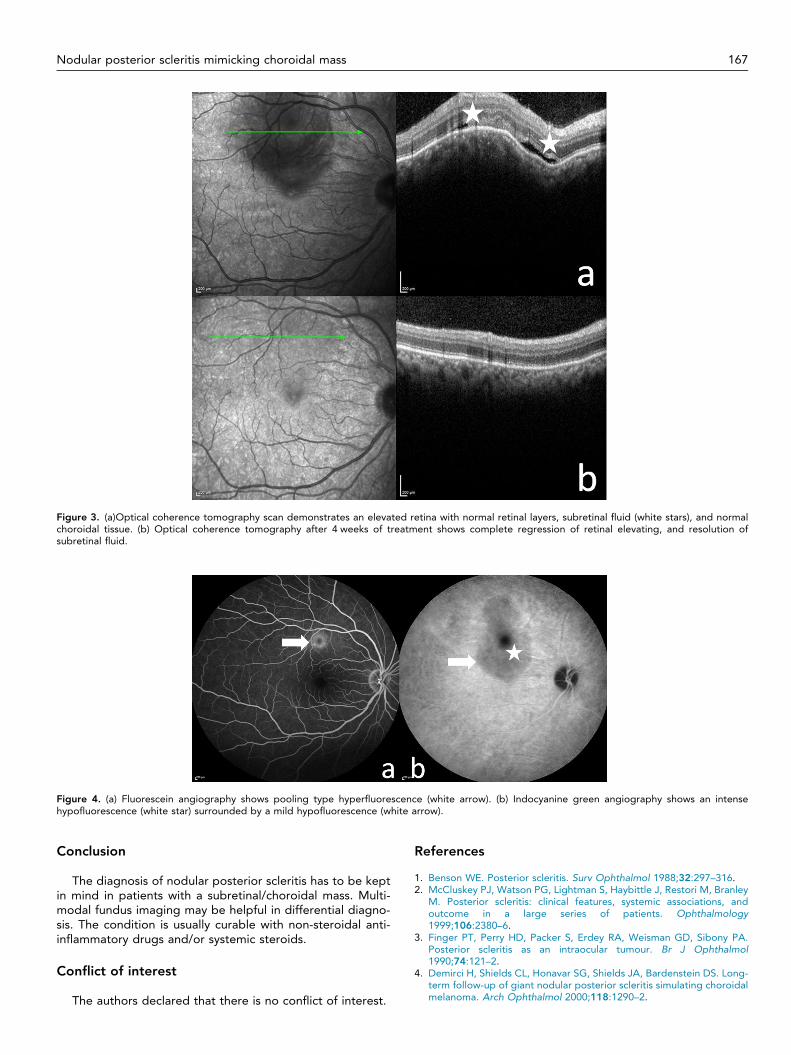
Figure 3. (a)Optical coherence tomography scan demonstrates an elevated retina with normal retinal layers, subretinal fluid (white stars), and normalchoroidal tissue. (b) Optical coherence tomography after 4 weeks of treatment shows complete regression of retinal elevating, and resolution ofsubretinal fluid.
Figure 4. (a) Fluorescein angiography shows pooling type hyperfluorescence (white arrow). (b) Indocyanine green angiography shows an intensehypofluorescence (white star) surrounded by a mild hypofluorescence (white arrow).
Nodular posterior scleritis mimicking choroidal mass 167
Conclusion
The diagnosis of nodular posterior scleritis has to be keptin mind in patients with a subretinal/choroidal mass. Multi-modal fundus imaging may be helpful in differential diagno-sis. The condition is usually curable with non-steroidal anti-inflammatory drugs and/or systemic steroids.
Conflict of interest
The authors declared that there is no conflict of interest.
References
1. Benson WE. Posterior scleritis. Surv Ophthalmol 1988;32:297–316.2. McCluskey PJ, Watson PG, Lightman S, Haybittle J, Restori M, Branley
M. Posterior scleritis: clinical features, systemic associations, andoutcome in a large series of patients. Ophthalmology1999;106:2380–6.
3. Finger PT, Perry HD, Packer S, Erdey RA, Weisman GD, Sibony PA.Posterior scleritis as an intraocular tumour. Br J Ophthalmol1990;74:121–2.
4. Demirci H, Shields CL, Honavar SG, Shields JA, Bardenstein DS. Long-term follow-up of giant nodular posterior scleritis simulating choroidalmelanoma. Arch Ophthalmol 2000;118:1290–2.

168 A. Ozkaya et al.
5. Pérez-Campagne E, Guex-Crosier Y, Schalenbourg A, Uffer S,Zografos L. Giant nodular posterior scleritis compatible with ocularsarcoidosis simulating choroidal melanoma. Arch Soc Esp Oftalmol2007;82:563–6 Article in Spanish.
6. Hage R, Jean-Charles A, Guyomarch J, Rahimian O, Donnio A, MerleH. Nodular posterior scleritis mimicking choroidal metastasis: a reportof two cases. Clin Ophthalmol 2011;5:877–80.
7. Shukla D, Kim R. Giant nodular posterior scleritis simulating choroidalmelanoma. Indian J Ophthalmol 2006;54:120–2.
8. Arevalo JF, Shields CL, Shields JA. Giant nodular posterior scleritissimulating choroidal melanoma and birdshot retinochoroidopathy.Ophthal Surg Las Imag 2003;34:403–5.