A Case of Acrodermatitis Continua of Hallopeau Following ... · CASE REPORT A Case of...
6
CASE REPORT A Case of Acrodermatitis Continua of Hallopeau Following Chronic Pustular Cheilitis Mariachiara Arisi . Maria Teresa Rossi . Raffaella Sala . Giulia Petrilli . Daniela Marocolo . Marco Ungari . Fabio Facchetti . Pier Giacomo Calzavara-Pinton To view enhanced content go to www.dermtherapy-open.com Received: November 26, 2015 / Published online: February 26, 2016 Ó The Author(s) 2016. This article is published with open access at Springerlink.com ABSTRACT We describe the case of a young male affected by chronic pustular psoriasis of the lips that remained the only manifestation of acrodermatitis continua of Hallopeau (ACH) for years before the onset of the characteristic hand lesions. Keywords: Acrodermatitis continua; Cheilitis; Hallopeau; Psoriasis INTRODUCTION Acrodermatitis continua of Hallopeau (ACH), also known as dermatitis repens or pustular acrodermatitis, is an uncommon clinical variant of pustular psoriasis. Clinical presentation is quite monotonous with chronic recurrent pustulation of the nail fold, nail bed and tip of one or more fingers or toes. With time, it spreads proximally with destruction of the nail matrix, and sometimes it causes osteolysis and bone reabsorption. Other modalities of clinical presentation have not been reported so far [1]. We describe the case of a 25-year-old male affected by chronic pustular psoriasis of the lips that remained the only manifestation of ACH for years before the onset of the characteristic clinical hand lesions. CASE REPORT A 25-year-old male presented with a 2-year history of erythematous and desquamative cheilitis with pustules. The lesions were not responsive to several treatment cycles with corticosteroids and emollients. Clinical examination showed that the upper lip was erythematous, fissured and covered by thick white-yellowish scales with small noncoalescing pustules. Skin examination was M. Arisi (&) Á M. T. Rossi Á R. Sala Á P. G. Calzavara-Pinton Department of Dermatology, Spedali Civili di Brescia, University of Brescia, Brescia, Italy e-mail: [email protected] G. Petrilli Á D. Marocolo Á F. Facchetti Department of Pathology, Spedali Civili di Brescia, University of Brescia, Brescia, Italy M. Ungari Department of Pathology, Ospedale di Cremona, Cremona, Italy Dermatol Ther (Heidelb) (2016) 6:89–94 DOI 10.1007/s13555-016-0100-2
Transcript of A Case of Acrodermatitis Continua of Hallopeau Following ... · CASE REPORT A Case of...
CASE REPORT
A Case of Acrodermatitis Continua of HallopeauFollowing Chronic Pustular Cheilitis
Mariachiara Arisi . Maria Teresa Rossi . Raffaella Sala .
Giulia Petrilli . Daniela Marocolo . Marco Ungari . Fabio Facchetti .
Pier Giacomo Calzavara-Pinton
To view enhanced content go to www.dermtherapy-open.comReceived: November 26, 2015 / Published online: February 26, 2016� The Author(s) 2016. This article is published with open access at Springerlink.com
ABSTRACT
We describe the case of a young male affected
by chronic pustular psoriasis of the lips that
remained the only manifestation of
acrodermatitis continua of Hallopeau (ACH)
for years before the onset of the characteristic
hand lesions.
Keywords: Acrodermatitis continua; Cheilitis;
Hallopeau; Psoriasis
INTRODUCTION
Acrodermatitis continua of Hallopeau (ACH),
also known as dermatitis repens or pustular
acrodermatitis, is an uncommon clinical variant
of pustular psoriasis.
Clinical presentation is quite monotonous
with chronic recurrent pustulation of the nail
fold, nail bed and tip of one or more fingers or
toes.
With time, it spreads proximally with
destruction of the nail matrix, and sometimes
it causes osteolysis and bone reabsorption.
Other modalities of clinical presentation have
not been reported so far [1].
We describe the case of a 25-year-old male
affected by chronic pustular psoriasis of the lips
that remained the only manifestation of ACH
for years before the onset of the characteristic
clinical hand lesions.
CASE REPORT
A 25-year-old male presented with a 2-year
history of erythematous and desquamative
cheilitis with pustules. The lesions were not
responsive to several treatment cycles with
corticosteroids and emollients.
Clinical examination showed that the upper
lip was erythematous, fissured and covered by
thick white-yellowish scales with small
noncoalescing pustules. Skin examination was
M. Arisi (&) � M. T. Rossi � R. Sala �P. G. Calzavara-PintonDepartment of Dermatology, Spedali Civili diBrescia, University of Brescia, Brescia, Italye-mail: [email protected]
G. Petrilli � D. Marocolo � F. FacchettiDepartment of Pathology, Spedali Civili di Brescia,University of Brescia, Brescia, Italy
M. UngariDepartment of Pathology, Ospedale di Cremona,Cremona, Italy
Dermatol Ther (Heidelb) (2016) 6:89–94
DOI 10.1007/s13555-016-0100-2
otherwise normal, and the oral mucosa, tongue
and nails did not present any lesions (Fig. 1).
The patient was otherwise in good health. He
reported no personal or family history of
psoriasis or any other dermatological
conditions and no history of smoking. He
denied any history of lip licking or contact
with lipstick, topical antibiotics and steroids, or
other cosmetics.
Findings of routine hematology and
biochemistry tests, total IgE serum level and
IgE microarray ImmunoCap assay were within
normal ranges. Bacteriological investigations of
the pustules were negative as well.
Patch testing with the standard series and
series of dental materials recommended by the
Italian Society of Occupational and
Environmental Allergological Dermatology
(SIDAPA) did not show contact allergic
sensitization [2].
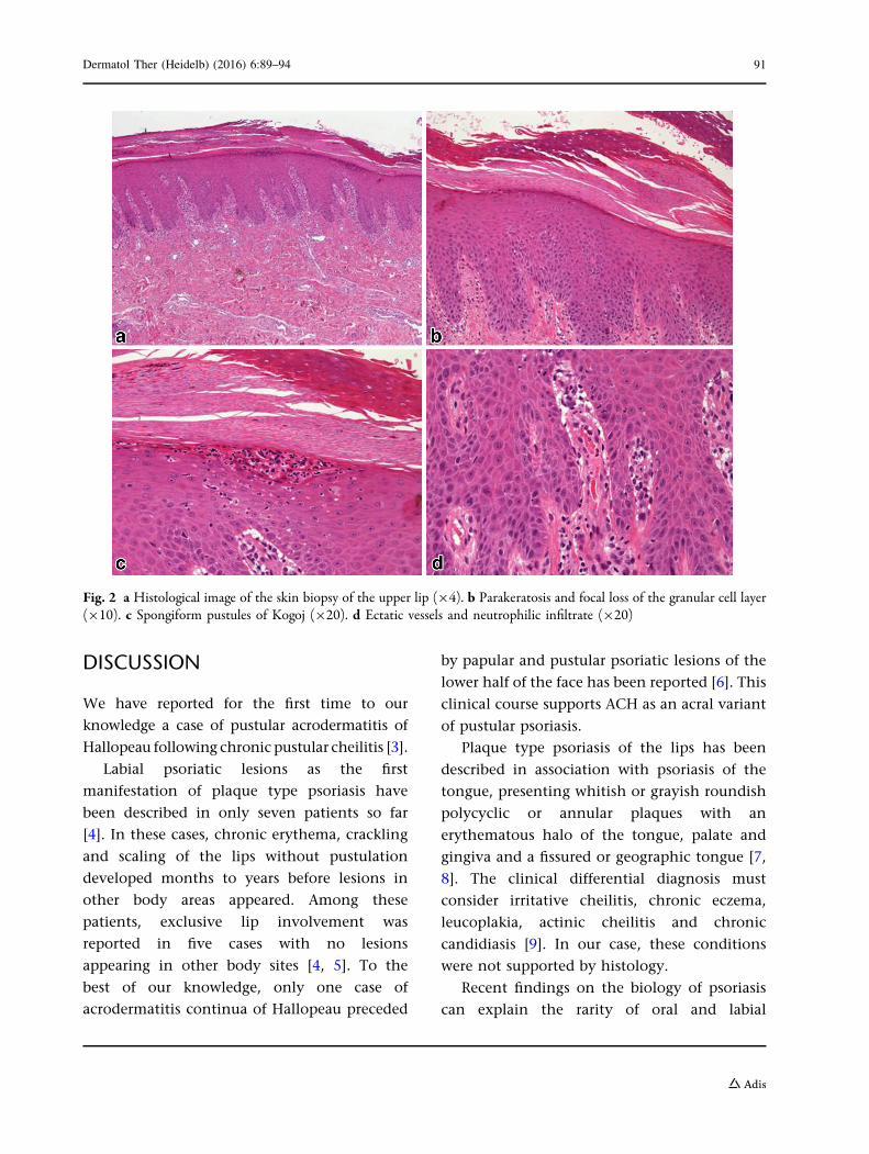
A biopsy sample of the upper lip was
obtained for histopathologic evaluation; the
analysis showed a stratified squamous
epithelium with confluent parakeratosis and
focal loss of the granular cell layer. Acanthosis
and irregular elongation of the rete ridges were
also noted, with ectatic vessels and a
lymphocytic inflammatory cell infiltrate. There
were collections of neutrophils, with the aspect
of both Munro microabscesses (accumulation of
polymorphs within the parakeratotic stratum
corneum) and spongiform pustules of Kogoj
(small accumulation of neutrophils and
occasional lymphocytes beneath the keratin
layer). No fungi or spirochetes were found
with specific stainings (Fig. 2a–c).
A diagnosis of psoriasiform cheilitis was
made.
The lip lesions were treated with calcipotriol
ointment twice daily for a month without any
improvement. Topical tacrolimus 0.1%
ointment twice daily for a month was
ineffective as well.
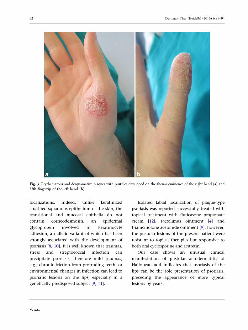
At the 4-month follow-up erythematous and
desquamative plaques with pustules developed
on the thenar eminence of the right hand and
fifth fingertip of the left hand (Fig. 3a, b). A skin
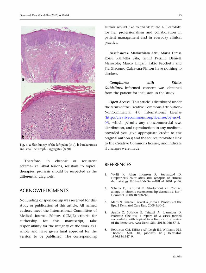
biopsy of the palm showed regular acanthosis of
the epidermis, with elongation of the rete
ridges, which tended to fuse with each other.
The overlying hyperkeratosis, ortho- and para-,
was filled with small aggregates of neutrophils,
observed even in the subcorneal location. Some
suprapapillary plates were thin, and the dermal
papillae were expanded by tortuous vessels;
there was an inflammatory infiltrate composed
of small lymphocytes, with mild exocytosis and
perivascular disposition (Fig. 4a, b).
The histological findings were consistent
with psoriasis.
Oral cyclosporine (200 mg/day) for 6 months
led to a complete remission of the lesions of the
lip and hands. However, 2 months after
interruption a relapse was seen. Low-dose oral
acitretin (25 mg/day) was administered, which
quickly cleared the pustular lesions of the lips
and hands, although a mild xerotic cheilitis
developed.
Compliance with Ethics Guidelines
Informed consent was obtained from the
patient for inclusion in the study.
Fig. 1 Eryrthema, edema and scaling with small pustularlesions of the lips
90 Dermatol Ther (Heidelb) (2016) 6:89–94
DISCUSSION
We have reported for the first time to our
knowledge a case of pustular acrodermatitis of
Hallopeau following chronic pustular cheilitis [3].
Labial psoriatic lesions as the first
manifestation of plaque type psoriasis have
been described in only seven patients so far
[4]. In these cases, chronic erythema, crackling
and scaling of the lips without pustulation
developed months to years before lesions in
other body areas appeared. Among these
patients, exclusive lip involvement was
reported in five cases with no lesions
appearing in other body sites [4, 5]. To the
best of our knowledge, only one case of
acrodermatitis continua of Hallopeau preceded
by papular and pustular psoriatic lesions of the
lower half of the face has been reported [6]. This
clinical course supports ACH as an acral variant
of pustular psoriasis.
Plaque type psoriasis of the lips has been
described in association with psoriasis of the
tongue, presenting whitish or grayish roundish
polycyclic or annular plaques with an
erythematous halo of the tongue, palate and
gingiva and a fissured or geographic tongue [7,
8]. The clinical differential diagnosis must
consider irritative cheilitis, chronic eczema,
leucoplakia, actinic cheilitis and chronic
candidiasis [9]. In our case, these conditions
were not supported by histology.
Recent findings on the biology of psoriasis
can explain the rarity of oral and labial
Fig. 2 a Histological image of the skin biopsy of the upper lip (94). b Parakeratosis and focal loss of the granular cell layer(910). c Spongiform pustules of Kogoj (920). d Ectatic vessels and neutrophilic infiltrate (920)
Dermatol Ther (Heidelb) (2016) 6:89–94 91
localizations. Indeed, unlike keratinized
stratified squamous epithelium of the skin, the
transitional and mucosal epithelia do not
contain corneodesmosin, an epidermal
glycoprotein involved in keratinocyte
adhesion, an allelic variant of which has been
strongly associated with the development of
psoriasis [8, 10]. It is well known that traumas,
stress and streptococcal infection can
precipitate psoriasis; therefore mild traumas,
e.g., chronic friction from protruding teeth, or
environmental changes in infection can lead to
psoriatic lesions on the lips, especially in a
genetically predisposed subject [9, 11].
Isolated labial localization of plaque-type
psoriasis was reported successfully treated with
topical treatment with fluticasone propionate
cream [12], tacrolimus ointment [4] and
triamcinolone acetonide ointment [9]; however,
the pustular lesions of the present patient were
resistant to topical therapies but responsive to
both oral cyclosporine and acitretin.
Our case shows an unusual clinical
manifestation of pustular acrodermatitis of
Hallopeau and indicates that psoriasis of the
lips can be the sole presentation of psoriasis,
preceding the appearance of more typical
lesions by years.
Fig. 3 Erythematous and desquamative plaques with pustules developed on the thenar eminence of the right hand (a) andfifth fingertip of the left hand (b)
92 Dermatol Ther (Heidelb) (2016) 6:89–94
Therefore, in chronic or recurrent
eczema-like labial lesions, resistant to topical
therapies, psoriasis should be suspected as the
differential diagnosis.
ACKNOWLEDGMENTS
No funding or sponsorship was received for this
study or publication of this article. All named
authors meet the International Committee of
Medical Journal Editors (ICMJE) criteria for
authorship for this manuscript, take
responsibility for the integrity of the work as a
whole and have given final approval for the
version to be published. The corresponding
author would like to thank nurse A. Bertolotti
for her professionalism and collaboration in
patient management and in everyday clinical
practice.
Disclosures. Mariachiara Arisi, Maria Teresa
Rossi, Raffaella Sala, Giulia Petrilli, Daniela
Marocolo, Marco Ungari, Fabio Facchetti and
PierGiacomo Calzavara-Pinton have nothing to
disclose.
Compliance with Ethics
Guidelines. Informed consent was obtained
from the patient for inclusion in the study.
Open Access. This article is distributed under
the terms of the Creative Commons Attribution-
NonCommercial 4.0 International License
(http://creativecommons.org/licenses/by-nc/4.
0/), which permits any noncommercial use,
distribution, and reproduction in any medium,
provided you give appropriate credit to the
original author(s) and the source, provide a link
to the Creative Commons license, and indicate
if changes were made.
REFERENCES
1. Wolff K, Allen Jhonson R, Suurmond D.Fitzpatrick’s color atlas and synopsis of clinicaldermatology: Fifth ed. McGraw-Hill ed. 2001. p. 66.
2. Schena D, Fantuzzi F, Girolomoni G. Contactallergy in chronic eczematous lip dermatitis. Eur JDermatol. 2008;18:688–92.
3. Martı N, Pinazo I, Revert A, Jorda E. Psoriasis of thelips. J Dermatol Case Rep. 2009;3:50–2.
4. Apalla Z, Sotiriou E, Trigoni A, Ioannides D.Psoriatic Cheilitis: a report of 2 cases treatedsuccessfully with topical tacrolimus and a reviewof the literature. Acta Derm Sifil. 2015;106:687–8.
5. Robinson CM, DiBiase AT, Leigh IM, Williams DM,Thornhill MH. Oral psoriasis. Br J Dermatol.1996;134:347–9.
Fig. 4 a Skin biopsy of the left palm (94). b Parakeratosisand small neutrophil aggregates (920)
Dermatol Ther (Heidelb) (2016) 6:89–94 93

6. Iijima S, Okazaki Y, Watanabe S, Maruyama Y. Caseof acrodermatitis continua of Hallopeau followingpsoriasis with atypical clinical presentation.J Dermatol. 2014;41:1006–8.
7. Prignano F, D’Erme AM, Bonciolini V, Lotti T.Mucosal psoriasis: a new insight toward a systemicinflammatory disease. Int J Dermatol.2011;50:1579–81.
8. Yesudian PD, Chalmers RJG, Warren RB, GriffithsCEM. In search of oral psoriasis. Arch Dermatol Res.2012;304:1–5.
9. Rahman MA, Fikree M. Perioral psoriasis. J Eur AcadDermatol Venereol. 2000;14:513–22.
10. Allen M, Ishida-Yamamoto A, McGrath J, DavisonS, Iizuka H, Simon M, et al. Corneodesmosinexpression in psoriasis vulgaris differs fromnormal skin and other inflammatory skindisorders. Lab Invest. 2001;81:969–76.
11. Brenner S, Lipitz R, Illie B, Krakowski A. Psoriasis ofthe lips: the unusual Koebner phenomenon causedby protruding upper teeth. Dermatologica.1982;164:413–6.
12. Ersoy-Evans S, Nuralina L, Erkin G, Ozkaya O.Psoriasis of the lips: a rare entity. J Eur AcadDermatol Venereol. 2007;21:142–3.
94 Dermatol Ther (Heidelb) (2016) 6:89–94