A baculovirus-encoded miRNA suppresses its host miRNAs
13
A Baculovirus-Encoded MicroRNA (miRNA) Suppresses Its Host miRNA Biogenesis by Regulating the Exportin-5 Cofactor Ran C. P. Singh, J. Singh, and J. Nagaraju Centre of Excellence for Genetics and Genomics of Silkmoths, Laboratory of Molecular Genetics, Centre for DNA Fingerprinting and Diagnostics, Tuljaguda Complex, Nampally, Hyderabad, India MicroRNAs have emerged as key players in the regulation of various biological processes in eukaryotes, including host-pathogen interactions. Recent studies suggest that viruses encode miRNAs to manipulate their host gene expression to ensure their effec- tive proliferation, whereas the host limits virus infection by differentially expressing miRNAs that target essential viral genes. Here, we demonstrate that an insect virus, Bombyx mori nucleopolyhedrosis virus (BmNPV), modulates the small-RNA-medi- ated defense of its host, B. mori, by encoding an miRNA (bmnpv-miR-1) that downregulates the expression of the host GTP- binding nuclear protein Ran, an essential component of the exportin-5-mediated nucleocytoplasmic transport machinery mainly involved in small-RNA transport from the nucleus to the cytoplasm. We demonstrate the sequence-dependent interac- tion of bmnpv-miR-1 with Ran mRNA using cell culture and in vivo assays, including RNA interference (RNAi) of Ran. Our re- sults clearly show that bmnpv-miR-1 represses Ran, leading to reduction in the host small-RNA population, and consequently, the BmNPV load increases in the infected larvae. Blocking of bmnpv-miR-1 resulted in higher expression levels of Ran and a de- crease in BmNPV proliferation. In contrast, blockage of host miRNA, bmo-miR-8, which targets the immediate-early gene of the virus and whose production was repressed upon bmnpv-miR-1 and Ran dsRNA administration, resulted in a significant increase in the virus load in the infected B. mori larvae. The present study provides an insight into one of the evasion strategies used by the virus to counter the host defense for its effective proliferation and has relevance to the development of insect virus control strategies. M icroRNAs (miRNAs) are noncoding RNAs that regulate gene expression by binding to the complementary sequence of the target mRNAs. Canonically, miRNAs are transcribed by RNA polymerase II as long primary transcripts (pri-miRNAs), which are processed by a nuclear RNase III called Drosha to gen- erate 70-nucleotide (nt) imperfectly complementary stem-loop precursor miRNAs (pre-miRNAs). The pre-miRNAs are then ex- ported to the cytoplasm by RanGTP-dependent exportin-5 (6, 54) for further processing by the cytoplasmic RNase III Dicer into 22-nt miRNA-miRNA* duplexes, followed by incorporation of one of the strands into an RNA-induced silencing complex (RISC). Finally, the whole RISC assembly, loaded with miRNA, targets the specific mRNAs, leading to either their translational repression or degradation, depending upon the extent of comple- mentarity between the miRNA and its target mRNA (3, 27). Since the discovery of miRNAs (29, 40), a number of studies have been carried out to understand the role of miRNAs in almost all the biological processes in eukaryotes. Apart from eukaryotes, viruses have also been shown to encode miRNAs (18, 38). Re- cently, miRNAs have been shown to play a critical role in intricate host-virus interactions (13, 15). Its small size, nonantigenic na- ture, and target specificity make viral miRNA a potential weapon against the host defense machinery. Some of the well-known strat- egies adopted by viruses to achieve their establishment and persis- tent infection in the host cells are modulation of host immune response and escaping recognition by the host immune system (48, 51), antiapoptosis (9, 52), cell cycle arrest (14, 17), and mim- icking of host miRNAs (16, 47, 56). So far, a limited number of studies have been carried out on avoidance of host defenses by viral miRNAs, but they are confined to mammalian viruses. Insect viruses have hardly been the subject of such investigations, except for a recent report of autoregulation of viral gene expression by virus-encoded miRNAs in the Heliothis virescens ascovirus (21). Here, we show that Bombyx mori nucleo- polyhedrosis virus (BmNPV) encodes an miRNA that modulates the host defense machinery by regulating the production of the host GTP-binding nuclear protein Ran (Ras-related nuclear pro- tein). Ran is a 25-kDa protein that plays an important role in nucleocytoplasmic transport of various noncoding RNAs and proteins (5). During nuclear export of pre-miRNAs, exportin-5 binds to the pre-miRNAs in a RanGTP-dependent manner, and depletion of Ran results in a severe reduction of pre-miRNA ex- port (6, 33, 50, 54). In humans, Ran also interacts with other transporters, like exportin-1 and exportin-t, which are involved in the transport of small nuclear RNAs (snRNAs) and tRNAs, re- spectively (44). In Drosophila, exportin-5 is known to transport both pre-miRNAs and tRNAs (44), whereas in B. mori, small- RNA export pathways and factors have yet to be investigated. BmNPV, a double-stranded DNA (dsDNA) virus, belongs to the family Baculoviridae, which includes various pathogens of Lepidoptera, one of the largest and most economically important insect orders (41). BmNPV, a natural pathogen of the domesti- cated silk moth, B. mori, inflicts heavy larval mortality and thus causes severe economic damage to silk production. In our previ- ous study, we discovered four BmNPV-encoded miRNAs, and in Received 10 January 2012 Accepted 9 May 2012 Published ahead of print 16 May 2012 Address correspondence to J. Nagaraju, [email protected]. Supplemental material for this article may be found at http://jvi.asm.org/. Copyright © 2012, American Society for Microbiology. All Rights Reserved. doi:10.1128/JVI.00064-12 August 2012 Volume 86 Number 15 Journal of Virology p. 7867–7879 jvi.asm.org 7867 Downloaded from https://journals.asm.org/journal/jvi on 28 November 2021 by 91.246.209.153.
Transcript of A baculovirus-encoded miRNA suppresses its host miRNAs

A Baculovirus-Encoded MicroRNA (miRNA) Suppresses Its HostmiRNA Biogenesis by Regulating the Exportin-5 Cofactor Ran
C. P. Singh, J. Singh, and J. Nagaraju
Centre of Excellence for Genetics and Genomics of Silkmoths, Laboratory of Molecular Genetics, Centre for DNA Fingerprinting and Diagnostics, Tuljaguda Complex,Nampally, Hyderabad, India
MicroRNAs have emerged as key players in the regulation of various biological processes in eukaryotes, including host-pathogeninteractions. Recent studies suggest that viruses encode miRNAs to manipulate their host gene expression to ensure their effec-tive proliferation, whereas the host limits virus infection by differentially expressing miRNAs that target essential viral genes.Here, we demonstrate that an insect virus, Bombyx mori nucleopolyhedrosis virus (BmNPV), modulates the small-RNA-medi-ated defense of its host, B. mori, by encoding an miRNA (bmnpv-miR-1) that downregulates the expression of the host GTP-binding nuclear protein Ran, an essential component of the exportin-5-mediated nucleocytoplasmic transport machinerymainly involved in small-RNA transport from the nucleus to the cytoplasm. We demonstrate the sequence-dependent interac-tion of bmnpv-miR-1 with Ran mRNA using cell culture and in vivo assays, including RNA interference (RNAi) of Ran. Our re-sults clearly show that bmnpv-miR-1 represses Ran, leading to reduction in the host small-RNA population, and consequently,the BmNPV load increases in the infected larvae. Blocking of bmnpv-miR-1 resulted in higher expression levels of Ran and a de-crease in BmNPV proliferation. In contrast, blockage of host miRNA, bmo-miR-8, which targets the immediate-early gene of thevirus and whose production was repressed upon bmnpv-miR-1 and Ran dsRNA administration, resulted in a significant increasein the virus load in the infected B. mori larvae. The present study provides an insight into one of the evasion strategies used bythe virus to counter the host defense for its effective proliferation and has relevance to the development of insect virus controlstrategies.
MicroRNAs (miRNAs) are noncoding RNAs that regulategene expression by binding to the complementary sequence
of the target mRNAs. Canonically, miRNAs are transcribed byRNA polymerase II as long primary transcripts (pri-miRNAs),which are processed by a nuclear RNase III called Drosha to gen-erate �70-nucleotide (nt) imperfectly complementary stem-loopprecursor miRNAs (pre-miRNAs). The pre-miRNAs are then ex-ported to the cytoplasm by RanGTP-dependent exportin-5 (6, 54)for further processing by the cytoplasmic RNase III Dicer into�22-nt miRNA-miRNA* duplexes, followed by incorporation ofone of the strands into an RNA-induced silencing complex(RISC). Finally, the whole RISC assembly, loaded with miRNA,targets the specific mRNAs, leading to either their translationalrepression or degradation, depending upon the extent of comple-mentarity between the miRNA and its target mRNA (3, 27).
Since the discovery of miRNAs (29, 40), a number of studieshave been carried out to understand the role of miRNAs in almostall the biological processes in eukaryotes. Apart from eukaryotes,viruses have also been shown to encode miRNAs (18, 38). Re-cently, miRNAs have been shown to play a critical role in intricatehost-virus interactions (13, 15). Its small size, nonantigenic na-ture, and target specificity make viral miRNA a potential weaponagainst the host defense machinery. Some of the well-known strat-egies adopted by viruses to achieve their establishment and persis-tent infection in the host cells are modulation of host immuneresponse and escaping recognition by the host immune system(48, 51), antiapoptosis (9, 52), cell cycle arrest (14, 17), and mim-icking of host miRNAs (16, 47, 56).
So far, a limited number of studies have been carried out onavoidance of host defenses by viral miRNAs, but they are confinedto mammalian viruses. Insect viruses have hardly been the subjectof such investigations, except for a recent report of autoregulation
of viral gene expression by virus-encoded miRNAs in the Heliothisvirescens ascovirus (21). Here, we show that Bombyx mori nucleo-polyhedrosis virus (BmNPV) encodes an miRNA that modulatesthe host defense machinery by regulating the production of thehost GTP-binding nuclear protein Ran (Ras-related nuclear pro-tein). Ran is a 25-kDa protein that plays an important role innucleocytoplasmic transport of various noncoding RNAs andproteins (5). During nuclear export of pre-miRNAs, exportin-5binds to the pre-miRNAs in a RanGTP-dependent manner, anddepletion of Ran results in a severe reduction of pre-miRNA ex-port (6, 33, 50, 54). In humans, Ran also interacts with othertransporters, like exportin-1 and exportin-t, which are involved inthe transport of small nuclear RNAs (snRNAs) and tRNAs, re-spectively (44). In Drosophila, exportin-5 is known to transportboth pre-miRNAs and tRNAs (44), whereas in B. mori, small-RNA export pathways and factors have yet to be investigated.
BmNPV, a double-stranded DNA (dsDNA) virus, belongs tothe family Baculoviridae, which includes various pathogens ofLepidoptera, one of the largest and most economically importantinsect orders (41). BmNPV, a natural pathogen of the domesti-cated silk moth, B. mori, inflicts heavy larval mortality and thuscauses severe economic damage to silk production. In our previ-ous study, we discovered four BmNPV-encoded miRNAs, and in
Received 10 January 2012 Accepted 9 May 2012
Published ahead of print 16 May 2012
Address correspondence to J. Nagaraju, [email protected].
Supplemental material for this article may be found at http://jvi.asm.org/.
Copyright © 2012, American Society for Microbiology. All Rights Reserved.
doi:10.1128/JVI.00064-12
August 2012 Volume 86 Number 15 Journal of Virology p. 7867–7879 jvi.asm.org 7867
Dow
nloa
ded
from
http
s://j
ourn
als.
asm
.org
/jour
nal/j
vi o
n 28
Nov
embe
r 20
21 b
y 91
.246
.209
.153
.

silico prediction of their targets showed Ran to be one of the po-tential targets of bmnpv-miR-1 (45). Here, we carried out func-tional analysis of bmnpv-miR-1 to show that it acts as a suppressorof Ran both in cell culture and in vivo in B. mori. We have foundthat bmnpv-miR-1 significantly represses the expression of Ran atboth the transcript and protein levels, resulting in the impairmentof RanGTP-mediated small-RNA transport in the host.
Further, we also analyzed the effect of bmnpv-miR-1-mediateddownregulation of Ran on viral proliferation and found a signifi-cant increase in the viral load upon Ran knockdown. Consistentwith this result, blocking of bmnpv-miR-1 by specific locked nu-cleic acid (LNA) resulted in higher Ran transcript levels and adecrease in the virus titer in BmNPV-infected larvae. Further, weassessed the virus load by blocking one of the host miRNAs, bmo-miR-8, which was suppressed by administration of bmnpv-miR-1and Ran dsRNA, by specific LNA. The virus load was dramaticallyincreased upon inhibition of this host miRNA. These results pro-vide convincing evidence that BmNPV suppresses the small-RNA-mediated host defense to successfully proliferate in the hostcells by employing bmnpv-miR-1. The present study thus pro-vides insight into yet another layer of complex host-virus interac-tions mediated by a virus-encoded miRNA.
MATERIALS AND METHODSIn silico prediction of a bmnpv-miR-1 binding site on Ran transcript.The targets of bmnpv-miR-1 were initially predicted by using themiRanda program (11), along with many screening parameters, as de-scribed in our previous report (45). The predicted binding site of bmnpv-miR-1 on the 3= untranslated region (UTR) of Ran was reconfirmed byemploying the RNAhybrid program, which calculates the minimum freeenergy of the miRNA-mRNA duplex.
B. mori strain and BmNPV infection. The SBNP-1 strain of B. moriwas provided by the Andhra Pradesh State Sericulture Research and De-velopment Institute (APSSRDI), Hindupur, India. The larvae were reared
at 27°C on fresh mulberry leaves. For infection study, fifth-instar larvaewere fed per os with a purified suspension of BmNPV occlusion bodies(OBs) (20,000 OBs/larva).
RNA isolation and RT-qPCR analysis. Total RNA was extracted fromfat body tissues of uninfected and BmNPV-infected B. mori larvae usingTRIzol reagent (Invitrogen), followed by DNase I treatment (Invitrogen).RNA (2 �g) was reverse transcribed using the SuperScript III First-StrandSynthesis System (Invitrogen) in a 20-�l reaction mixture. One microliterof the reverse transcription (RT) product was used to perform quantita-tive-PCR (qPCR) amplification on the RT-7500 system (Applied Biosys-tems) using primers specific to Ran, as described in the supplementalmaterials and methods. RT-qPCR data were analyzed by using the RT-7500 system (Applied Biosystems) software and were further verified byusing the standard ��CT method.
Counting of OBs. To determine the BmNPV load in B. mori larvae,OBs were isolated from the hemolymph of infected larvae by centrifuga-tion (1,000 rpm for 1 min), followed by resuspending the viral pellet in 1�PBS. The OBs were counted using an automated cell counter (Countess;Invitrogen).
HeLa cell culture and luciferase assays. HeLa cells were maintained inDulbecco’s modified Eagle’s medium (Thermo Scientific) containing 2mM L-glutamine and 10% fetal bovine serum (FBS) (Invitrogen) at 37°Cin 5% CO2. The luciferase construct was prepared using PmirGLO, adual-luciferase miRNA target expression vector (Promega) fused with thebmnpv-miR-1 target site on the 3= UTR of Ran, as detailed in the supple-mental materials and methods. Around 105 HeLa cells/well were seeded ina 24-well plate a day prior to transfection. The cells were first transfectedwith 300 ng of PmirGLO-Ran–3= UTR construct, and after 24 h, 200 nMperfectly complementary bmnpv-miR-1 duplexes was added. For miRNAinhibitor assays, the cells were cotransfected with bmnpv-miR-1 duplexes,along with its 200 nM antagomir (synthetic oligonucleotides antisense tomiRNA), LNA-1, i.e., miRcury LNA knockdown probe (Exiqon), whereasscrambled LNA was used as a negative control. All the transfections weredone using Lipofectamine (Invitrogen) according to the manufacturer’sprotocol. A luciferase assay was performed 48 h posttransfection using theDual Luciferase Reporter Assay System (Promega) according to the man-
FIG 1 Schematic representation of the bmnpv-miR-1 binding site on Ran mRNA and the luciferase constructs. (A) Ran mRNA showing the putative binding siteof bmnpv-miR-1. (B) Luciferase reporter constructs containing the Ran 3= UTR (as shown in panel A) and the 3= UTR of the unrelated target PPO (proph-enoloxidase) fused at the 3= end of the firefly luciferase (luc) gene. The firefly and Renilla luciferase genes are driven by human phosphoglycerate kinase (PGK)and simian virus 40 (SV40) promoters, respectively. (C) The WT or the Mut 3= UTR of Ran encompassing the binding site of bmnpv-miR-1 was cloned into thePmirGLO vector downstream of the firefly luciferase gene (PmirGLO-Ran-WT and PmirGLO-Ran-Mut, respectively). The 4 mutated nucleotides (PmirGLO-Ran-Mut) in the seed region of the bmnpv-miR-1 binding site are highlighted in red.
Singh et al.
7868 jvi.asm.org Journal of Virology
Dow
nloa
ded
from
http
s://j
ourn
als.
asm
.org
/jour
nal/j
vi o
n 28
Nov
embe
r 20
21 b
y 91
.246
.209
.153
.

ufacturer’s instructions. Luciferase activity was normalized against thecontrol plasmid harboring the unrelated insert (prophenoloxidase) with-out the bmnpv-miR-1 binding site. Assays were done independently threetimes in triplicate for each sample. The following miRNA duplexes andLNA sequences were used for transfections: bmnpv-miR-1, sense, 5=-AAAUGGGCGGCGUACAGCUGG-3=, and antisense, 5=-AGCUGUACGCCGCCCAUUUGG-3=; bmnpv-miR-3, sense, 5=-GAAAGCCAAACGAGGGCAGGCG-3=, and antisense, 5=-CCUGCCCUCGUUUGGCUUUCCG-3=; LNA-1, 5=-CCAGCTGTACGCCGCCCATTT-3=; and scrambledLNA, 5=-CATGAGCTGACCGGAACAGCT-3=.
Ran 3= UTR mutational analysis. To perform mutational analysis ofthe bmnpv-miR-1 binding site on Ran mRNA, we designed oligonucleo-tides with either the intact (wild-type [WT]) or mutated (Mut) target siteof bmnpv-miR-1 (Fig. 1C). The restriction sites for SacI and XhoI wereadded to these oligonucleotides at their 5= and 3= ends, respectively, andwere digested with the same enzymes and ligated to the SacI- and XhoI-digested PmirGLO vector. The mutations in the target site were con-firmed by sequencing. Transfections and luciferase assays were done inHeLa cells as described previously.
BmN cell culture and transfections. BmN cells were cultured in TC-100 insect medium (PAN Biotech GmbH) supplemented with 10% FBS at27°C. Around 106 cells were plated in a six-well plate overnight prior totransfection; 1 pmol of bmnpv-miR-1 perfect duplexes and small inter-fering RNA (siRNA) duplexes against green fluorescent protein (GFP)(sense, 5= GCUACCUGUUCCAUGGCCATT 3=, and antisense, 5= UGGCCAUGGAACAGGUAGCTT 3=) were transfected using Cellfectin (In-vitrogen). After 72 h of transfection, the cells were harvested, and RNAwas isolated by the TRizol method (Invitrogen).
In vivo assay. bmnpv-miR-1 was administered to B. mori larvae (5thinstar, 2nd day) by injecting 100 pmol of bmnpv-miR-1 perfect duplexeswith 100 pmol of specific or nonspecific LNA as an antagomir. Larvaeinjected only with 10 �l diethyl pyrocarbonate (DEPC)-treated waterwere taken as the negative control. After 60 h, fat body tissues were ex-tracted from the injected larvae, and RNA was isolated. For blocking theendogenous expression of bmnpv-miR-1, 100 pmol (each) of specificLNA of bmnpv-miR-1 (LNA-1) and scrambled LNA was injected 24 hpost-BmNPV infection into larvae on the 2nd day of the 5th instar. Cel-lular miRNA (bmo-miR-8) inhibition was also done by injecting 100
FIG 2 bmnpv-miR-1 negatively regulates Ran expression by binding to its 3= UTR in HeLa cells. (A) Luciferase assay showing that bmnpv-miR-1 repressesluciferase activity when it binds to the Ran 3= UTR and that this repression is rescued when the cells are treated with a bmnpv-miR-1-specific antagomir, LNA-1,but the expression remains repressed upon scrambled-LNA treatment. (B) Mutation in the seed region of the bmnpv-miR-1 binding site on the 3= UTR of RanmRNA leads to derepression of luciferase activity. (C) To determine the specificity of bmnpv-miR-1–Ran 3= UTR interaction, a PmirGLO-Ran construct wastransfected with an unrelated BmNPV miRNA (bmnpv-miR-3). Also, bmnpv-miR-1 was subjected to an unrelated target site containing the reporter vector(PmirGLO-PPO). In both cases, no significant change was observed in luciferase activity. The assays were done independently three times in triplicate for eachsample. Significant differences (P values of �0.05) are denoted by different letters above the error bars. The data are presented as means and standard deviations(SD) (n � 3).
Viral miRNA Suppresses Its Host’s miRNA Biogenesis
August 2012 Volume 86 Number 15 jvi.asm.org 7869
Dow
nloa
ded
from
http
s://j
ourn
als.
asm
.org
/jour
nal/j
vi o
n 28
Nov
embe
r 20
21 b
y 91
.246
.209
.153
.

pmol LNA specific to bmo-miR-8 (5=-GACATCTTTACCTGACAGTATTA-3=) into BmNPV-infected larvae on the 2nd day of the 5th instar. Thescrambled LNA (5=-GTGTAACACGTCTATACGCCTA-3=) was used as acontrol. Three independent experiments were carried out in three repli-cates each with a set of 3 larvae.
RNA interference (RNAi) analysis. Double-stranded RNAs for Ran,Dicer-2, and GFP were synthesized by using an in vitro transcription kit(Megascript; Ambion) as described in the supplemental materials andmethods. Ten micrograms of dsRNA was injected into 2-day-old 5th-instar larvae at the fourth thoracic abdominal leg. GFP dsRNA-injectedlarvae at the same developmental stage were maintained as negative con-trols. After 3 days of injection, fat body tissues were extracted, and RNAwas isolated from both injected and control larvae.
Northern blot analysis. For host miRNA detection by Northern blot-ting, 20 �g of total RNA (for bmnpv-miR-1, 50 �g of total RNA was used)was resolved on a 15% denaturing polyacrylamide gel and transferred to anylon Hybond� membrane (Amersham) using a semidry transfer cell(Trans-blot SD; Bio-Rad). UV-cross-linked membranes were hybridizedovernight at 37°C in Ultrahyb-Oligo hybridization buffer (Ambion) with�32P-ATP (specific activity, 1 � 108 cpm/pmol)-labeled probes (thesequences of DNA probes are given in Table S1 in the supplemental ma-terial) and scanned using a FLA-9000 Starion phosphorimager (FujifilmGlobal).
Western blot assay. Fifty micrograms of protein from samples admin-istered Ran knockdown and bmnpv-miR-1 was resolved on a 12% SDS-polyacrylamide gel and transferred to polyvinylidene difluoride (PVDF)membranes with a semidry transfer cell (Trans-blot SD; Bio-Rad), fol-lowed by blocking in NAP-Blocker (G-Biosciences) overnight at 4°C. Themembrane was then incubated for 2 h at room temperature with poly-clonal antibody (1:1,000 dilution) generated in rabbits against Ran (SCLGroup). Detection was done with donkey anti-rabbit IgG conjugated tohorseradish peroxidase (HRP) (1/2,000 dilution) and ECL detection re-agent (Amersham).
Statistical analysis. We carried out three independent experiments,each with three replicates for every assay. The results obtained were ana-lyzed using a one-way analysis of variance (ANOVA). This was followedby a post hoc test, i.e., Tukey’s test for pairwise comparison, between eachpair of treatments. All the tests were performed at a P value of �0.05. In allthe histograms representing qPCR, the results were normalized againstcontrol samples.
RESULTS AND DISCUSSIONIn silico prediction of Ran as a target of bmnpv-miR-1. Ran waspredicted to be one of the potential targets of bmnpv-miR-1 in ourprevious study (45). The viral miRNA bmnpv-miR-1 was found to
FIG 3 Expression of Ran decreases as the bmnpv-miR-1 level increases post-BmNPV infection in B. mori larvae. (A) RT-qPCR analysis of Ran transcripts fromcontrol fat body (CFB) tissues and BmNPV IFB tissues 4 days postinfection. (B) Ran transcript levels upon BmNPV infection at different time points examinedby RT-qPCR in larvae. Each of the reactions was performed in triplicate three times independently, and the results were normalized against endogenous 18SrRNA. The data are presented as means SD (n � 3). (C) bmnpv-miR-1 expression analyzed by Northern blotting in BmNPV-infected RNA samples used inthe experiment shown in panel B. The blots were probed with bmnpv-miR-1 (top) and 5S rRNA (bottom), an endogenous control.
Singh et al.
7870 jvi.asm.org Journal of Virology
Dow
nloa
ded
from
http
s://j
ourn
als.
asm
.org
/jour
nal/j
vi o
n 28
Nov
embe
r 20
21 b
y 91
.246
.209
.153
.

have a binding site in the 3= UTR of Ran transcript with completecomplementarity in the seed region (24, 30), i.e., consecutive Wat-son-Crick matches from positions 2 to 8 at the 5= end of themiRNA (Fig. 1A). The thermodynamic stability of the bmnpv-miR1–Ran mRNA duplex was further confirmed using anothertarget prediction program, RNAhybrid (39), which gave a value of�26.7 kcal/mol as the minimum free energy of hybridization.
bmnpv-miR-1 negatively regulates Ran expression by bind-ing to the 3= UTR of Ran mRNA. To determine whether bmnpv-miR-1 binds to its predicted target site on the 3= UTR of the RanmRNA sequence specifically and regulates its expression (Fig. 1A),we constructed a dual-luciferase reporter vector (firefly and Re-nilla) by inserting 186 bases (631 to 816) of Ran cDNA sequence
comprising the bmnpv-miR-1 binding site downstream of thefirefly luciferase gene (Fig. 1B). Renilla luciferase served as an en-dogenous control. The vector was transfected into HeLa cells, andbmnpv-miR-1 duplexes were added after 24 h. Luciferase activitywas found to be reduced by 36% in these cells, which was sig-nificantly rescued when bmnpv-miR-1 was blocked by its specificantagomir LNA (LNA-1). The addition of scrambled LNA se-quences, on the other hand, did not alter luciferase activity (Fig.2A). To confirm that the inhibitory effect on luciferase activity wasdue to sequence-specific binding of bmnpv-miR-1 to its target siteon Ran, we mutated 4 nucleotides on the binding site of RanmRNA corresponding to the seed region of bmnpv-miR-1 (Fig.1C) and analyzed the interaction using a luciferase assay. For the
FIG 4 bmnpv-miR-1 downregulates Ran expression in B. mori larvae and in BmN cells. (A) RT-qPCR analysis of Ran transcripts in the fat body tissues of B. morilarvae administered bmnpv-miR-1 with or without its specific LNA. Larvae administered DEPC-H2O were used as a negative control. (B) Western blots showinga decrease in the protein level of Ran in larvae administered bmnpv-miR-1 compared to control larvae (DEPC-H2O administration). The band ratio wasdetermined by densitometry and normalized against internal control �-tubulin. (C) Ran transcript analysis by RT-qPCR in BmN cells upon bmnpv-miR-1transfection. For transfection, siRNA against GFP (siGFP) was used as a negative control. For RT-qPCR analysis, three independent experiments were carried outin three replicates, each with a set of 3 larvae, and the results were normalized against endogenous 18S rRNA. The data are presented as means SD (n � 3).
Viral miRNA Suppresses Its Host’s miRNA Biogenesis
August 2012 Volume 86 Number 15 jvi.asm.org 7871
Dow
nloa
ded
from
http
s://j
ourn
als.
asm
.org
/jour
nal/j
vi o
n 28
Nov
embe
r 20
21 b
y 91
.246
.209
.153
.

luciferase assay, the HeLa cells were transfected either with thePmirGLO-Ran-WT or with the PmirGLO-Ran-Mut construct,followed by bmnpv-miR-1 transfection. We did not observe anysignificant reduction in luciferase activity in the cells trans-fected with PmirGLO-Ran-Mut, whereas 50% repressionwas observed in the cells harboring PmirGLO-Ran-WT (Fig.2B). We also analyzed the effect of an unrelated miRNA(bmnpv-miR-3) on Ran and the bmnpv-miR-1 effect on anunrelated target (prophenoloxidase 3= UTR) (Fig. 1B) by lucif-erase assay. As shown in Fig. 2C, unrelated miRNA did not alterthe luciferase activity of a reporter vector carrying a bmnpv-miR-1 binding site. Similarly, upon introduction of bmnpv-miR-1 duplexes, no effect was observed in the luciferase level ofthe reporter vector lacking a bmnpv-miR-1 binding site (car-rying the prophenoloxidase 3= UTR). From these results, weconclude that bmnpv-miR-1 indeed binds sequence specifi-cally to its target site located in the 3= UTR of the Ran mRNAand represses its translation.
Ran and bmnpv-miR-1 levels are inversely correlated inBmNPV-infected larvae. In order to determine the expressionlevel of Ran upon BmNPV infection, we first analyzed the tran-
script levels of Ran upon BmNPV infection. RT-qPCR was carriedout using RNA isolated from the fat body tissues from uninfectedlarvae and larvae 4 days post-BmNPV infection. The Ran tran-script level was found to decrease 3-fold upon BmNPV infection(Fig. 3A). Then, we analyzed the expression of Ran in BmNPV-infected samples at different time points after infection by RT-qPCR, and we observed a gradual decrease in Ran expression withthe increase of BmNPV infection (Fig. 3B). To establish thatdownregulation of Ran upon BmNPV infection is due to bmnpv-miR-1, we followed bmnpv-miR-1 expression, as determined byNorthern blotting, and noted that its expression was enhanced inthe late stages of infection (Fig. 3C). This contradiction may beattributed to the saturation of Ran-mediated transport by pre-bmnpv-miR-1 (see below). These results indicate that Ran andbmnpv-miR-1 expression levels are inversely correlated.
bmnpv-miR-1 downregulates Ran in B. mori larvae and cells.To check the effect of bmnpv-miR-1 on Ran expression in B. morilarvae, double-stranded bmnpv-miR-1 perfect duplexes with orwithout its LNA were administered to the larvae, and the Rantranscript and protein levels in the fat body tissues were deter-mined by RT-qPCR and Western blotting, respectively. The re-
FIG 5 Functional analysis of Ran by RNAi in B. mori larvae. (A) dsRNA-mediated knockdown of Ran in larvae analyzed by RT-qPCR. Larvae injected with GFPdsRNA were used as a negative control for all the knockdown experiments. Three independent experiments were carried out in three replicates each with a set of3 larvae, and the results were normalized against endogenous 18S rRNA. The data are presented as means SD (n � 3). (B) Western blot showing a markeddecrease in the Ran protein level upon dsRNA injection in larvae. �-Tubulin was used as an internal control. (C) Downregulation of host miRNAs upon Ranknockdown as determined by Northern blot analysis. (D) Northern blot showing expression of lysine tRNA in Ran knockdown larvae. The probes used are shownbeneath each of the blots. The band ratio was obtained by densitometry and normalized against endogenous control 5S rRNA.
Singh et al.
7872 jvi.asm.org Journal of Virology
Dow
nloa
ded
from
http
s://j
ourn
als.
asm
.org
/jour
nal/j
vi o
n 28
Nov
embe
r 20
21 b
y 91
.246
.209
.153
.

sults showed a 4-fold decrease in the Ran transcript level in thelarvae administered bmnpv-miR-1 compared to that in the con-trol larvae (administered DEPC-H2O) (Fig. 4A), whereas no sig-nificant change was observed in the expression level of Ran in thelarvae treated with both bmnpv-miR-1 and its specific LNA(LNA-1) (Fig. 4A). However, no inhibition of bmnpv-miR-1 wasobserved in larvae administered scrambled LNA. Inhibition ofRan expression by bmnpv-miR-1 was also confirmed at the pro-tein level by Western blotting (Fig. 4B). bmnpv-miR-1-mediatednegative regulation of Ran was also observed in B. mori cells (theBmN cell line) by analyzing the transcript level of Ran upon trans-fection of bmnpv-miR-1 (Fig. 4C). Taken together, these resultspermit us to conclude that bmnpv-miR-1 effectively represses theexpression of Ran in B. mori larvae, as well as in cells.
Abolition of Ran function by RNAi results in suppression ofthe host small-RNA population. Ran is known to be involved inpre-miRNA transport from the nucleus to the cytoplasm by bind-ing and inducing a conformational change in the key export pro-tein, exportin-5 (33). In B. mori, the function of Ran has not yetbeen demonstrated, and hence, in order to establish its role inpre-miRNA transport, we carried out dsRNA-mediated knock-down of Ran and analyzed its effects on the expression of the hostmiRNAs. As a result of RNAi, the Ran transcript level was mark-edly reduced (Fig. 5A). Western blot analysis also confirmed the
knockdown of Ran (Fig. 5B). Next, we assessed the effect of Ranrepression on host miRNA expression. Northern blot analysis wascarried out for 4 randomly selected, highly expressed known hostmiRNAs, i.e., bmo-let-7, bmo-miR-1, bmo-miR-276a, and bmo-miR-8 (22). As expected, the expression levels of all 4 miRNAswere found to be reduced compared with their respective GFPdsRNA-injected control larvae (Fig. 5C). As a positive control formiRNA processing, we performed knockdown of Dicer-1 (49) inB. mori larvae and checked the expression levels of two of thesemiRNAs (bmo-miR-1 and bmo-miR-8) by Northern blotting, butunexpectedly, we could not see any changes in the levels of thesemiRNAs, although Dicer-1 knockdown was efficient (data notshown). Then, we analyzed the host miRNA expression upon Di-cer-2 (NM_001193614) knockdown, and we found reduced levelsof host miRNA expression (see Fig. S1 in the supplemental mate-rial) similar to that observed in Ran knockdown larvae, clearlyimplying a role for Ran in miRNA biogenesis.
In addition to pre-miRNAs, Ran is also known to transportother small RNAs, like tRNAs (2, 26). Hence, we checked the effectof Ran knockdown on lysine tRNA expression and did not findany significant change. This might be due to the fact that matura-tion of tRNAs occurs in the nucleus, and if their transport is ab-rogated, the tRNAs may accumulate in the nucleus, which in turnmay inhibit tRNA production; consequently, the amount of
FIG 6 bmnpv-miR-1-mediated repression of Ran hampers small-RNA transport in B. mori larvae and in BmN cells. (A) Northern blot analysis of B. morimiRNAs and lysine tRNA expression in larvae administered bmnpv-miR-1. Larvae administered DEPC-H2O were used as a negative control. (B) bmnpv-miR-1effects on host miRNAs, bmo-miR-1, bmo-let-7, and lysine tRNA were analyzed by Northern blotting. siGFP- and bmnpv-miR-1-transfected BmN cells arerepresented as siGFP and bmnpv-miR-1, respectively. The probes used are shown beneath each of the blots. The band ratio was determined by densitometry andnormalized against endogenous control 5S rRNA.
Viral miRNA Suppresses Its Host’s miRNA Biogenesis
August 2012 Volume 86 Number 15 jvi.asm.org 7873
Dow
nloa
ded
from
http
s://j
ourn
als.
asm
.org
/jour
nal/j
vi o
n 28
Nov
embe
r 20
21 b
y 91
.246
.209
.153
.

tRNAs may decrease to some extent (Fig. 5D). However, Dicer-2knockdown did not alter the expression of lysine tRNA (see Fig. S1in the supplemental material), as it is known to specifically processstem-loop pre-miRNAs and dsRNAs into miRNAs and siRNAs,respectively, and not tRNAs (34).
bmnpv-miR-1 inhibits Ran function. To demonstrate thatbmnpv-miR-1-mediated downregulation of Ran mRNA indeedresults in impairment of its function, we checked, by Northernblot analysis, the expression levels of 4 host miRNAs that we hadselected earlier, as well as lysine tRNA, in larvae administered bm-npv-miR-1. Again, we noticed a reduction in the expression levelsof all 4 host miRNAs, but the lysine tRNA expression level re-mained unchanged (Fig. 6A). The experiments carried out in theBmN cell line also yielded results consistent with the in vivo ex-periments (Fig. 6B). Expression of these 4 host miRNAs and lysinetRNA were also analyzed 4 days after bmnpv-miR-1 administra-tion, and we found results for all 4 miRNAs similar to those weobserved after 60 h of treatment (see Fig. S2 in the supplementalmaterial), whereas no noticeable change was observed in the ex-pression level of lysine tRNA (see Fig. S3 in the supplemental
material). Together, these results suggest that BmNPV utilizes itsmiRNA to repress the host miRNAome. Then, we asked whatadvantage the virus derives by suppressing the production of hostmiRNAs.
Blocking of a cellular miRNA, bmo-miR-8, results in a largerBmNPV load. To answer the above question and to further un-derstand the functional importance of bmnpv-miR-1-mediatedRan inhibition in the host-pathogen interaction, we looked for thetargets of the 4 selected host miRNAs, which were found to berepressed upon inhibition of Ran by bmnpv-miR-1 and RandsRNA (Fig. 5C and 6A), both in the virus (trans) and in the host(cis), using the miRanda program (as described in reference 45).The putative functions of some of these targets are listed in TableS2 in the supplemental material. The known biological functionsof these targets clearly indicate that the role of host miRNAs is notrestricted to regulating its own immune genes but also includesvarious viral genes required for viral entry and establishment in-side the host, as demonstrated in various mammalian systems (20,28, 36, 37, 55).
To discover the direct role of cellular miRNAs in viral prolif-
FIG 7 Inhibition of cellular miRNA, bmo-miR-8, results in greater BmNPV proliferation in B. mori larvae. (A) Transcript analysis of BmNPV ie-1 by RT-qPCRin BmNPV-infected larvae administered LNA-8 and scrambled LNA. (B) BmNPV loads in BmNPV-infected larvae administered LNA-8 and scrambled LNAwere determined by qPCR of viral DNA using BmNPV ie-1-specific primers. For qPCR analysis, three independent experiments were carried out in threereplicates each with a set of 3 larvae, and the results were normalized against endogenous 18S rRNA. The data are presented as means SD (n � 3). (C) Northernblots showing reduction in expression of bmo-miR-8 upon BmNPV infection in B. mori larvae. The band ratio was determined by densitometry and normalizedagainst endogenous 5S rRNA.
Singh et al.
7874 jvi.asm.org Journal of Virology
Dow
nloa
ded
from
http
s://j
ourn
als.
asm
.org
/jour
nal/j
vi o
n 28
Nov
embe
r 20
21 b
y 91
.246
.209
.153
.
eration, we analyzed the effect of one of the host miRNAs, bmo-miR-8, which was found to be repressed in bmnpv-miR-1 and Ranknockdown samples on virus proliferation. bmo-miR-8 has mul-tiple putative binding sites on BmNPV immediate-early gene(ie-1) mRNA, as well as on other important genes of BmNPV. Theie-1 gene encodes a trans-activator protein required for initial es-tablishment of the virus in host cells. On blocking of bmo-miR-8by specific LNA, we observed an �3-fold increase in the ie-1 tran-script level and an �8-fold increase in BmNPV accumulation infat body tissues of infected larvae, as quantified by RT-qPCR andqPCR, respectively, whereas no such effect was seen in the sampleadministered scrambled LNA (Fig. 7A and B). The marked effectof bmo-miR-8 on the increase of the virus load might be becauseof its multiple targets in the viral genome. As expected, bmo-miR-8 expression was found to be repressed upon BmNPV infec-tion (Fig. 7C). Taken together, these results suggest that in orderto counterattack the miRNA-mediated host defense, the virus usesits miRNA resources to block the host miRNAome by modulating
some of the key components of the miRNA biogenesis pathway inits own favor. Further, the functional basis of Ran inhibition bybmnpv-miR-1 was determined by analyzing the virus load uponrepression of Ran by bmnpv-miR-1 and Ran dsRNA administra-tion in BmNPV-infected B. mori larvae.
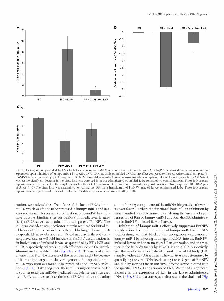
Inhibition of bmnpv-miR-1 effectively suppresses BmNPVproliferation. To confirm the role of bmnpv-miR-1 in BmNPVproliferation, we first blocked the endogenous expression ofbmnpv-miR-1 by injecting its antagomir, LNA, into the BmNPV-infected larvae and then measured Ran expression and the viraltiter in the fat body tissues by RT-qPCR and qPCR, respectively,and the results were normalized against infected fat body (IFB)samples without LNA treatment. The viral titer was determined byquantifying the viral DNA levels using the ie-1 gene of BmNPVand also by scoring OBs in BmNPV-infected larvae injected withthe specific (LNA-1) and scrambled LNA. We found a significantincrease in the expression of Ran in the larvae administeredLNA-1 (Fig. 8A) and a consequent decrease in the viral load (Fig.
FIG 8 Blocking of bmnpv-miR-1 by LNA leads to a decrease in BmNPV accumulation in B. mori larvae. (A) RT-qPCR analysis shows an increase in Ranexpression upon inhibition of bmnpv-miR-1 by specific LNA (LNA-1), while scrambled LNA has no effect compared to the respective control samples. (B)BmNPV titers, determined by qPCR using ie-1 of BmNPV, showed drastic reduction in the virus load when bmnpv-miR-1 was blocked by specific LNA (LNA-1),whereas no significant decrease in the virus load was observed in larvae administered scrambled LNA compared to control samples. Three independentexperiments were carried out in three replicates each with a set of 3 larvae, and the results were normalized against the constitutively expressed 18S rRNA geneof B. mori. (C) The virus load was determined by scoring the OBs from hemolymph of BmNPV-infected larvae administered LNA. Three independentexperiments were performed with a set of 3 larvae. The data are presented as means SD (n � 3).
Viral miRNA Suppresses Its Host’s miRNA Biogenesis
August 2012 Volume 86 Number 15 jvi.asm.org 7875
Dow
nloa
ded
from
http
s://j
ourn
als.
asm
.org
/jour
nal/j
vi o
n 28
Nov
embe
r 20
21 b
y 91
.246
.209
.153
.

8B and C). No such change was observed either in Ran expressionlevels or on viral accumulation in the infected larvae treated withscrambled LNA (Fig. 8B and C).
BmNPV proliferates upon Ran knockdown. To check viralproliferation upon Ran knockdown, dsRNAs against GFP andRan were injected into the BmNPV-infected larvae, and ie-1 ex-pression was quantified by qPCR. As shown in Fig. 9A, a very highviral load was observed in the larvae injected with Ran dsRNAcompared to that seen in the control larvae, and a similar resultwas also observed when OBs were scored in the same samples (Fig.9B). To further confirm that the higher virus load upon Ranknockdown is due to inhibition of the miRNAome of the host andnot to some other function of Ran, we also checked the virus titerin the Dicer-2 knockdown larvae. We noticed a significant in-crease in the virus load in the Dicer-2 knockdown larvae, as wasalso observed upon Ran knockdown (Fig. 9B), suggesting that thehigher virus accumulation is due to suppression of the hostmiRNAs. No observable difference was noticed in the phenotypesof Ran and Dicer-2 knockdown larvae (see Fig. S4 in the supple-mental material). Interestingly, blocking of bmnpv-miR-1 re-sulted in a marked decrease (�8-fold) in the BmNPV load (Fig.8B), whereas knockdown of Ran and Dicer-2 larvae showed only�1.5 and 2.5-fold increases in virus accumulation (Fig. 9B), re-spectively. This may possibly be due to bmnpv-miR-1 targetingother host genes that are involved in host defense. Based on theseresults, we conclude that BmNPV-encoded miRNA, bmnpv-miR-1, combats the host small-RNA-mediated antiviral attack bysuppressing the expression of Ran, an important component ofthe small-RNA transport machinery of the cell, as proposed in themodel shown in Fig. 10.
Viruses are known to evade host defenses in different ways.
Recently, virus-derived miRNAs have emerged as potential mod-ulators of the host immune system. Many mammalian viralmiRNAs have been shown to regulate both the host’s and theirown sets of genes, which otherwise are obstacles to virus entry orproliferation inside the host (15). Similarly, there are many re-ports of cellular miRNAs of the host modulating the expression ofvarious viral genes (20, 28, 36, 37, 55); hence, it is likely that B.mori miRNAs can directly or indirectly inhibit BmNPV prolifer-ation. In the present study, we have shown that Ran is involved insmall-RNA trafficking in B. mori and that BmNPV-encodedmiRNA, bmnpv-miR-1, controls Ran expression by destabilizingits mRNA, followed by translation repression that leads to sup-pression of cellular miRNA production, which in turn results inenhanced virus proliferation.
The contradiction that we came across in our study is a higherlevel of expression of bmnpv-miR-1 in the late stage of infectiondespite effective repression of Ran (Fig. 3B and C). The pertinentquestion here is, when the bmnpv-miR-1 pre-miRNA is not trans-ported out of the nucleus as a result of Ran repression, how such ahigh load of processed bmnpv-miR-1 could still be seen in the cell.This result suggests that BmNPV may deploy a variety of strategiesto effectively replicate in the host cell. One such strategy we pro-pose here is that the baculovirus may not fully block RanGTP-mediated transport by its bmnpv-miR-1, and as a result, transportof host pre-miRNAs in small quantities may still occur in theinfected larvae. This strategy allows the virus to proliferate in thehost cell by keeping the small-RNA-mediated host defense incheck and, at the same time, keeping the larvae viable to enhanceits proliferation. During the late stage of infection, a massivequantity of virus is produced in the host cell, giving rise to a largeramount of virus-derived pre-bmnpv-miR-1, which saturates the
FIG 9 B. mori miRNA suppression increases BmNPV proliferation in larvae. (A) Quantitative-PCR analysis of ie-1 of BmNPV to determine the viral load in Ranand Dicer-2 knockdown larvae. Larvae injected with GFP dsRNA were used as a negative control. Three independent experiments were carried out in threereplicates each with a set of 3 larvae, and the results were normalized against endogenous 18S rRNA. (B) The virus load was determined by scoring the OBs fromhemolymph of Ran knockdown BmNPV-infected larvae. Three independent experiments were performed with a set of 3 larvae. The data are presented asmeans SD (n � 3).
Singh et al.
7876 jvi.asm.org Journal of Virology
Dow
nloa
ded
from
http
s://j
ourn
als.
asm
.org
/jour
nal/j
vi o
n 28
Nov
embe
r 20
21 b
y 91
.246
.209
.153
.

Ran-mediated transport machinery, resulting in higher-leveltransport of pre-bmnpv-miR-1 into the cytoplasm and, at thesame time, limiting the transport of host pre-miRNAs. Thesespeculations draw credence from recent studies that have uncov-ered ingenious and complex strategies employed by the viruses toenhance their accumulation in host cells. For example, adenovi-rus-derived small RNAs massively produced in the late stage ofinfection are able to deplete Dicer expression by saturating expor-tin-5 and abrogating the nuclear export of exportin-5-dependentDicer mRNA. As a result, maturation of small RNA is terminatedby the virus using this strategy (1, 4, 7, 31). Similarly, influenzavirus-encoded NS1 protein has been shown to inhibit the hostmRNA export pathway to produce greater permissiveness by hostcells for influenza virus replication (32). It is also possible that
BmNPV employs alternative strategies, including utilization of anmRNA transport pathway that is independent of Ran (23). In ourprevious study (45), we showed that bmnpv-miR-1 is derivedfrom the coding region of cathepsin, which is expressed heavilyduring the late stage of infection, as it is required for liquefactionof infected larvae (19), and hence, bmnpv-miR-1 may escapeDrosha cleavage and be transported as mRNA to be processedsubsequently by canonical or noncanonical pathways in the cyto-plasm to give rise to mature bmnpv-miR-1 (8, 10, 43, 46, 53).
The important question that remains to be addressed further ishow the virus controls the expression of bmnpv-miR-1. Ran hasbeen shown to be an important component of nucleocytoplasmictransport of small RNAs, as well as proteins (5, 6, 33, 50, 54), andits complete depletion may be lethal, but we did not observe such
FIG 10 The proposed model for bmnpv-miR-1-mediated evasion of the host defense response mounted by small RNAs for effective proliferation of BmNPV ininfected larvae. The model proposed here shows that BmNPV, upon infection of silkworm larvae, encodes an miRNA, bmnpv-miR-1, which represses Ran, animportant component of small-RNA export from the nucleus to the cytoplasm, by binding its 3= UTR. As a result, the small-RNA population is reduced in thecytoplasm, leading to proliferation of the virus.
Viral miRNA Suppresses Its Host’s miRNA Biogenesis
August 2012 Volume 86 Number 15 jvi.asm.org 7877
Dow
nloa
ded
from
http
s://j
ourn
als.
asm
.org
/jour
nal/j
vi o
n 28
Nov
embe
r 20
21 b
y 91
.246
.209
.153
.

a drastic effect in either bmnpv-miR-1- or dsRNA-mediatedknockdown of Ran, although the treated larvae showed stuntedgrowth (see Fig. S4 in the supplemental material). These observa-tions point to the role of miRNAs as fine-tuners of gene expressionthat may not act as phenotype switchers (12, 25, 35, 42). Sinceviruses do not possess any miRNA biogenesis component in theirgenomes, it is interesting to investigate how virus generates itsown miRNAs when it blocks its host’s miRNA transport.
Can we generalize this strategy to other viruses, especially la-tent viruses? As miRNAs are also required for the regulation ofmany cellular genes, which are important for the survival of thehost, inhibition of miRNA processing would have a serious effecton these genes and might lead to cell death. Hence, in cases ofpersistent or latent infection, instead of blocking miRNA biogen-esis, the virus may try to escape the silencing machinery. The an-swers to all these questions might also reveal some of the verycomplicated aspects of virus-governed fine-tuning of host genes.
In summary, our results revealed one of the strategies em-ployed by the virus to modulate miRNA-mediated antiviral hostdefense by producing an miRNA that suppresses Ran, an impor-tant component of miRNA biogenesis. Our results thus provideuseful insights into the multilayered complexity of intricate host-virus interactions and have implications for future applications ininsect virus control and possibly a novel vision for therapeuticstrategies.
ACKNOWLEDGMENTS
J.N. acknowledges financial support from the Department of Biotechnol-ogy, Government of India, under the Centre of Excellence for Geneticsand Genomics of Silkmoths (grant number BT/01/COE/05/12). C.P.S.and J.S. are recipients of senior research fellowships from the Council ofScientific and Industrial Research, India, and the Department of Biotech-nology, Government of India, respectively.
REFERENCES1. Andersson MG, et al. 2005. Suppression of RNA interference by adeno-
virus virus-associated RNA. J. Virol. 79:9556 –9565.2. Arts GJ, Fornerod M, Mattaj IW. 1998. Identification of a nuclear export
receptor for tRNA. Curr. Biol. 8:305–314.3. Bartel DP. 2004. MicroRNAs: genomics, biogenesis, mechanism, and
function. Cell 116:281–297.4. Bennasser Y, et al. 2011. Competition for XPO5 binding between Dicer
mRNA, pre-miRNA and viral RNA regulates human Dicer levels. Nat.Struct. Mol. Biol. 18:323–327.
5. Bischoff FR, Ponstingl H. 1991. Catalysis of guanine nucleotide exchangeon Ran by the mitotic regulator RCC1. Nature 354:80 – 82.
6. Bohnsack MT, Czaplinski K, Gorlich D. 2004. Exportin 5 is a RanGTP-dependent dsRNA-binding protein that mediates nuclear export of pre-miRNAs. RNA 10:185–191.
7. Carnero E, Sutherland JD, Fortes P. 2011. Adenovirus and miRNAs.Biochim. Biophys. Acta 1809:660 – 667.
8. Cheloufi S, Dos Santos CO, Chong MM, Hannon GJ. 2010. A dicer-independent miRNA biogenesis pathway that requires Ago catalysis. Na-ture 465:584 –589.
9. Choy EY, et al. 2008. An Epstein-Barr virus-encoded microRNA targetsPUMA to promote host cell survival. J. Exp. Med. 205:2551–2560.
10. Cullen BR. 2011. Viruses and microRNAs: RISCy interactions with seri-ous consequences. Genes Dev. 25:1881–1894.
11. Enright AJ, et al. 2003. MicroRNA targets in Drosophila. Genome Biol.5:R1.
12. Flynt AS, Lai EC. 2008. Biological principles of microRNA-mediatedregulation: shared themes amid diversity. Nat. Rev. Genet. 9:831– 842.
13. Ghosh Z, Mallick B, Chakrabarti J. 2009. Cellular versus viral microRNAs inhost-virus interaction. Nucleic Acids Res. 37:1035–1048.
14. Gottwein E, Cullen BR. 2010. A human herpesvirus microRNA inhibits
p21 expression and attenuates p21-mediated cell cycle arrest. J. Virol.84:5229 –5237.
15. Gottwein E, Cullen BR. 2008. Viral and cellular microRNAs as determi-nants of viral pathogenesis and immunity. Cell Host Microbe 3:375–387.
16. Gottwein E, et al. 2007. A viral microRNA functions as an orthologue ofcellular miR-155. Nature 450:1096 –1099.
17. Grey F, et al. 2010. A viral microRNA down-regulates multiple cell cyclegenes through mRNA 5=UTRs. PLoS Pathog. 6:e1000967. doi:10.1371/journal.ppat.1000967.
18. Grundhoff A, Sullivan CS. 2011. Virus-encoded microRNAs. Virology411:325–343.
19. Hawtin RE, et al. 1997. Liquefaction of Autographa californica nucleopo-lyhedrovirus-infected insects is dependent on the integrity of virus-encoded chitinase and cathepsin genes. Virology 238:243–253.
20. Hussain M, Asgari S. 2010. Functional analysis of a cellular microRNA ininsect host-ascovirus interaction. J. Virol. 84:612– 620.
21. Hussain M, Taft RJ, Asgari S. 2008. An insect virus-encoded microRNAregulates viral replication. J. Virol. 82:9164 –9170.
22. Jagadeeswaran G, et al. 2010. Deep sequencing of small RNA librariesreveals dynamic regulation of conserved and novel microRNAs and mi-croRNA-stars during silkworm development. BMC Genomics 11:52.
23. Kohler A, Hurt E. 2007. Exporting RNA from the nucleus to the cyto-plasm. Nat. Rev. Mol. Cell Biol. 8:761–773.
24. Krek A, et al. 2005. Combinatorial microRNA target predictions. Nat.Genet. 37:495–500.
25. Krol J, Loedige I, Filipowicz W. 2010. The widespread regulation ofmicroRNA biogenesis, function and decay. Nat. Rev. Genet. 11:597– 610.
26. Kutay U, et al. 1998. Identification of a tRNA-specific nuclear exportreceptor. Mol. Cell 1:359 –369.
27. Lau NC, Lim LP, Weinstein EG, Bartel DP. 2001. An abundant class oftiny RNAs with probable regulatory roles in Caenorhabditis elegans. Sci-ence 294:858 – 862.
28. Lecellier CH, et al. 2005. A cellular microRNA mediates antiviral defensein human cells. Science 308:557–560.
29. Lee RC, Feinbaum RL, Ambros V. 1993. The C. elegans heterochronicgene lin-4 encodes small RNAs with antisense complementarity to lin-14.Cell 75:843– 854.
30. Lewis BP, Burge CB, Bartel DP. 2005. Conserved seed pairing, oftenflanked by adenosines, indicates that thousands of human genes are mi-croRNA targets. Cell 120:15–20.
31. Lu S, Cullen BR. 2004. Adenovirus VA1 noncoding RNA can inhibitsmall interfering RNA and MicroRNA biogenesis. J. Virol. 78:12868 –12876.
32. Lu Y, Qian XY, Krug RM. 1994. The influenza virus NS1 protein: a novelinhibitor of pre-mRNA splicing. Genes Dev. 8:1817–1828.
33. Lund E, Guttinger S, Calado A, Dahlberg JE, Kutay U. 2004. Nuclearexport of microRNA precursors. Science 303:95–98.
34. Macrae IJ, et al. 2006. Structural basis for double-stranded RNA process-ing by Dicer. Science 311:195–198.
35. Mukherji S, et al. 2011. MicroRNAs can generate thresholds in target geneexpression. Nat. Genet. 43:854 – 859.
36. Nuovo GJ, et al. 2010. Strong inverse correlation between microRNA-125b and human papillomavirus DNA in productive infection. Diagn.Mol. Pathol. 19:135–143.
37. Pedersen IM, et al. 2007. Interferon modulation of cellular microRNAs asan antiviral mechanism. Nature 449:919 –922.
38. Pfeffer S, et al. 2004. Identification of virus-encoded microRNAs. Science304:734 –736.
39. Rehmsmeier M, Steffen P, Hochsmann M, Giegerich R. 2004. Fast andeffective prediction of microRNA/target duplexes. RNA 10:1507–1517.
40. Reinhart BJ, et al. 2000. The 21-nucleotide let-7 RNA regulates develop-mental timing in Caenorhabditis elegans. Nature 403:901–906.
41. Rohrmann GF. 2008. Baculovirus molecular biology. National Library ofMedicine, NCBI, Bethesda, MD.
42. Sevignani C, Calin GA, Siracusa LD, Croce CM. 2006. MammalianmicroRNAs: a small world for fine-tuning gene expression. Mamm. Ge-nome 17:189 –202.
43. Shapiro JS, Varble A, Pham AM, Tenoever BR. 2010. Noncanonicalcytoplasmic processing of viral microRNAs. RNA 16:2068 –2074.
44. Shibata S, et al. 2006. Exportin-5 orthologues are functionally divergentamong species. Nucleic Acids Res. 34:4711– 4721.
45. Singh J, Singh CP, Bhavani A, Nagaraju J. 2010. Discovering microRNAsfrom Bombyx mori nucleopolyhedrosis virus. Virology 407:120 –128.
Singh et al.
7878 jvi.asm.org Journal of Virology
Dow
nloa
ded
from
http
s://j
ourn
als.
asm
.org
/jour
nal/j
vi o
n 28
Nov
embe
r 20
21 b
y 91
.246
.209
.153
.
46. Skalsky RL, Cullen BR. 2010. Viruses, microRNAs, and host interactions.Annu. Rev. Microbiol. 64:123–141.
47. Skalsky RL, et al. 2007. Kaposi’s sarcoma-associated herpesvirus encodesan ortholog of miR-155. J. Virol. 81:12836 –12845.
48. Stern-Ginossar N, et al. 2007. Host immune system gene targeting by aviral miRNA. Science 317:376 –381.
49. Swevers L, Liu J, Huvenne H, Smagghe G. 2011. Search for limitingfactors in the RNAi pathway in silkmoth tissues and the Bm5 cell line: theRNA-binding proteins R2D2 and Translin. PLoS One 6:e20250. doi:10.1371/journal.pone.0020250.
50. Wang X, et al. 2011. Dynamic mechanisms for pre-miRNA binding andexport by Exportin-5. RNA 17:1511–1528.
51. Xia T, et al. 2008. EBV microRNAs in primary lymphomas and targetingof CXCL-11 by ebv-mir-BHRF1-3. Cancer Res. 68:1436 –1442.
52. Xu S, Xue C, Li J, Bi Y, Cao Y. 2011. Marek’s disease virus type 1microRNA miR-M3 suppresses cisplatin-induced apoptosis by targetingSmad2 of the transforming growth factor beta signal pathway. J. Virol.85:276 –285.
53. Yang JS, Lai EC. 2011. Alternative miRNA biogenesis pathways andthe interpretation of core miRNA pathway mutants. Mol. Cell 43:892–903.
54. Yi R, Qin Y, Macara IG, Cullen BR. 2003. Exportin-5 mediates thenuclear export of pre-microRNAs and short hairpin RNAs. Genes Dev.17:3011–3016.
55. Zhang GL, et al. 2010. Suppression of hepatitis B virus replication bymicroRNA-199a–3p and microRNA-210. Antiviral Res. 88:169 –175.
56. Zhao Y, et al. 2009. A functional microRNA-155 ortholog encoded by theoncogenic Marek’s disease virus. J. Virol. 83:489 – 492.
Viral miRNA Suppresses Its Host’s miRNA Biogenesis
August 2012 Volume 86 Number 15 jvi.asm.org 7879
Dow
nloa
ded
from
http
s://j
ourn
als.
asm
.org
/jour
nal/j
vi o
n 28
Nov
embe
r 20
21 b
y 91
.246
.209
.153
.