
9: Infections in the returned traveller · Pre-travel health assessment and planning can ......
8
212 MJA Vol 177 19 August 2002 Remember to consider serious yet treatable diseases, such as malaria STUDIES SHOW that almost 50% of travellers returning to Europe and North America from the tropics experience health problems related to their travel, and that 8% consult doctors either abroad or on return. 1 Figures are not availa- ble for Australian travellers but are probably similar. The most common diseases in travellers to the tropics are travellers’ diarrhoea (35% incidence per month of travel), followed by acute respiratory infections (1.4%), giardiasis (0.7%) and other infections (including hepatitis, amoebiasis and gonorrhoea). 1 Australian travellers increasingly visit tropical and developing countries and may present with infections that are rare in Australia. Pre-travel health assessment and planning can prevent or ameliorate some of these problems (Box 1). Useful informa- tion about travel-related infections can be found at the websites of the World Health Organization (www.who.int/ ith), United States Centers for Disease Control and Preven- tion (www.cdc.gov/travel) and the Department of Public Health and Tropical Medicine at James Cook University, Townsville (www.jcu.edu.au/school/phtm/PHTM/ putravel.htm). Clinical presentations Over 90% of infections related to short-term travel present within six months of return. Diseases with long latent periods or potential for lifetime persistence are rare after short-term travel, but more common in those who have lived abroad or were born overseas. The usual presentation of a returned traveller is with a particular syndrome — fever, respiratory infection, diarrhoea, eosinophilia, or skin or soft tissue infection — or for screening for asymptomatic infec- tion. When assessing a returned traveller, it is important to obtain: ■ a complete travel history, including dates and places visited, and potential exposures to exotic diseases (eg, travel to rural areas, freshwater exposure, insect bites and sexual contacts); and ■ time of symptom onset in relation to travel, which may help eliminate diseases with different incubation periods (eg, fever beginning over 10 days after departure from an endemic area is unlikely to be dengue, which has a short incubation period). Incubation periods of common travel- related infections are shown in Box 2. Fever Between 2% and 12% of travellers suffer from a febrile illness while away or on return. 1,3 Malaria is the most common diagnosis 3-5 and is particularly frequent in travellers to West Africa who have taken no prophylaxis (2%). 1 Among people arriving in Australia, the incidence of malaria is highest in those who have been in Nigeria (1.3%), the Solomon Islands (1.1%), Ghana (0.6%) and Papua New Guinea (0.4%). 6 Other causes of fever are shown in Box 3. Younger travellers are more likely to have an exotic infection than the elderly, as their travel tends to be more adventurous. Older travellers are more likely to have a febrile complication of underlying disease, such as endocarditis or pneumonia. Evaluation and initial management of fever in a returned traveller is outlined in Box 4. It is important to exclude falciparum malaria, which is potentially fatal but treatable. Once this is excluded, patients require hospital admission only if severely ill. Viral haemorrhagic fevers (eg, Lassa fever, and Marburg and Ebola virus infections) usually present as undifferentiated fever, but are rare in returned travellers. If suspected (recent travel to an African country with endemic 9: Infections in the returned traveller David F M Looke and Jennifer M B Robson Series Editors: M Lindsay Grayson, Steven L Wesselingh Infection Management Services – Southern Queensland, Princess Alexandra Hospital, Woolloongabba, QLD. David F M Looke, FRACP, FRCPA, MMedSci (Clin Epidemiol), Infectious Diseases Physician. Sullivan Nicolaides Pathology, Taringa, QLD. Jennifer M B Robson, FRACP, FRCPA, FACTM, Microbiologist. Reprints will not be available from the authors. Correspondence: Dr J M B Robson, Sullivan Nicolaides Pathology, 134 Whitmore Street, Taringa, QLD 4068. [email protected] Abstract ■ The usual presentation of a returned traveller is with a particular syndrome — fever, respiratory infection, diarrhoea, eosinophilia, or skin or soft tissue infection — or for screening for asymptomatic infection. ■ Fever in a returned traveller requires prompt investigation to prevent deaths from malaria; diagnosis of malaria may require up to three blood films over 36–48 hours. ■ Diarrhoea is the most common health problem in travellers and is caused predominantly by bacteria; persistent diarrhoea is less likely to have an infectious cause, but its prognosis is usually good. ■ While most travel-related infections present within six months of return, some important chronic infections may present months or years later (eg, strongyloidiasis, schistosomiasis). ■ Travellers who have been bitten by an animal require MJA 2002; 177: 212-219 evaluation for rabies prophylaxis. MJA Practice Essentials Infectious Diseases
-
Upload
nguyenkhanh -
Category
Documents
-
view
226 -
download
0
Transcript of 9: Infections in the returned traveller · Pre-travel health assessment and planning can ......

212 MJA Vol 177 19 August 2002
Infectious Diseases MJA Practice Essentials
The Medical Journal of Australia ISSN: 0025-729X 19 August 2002177 4 212-219©The Medical Journal of Australia 2002 www.mja.com.auMJA Practice Essentials — Infectious Diseases
Remember to consider serious yet treatable diseases, such as malaria
STUDIES SHOW that almost 50% of travellers returning toEurope and North America from the tropics experiencehealth problems related to their travel, and that 8% consultdoctors either abroad or on return.1 Figures are not availa-ble for Australian travellers but are probably similar. Themost common diseases in travellers to the tropics aretravellers’ diarrhoea (35% incidence per month of travel),followed by acute respiratory infections (1.4%), giardiasis(0.7%) and other infections (including hepatitis, amoebiasisand gonorrhoea).1 Australian travellers increasingly visittropical and developing countries and may present withinfections that are rare in Australia.
Pre-travel health assessment and planning can prevent orameliorate some of these problems (Box 1). Useful informa-tion about travel-related infections can be found at thewebsites of the World Health Organization (www.who.int/ith), United States Centers for Disease Control and Preven-tion (www.cdc.gov/travel) and the Department of PublicHealth and Tropical Medicine at James Cook University,Townsville (www.jcu.edu.au/school/phtm/PHTM/putravel.htm).
Clinical presentations
Over 90% of infections related to short-term travel presentwithin six months of return. Diseases with long latentperiods or potential for lifetime persistence are rare aftershort-term travel, but more common in those who have livedabroad or were born overseas. The usual presentation of areturned traveller is with a particular syndrome — fever,respiratory infection, diarrhoea, eosinophilia, or skin or softtissue infection — or for screening for asymptomatic infec-tion.
When assessing a returned traveller, it is important toobtain:■ a complete travel history, including dates and placesvisited, and potential exposures to exotic diseases (eg, travelto rural areas, freshwater exposure, insect bites and sexualcontacts); and
■ time of symptom onset in relation to travel, which mayhelp eliminate diseases with different incubation periods(eg, fever beginning over 10 days after departure from anendemic area is unlikely to be dengue, which has a shortincubation period). Incubation periods of common travel-related infections are shown in Box 2.
Fever
Between 2% and 12% of travellers suffer from a febrile illnesswhile away or on return.1,3 Malaria is the most commondiagnosis3-5 and is particularly frequent in travellers to WestAfrica who have taken no prophylaxis (2%).1 Among peoplearriving in Australia, the incidence of malaria is highest inthose who have been in Nigeria (1.3%), the Solomon Islands(1.1%), Ghana (0.6%) and Papua New Guinea (0.4%).6
Other causes of fever are shown in Box 3. Younger travellersare more likely to have an exotic infection than the elderly, astheir travel tends to be more adventurous. Older travellers aremore likely to have a febrile complication of underlyingdisease, such as endocarditis or pneumonia.
Evaluation and initial management of fever in a returnedtraveller is outlined in Box 4. It is important to excludefalciparum malaria, which is potentially fatal but treatable.Once this is excluded, patients require hospital admissiononly if severely ill. Viral haemorrhagic fevers (eg, Lassa fever,and Marburg and Ebola virus infections) usually present asundifferentiated fever, but are rare in returned travellers. Ifsuspected (recent travel to an African country with endemic
9: Infections in the returned traveller
David F M Looke and Jennifer M B Robson
Series Editors: M Lindsay Grayson, Steven L Wesselingh
Infection Management Services – Southern Queensland, Princess Alexandra Hospital, Woolloongabba, QLD.David F M Looke, FRACP, FRCPA, MMedSci (Clin Epidemiol), Infectious Diseases Physician. Sullivan Nicolaides Pathology, Taringa, QLD.Jennifer M B Robson, FRACP, FRCPA, FACTM, Microbiologist. Reprints will not be available from the authors. Correspondence: Dr J M B Robson, Sullivan Nicolaides Pathology, 134 Whitmore Street, Taringa, QLD 4068. [email protected]
Abstract
■ The usual presentation of a returned traveller is with a particular syndrome — fever, respiratory infection, diarrhoea, eosinophilia, or skin or soft tissue infection — or for screening for asymptomatic infection.
■ Fever in a returned traveller requires prompt investigation to prevent deaths from malaria; diagnosis of malaria may require up to three blood films over 36–48 hours.
■ Diarrhoea is the most common health problem in travellers and is caused predominantly by bacteria; persistent diarrhoea is less likely to have an infectious cause, but its prognosis is usually good.
■ While most travel-related infections present within six months of return, some important chronic infections may present months or years later (eg, strongyloidiasis, schistosomiasis).
■ Travellers who have been bitten by an animal require
MJA 2002; 177: 212-219
evaluation for rabies prophylaxis.
MJA Practice Essentials Infectious Diseases

MJA Vol 177 19 August 2002 213
MJA Practice Essentials Infectious Diseases
disease or a known outbreak), then public health authoritiesmust be notified to allow appropriate action.7
Malaria
Malaria caused by Plasmodium falciparum generally presentswithin six weeks of return from an endemic area, althoughonset may be delayed by prophylactic mefloquine.8 Incontrast, malaria caused by Plasmodium vivax may presentmany months after exposure. No signs or symptoms arespecific to malaria, although fever is invariable. Occasion-ally, malaria mimics acute gastroenteritis, acute abdomen,pneumonia and meningoencephalitis. As prophylactic anti-malarials do not eliminate the possibility of malaria, appro-priate testing should always be done.
Malaria is diagnosed by detecting the parasite in a bloodfilm or circulating malarial antigen. Initial blood films may benegative, and repeat films should be taken every six to 12hours for 36–48 hours before malaria can be confidentlyexcluded. As untreated falciparum malaria can be fatal within24–48 hours of presentation, particularly in children, thediagnosis should be pursued urgently (case history, Box 5).
Patients with falciparum malaria require hospital admis-sion until the disease is clearly under control. All should betreated as if they have chloroquine-resistant disease.Uncomplicated disease is treated with oral quinine pluseither doxycycline or pyrimethamine–sulfadoxine, or, alter-natively, mefloquine as a single agent, or the combination ofatovaquone and proguanil.9 Severe malaria requires intrave-nous quinine.
Malaria caused by the Plasmodium species vivax, ovale andmalariae can still usually be treated effectively with oral
chloroquine followed by primaquine to eradicate the hepaticstages of P. vivax and P. ovale. Patients receiving primaquineshould have prior testing for glucose-6-phosphate dehydro-genase deficiency, as the drug may cause severe haemolyticanaemia if this is present. However, as chloroquine-resistantP. vivax has now been reported throughout South-EastAsia,10 specialist advice may be required, particularly forrelapsing disease.9
Dengue
Infected travellers have periodically introduced this commonmosquito-borne flavivirus disease to northern Australia. Itsrange is currently spreading as one of its vectors, Aedesalbopictus, becomes acclimatised to the urban environment.11
Clues to diagnosis of dengue are the short incubationperiod (four to seven days), maculopapular rash, thrombo-
1: Pre-travel health assessment and planning
This is recommended at least six weeks before departure and should include:Assessment of fitness to travel: pre-existing conditions (eg, diabetes, heart disease, pregnancy, immunocompromise); psychological state (eg, depression, seeking “way-out” of problems, alcohol, drugs); need for specialised medications, with doctor’s letter specifying this.Full travel history and risk assessmentUpdate of routine vaccinations: measles–mumps–rubella; diphtheria; pertussis; tetanus; hepatitis B; poliomyelitis; influenza; and pneumococcal vaccines.Routine vaccinations for travel to developing countries: hepatitis A and (if travelling for more than two weeks) typhoid.“Special circumstance” vaccinations: yellow fever; meningococcus; rabies; and Japanese B encephalitis.Prophylactic drugs when indicated: antimalarials (mefloquine, doxycycline, chloroquine, proguanil or malarone); and antibiotics for travellers’ diarrhoea (if immunocompromised or other special circumstances).Advice on prevention: travellers’ diarrhoea; schistosomiasis and fresh-water exposure in Africa; traffic-accident injuries; sexually transmitted diseases; sunburn and sunstroke; insect bites; scuba-diving accidents; deep venous thrombosis; and jet lag.Advice and drugs for medical or first-aid kit: including diarrhoea kit (loperamide, antibiotic [norfloxacin], thermometer and [for long-term traveller or trekker] tinidazole).Advice on travel health insurance and assistance
2: Incubation periods of travel-related infections2
Short (<10 days)
Intermediate (10–21 days)
Long (>21 days)
InfluenzaDengueYellow feverPlagueParatyphoid feverMediterranean spotted feverAfrican tick-bite feverRocky Mountain spotted fever
MalariaViral haemorrhagic fevers
Typhoid feverScrub typhusQ feverRelapsing fever (Borrelia spp.)
African trypanosomiasis
MalariaHepatitis A, B, C, ERabiesSchistosomiasisLeishmaniasisAmoebic liver abscess
TuberculosisFilariasisBrucellosis
3: Causes of fever in Canadian and Australian studies of returned travellers
Percentage of fever cases
DiagnosisCanada(n=587)4
Australia(n=232)5
Undiagnosed 25 9
Malaria 32 27
Respiratory infection* 11 18
Hepatitis 6 3
Diarrhoeal illness 5 14
Pyelonephritis 4 1
Dengue 2 8
Enteric fever 2 3
Epstein–Barr virus infection 2 0.4
Rickettsial infection 1 2
Amoebiasis 1 1
Tuberculosis 1 0.4
Acute HIV infection 0.3 0.4
Other infections 4 10
Other causes 4 3
* Including upper respiratory tract infection, bronchitis and pneumonia.

214 MJA Vol 177 19 August 2002
Infectious Diseases MJA Practice Essentials
cytopenia and leukopenia. The last three features also occurin some rickettsial diseases (eg, scrub typhus), HIV serocon-version (Box 6), Q fever, meningococcal disease, toxic shocksyndrome, measles, secondary syphilis and rubella. Diagno-sis depends on virus isolation from blood, amplification ofviral nucleic acid or serological results (dengue IgM or IgGseroconversion).
There is as yet no effective antiviral therapy, and support-ive treatment is all that can be offered. Spontaneous resolu-tion is usual. Occasionally, severe depression complicatesthe recovery phase.
Enteric fever (typhoid and paratyphoid)
Enteric fever is the clinical syndrome caused by Salmonellatyphi or “paratyphi” Salmonella species. Fever is the domi-nant symptom, with gastrointestinal symptoms variablypresent. Typhoid vaccine confers about 70% protectionagainst S. typhi, so does not rule out the diagnosis. Diagno-sis depends on culture, as serological testing is unreliable,particularly if vaccine has been given.
In a patient with prolonged, undifferentiated fever, empir-ical therapy for enteric fever with quinolones (eg, cipro-floxacin) can be considered once malaria has been ruled out.Quinolone-resistant strains of Salmonella have now beenreported, and, in children, third-generation cephalosporins(eg, ceftriaxone or cefotaxime) may be required.9
Rickettsial diseases
These include scrub typhus, African tick bite fever andMediterranean and Rocky Mountain spotted fever. All aretransmitted by arthropod (mostly tick) bites, although manypatients have not noted a bite. Rickettsial disease should besuspected in patients with fever and a rash (particularly if aneschar is present) who have travelled to an endemic area.Diagnosis is by serological testing. As acute serologicaltesting is usually negative, empirical therapy with doxycy-cline should be considered while awaiting results of tests onconvalescent sera.
Respiratory infections
About 20%–40% of travellers have respiratory symptomswhile away or on return.12,13 The most common causes areallergy and viral infection of the upper respiratory tract.
Tuberculosis (defined as skin test conversion) occurs in anestimated 2% of long-term travellers to high risk areas, suchas sub-Saharan and North Africa, South and East Asia, theMiddle East and Central and South America.14 Risk is lowerduring short-term holidays. Infants and children staying inthese areas for more than three months are recommended tohave bacille Calmette–Guérin (BCG) vaccination, as itreduces disease severity and mortality. For adults, therecommendations are less clear. Possible strategies include
4: Evaluation and initial management of fever in a returned traveller*
PCR=polymerase chain reaction.*Evaluation should also include the differential diagnoses that would be considered in a non-traveller with fever.†Travel to high-risk area, rural or prolonged travel, non-compliance with prophylaxis.
Suspected febrile illness in a returned traveller
Confirm fever
Severe sepsis(confusion, collapse, cyanosis, tachypnoea, hypotension,
neck stiffness, peritonism or digital gangrene)No features of severe sepsis
Resuscitation if shockedBlood culturesMalaria filmsPenicillin or ceftriaxone (if meningococcal disease likely)
History: Travel and fever onset (compare with typical incubation periods)
Pattern of fever: Occasionally helpful (eg, second-daily paroxysm in vivax malaria)
Focal features: Neck stiffness, cellulitis, abdominal tenderness, pulmonary consolidation
Investigations: Full blood count, liver function tests, blood cultures (two), chest x-ray, urine microscopy and culture, baseline serologicaltests, specific investigations for focal disease
Malaria possible† Rash Respiratory symptoms Fever > 7 days,malaria ruled out
Thick and thin malariafilms (if initially negative,repeat 3 times)
Urgent hospital transfer
Consider dengue orrickettsial disease- Serological tests- Consider empirical doxycycline for rickettsia Pulmonary
consolidation
Return within 3days from countrywith acute influenza
Consider unusual causes of pneumonia(eg, Legionnaire's disease, melioidosis)- If severe, give empirical treatment, including macrolide or quinolone
Consider enteric fever- Blood, stool and urine cultures- Consider empirical quinolone or third- generation cephalosporin
Consider influenza- Rapid antigen or PCR test
Jaundice
Consider- Acute hepatitis (eg, A, B, C, E, Epstein-Barr virus, dengue, Q fever): serological tests- Acute cholangitis (stones, liver fluke): blood cultures, ultrasound and stool examination- Liver abscess (amoebic, pyogenic): blood cultures, serological tests
Plasmodium vivax,ovale or malariae
Plasmodium falciparum

MJA Vol 177 19 August 2002 215
MJA Practice Essentials Infectious Diseases
pre-travel BCG vaccination or Mantoux screening beforeand after travel, followed by prophylactic isoniazid for thosewhose skin test has converted. However, a recent analysissuggested that, given the problems of compliance andadverse effects with these strategies, it is more rational toawait the rare cases of clinically overt travel-associatedtuberculosis and treat appropriately as they arise.15
Travellers’ diarrhoea
Acute diarrhoea is common in travellers to developingcountries.16 Despite the advice “cook it, boil it, peel it orforget it”, dietary transgressions are usually unavoidable.Most cases of travellers’ diarrhoea are caused by bacterialcontamination of food, particularly with enterotoxigenicEscherichia coli (ETEC). Other causes are shown in Box 7. Itis rarely life-threatening. Mean duration of symptoms is fourdays. Up to 60% of people complain of cramps, 15% ofbloody stools, and 10% of fever or vomiting.
Preventive strategies include education about safe foodand beverages, water purification, and chemoprophylaxiswith antibiotics (eg, trimethoprim–sulfamethoxazole or nor-
floxacin administered once daily). Antibiotics are 70%–95%effective in preventing travellers’ diarrhoea,16 but should beconsidered only in selected high-risk, short-term travellers— those who cannot tolerate a brief illness (eg, athletes,business and political travellers) and those with increasedsusceptibility (eg, because of achlorhydria, gastrectomy,history of repeated severe travellers’ diarrhoea, immuno-compromise or chronic illness). Prophylaxis is not recom-mended for healthy travellers. As most travellers’ diarrhoea
6: Case history — fever with rash after a tropical holiday
History: A 34-year-old man presented 10 days after returning from a six-week holiday in South-East Asia. He had an eight-day history of malaise, chills, headache, sore throat and generalised rash. He had stayed mostly at five-star hotels, but reported many mosquito bites.Examination: He had fever, a macular rash and generalised lymphadenopathy with mild splenomegaly, but no meningism and no eschar present.Investigations: Full blood examination revealed lymphocytosis with numerous atypical lymphocytes and thrombocytopenia. Blood cultures and malaria films were negative. Liver function tests revealed marginally elevated serum transaminase levels. Serological testing revealed past infection with Epstein–Barr virus and cytomegalovirus and was negative for Q fever, dengue, rubella, measles and rickettsial infection.Management: The presumptive diagnosis was dengue, and supportive treatment was instituted. However, symptoms persisted for the next three weeks.Dengue serological testing was repeated and found to be still negative.A repeat sexual history revealed unprotected intercourse with a number of men during the holiday. HIV antibody tests were positive on enzyme immunoassay and indeterminate on western blot. A p24 antigen test was positive. HIV seroconversion illness was diagnosed, and appropriate counselling and therapy offered.■ Acute HIV infection may mimic several tropical diseases,
including dengue and typhus, as well as infectious mononucleosis.
■ A sexual history is important in assessing infection risk in any traveller with a febrile illness.
■ Early diagnosis of HIV infection is important in preventing transmission, as well as allowing the correct timing of antiretroviral therapy.
5: Case history — fever and confusion in a traveller to Papua New Guinea
History: A 42-year-old geologist presented 14 days after return from a 6-week field trip to Papua New Guinea. He had a six day history of high fevers and rigors. On the day of presentation, he had become vague and confused. He had taken antimalarials as prophylaxis, but ceased when he found that local people did not take them.Examination: His temperature was 40oC, pulse rate 140 bpm, respiratory rate 28 per minute, and blood pressure 100/60 mmHg. He had dry mucous membranes, mild jaundice, pallor, spleno-megaly and generalised crackles in both lungs. He was drowsy and disoriented but had no meningism.Investigations: Full blood examination revealed reduced haemo-globin concentration (65 g/L; reference range [RR], 130–180 g/L), leukopenia (2.5 x 10⁹/L; RR, 3.5–10 x 109/L) and thrombocytopenia (10 x 109/L; RR, 150–400 x 109/L). Malaria films were positive for Plasmodium falciparum, with 18% of red blood cells parasitised. Liver function tests revealed raised levels of bilirubin (60 �mol/L; RR, 3–20 �mol/L) and lactate dehydrogenase (489 U/L; RR, 100–225 U/L). Serum creatinine level was also raised (0.28 mmol/L [RR, 0.04–0.12 mmol/L]). Blood gas measurement revealed pH 7.1, PO2 110 mmHg (RR, 70–80 mmHg) and PCO2 15 mmHg (RR, 36–45 mmHg).Management: Severe falciparum malaria was diagnosed. The patient was admitted to the intensive care unit, treated with ventilation, renal dialysis, intravenous quinine and exchange transfusion. Despite therapy and full support, he died on Day 5 of the admission.■ Malaria should be excluded in any traveller with fever, even if
prophylactic antimalarials have been taken, as their efficacy is less than 100%.
■ Severe malaria is defined by any degree of altered consciousness, focal neurological signs, jaundice, oliguria, severe anaemia, hypoglycaemia, hypotension, >2% of red blood cells parasitised or absolute parasite count >100000/mL, vomiting or acidosis.
■ Severe malaria requires treatment with intravenous quinine and admission to an intensive care unit, as complications may occur suddenly, and quinine therapy must be monitored closely. Even with intensive-care treatment, mortality is high.
7: Causes of travellers’ diarrhoea (% of cases)16
Bacteria (> 80%)Salmonella spp.Campylobacter spp.Shigella spp.Aeromonas spp.Plesiomonas spp.Yersinia spp.Vibrio choleraeVibrio parahaemolyticus and other non-cholera vibriosEscherichia coli (eg, enterotoxigenic strains)Clostridium perfringensClostridium difficileEdwardsiella tarda
Viruses (0–36%)RotavirusAdenovirusCalicivirusesAstrovirusesNorwalk virus
Parasites (0–20%)Giardia lambliaCryptosporidium parvumCyclospora cayetanensisEntamoeba histolyticaIsospora belliMicrosporidiaBlastocystis hominis (pathogenicity controversial)17

216 MJA Vol 177 19 August 2002
Infectious Diseases MJA Practice Essentials
occurs during travel, patients should be instructed in self-management (Box 8).
When diarrhoea persists for 14 days or more (1%–3% ofcases18), bacterial causes are less likely. Parasite infestationsare more common, but the spectrum of causes is muchwider and includes non-infective causes, such as lactoseintolerance, irritable-bowel syndrome, post-infectious mal-absorption, bile-salt enteritis, inflammatory bowel disease,malignancy, coeliac disease and drugs. An approach toevaluation of persistent diarrhoea in travellers is shown inBox 9. It is important to consult with the pathologylaboratory, as tests for some potential causes may not beroutine.
Some patients have continuing symptoms (loose stools,urgency, and sensitivity to certain foods resulting in flatus ordiarrhoea, similar to irritable-bowel syndrome) despiteempirical therapy and negative results on gastroenterologi-cal workup. They are best treated symptomatically andreassured that the prognosis is good.18
Eosinophilia
Eosinophilia (blood eosinophil count �0.6 x 10⁹/L) in areturned traveller may be an isolated finding or associatedwith symptoms such as cough, fatigue, skin lesions (urti-caria), abdominal discomfort and diarrhoea. A recent Ger-man study of 14 000 travellers returning from the tropicsfound eosinophilia in almost 5%, and a definite diagnosiswas made in a third of these.19 The higher the grade ofeosinophilia, the more likely a travel-related diagnosis, par-
ticularly helminth infestation (eg, strongyloidiasis, filariasis,hookworm, schistosomiasis, cutaneous larva migrans andascariasis).19 However, less than half of those with helminthinfestation in the total population had blood eosinophilia.The absence of eosinophilia does not exclude parasites, aseosinophilia generally occurs only during the tissue invasivestages of worm development.
Initial investigation of eosinophilia should include threestool examinations for ova, cysts and parasites to detect themore common gastrointestinal helminths, whose eggs maybe excreted intermittently. Patients may report passingworms in faeces or coughing them up, and any macroscop-ically visible worms (likely to be ascarids or tapeworm)should be sent for laboratory identification. Specific sero-logical testing is available for schistosomiasis, strongyloid-iasis, filariasis, echinococcosis, toxocariasis and angio-strongyliasis.
Initial empirical treatment with a broad-spectrum ant-helmintic is reasonable after stool collection. Optionsinclude mebendazole, pyrantel and albendazole.9 As the firsttwo are not effective for strongyloidiasis, tapeworm infectionor schistosomiasis, a specific diagnosis should be attemptedif the eosinophil count does not return to normal andtreated appropriately. Albendazole is available from thePharmaceutical Benefits Scheme on authority for specificindications.
Strongyloidiasis
Infection is acquired by penetration through intact skin offilariform larvae of Strongyloides stercoralis in faecally con-
8: Self-management of acute travellers’ diarrhoea
*Contraindicated in children under 16 years and pregnant women. However, for children with severe diarrhoea, the benefits of quinolones used for 1–3 days may outweigh the risks.†Although resistance to trimethoprim–sulfamethoxazole is widespread in some areas, prescription is less restricted for these antibiotics than for quinolones.
Sudden onset of nausea, diarrhoea and abdominal cramps
Diarrhoea persists, particularly if severe, worsening or bloody
Rehydration (eg, oral rehydration solutions)
Seek medical advice (consider protozoal infection, such as Cyclospora spp. or amoebae, drug-resistant organism or other cause)
Antimicrobial therapy for 3 days- Norfloxacin (400 mg twice daily);*- Ciprofloxacin (500 mg twice daily);* - Trimethoprim-sulfamethoxazole (160 mg/800 mg twice daily);† or- Azithromycin (500 mg [10 mg/kg in children] once daily) (alternative for quinolone-resistant bacteria, children and pregnant women)
Symptoms tolerable Symptoms distressing
Moderate (�3 loose stools / 24h)Mild(1-2 loose stools / 24h,
symptoms tolerable)
Moderate-severe (�3 loosestools / 24h, incapacitating symptoms,
fever or bloody diarrhoea)
No treatment or loperamide (4 mg,then 2 mg after each loose stool to amaximum of 16 mg/day; do not usein children aged under 2 years)
Loperamide
Symptoms persist over 2 days
Loperamide plus antimicrobialtherapy (double dose as initial dose;if no improvement after 12h, continuewith standard dose for 3 days)
Antimicrobial therapy (double doseinitially, then standard dose for3 days)Avoid loperamide if dysentericsymptoms (fever, bloody diarrhoea)

MJA Vol 177 19 August 2002 217
MJA Practice Essentials Infectious Diseases
9: Suggested investigation and management of persistent diarrhoea in a returned traveller
Diarrhoea persisting over 14 days
History and examination- Travel history and symptom onset in relation to travel- Antimicrobial therapy in previous month (consider Clostridium difficile), other medications, laxative abuse- Underlying conditions (eg, HIV infection)- Fever (consider malaria)- Stool quality (eg, semiformed, bulky, greasy and malodorous suggests malabsorption; bloody stools suggest amoebiasis, inflammatory bowel disease)- Relationship of symptoms to food (eg, lactose, fatty foods)- Weight loss- Associated symptoms (eg, extraintestinal manifestations of inflammatory bowel disease)
Investigations- Microscopy and culture of three stool samples- Test for Clostridium difficile toxin- Full blood count, blood film and differential, erythrocyte sedimentation rate, serum electrolyte levels, renal and liver function tests- Consider serological tests for parasites (strongyloidiasis, amoebiasis, schistosomiasis) and coeliac disease; blood cultures; malaria blood films; thyroid function tests
Positive test results* Negative test results
Appropriate treatment- Antibacterial: norfloxacin (400 mg twice daily for 5 days)† or - Antigiardial: tinidazole (2 g [50 mg/kg in children] as single dose) or metronidazole (400 mg [5-10 mg/kg in children] three times daily for 5-7 days)
(Administer antibacterial therapy first if both are to be tried)
Empirical therapy
No response
Refer for gastrointestinal assessment to exclude non-infectious causes(eg, bowel malignancy, especially if patient aged over 50 years)
Symptoms persist
Treat symptomatically and reassure of good long-term prognosis
No cause found
Modify diet (avoid all lactose or pre-treat with lactase; reduce caffeine intake; increase dietary bulk)
* Often Blastocystis hominis is the only positive finding, but its pathogenicity is controversial. If symptoms persist after other causes have been excluded, a trial of therapy for this organism may be indicated: metronidazole 400 mg (10 mg/kg in children) three times daily for 7–10 days or trimethoprim–sulfamethoxazole 160/800 mg orally twice daily for 7 days.† In children, trimethoprim–sulfamethoxazole (2.5–10 mL [concentration, 40/200 mg per 5 mL] twice daily) or azithromycin (10 mg/kg once daily) for 5 days.
10: Case history — genital ulceration two years after a trip across Africa
Presentation: A 28-year-old woman presented with a non-healing genital ulcer. She had travelled overland through Africa two years previously.
Examination: An indurated 1 cm ulcer was present on the inner aspect of the left labia majoris.
Investigations: Polymerase chain reaction tests were negative for herpes simplex, gonorrhoea and chlamydia; syphilis serological testing and a swab for Haemophilus ducreyi (chancroid) were also negative. Skin biopsy of the lesion revealed a granuloma surrounding a schistosome egg (pictured). Schistosoma haematobium eggs were detected in terminal urine collected between midday and 2 pm (pictured). Eosinophil count in blood was 1.2 x 10⁹/L (reference range, < 0.6 x 10⁹/L).Serological testing by indirect hemagglutination assay was positive for schistosomiasis (titre, 512).
Management: She was treated with praziquantel and six months later remained well. Her asymptomatic travelling companions were found to be positive for schistosomiasis on serological screening and were also treated.
Biopsy of genital ulcer, showing granuloma-tous reaction surrounding a schistosome egg (haematoxylin and eosin stain; original magnification, x 400).
Schistosoma haematobium egg in urine sediment (phase contrast; original magnifi-cation x 100).
■ Schistosomal infestation may persist and present long after leaving an endemic area.
■ Attack rates for infection are high, and diagnosis in a traveller should prompt screening of all fellow travellers, regardless of presence or absence of symptoms.
■ Serological tests are the most sensitive method of screening, but are not species-specific. Indirect hemagglutination titres �64 suggest infection, although antibodies may take months or years to fall to low or undetectable levels.
■ Diagnosis is complicated by the long pre-patent interval (time between infection and detection of eggs in urine or faeces). As antibodies develop against egg antigens, not adult worms, antibody testing gives false negative results if done too early. Tests may need to be repeated for up to six months after exposure.
■ Identification of schistosome eggs in terminal urine or faeces is required to differentiate the species causing infection. At least two specimens are recommended.
218 MJA Vol 177 19 August 2002
Infectious Diseases MJA Practice Essentials
taminated soil. Autoinfection may occur, and no intermedi-ate host is necessary. Infection can persist for decades andoccasionally presents as hyperinfection syndrome afterimmunosuppression.
Strongyloidiasis may be diagnosed with stool microscopyand special culture techniques, as well as serological testing.Effective treatment includes ivermectin, albendazole andthiabendazole.9 Repeat or prolonged treatment may benecessary in patients who are immunosuppressed.
Schistosomiasis
Schistosomiasis detection is increasingly common in Aus-tralian travellers.20 Although the parasite is endemic inAfrica, the Middle East, Latin America, and limited areas inAsia, the greatest risk of infection appears to come fromexposure to freshwater in Africa, especially Lake Malawi.Infection results from penetration of intact skin by larvaeliberated from snail intermediate hosts.
Most infected travellers are asymptomatic and at low riskof complications because of low parasite burdens, althoughend-organ damage may result from egg deposition. Otherpresentations include swimmers’ itch (a papular pruriticdermatitis at the time of exposure) and Katayama fever,which is associated with the migration of schistosome larvaethroughout the body (symptoms include fever, chills,malaise, headache, cough, abdominal pain, diarrhoea, urti-caria, and occasionally neurological disorders).
Once egg deposition begins, clinical features includehaematuria, haematospermia, urgency, frequency, terminaldysuria, salpingitis, prostatitis and genital ulcers (case his-tory, Box 10). More serious but rare complications areparaparesis, paraplegia and mass cerebral lesions. Immi-grants and refugees from endemic countries are likely tohave much heavier infection loads and may present with thecomplications of chronic fibrosis, such as ureteric obstruc-tion and portal hypertension.
The standard treatment is praziquantel (20 mg/kg orallyfor two doses, four hours apart). About 10%–15% ofpatients require re-treatment.
Skin and soft tissue infections
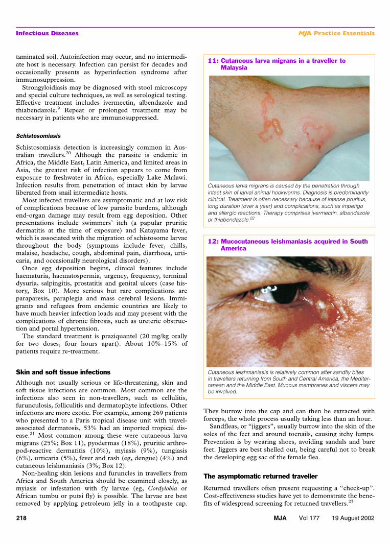
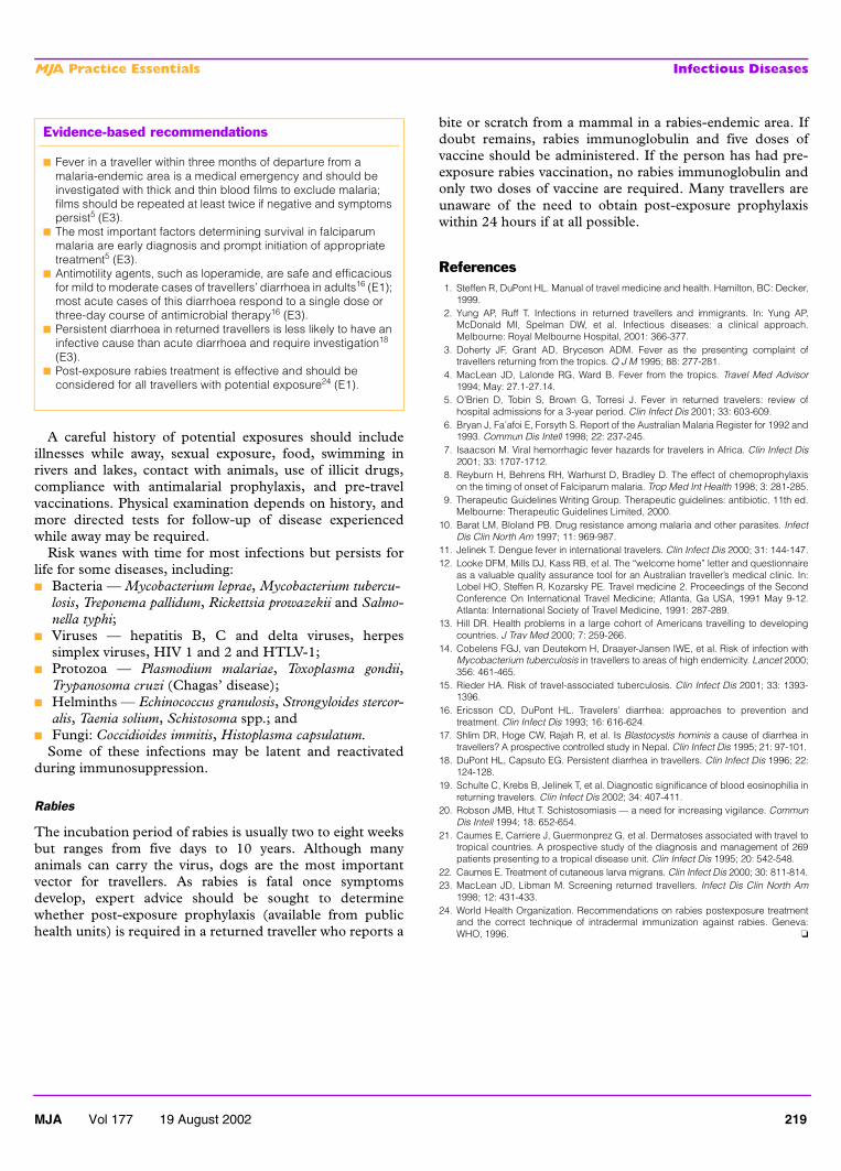
Although not usually serious or life-threatening, skin andsoft tissue infections are common. Most common are theinfections also seen in non-travellers, such as cellulitis,furunculosis, folliculitis and dermatophyte infections. Otherinfections are more exotic. For example, among 269 patientswho presented to a Paris tropical disease unit with travel-associated dermatosis, 53% had an imported tropical dis-ease.21 Most common among these were cutaneous larvamigrans (25%; Box 11), pyodermas (18%), pruritic arthro-pod-reactive dermatitis (10%), myiasis (9%), tungiasis(6%), urticaria (5%), fever and rash (eg, dengue) (4%) andcutaneous leishmaniasis (3%; Box 12).
Non-healing skin lesions and furuncles in travellers fromAfrica and South America should be examined closely, asmyiasis or infestation with fly larvae (eg, Cordylobia orAfrican tumbu or putsi fly) is possible. The larvae are bestremoved by applying petroleum jelly in a toothpaste cap.
They burrow into the cap and can then be extracted withforceps, the whole process usually taking less than an hour.
Sandfleas, or “jiggers”, usually burrow into the skin of thesoles of the feet and around toenails, causing itchy lumps.Prevention is by wearing shoes, avoiding sandals and barefeet. Jiggers are best shelled out, being careful not to breakthe developing egg sac of the female flea.
The asymptomatic returned traveller
Returned travellers often present requesting a “check-up”.Cost-effectiveness studies have yet to demonstrate the bene-fits of widespread screening for returned travellers.23
12: Mucocutaneous leishmaniasis acquired in South America
Cutaneous leishmaniasis is relatively common after sandfly bites in travellers returning from South and Central America, the Mediter-ranean and the Middle East. Mucous membranes and viscera may be involved.
11: Cutaneous larva migrans in a traveller to Malaysia
Cutaneous larva migrans is caused by the penetration through intact skin of larval animal hookworms. Diagnosis is predominantly clinical. Treatment is often necessary because of intense pruritus, long duration (over a year) and complications, such as impetigo and allergic reactions. Therapy comprises ivermectin, albendazole or thiabendazole.22
MJA Vol 177 19 August 2002 219
MJA Practice Essentials Infectious Diseases
A careful history of potential exposures should includeillnesses while away, sexual exposure, food, swimming inrivers and lakes, contact with animals, use of illicit drugs,compliance with antimalarial prophylaxis, and pre-travelvaccinations. Physical examination depends on history, andmore directed tests for follow-up of disease experiencedwhile away may be required.
Risk wanes with time for most infections but persists forlife for some diseases, including:■ Bacteria — Mycobacterium leprae, Mycobacterium tubercu-
losis, Treponema pallidum, Rickettsia prowazekii and Salmo-nella typhi;
■ Viruses — hepatitis B, C and delta viruses, herpessimplex viruses, HIV 1 and 2 and HTLV-1;
■ Protozoa — Plasmodium malariae, Toxoplasma gondii,Trypanosoma cruzi (Chagas’ disease);
■ Helminths — Echinococcus granulosis, Strongyloides stercor-alis, Taenia solium, Schistosoma spp.; and
■ Fungi: Coccidioides immitis, Histoplasma capsulatum.Some of these infections may be latent and reactivated
during immunosuppression.
Rabies
The incubation period of rabies is usually two to eight weeksbut ranges from five days to 10 years. Although manyanimals can carry the virus, dogs are the most importantvector for travellers. As rabies is fatal once symptomsdevelop, expert advice should be sought to determinewhether post-exposure prophylaxis (available from publichealth units) is required in a returned traveller who reports a
bite or scratch from a mammal in a rabies-endemic area. Ifdoubt remains, rabies immunoglobulin and five doses ofvaccine should be administered. If the person has had pre-exposure rabies vaccination, no rabies immunoglobulin andonly two doses of vaccine are required. Many travellers areunaware of the need to obtain post-exposure prophylaxiswithin 24 hours if at all possible.
References1. Steffen R, DuPont HL. Manual of travel medicine and health. Hamilton, BC: Decker,
1999.2. Yung AP, Ruff T. Infections in returned travellers and immigrants. In: Yung AP,
McDonald MI, Spelman DW, et al. Infectious diseases: a clinical approach.Melbourne: Royal Melbourne Hospital, 2001: 366-377.
3. Doherty JF, Grant AD, Bryceson ADM. Fever as the presenting complaint oftravellers returning from the tropics. Q J M 1995; 88: 277-281.
4. MacLean JD, Lalonde RG, Ward B. Fever from the tropics. Travel Med Advisor1994; May: 27.1-27.14.
5. O’Brien D, Tobin S, Brown G, Torresi J. Fever in returned travelers: review ofhospital admissions for a 3-year period. Clin Infect Dis 2001; 33: 603-609.
6. Bryan J, Fa’afoi E, Forsyth S. Report of the Australian Malaria Register for 1992 and1993. Commun Dis Intell 1998; 22: 237-245.
7. Isaacson M. Viral hemorrhagic fever hazards for travelers in Africa. Clin Infect Dis2001; 33: 1707-1712.
8. Reyburn H, Behrens RH, Warhurst D, Bradley D. The effect of chemoprophylaxison the timing of onset of Falciparum malaria. Trop Med Int Health 1998; 3: 281-285.
9. Therapeutic Guidelines Writing Group. Therapeutic guidelines: antibiotic. 11th ed.Melbourne: Therapeutic Guidelines Limited, 2000.
10. Barat LM, Bloland PB. Drug resistance among malaria and other parasites. InfectDis Clin North Am 1997; 11: 969-987.
11. Jelinek T. Dengue fever in international travelers. Clin Infect Dis 2000; 31: 144-147.12. Looke DFM, Mills DJ, Kass RB, et al. The “welcome home” letter and questionnaire
as a valuable quality assurance tool for an Australian traveller’s medical clinic. In:Lobel HO, Steffen R, Kozarsky PE. Travel medicine 2. Proceedings of the SecondConference On International Travel Medicine; Atlanta, Ga USA, 1991 May 9-12.Atlanta: International Society of Travel Medicine, 1991: 287-289.
13. Hill DR. Health problems in a large cohort of Americans travelling to developingcountries. J Trav Med 2000; 7: 259-266.
14. Cobelens FGJ, van Deutekom H, Draayer-Jansen IWE, et al. Risk of infection withMycobacterium tuberculosis in travellers to areas of high endemicity. Lancet 2000;356: 461-465.
15. Rieder HA. Risk of travel-associated tuberculosis. Clin Infect Dis 2001; 33: 1393-1396.
16. Ericsson CD, DuPont HL. Travelers’ diarrhea: approaches to prevention andtreatment. Clin Infect Dis 1993; 16: 616-624.
17. Shlim DR, Hoge CW, Rajah R, et al. Is Blastocystis hominis a cause of diarrhea intravellers? A prospective controlled study in Nepal. Clin Infect Dis 1995; 21: 97-101.
18. DuPont HL, Capsuto EG. Persistent diarrhea in travellers. Clin Infect Dis 1996; 22:124-128.
19. Schulte C, Krebs B, Jelinek T, et al. Diagnostic significance of blood eosinophilia inreturning travelers. Clin Infect Dis 2002; 34: 407-411.
20. Robson JMB, Htut T. Schistosomiasis — a need for increasing vigilance. CommunDis Intell 1994; 18: 652-654.
21. Caumes E, Carriere J, Guermonprez G, et al. Dermatoses associated with travel totropical countries. A prospective study of the diagnosis and management of 269patients presenting to a tropical disease unit. Clin Infect Dis 1995; 20: 542-548.
22. Caumes E. Treatment of cutaneous larva migrans. Clin Infect Dis 2000; 30: 811-814.23. MacLean JD, Libman M. Screening returned travellers. Infect Dis Clin North Am
1998; 12: 431-433.24. World Health Organization. Recommendations on rabies postexposure treatment
and the correct technique of intradermal immunization against rabies. Geneva:WHO, 1996. ❏
Evidence-based recommendations
■ Fever in a traveller within three months of departure from a malaria-endemic area is a medical emergency and should be investigated with thick and thin blood films to exclude malaria; films should be repeated at least twice if negative and symptoms persist5 (E3).
■ The most important factors determining survival in falciparum malaria are early diagnosis and prompt initiation of appropriate treatment5 (E3).
■ Antimotility agents, such as loperamide, are safe and efficacious for mild to moderate cases of travellers’ diarrhoea in adults16 (E1); most acute cases of this diarrhoea respond to a single dose or three-day course of antimicrobial therapy16 (E3).
■ Persistent diarrhoea in returned travellers is less likely to have an infective cause than acute diarrhoea and require investigation18 (E3).
■ Post-exposure rabies treatment is effective and should be considered for all travellers with potential exposure24 (E1).