8LECTURES - KSUMSCksumsc.com/download_center/2nd/2) GNT Block/Female... · • Causes diarrhea,...
68
8 LECTURES Gastroesophageal reflux disease Pep4c Ulcer Disease Inflammatory bowel disease1 Malabsorp4on Diarrhea Colonic polyps and carcinoma1 Inflammatory bowel disease2 Colonic polyps and carcinoma2
Transcript of 8LECTURES - KSUMSCksumsc.com/download_center/2nd/2) GNT Block/Female... · • Causes diarrhea,...

8 LECTURES
Gastro-‐esophageal reflux disease Pep4c Ulcer Disease
Inflammatory bowel disease-‐1
Malabsorp4on
Diarrhea
Colonic polyps and carcinoma-‐1
Inflammatory bowel disease-‐2
Colonic polyps and carcinoma-‐2

8 LECTURES
Malabsorp4on
Diarrhea

DIARREAHA

Physiology of Fluid and small intes4ne

DIARREAHA DEFINITION
• World Health Organiza4on Ø 3 or more loose or liquid stools per day
• Abnormally high fluid content of stool > 200-‐300 gm/day

Fecal osmolarity • As stool leaves the colon, fecal osmolality is equal to the serum osmolality i.e. 290 mosm/kg.
• Under normal circumstances, the major osmoles are Na+, K+, Cl–, and HCO3
–.

CLASSIFICATION
1. Acute …………….if 2 weeks,
2. Persistent ……. if 2 to 4 weeks,
3. Chronic ………..if 4 weeks in dura4on.

Why important?
• The loss of fluids through diarrhea can cause dehydra4on and electrolyte imbalances
• Easy to treat but if untreated, may lead to death especially in children
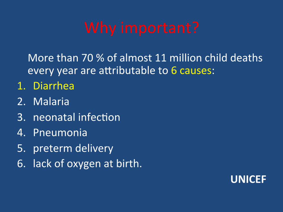
Why important?
More than 70 % of almost 11 million child deaths every year are a`ributable to 6 causes:
1. Diarrhea 2. Malaria 3. neonatal infec4on 4. Pneumonia 5. preterm delivery 6. lack of oxygen at birth.
UNICEF

Pathophysiology Categories of diarrhea
1. Secretory 2. Osmo<c 3. Exuda<ve (inflammatory ) 4. Mo<lity-‐related

Secretory
• There is an increase in the ac4ve secre4on • High stool output • Lack of response to fas4ng • Normal stool osmo4c gap < 100 mOsm/kg • The most common cause of this type of diarrhea is a bacterial toxin ( E. coli , cholera) that s4mulates the secre4on of anions.
• Also seen in Endocrine tumours

Osmo<c
• Excess amount of poorly absorbed substances that exert osmo4c effect………water is drawn into the bowels……diarrhea
• Stool output is usually not massive • Fas4ng improve the condi4on • Stool osmo4c gap is high, > 125 mOsm/kg • Can be the result of 1. Malabsorp4on in which the nutrients are lej in the
lumen to pull in water e.g. lactose intolerance 2. osmo4c laxa4ves.

Exuda<ve (inflammatory )
• Results from the outpouring of blood protein, or mucus from an inflamed or ulcerated mucosa
• Presence of blood and pus in the stool. • Persists on fas4ng • Occurs with inflammatory bowel diseases, and invasive infec4ons.

Mo<lity-‐related
• Caused by the rapid movement of food through the intes4nes (hypermo4lity).
• Irritable bowel syndrome (IBS) – a motor disorder that causes abdominal pain and altered bowel habits with diarrhea predomina4ng

Pathophysiology Categories of diarrhea
1. Secretory 2. Osmo<c 3. Exuda<ve (inflammatory ) 4. Mo<lity-‐related

Ae4ology Acute diarrhea?
• Approximately 80% of acute diarrheas are due to infec3ons (viruses, bacteria, helminths, and protozoa).
• Viral gastroenteri4s (viral infec4on of the stomach and the small intes4ne) is the most common cause of acute diarrhea worldwide.
• Food poisoning • Drugs • Others
Rotavirus the cause of nearly 40% of hospitaliza4ons from diarrhea in children under 5

An<bio<c-‐Associated Diarrheas
• Diarrhea occurs in 20% of pa<ents receiving broad-‐spectrum an<bio<cs; about 20% of these diarrheas are due to Clostridium difficile

Ae4ology • Chronic diarrhea? 1. Infec<on -‐-‐-‐-‐-‐-‐-‐-‐-‐-‐-‐-‐-‐-‐-‐-‐-‐-‐ e.g.Giardia lamblia . AIDS
ojen have chronic infec4ons of their intes4nes that cause diarrhea.
2. Post-‐infec<ous. Following acute viral, bacterial or parasi4c infec4ons
3. Malabsorp<on 4. Inflammatory bowel disease (IBD) 5. Endocrine diseases. 6. Colon cancer 7. Irritable bowel syndrome.

Complica<ons
1. Fluids ………………Dehydra4on 2. Electrolytes …………….. Electrolytes imbalance 3. Sodium bicarbonate……. Metabolic acidosis 4. If persistent ……Malnutri4on

Tests useful in the evalua<on of diarrhea Acute diarrhea
Noninflammatory Diarrhea Inflammatory Diarrhea
Fecal leukocytes
Suggests a small bowel source Or colon but without mucosal injury Suggests colonic mucosa damage
caused by invasion Ø shigellosis, salmonellosis, Campylobacter or Yersinia infec4on, amebiasis) Ø toxin (C difficile, E coli O157:H7). Ø Inflammatory bowel diseases
not present present

Chronic diarrhea
Stool analysis Ova, parasites
+
-‐
Infec4on
Stool fat test Secretory or Noninfec4ous inflammatory diarrhea
Malabsorp4on
-‐
+
(normal <20%)

8 LECTURES
Malabsorp4on
Diarrhea

Malabsorption Syndrome
Inability of the intes4ne to absorb nutrients adequately into the bloodstream.
Impairment can be of single or mul4ple nutrients depending on the abnormality.

Physiology
– The main purpose of the gastrointes4nal tract is to digests and absorbs nutrients (fat, carbohydrate, and protein), micronutrients (vitamins and trace minerals), water, and electrolytes.

Mechanisms and Causes of Malabsorp4on Syndrome
Inadequate diges<on Postgastrectomy Deficiency of pancrea4c lipase Chronic pancrea44s Cys4c fibrosis Pancrea4c resec4on Zollinger-‐Ellison syndrome Deficient bile salt Obstruc4ve jaundice Bacterial overgrowth Stasis in blind loops, diver4cula Fistulas Hypomo4lity states (diabetes) Terminal ileal resec4on Crohns' disease Precipita4on of bile salts (neomycin)
Primary mucosal abnormali<es Celiac disease Tropical sprue Whipple's disease Amyloidosis Radia4on enteri4s Abetalipoproteinemia Giardiasis Inadequate small intes<ne Intes4nal resec4on Crohn's disease Mesenteric vascular disease with infarc4on Jejunoileal bypass Lympha<c obstruc<on Intes4nal lymphangiectasia Malignant lymphoma Macroglobulinemia

Pathophysiology
Small intes<ne abnormali<es Inadequate diges<on Or Malabsorption =

Pathophysiology
Inadequate diges<on
Small intes<ne abnormali<es
Pancrease
Bile
Stomach
mucosa
Inadequate small intes<ne
Lympha<c obstruc<on
Postgastrectomy
Deficiency of pancrea4c lipase Chronic pancrea44s Cys4c fibrosis Pancrea4c resec4on
Obstruc4ve jaundice Terminal ileal resec4on

Pathophysiology
Inadequate diges<on
Small intes<ne abnormali<es
Pancrease
Bile
Stomach
mucosa
Inadequate small intes<ne
Lympha<c obstruc<on
Celiac disease Tropical sprue Whipple's disease Giardiasis
Intes4nal resec4on Crohn's disease
Intes4nal lymphangiectasia Malignant lymphoma

Pathophysiology
Pancrease
Bile
mucosa
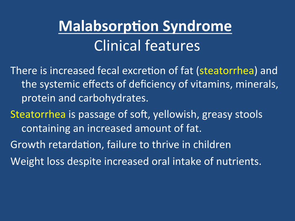
Malabsorp<on Syndrome Clinical features
There is increased fecal excre4on of fat (steatorrhea) and the systemic effects of deficiency of vitamins, minerals, protein and carbohydrates.
Steatorrhea is passage of soj, yellowish, greasy stools containing an increased amount of fat.
Growth retarda4on, failure to thrive in children Weight loss despite increased oral intake of nutrients.

Clinical features

Malabsorp<on Syndrome Clinical features
Protein Swelling or oedema
B12, folic acid and iron deficiency Anaemias (fa4gue and weakness)
vitamin D, calcium Muscle cramp Osteomalacia and osteoporosis
vitamin K and other coagula4on factor Bleeding tendencies
Depend on the deficient nutrient

Diagnosis
There is no specific test for malabsorp4on. Inves4ga4on is guided by symptoms and signs. 1. Fecal fat study to diagnose steatorrhoea 2. Blood tests 3. Stool studies 4. Endoscopy Biopsy of small bowel

Malabsorp<on Syndrome Celiac disease
An immune reac4on to gliadin frac4on of the wheat protein gluten
Usually diagnosed in childhood – mid adult. Pa4ents have raised an4bodies to gluten autoan4bodies Highly specific associa4on with class II HLA DQ2 (haplotypes DR-‐17 or DR5/7) and, to a lesser extent, DQ8 (haplotype DR-‐4).

Clinical features Celiac disease
Typical presenta<on GI symptoms that characteris4cally appear at age 9-‐24 months. Symptoms begin at various 4mes ajer the introduc4on of foods that contain gluten. A rela<onship between the age of onset and the type of presenta<on; Infants and toddlers….GI symptoms and failure to thrive Childhood…………………minor GI symptoms, inadequate rate of weight gain, Young adults……………anemia is the most common form of presenta4on. Adults and elderly…...GI symptoms are more prevalent

Endoscopy Normal
Celiac disease

Histology • Mucosa is flattened with marked villous atrophy.
• Increased intraepithelial lymphocytosis
Celiac Disease
Normal

Celiac Disease
Diagnosis Clinical documenta4ons of malabsorp4on. Stool ………. fat Small intes4ne biopsy demonstrate villous atrophy. Improvement of symptom and mucosal histology on gluten
withdrawal from diet.
wheat, barley, flour Other grains, such as rice and corn flour, do not have such an effect.

Celiac Disease
Complica4ons Osteopenia , osteoporosis Infer4lity in women Short stature, delayed puberty, anemia, Malignancies,[ intes4nal T-‐cell lymphoma] 10 to 15% risk of developing GI lymphoma.

Lactose Intolerance

Lactose Intolerance Pathophysiology
Lactose glucose + galactose At the brush border of enterocytes
lactase
Low or absent ac4vity of the enzyme lactase Lactose Intolerance
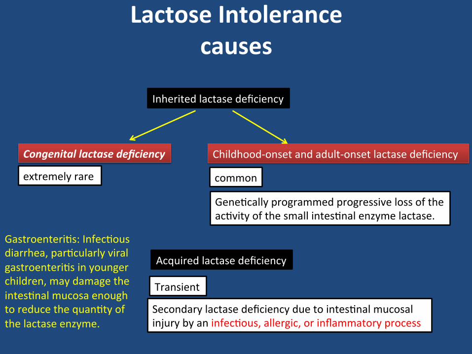
Lactose Intolerance causes
Congenital lactase deficiency Childhood-‐onset and adult-‐onset lactase deficiency
Gene4cally programmed progressive loss of the ac4vity of the small intes4nal enzyme lactase.
Secondary lactase deficiency due to intes4nal mucosal injury by an infec4ous, allergic, or inflammatory process
Acquired lactase deficiency
Inherited lactase deficiency
extremely rare common
Transient
Gastroenteri4s: Infec4ous diarrhea, par4cularly viral gastroenteri4s in younger children, may damage the intes4nal mucosa enough to reduce the quan4ty of the lactase enzyme.

Clinical
Bloa4ng, abdominal discomfort, and flatulence ……………1 hour to a few hours ajer inges4on of milk products


Lactose Intolerance Diagnosis
Empirical treatment with a lactose-‐free diet, which results in resolu4on of symptoms;
Hydrogen breath test

Hydrogen breath test .
• An oral dose of lactose is administered • The sole source of H2 is bacterial
fermentation; • Unabsorbed lactose makes its way to
colonic bacteria, resulting in excess breath H2.
• Increased exhaled H2 after lactose ingestion suggests lactose malabsorption.

A 3-‐week trial of a diet that is free of milk and milk products is a sa4sfactory trial to diagnose lactose intolerance

Lactose Intolerance summary
• Deficiency/absence of the enzyme lactase in the brush border of the intestinal mucosa → maldigestion and malabsorption of lactose
• Unabsorbed lactose draws water in the intestinal lumen
• In the colon, lactose is metabolized by bacteria to organic acid, CO2 and H2; acid is an irritant and exerts an osmotic effect
• Causes diarrhea, gaseousness, bloating and abdominal cramps

A 1. Fas4ng improve the
condi4on 2. inflammatory bowel
diseases 3. High stool output 4. Presence of WBC in stool 5. Irritable bowel syndrome 6. bacterial toxin 7. Malabsorp4on 8. High fecal osmo4c gap
B a) Secretory b) Osmo<c c) Exuda<ve (inflammatory ) d) Mo<lity-‐related
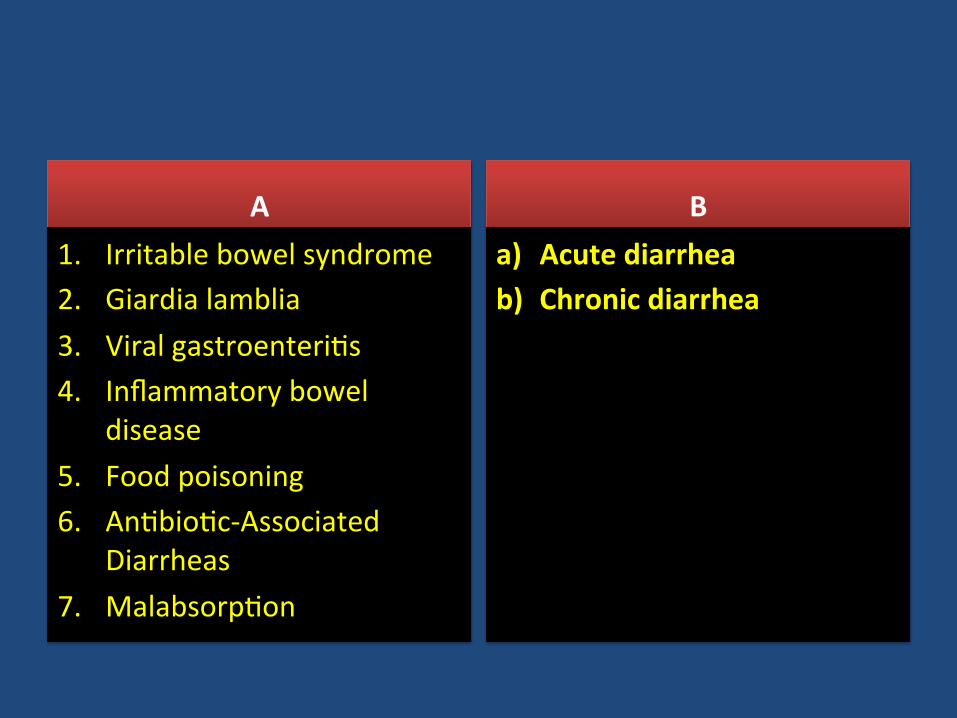
A 1. Irritable bowel syndrome 2. Giardia lamblia 3. Viral gastroenteri4s 4. Inflammatory bowel
disease 5. Food poisoning 6. An4bio4c-‐Associated
Diarrheas 7. Malabsorp4on
B a) Acute diarrhea b) Chronic diarrhea

CLASSIFICATION diarrhea
1. Acute
2. Persistent
3. Chronic
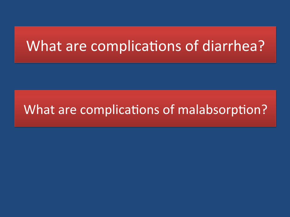
What are complica4ons of diarrhea?
What are complica4ons of malabsorp4on?

Pathophysiology of malabsorp4on ?

Clinical presenta4on of malabsorp4on ?

What is Celiac disease?


• A 10-‐month-‐old, previously healthy male infant develops a severe, watery diarrhea 2 days ajer visi4ng the pediatrician for a rou4ne checkup. The most likely diagnosis is
a. Rotavirus infec4on b. Enterotoxigenic E. coli infecAon c. Entamoeba histolyAca infecAon d. Lactase deficiency e. Ulcera4ve coli4s

Scenario A 44-‐year-‐old white male presented with a seven-‐month history of
diarrhea. The frequency of his bowel movements had increased to 5-‐7 per day, and his stools were yellow and floated at the top of the water in the toilet. He had occasional abdominal cramping, but no tenesmus, melena, or bleeding. His appe4te was good, but he had experienced gradual weight loss. His bowel movement frequency would decrease upon fas4ng and would increase with food intake.
Stool tests revealed a stool output of 4128 g/d (nl 100-‐200 g/d) with fat excre4on of 17 g/d (nl <5 g/d).
Microscopic examina4on for ova and parasites and cultures for bacterial pathogens and acid-‐fast bacilli were nega4ve.
Blood tes4ng showed mild anemia , hypoproteinemia (4.9 mg/dL), and hypoalbuminemia (3.4 mg/dL).

Duodenal biopsy
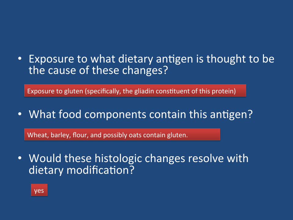
• Exposure to what dietary an4gen is thought to be the cause of these changes?
• What food components contain this an4gen?
• Would these histologic changes resolve with dietary modifica4on?
Exposure to gluten (specifically, the gliadin cons4tuent of this protein)
Wheat, barley, flour, and possibly oats contain gluten.
yes

A 6-‐year-‐old boy has been brought to outpa4ents by his mother because he has abdominal pain ajer some meals. This has been gexng increasingly frequent and it sounds, from his descrip4on, somewhat colicky in nature. You discover that he has always had very smelly, loose, pale bulky stools, which his parents have put down to the fact that he likes milk. On examina4on, he is pale, underweight, and of short stature.

• 1. What are the important differen4al diagnoses on presenta4on?
Celiac disease is the most likely diagnosis. Parasi4c infec4on (e.g. giardiasis) and pancrea4c insufficiency (e.g. due to chronic pancrea44s or cys4c fibrosis) may give rise to a similar presenta4on, but these are not supported by the results of the inves4ga4ons.

• 2. Blood tests reveal a mild macrocy4c anemia. There is a low level of vitamin B12, and folate is at the lower end of normal. Autoan4body screens reveal a posi4ve reac4on to an4gliadin an4bodies. Do these tests help to narrow down the diagnosis?
These results are very sugges4ve of celiac disease due to the low levels of vitamin B12 and the hypersensi4vity reac4on to α-‐gliadin, a component of gluten. The finding of villous atrophy would support the diagnosis, and this is achieved by endoscopic biopsy of the first part of the duodenum.

• 3. A duodenal biopsy shows
Normal

• The final diagnosis is celiac disease, provided the pa4ent’s symptoms respond to a gluten-‐free diet and the histological changes relapse on re-‐challenge. Such criteria are necessary before confining a pa4ent to a lifelong gluten-‐free diet.

• 4. What treatment op4ons are available?
Treatment is by adhering to a strict gluten-‐free diet.


• How to diagnose lactose intorelence ?
• How to treat lactose intorelence ?
Empirical treatment with a lactose-‐free diet, which results in resolu4on of symptoms; Hydrogen breath test
lactose-‐free diet