Daily Script - Movie Scripts and Movie ScreenplaysCreated Date 10/2/2005 8:58:57 PM
Upload
cecily-wheelerCategory
view
228download
0
8 - 1
Chapter 8Muscular System
8 - 2
Athletic Training and Muscular System Movie
3
8 - 4

8 - 5
8 - 6
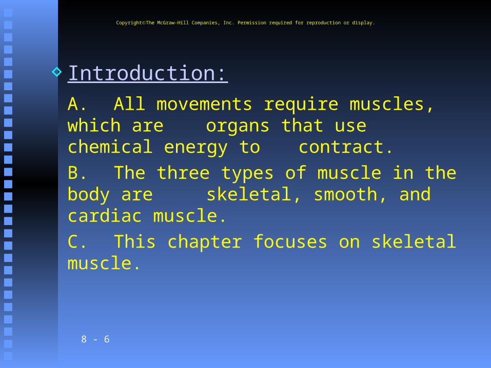
Introduction:A. All movements require muscles, which are organs that use chemical energy to contract.B. The three types of muscle in the body are skeletal, smooth, and cardiac muscle.C. This chapter focuses on skeletal muscle.
CopyrightThe McGraw-Hill Companies, Inc. Permission required for reproduction or display.
8 - 7
Structure of a Skeletal Muscle
A. Each muscle is an organ, comprised of skeletal muscle tissue, connective tissues, nervous tissue, and blood.
CopyrightThe McGraw-Hill Companies, Inc. Permission required for reproduction or display.
8 - 8
B. Connective Tissue Coverings 1. Layers of dense connective tissue, called fascia, surround and
separate each muscle.2. This connective tissue extends beyond the ends of the muscle and gives rise to tendons that are fused to the periosteum of bones.
CopyrightThe McGraw-Hill Companies, Inc. Permission required for reproduction or display.

8 - 9
3. Sometimes muscles are connected to each other by broad sheets of connective tissue called
aponeuroses.4. The layer of connective tissue around each whole muscle is the epimysium; the perimysium surrounds individual bundles
(fascicles) within each muscle; and each muscle cell (fiber) is covered by a connective tissue layer called endomysium.
CopyrightThe McGraw-Hill Companies, Inc. Permission required for reproduction or display.

8 - 10
CopyrightThe McGraw-Hill Companies, Inc. Permission required for reproduction or display.

8 - 11
Study Analogy
Pretend you are going to play a joke on someone and give them 100 pencils. The pencils will represent muscle fibers. First you wrap each individual pencil in tissue paper (dense tissue paper of course!). This would be endomysium. Then you take about 10 pencils in a bundle (a fascicle) and wrap them in paper (perimysium). After that you take all the bundles and wrap them in gift wrap (epimysium). But you are going to mail this joke, so you also have to wrap it in brown paper representing the fascia.
CopyrightThe McGraw-Hill Companies, Inc. Permission required for reproduction or display.
8 - 12
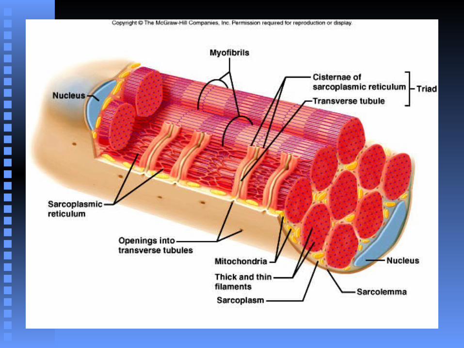
C. Skeletal Muscle Fibers 1. Each muscle fiber is a single, long, cylindrical muscle cell.2. Beneath the sarcolemma (cell membrane) lies sarcoplasm (cytoplasm) with many mitochondria and nuclei; the sarcoplasm contains myofibrils. Myofibrils are
separated into compartments called sarcomeres that contain thick filaments and thin filaments.
CopyrightThe McGraw-Hill Companies, Inc. Permission required for reproduction or display.
8 - 13
a. Thick filaments of myofibrils are made up of the protein myosin.b. Thin filaments of myofibrils are made up of the protein actin.c. The organization of these filaments produces striations.
CopyrightThe McGraw-Hill Companies, Inc. Permission required for reproduction or display.
8 - 14
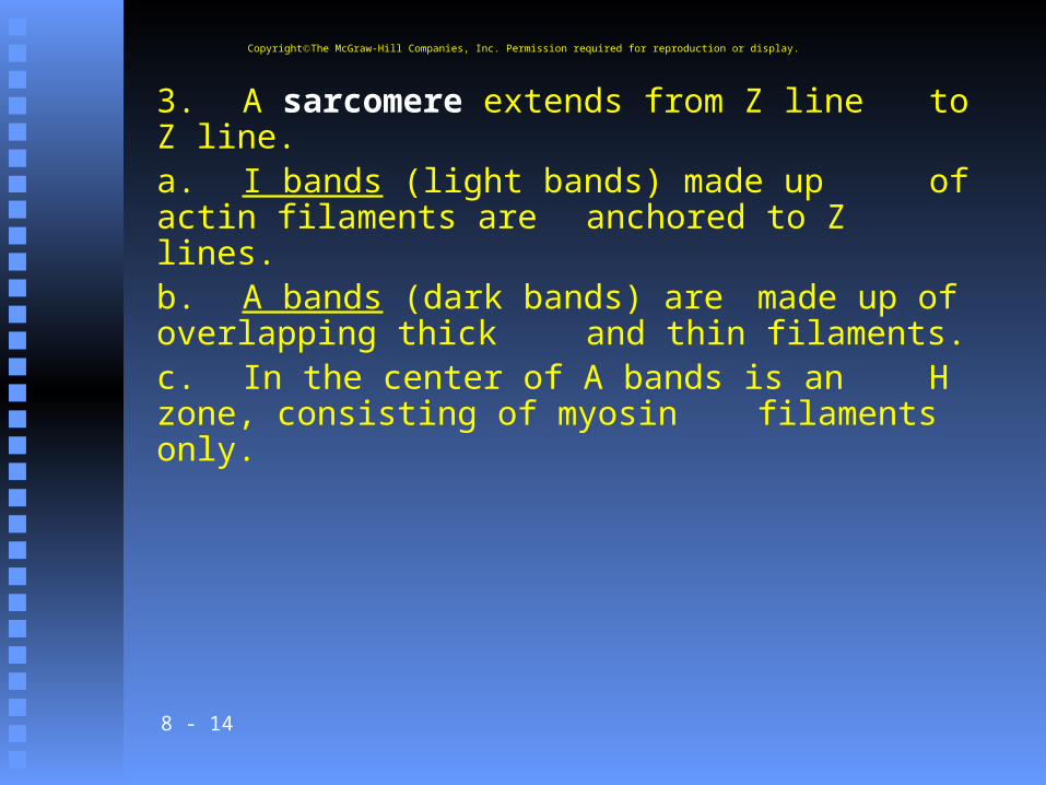
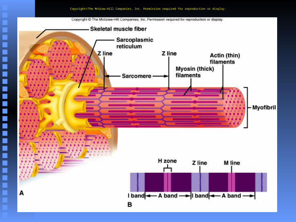
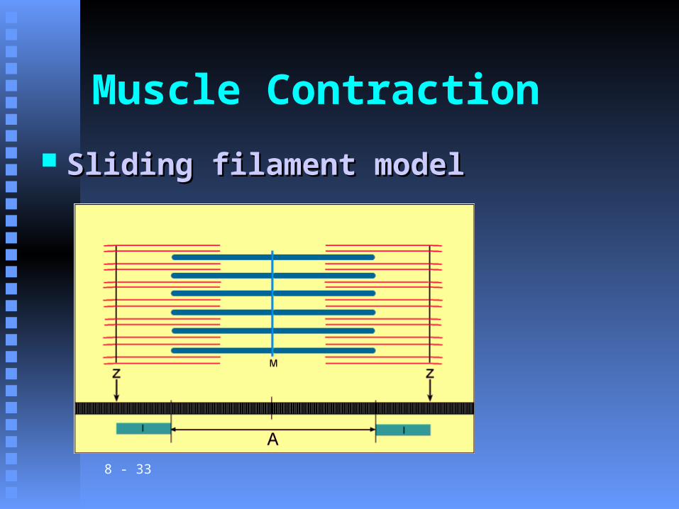
3. A sarcomere extends from Z line to Z line.a. I bands (light bands) made up of actin filaments are anchored to Z lines.b. A bands (dark bands) are
made up of overlapping thick and thin filaments.c. In the center of A bands is an H zone, consisting of myosin
filaments only.
CopyrightThe McGraw-Hill Companies, Inc. Permission required for reproduction or display.
8 - 15
CopyrightThe McGraw-Hill Companies, Inc. Permission required for reproduction or display.

8 - 16
Muscle Contraction
Sliding filament modelSliding filament model
8 - 17
4. Beneath the sarcolemma of a muscle fiber lies the sarcoplasmic reticulum (endoplasmic reticulum), which is associated with transverse (T) tubules (invaginations of the
sarcolemma).
CopyrightThe McGraw-Hill Companies, Inc. Permission required for reproduction or display.
8 - 18
a. Each T tubule lies between two cisternae of the sarcoplasmic reticulum and is open to the outside of the muscle fiber.b. The sarcoplasmic reticulum and transverse tubules activate the muscle contraction mechanism when the fiber is stimulated.
CopyrightThe McGraw-Hill Companies, Inc. Permission required for reproduction or display.

8 - 19
CopyrightThe McGraw-Hill Companies, Inc. Permission required for reproduction or display.
8 - 20
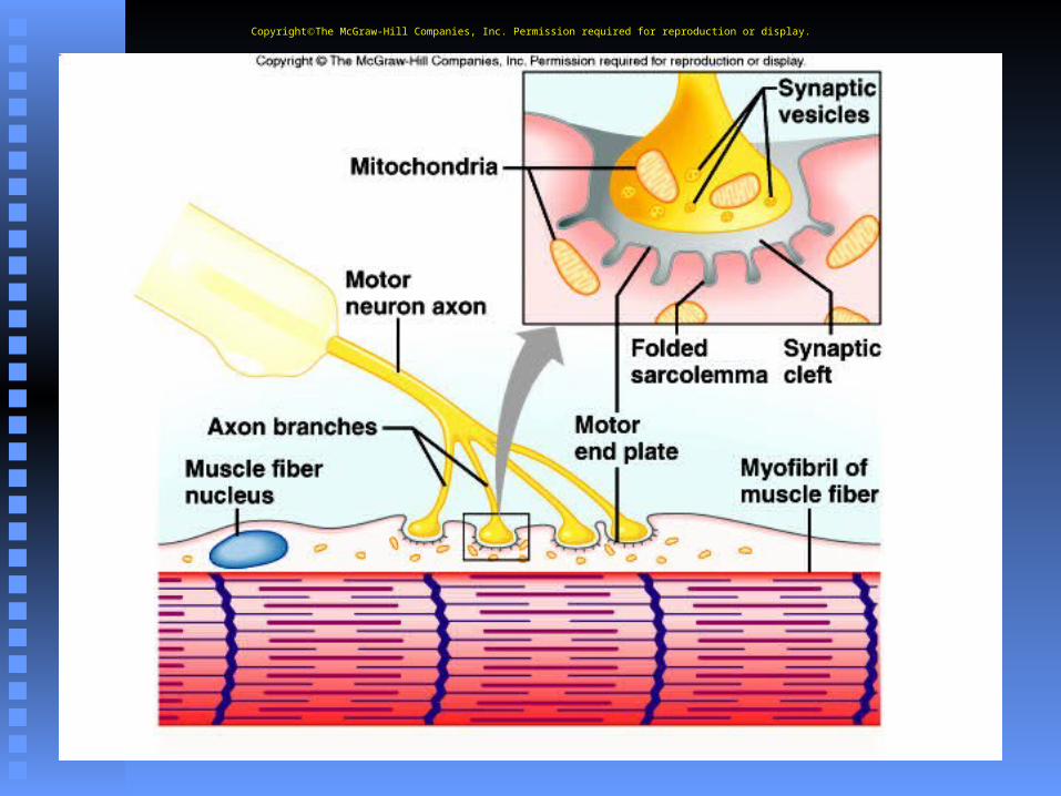
D. Neuromuscular Junction 1. The site where the motor neuron and muscle fiber meet is the neuromuscular
junction.a. The muscle fiber membrane forms a motor end plate in
which the sarcolemma is tightly folded and where nuclei and mitochondria are
abundant.b. The cytoplasm of the motor neuron contains numerous
mitochondria and synaptic vesicles storing neurotransmitters.
CopyrightThe McGraw-Hill Companies, Inc. Permission required for reproduction or display.
8 - 21
E. Motor Units 1. A motor neuron and the
muscle fibers it controls make up a motor unit; when stimulated to do so, the
muscle fibers of the motor unit contract all at
once.
CopyrightThe McGraw-Hill Companies, Inc. Permission required for reproduction or display.
8 - 22
CopyrightThe McGraw-Hill Companies, Inc. Permission required for reproduction or display.
8 - 23
Skeletal Muscle Contraction
A. Muscle contraction involves several components that result in the shortening of sarcomeres, and the pulling of the muscle against its attachments.
CopyrightThe McGraw-Hill Companies, Inc. Permission required for reproduction or display.
8 - 24
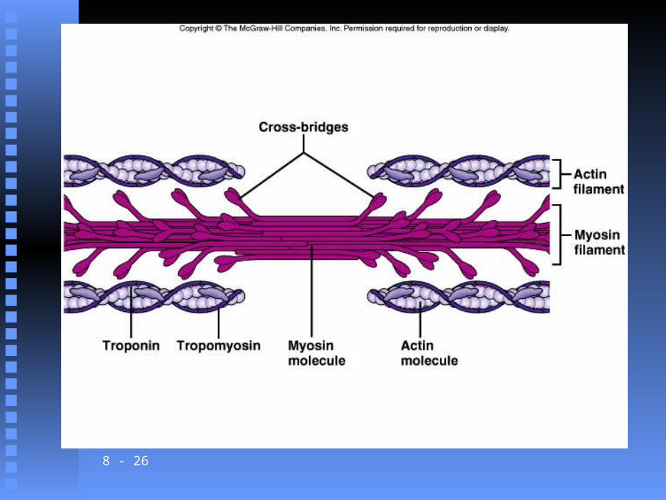
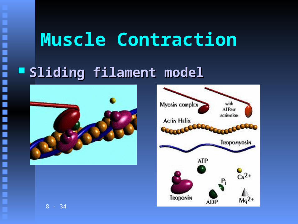
B. Role of Myosin and Actin 1. Myosin consists of two twisted
strands with globular cross-bridges projected outward along the strands.2. Actin is a globular protein with
myosin binding sites; tropomysosin and troponin are two proteins
associated with the surface of the actin filaments.
CopyrightThe McGraw-Hill Companies, Inc. Permission required for reproduction or display.
8 - 25
3. According to the sliding filament theory of muscle contraction, the myosin crossbridge attaches to the
binding site on the actin filament and bends, pulling on the actin filament; it then releases and attaches to the next binding site on the actin, pulling again.
4. Energy from the conversion of ATP to ADP is provided to the cross-bridges from the enzyme ATPase, causing them to be in a “cocked” position.
CopyrightThe McGraw-Hill Companies, Inc. Permission required for reproduction or display.
8 - 26
8 - 27
CopyrightThe McGraw-Hill Companies, Inc. Permission required for reproduction or display.

8 - 28
Click here to playSarcamere Shortening
Flash Animation
8 - 29
C. Stimulus for Contraction 1. The motor neuron must release the
neurotransmitter acetylcholine from its synaptic vesicles into the synaptic cleft in order to initiate a muscle contraction.
2. Protein receptors in the motor end plate detect the neurotransmitters, and a muscle impulse spreads over the surface of the sarcolemma and into
the T tubules, where it reaches the sarcoplasmic reticulum.
CopyrightThe McGraw-Hill Companies, Inc. Permission required for reproduction or display.
8 - 30
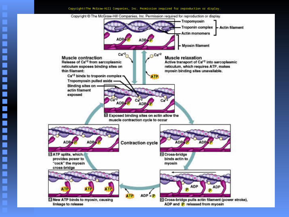
3. Upon receipt of the muscle impulse, the sarcoplasmic reticulum releases its stored calcium to the sarcoplasm of the muscle fiber.
4. The high concentration of calcium in the sarcoplasm interacts with the
troponin and tropomyosin molecules, which move aside, exposing the myosin binding sites on the actin filaments.
CopyrightThe McGraw-Hill Companies, Inc. Permission required for reproduction or display.
8 - 31
5. Myosin cross-bridges now bind and pull on the actin filaments, causing the sarcomeres to shorten.6. After the nervous impulse has been received, acetylcholinesterase rapidly decomposes the acetylcholine.7. Then, calcium is returned to the sarcoplasmic reticulum, and the linkages between myosin and actin are broken.
CopyrightThe McGraw-Hill Companies, Inc. Permission required for reproduction or display.
8 - 32
Myosin video clip
8 - 33
Muscle Contraction
Sliding filament modelSliding filament model
8 - 34
Muscle Contraction
Sliding filament modelSliding filament model
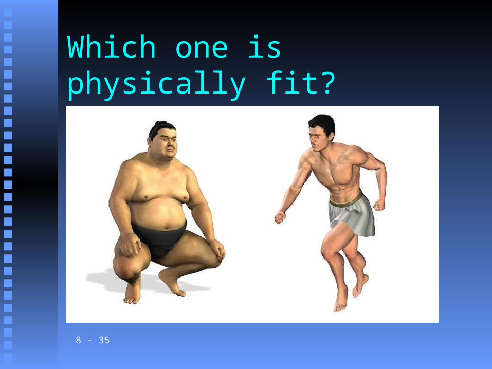
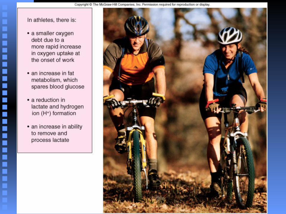
Which one is physically fit?
8 - 35
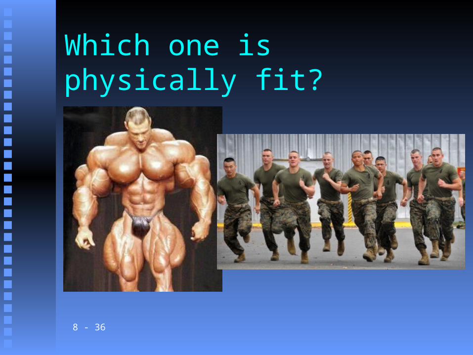
Which one is physically fit?
8 - 36

8 - 37
D. Energy Sources for Contraction 1. Energy for contraction comes from molecules of ATP. This
chemical is in limited supply and so must often be regenerated
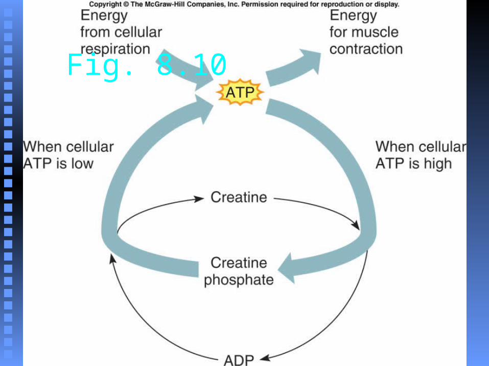
2. Creatine phosphate, which stores excess energy released by the mitochondria, is
present to regenerate ATP from ADP and phosphate.
CopyrightThe McGraw-Hill Companies, Inc. Permission required for reproduction or display.
8 - 38
3. Whenever the supply of ATP is sufficient, creatine
phosphokinase promotes the synthesis of creatine phosphate.
4. As ATP decomposes, the energy from creatine
phosphate can be transferred to ADP
molecules, converting them back to ATP.
CopyrightThe McGraw-Hill Companies, Inc. Permission required for reproduction or display.
8 - 39
Fig. 8.10
8 - 40
E. Oxygen Supply and Cellular Respiration
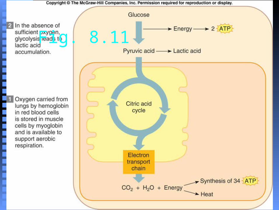
1. The early phase of cellular respiration yields few molecules of
ATP, so muscle has a high requirement for oxygen, which enables the complete breakdown of glucose in the mitochondria.2. Hemoglobin in red blood cells carries oxygen to muscle.3. The pigment myoglobin stores
oxygen in muscle tissue.
CopyrightThe McGraw-Hill Companies, Inc. Permission required for reproduction or display.
8 - 41
F. Oxygen Debt1. During rest or moderate activity,
there is enough oxygen to support aerobic respiration.2. Oxygen deficiency may develop
during strenuous exercise, and lactic acid accumulates as an end product of anaerobic respiration.
a. Lactic acid diffuses out of muscle cells and is carried in
the bloodstream to the liver.
CopyrightThe McGraw-Hill Companies, Inc. Permission required for reproduction or display.
8 - 42
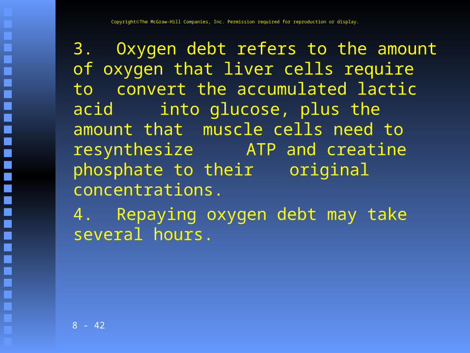
3. Oxygen debt refers to the amount of oxygen that liver cells require to convert the accumulated lactic acid into glucose, plus the amount that muscle cells need to resynthesize
ATP and creatine phosphate to their original concentrations.
4. Repaying oxygen debt may take several hours.
CopyrightThe McGraw-Hill Companies, Inc. Permission required for reproduction or display.
8 - 43
Fig. 8.11
8 - 44
45
Cellular Respiration
Food to ATP
8 - 46
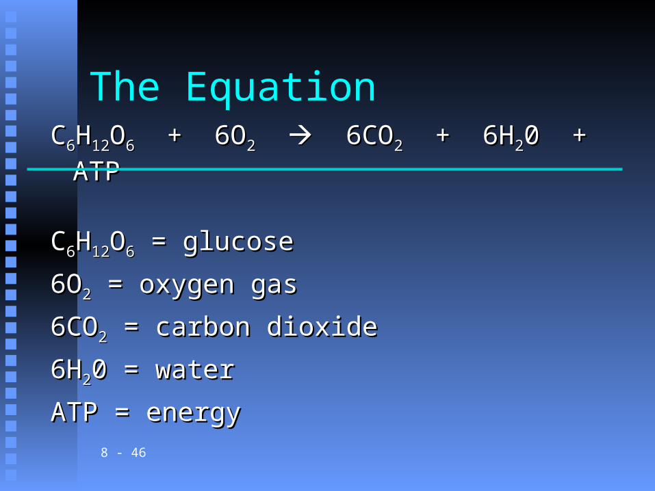
The EquationCC66HH1212OO66 + 6O + 6O22 6CO 6CO22 + 6H + 6H220 + ATP0 + ATP
CC66HH1212OO66 = glucose = glucose
6O6O22 = oxygen gas = oxygen gas
6CO6CO22 = carbon dioxide = carbon dioxide
6H6H220 = water0 = water
ATP = energyATP = energy

8 - 47
Mitochondria
Site of cellular respirationSite of cellular respiration StructureStructure
8 - 48
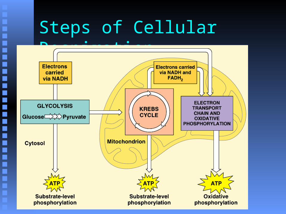
Steps of Cellular Respiration

8 - 49
Glycolysis

8 - 50
Krebs Cycle

8 - 51
Krebs Cycle
8 - 52
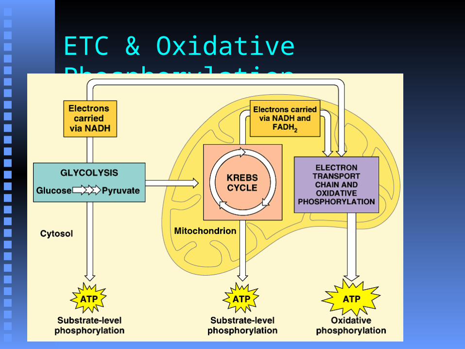
ETC & Oxidative Phosphorylation
8 - 53
ETC & Oxidative Phosphorylation

8 - 54
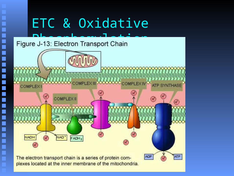
ETC & Oxidative Phosphorylation
Energy from electrons used to move H+ Energy from electrons used to move H+ into intermembrane spaceinto intermembrane space
Electrons accepted by oxygenElectrons accepted by oxygen
2e- + 2H+ + ½ O2e- + 2H+ + ½ O22 H H22OO

8 - 55
ATP Production
GlycolysisGlycolysis 4 ATP4 ATP (2 net) (2 net) Substrate level phosphorylationSubstrate level phosphorylation
KrebsKrebs 2 ATP2 ATP Substrate level phosphorylationSubstrate level phosphorylation

8 - 56
ATP Production
ETC & oxidative phosphorylationETC & oxidative phosphorylation 2 NADH from glycolysis = 2 NADH from glycolysis = 4 to 6 ATP4 to 6 ATP 2 NADH from acetyl CoA prep = 2 NADH from acetyl CoA prep = 6 ATP6 ATP 6 NADH from Krebs cycle = 6 NADH from Krebs cycle = 18 ATP18 ATP 2 FADH2 FADH22 from Krebs cycle = from Krebs cycle = 4 ATP4 ATP
8 - 57
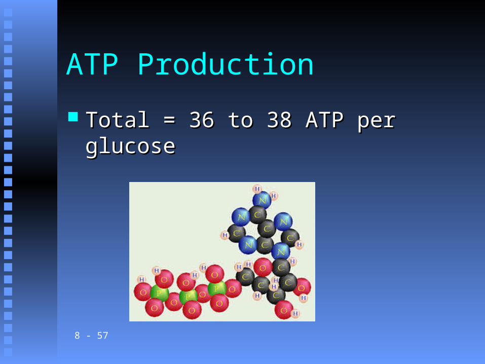
ATP Production
Total = 36 to 38 ATP per glucoseTotal = 36 to 38 ATP per glucose

8 - 58
LACK OF OXYGEN
8 - 59
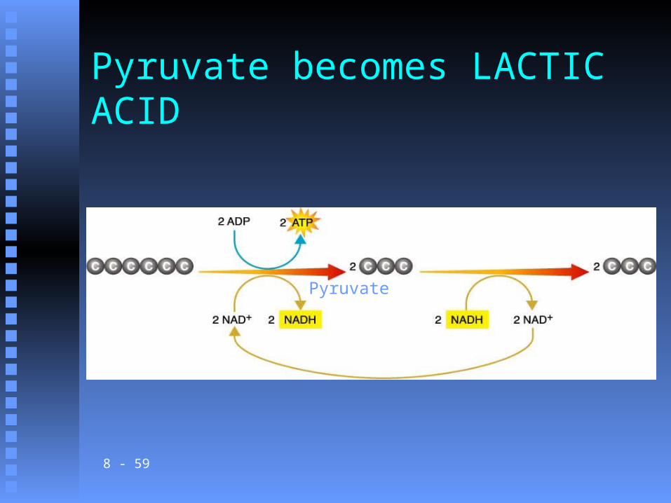
Pyruvate becomes LACTIC ACID
Pyruvate
8 - 60
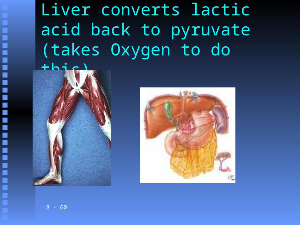
Liver converts lactic acid back to pyruvate (takes Oxygen to do this)
8 - 61
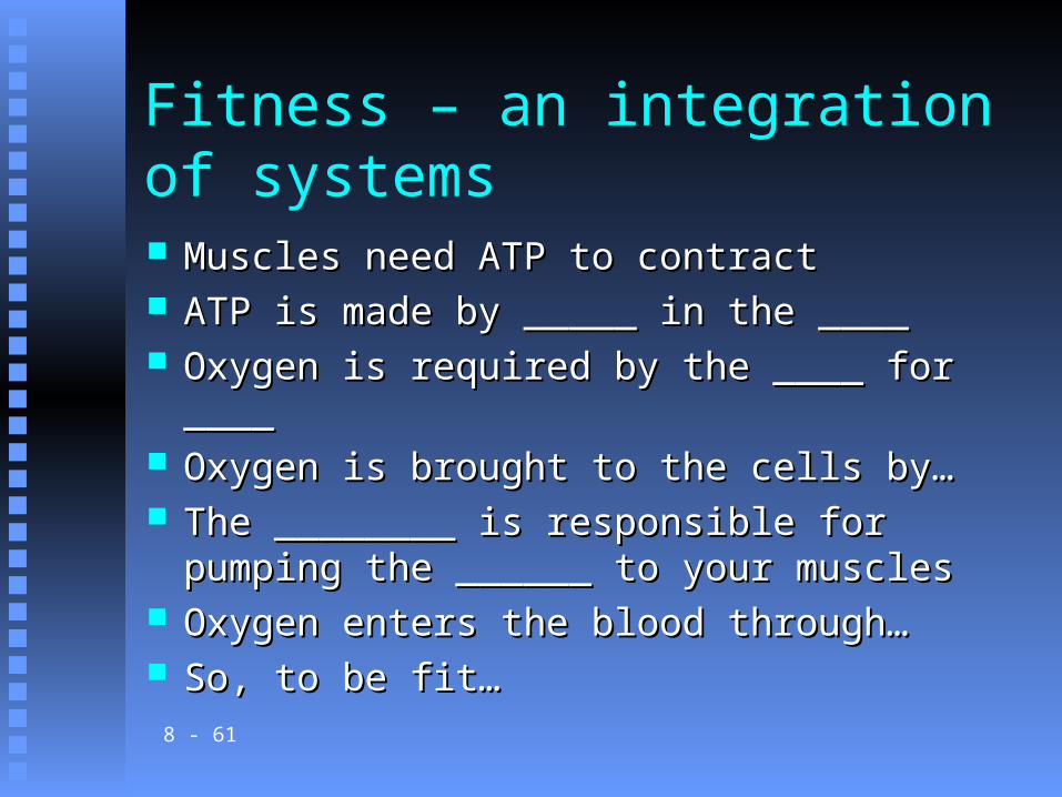
Fitness – an integration of systems
Muscles need ATP to contractMuscles need ATP to contract ATP is made by _____ in the ____ATP is made by _____ in the ____ Oxygen is required by the ____ for ____Oxygen is required by the ____ for ____ Oxygen is brought to the cells by…Oxygen is brought to the cells by… The ________ is responsible for pumping the The ________ is responsible for pumping the
______ to your muscles______ to your muscles Oxygen enters the blood through…Oxygen enters the blood through… So, to be fit…So, to be fit…
8 - 62
G. Muscle Fatigue 1. When a muscle loses its ability to
contract during strenuous exercise, it is referred to as fatigue.
2. Muscle fatigue usually arises from the accumulation of lactic acid in the muscle.a. A lowered pH as a result of
accumulated lactic acid prevents the muscle from contracting.
CopyrightThe McGraw-Hill Companies, Inc. Permission required for reproduction or display.
8 - 63
3. A muscle cramp occurs due to a lack of ATP required to return calcium ions back to the sarcoplasmic reticulum so muscle fibers can relax.
CopyrightThe McGraw-Hill Companies, Inc. Permission required for reproduction or display.
8 - 64
H. Heat Production 1. Contraction of skeletal muscle
represents an important source of heat for the body.
2. Much of the energy produced through the reactions of
cellular respiration is lost as heat (another source of heat for the body).
CopyrightThe McGraw-Hill Companies, Inc. Permission required for reproduction or display.
8 - 65
Muscular Responses A. One method of studying muscle
function is to remove a single fiber and connect it to a device that records its responses to electrical stimulation.B. Threshold Stimulus
1. A muscle fiber remains unresponsive to stimulation unless the stimulus is of a certain strength, called the threshold stimulus.
CopyrightThe McGraw-Hill Companies, Inc. Permission required for reproduction or display.
8 - 66
C. All-or-None Response1. When a muscle fiber
contracts, it contracts to its full extent (all-or- none response); it cannot contract partially.
CopyrightThe McGraw-Hill Companies, Inc. Permission required for reproduction or display.
8 - 67
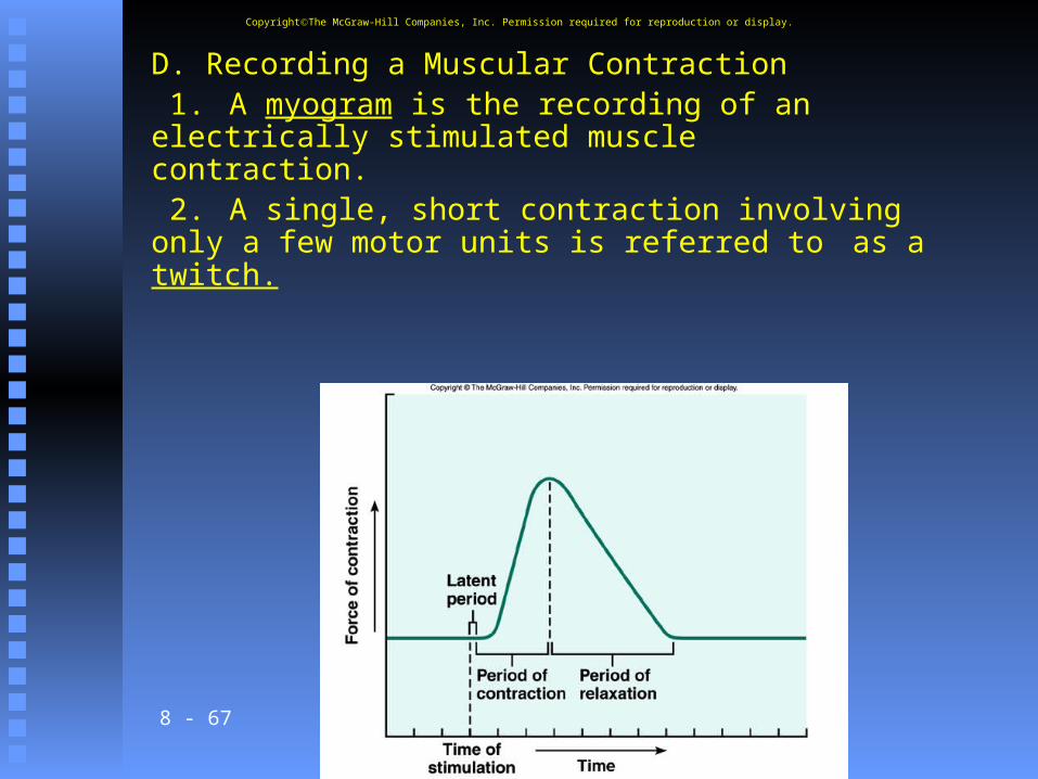
D. Recording a Muscular Contraction 1. A myogram is the recording of an electrically stimulated muscle contraction. 2. A single, short contraction involving only a few motor units is referred to as a twitch.
CopyrightThe McGraw-Hill Companies, Inc. Permission required for reproduction or display.
8 - 68
3. The time delay between when the stimulus is applied and when the muscle contracts is called the latent period, which is less than 0.01
second.4. The latent period is followed
by a period of contraction and a period of relaxation.
CopyrightThe McGraw-Hill Companies, Inc. Permission required for reproduction or display.
8 - 69
E. Summation 1. A muscle fiber receiving a series of stimuli of increasing frequency reaches a point when it is unable to relax completely and the force of individual twitches combine by the process of summation.2. If the sustained contraction lacks any relaxation, it is called a tetanic contraction.
CopyrightThe McGraw-Hill Companies, Inc. Permission required for reproduction or display.
8 - 70
F. Recruitment of Motor Units 1. An increase in the number of
activated motor units within a muscle at higher intensities of stimulation is called recruitment.
CopyrightThe McGraw-Hill Companies, Inc. Permission required for reproduction or display.
8 - 71
G. Sustained Contractions 1. Summation and recruitment
together can produce a sustained contraction of increasing strength.
2. Muscle tone is achieved by a continuous state of
sustained contraction of motor units within a muscle.
CopyrightThe McGraw-Hill Companies, Inc. Permission required for reproduction or display.
8 - 72
Smooth Muscles
A. Smooth Muscle Fibers1. Smooth muscle cells
are elongated with tapered ends, lack striations, and have a
relatively undeveloped sarcoplasmic reticulum.
CopyrightThe McGraw-Hill Companies, Inc. Permission required for reproduction or display.
8 - 73
2. Multiunit smooth muscle and visceral muscle are two types
of smooth muscles.a. In multiunit smooth
muscle, such as in the blood vessels and iris of the eye, fibers occur separately rather than as sheets.
CopyrightThe McGraw-Hill Companies, Inc. Permission required for reproduction or display.
8 - 74
b. Visceral smooth muscle occurs in sheets and is found in the walls of hollow organs; these fibers can stimulate one another and display rhythmicity, and are thus
responsible for peristalsis in hollow organs and
tubes.
CopyrightThe McGraw-Hill Companies, Inc. Permission required for reproduction or display.
8 - 75
B. Smooth Muscle Contraction1. The myosin-binding-to-actin
mechanism is mostly the same for smooth muscles and
skeletal muscles.2. Both acetylcholine and
norepinephrine stimulate and inhibit smooth muscle contraction, depending on the target muscle.
CopyrightThe McGraw-Hill Companies, Inc. Permission required for reproduction or display.
8 - 76
3. Hormones can also stimulate or inhibit contraction.
4. Smooth muscle is slower to contract and relax than is skeletal muscle, but can contract longer using the
same amount of ATP.
CopyrightThe McGraw-Hill Companies, Inc. Permission required for reproduction or display.
8 - 77
Cardiac Muscle A. The mechanism of contraction in
cardiac muscle is essentially the same as that for skeletal and smooth muscle, but with some differences.
B. Cardiac muscle has transverse tubules that supply extra calcium, and can thus contract for longer periods.
CopyrightThe McGraw-Hill Companies, Inc. Permission required for reproduction or display.
8 - 78
C. Complex membrane junctions, called intercalated disks,
join cells and transmit the force of contraction from one cell to the next, as well as aid in the rapid transmission of impulses throughout the heart.
D. Cardiac muscle is self-exciting and rhythmic, and the whole structure contracts as a unit.
CopyrightThe McGraw-Hill Companies, Inc. Permission required for reproduction or display.
8 - 79
Skeletal Muscle Actions A. Origin and Insertion
1. The immovable end of a muscle is the origin, while
the movable end is the insertion; contraction pulls the insertion toward the origin.
2. Some muscles have more than one insertion or origin.
CopyrightThe McGraw-Hill Companies, Inc. Permission required for reproduction or display.
8 - 80
B. Interaction of Skeletal Muscles
1. Of a group of muscles, the one doing the
majority of the work is the prime mover.
2. Helper muscles are called synergists; opposing
muscles are called antagonists.
CopyrightThe McGraw-Hill Companies, Inc. Permission required for reproduction or display.
8 - 81
Major Skeletal Muscles A. Muscles are named
according to any of the following criteria: size, shape, location, action, number of
attachments, or direction of its fibers.
CopyrightThe McGraw-Hill Companies, Inc. Permission required for reproduction or display.

8 - 82
F. Muscles that Move the Arm 1. Muscles connect the arm to the
pectoral girdle, ribs, and vertebral column, making the arm freely movable.
2. Flexors include the coracobrachialis and pectoralis
major.
CopyrightThe McGraw-Hill Companies, Inc. Permission required for reproduction or display.
8 - 83
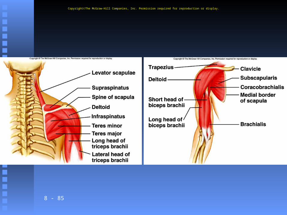
3. Extensors include the teres major
and latissimus dorsi.4. Abductors include the
supraspinatus and the deltoid.5. Rotators are the
subscapularis, infraspinatus, and teres minor.
CopyrightThe McGraw-Hill Companies, Inc. Permission required for reproduction or display.
8 - 84
G. Muscles that Move the Forearm 1. These muscles arise from the humerus or pectoral girdle and connect to the ulna and radius.
2. Flexors are the biceps brachii,
the brachialis, and the brachioradialis.
CopyrightThe McGraw-Hill Companies, Inc. Permission required for reproduction or display.
8 - 85
CopyrightThe McGraw-Hill Companies, Inc. Permission required for reproduction or display.
8 - 86
3. An extensor is the triceps brachii muscle.
4. Rotators include the supinator, pronator teres, and pronator quadratus.
CopyrightThe McGraw-Hill Companies, Inc. Permission required for reproduction or display.

8 - 87
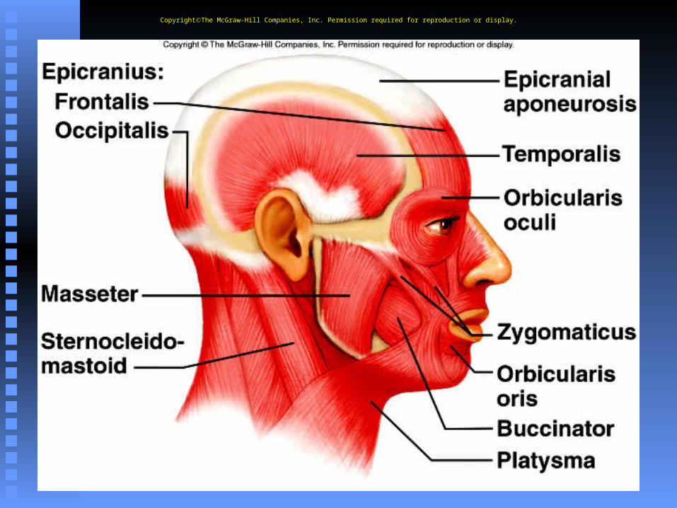
B. Muscles of Facial Expression 1. Muscles of facial expression
attach to underlying bones and overlying connective tissue of skin, and are responsible for the variety of
facial expressions possible in the human face.
2. Major muscles include the epicranius, orbicularis oculi, orbicularis oris, buccinator,
and zygomatigus.
CopyrightThe McGraw-Hill Companies, Inc. Permission required for reproduction or display.
8 - 88
CopyrightThe McGraw-Hill Companies, Inc. Permission required for reproduction or display.
8 - 89
C. Muscles of Mastication 1. Chewing movements
include up and down as well as side- to-side grinding motions of muscles attached to the skull and lower jaw.
2. Chewing muscles include masseter and temporalis.
CopyrightThe McGraw-Hill Companies, Inc. Permission required for reproduction or display.
8 - 90
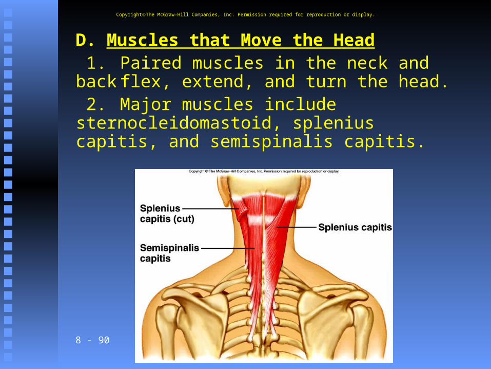
D. Muscles that Move the Head 1. Paired muscles in the neck and backflex, extend, and turn the head. 2. Major muscles include sternocleidomastoid, splenius capitis, and semispinalis capitis.
CopyrightThe McGraw-Hill Companies, Inc. Permission required for reproduction or display.
8 - 91
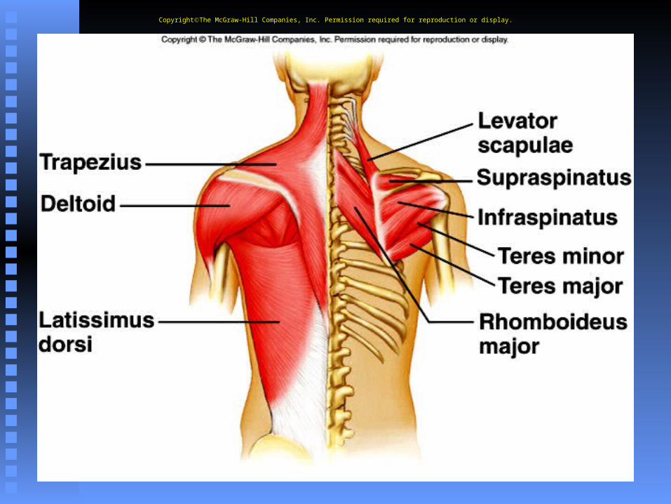
E. Muscles that Move the Pectoral Girdle 1. The chest and shoulder
muscles move the scapula. 2. Major muscles include the
trapezius, rhomboideus major, levator scapulae, serratus anterior,
and pectoralis minor.
CopyrightThe McGraw-Hill Companies, Inc. Permission required for reproduction or display.
8 - 92
CopyrightThe McGraw-Hill Companies, Inc. Permission required for reproduction or display.
8 - 93
F. Muscles that Move the Arm 1. Muscles connect the arm to the
pectoral girdle, ribs, and vertebral column, making the arm freely movable.
2. Flexors include the coracobrachialis and pectoralis
major.
CopyrightThe McGraw-Hill Companies, Inc. Permission required for reproduction or display.

8 - 94
3. Extensors include the teres major
and latissimus dorsi.4. Abductors include the
supraspinatus and the deltoid.5. Rotators are the
subscapularis, infraspinatus, and teres minor.
CopyrightThe McGraw-Hill Companies, Inc. Permission required for reproduction or display.

8 - 95
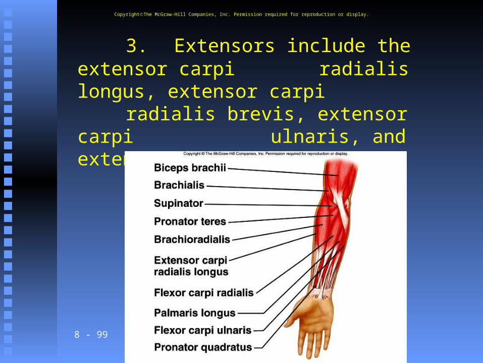
G. Muscles that Move the Forearm 1. These muscles arise from the humerus or pectoral girdle and connect to the ulna and radius.
2. Flexors are the biceps brachii,
the brachialis, and the brachioradialis.
CopyrightThe McGraw-Hill Companies, Inc. Permission required for reproduction or display.

8 - 96
CopyrightThe McGraw-Hill Companies, Inc. Permission required for reproduction or display.

8 - 97
3. An extensor is the triceps brachii muscle.
4. Rotators include the supinator, pronator teres, and pronator quadratus.
CopyrightThe McGraw-Hill Companies, Inc. Permission required for reproduction or display.
8 - 98
H. Muscles that Move the Wrist, Hand, and Fingers1. Movements of the hand are
caused by muscles originating from the distal zumerus, and the radius and ulna.
2. Flexors include the flexor carpi radialis, flexor carpi ulnaris,
palmaris longus, and flexor digitorum profundus.
CopyrightThe McGraw-Hill Companies, Inc. Permission required for reproduction or display.
8 - 99
3. Extensors include the extensor carpi radialis longus, extensor carpi radialis brevis, extensor carpi ulnaris, and extensor digitorum.
CopyrightThe McGraw-Hill Companies, Inc. Permission required for reproduction or display.
8 - 100
I. Muscles of the Abdominal Wall 1. This group of muscles connects the
rib cage and vertebral column to the pelvic girdle.a. A band of tough connective
tissue, the linea alba, extending from the xiphoid process to the symphysis pubis, serves as an
attachment for certain abdominal wall muscles.
CopyrightThe McGraw-Hill Companies, Inc. Permission required for reproduction or display.
8 - 101
2. These four muscles include:
external oblique, internal oblique, transverse abdominis, and rectus abdominis.
CopyrightThe McGraw-Hill Companies, Inc. Permission required for reproduction or display.
8 - 102
J. Muscles of the Pelvic Outlet 1. The superficial urogenital
diaphragm fills the space within the pubic arch, and the deeper pelvic diaphragm forms the floor of the pelvic cavity.
2. Pelvic diaphragm includes the levator ani.
CopyrightThe McGraw-Hill Companies, Inc. Permission required for reproduction or display.
8 - 103
3. Urogenital diaphragm: includes the superficial transversus, perinei, bulbospongiosus, and
ischiocavernosus.
CopyrightThe McGraw-Hill Companies, Inc. Permission required for reproduction or display.
8 - 104
K. Muscles that Move the Thigh 1. The muscles that move the
thigh are attached to the femur and to the pelvic girdle.
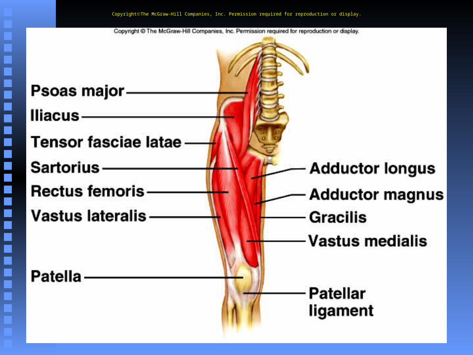
2. Anterior group includes the psoas major and iliacus.
CopyrightThe McGraw-Hill Companies, Inc. Permission required for reproduction or display.
8 - 105
CopyrightThe McGraw-Hill Companies, Inc. Permission required for reproduction or display.
8 - 106
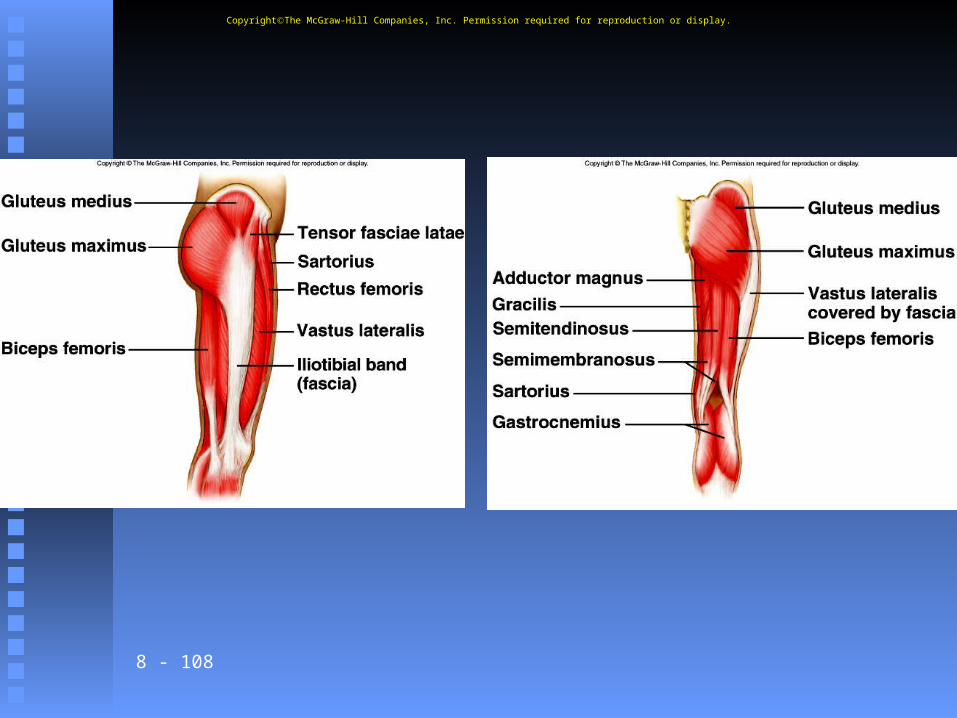
3. Posterior group is made up of the gluteus maximus, gluteus
medius, gluteus minimus, and tensor fasciae latae.
4. Thigh adductors include the adductor longus, adductor magnus, and gracilis.
CopyrightThe McGraw-Hill Companies, Inc. Permission required for reproduction or display.
8 - 107
L. Muscles that Move the Leg 1. This group connects the tibia or
fibula to the femur or pelvic girdle.2. Flexors are the biceps femoris,
semitendinosus semimembranosus, and sartorius.
3. An extensor is the quadruceps femoris group made up of four parts: rectus femoris, vastus
lateralis, vastus medialis, and vastus intermedius.
CopyrightThe McGraw-Hill Companies, Inc. Permission required for reproduction or display.
8 - 108
CopyrightThe McGraw-Hill Companies, Inc. Permission required for reproduction or display.
8 - 109
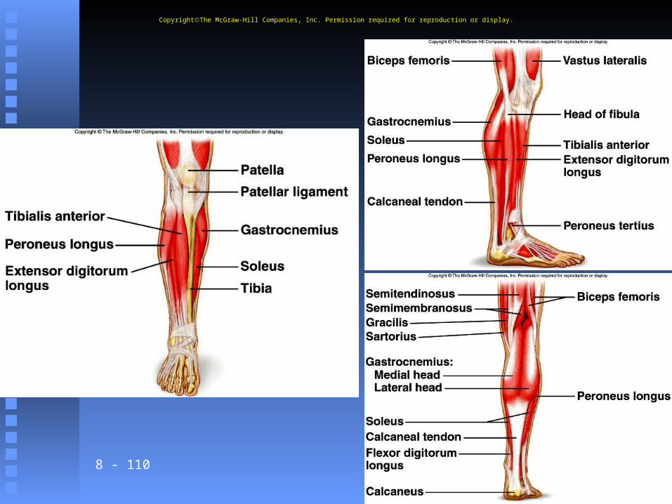
M. Muscles that Move the Ankle, Foot, and Toes 1. Muscles that move the foot are
attached to the femur, fibula, or tibia, and move the foot upward, downward, or in a turning motion.
2. Dorsal flexors include the tibialis anterior, peroneus tertius,
and extensor digitorum longus.
CopyrightThe McGraw-Hill Companies, Inc. Permission required for reproduction or display.
8 - 110
CopyrightThe McGraw-Hill Companies, Inc. Permission required for reproduction or display.
8 - 111
3. Plantar flexors are the gastrocnemius
soleus, and flexor digitorum longus.
4. An invertor is the tibialis posterior.
5. An evertor is the peroneus longus.
CopyrightThe McGraw-Hill Companies, Inc. Permission required for reproduction or display.
112
Cellular Respiration
Food to ATP

8 - 113
The EquationCC66HH1212OO66 + 6O + 6O22 6CO 6CO22 + 6H + 6H220 + ATP0 + ATP
CC66HH1212OO66 = glucose = glucose
6O6O22 = oxygen gas = oxygen gas
6CO6CO22 = carbon dioxide = carbon dioxide
6H6H220 = water0 = water
ATP = energyATP = energy
8 - 114
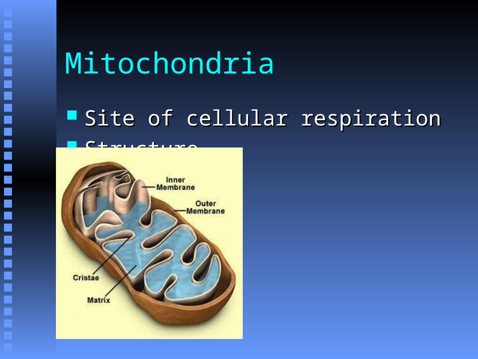
Mitochondria
Site of cellular respirationSite of cellular respiration StructureStructure

8 - 115
Steps of Cellular Respiration

8 - 116
Glycolysis

8 - 117
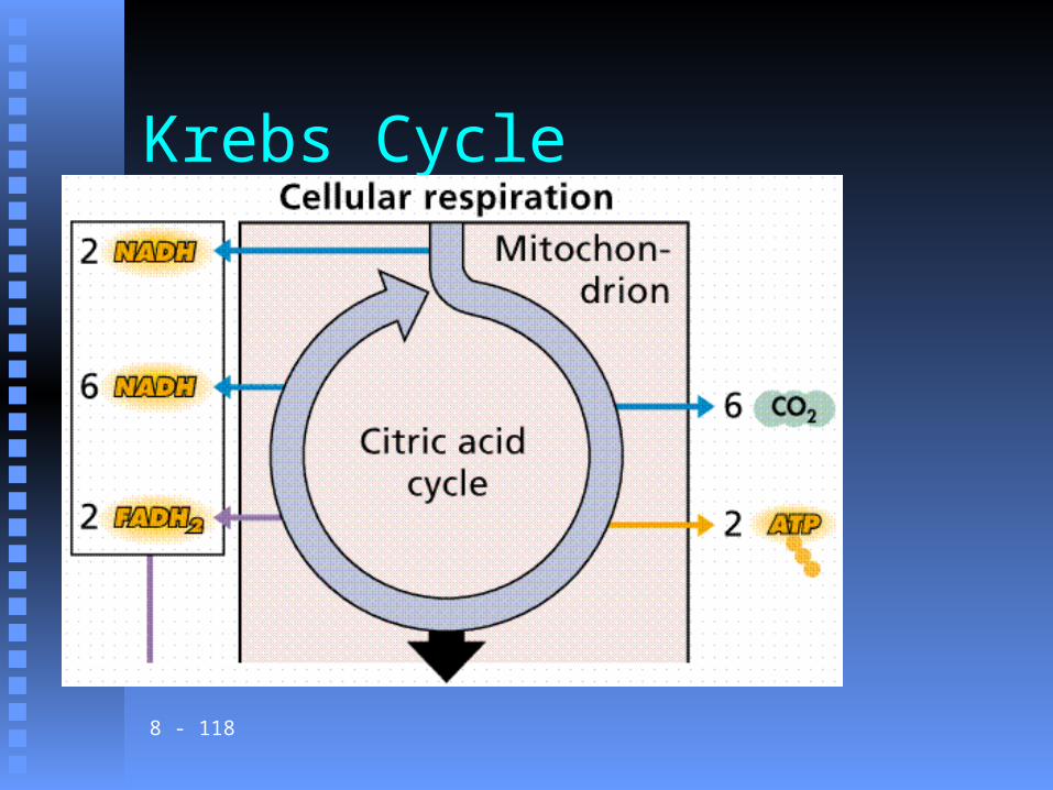
Krebs Cycle
8 - 118
Krebs Cycle

8 - 119
ETC & Oxidative Phosphorylation

8 - 120
ETC & Oxidative Phosphorylation
8 - 121
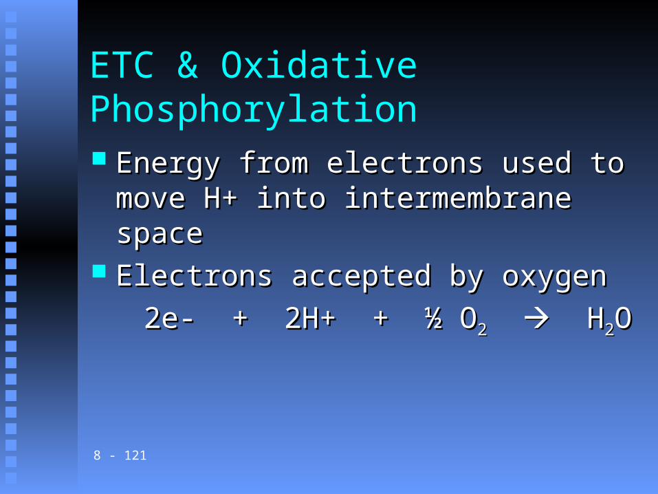
ETC & Oxidative Phosphorylation
Energy from electrons used to move H+ Energy from electrons used to move H+ into intermembrane spaceinto intermembrane space
Electrons accepted by oxygenElectrons accepted by oxygen
2e- + 2H+ + ½ O2e- + 2H+ + ½ O22 H H22OO
8 - 122
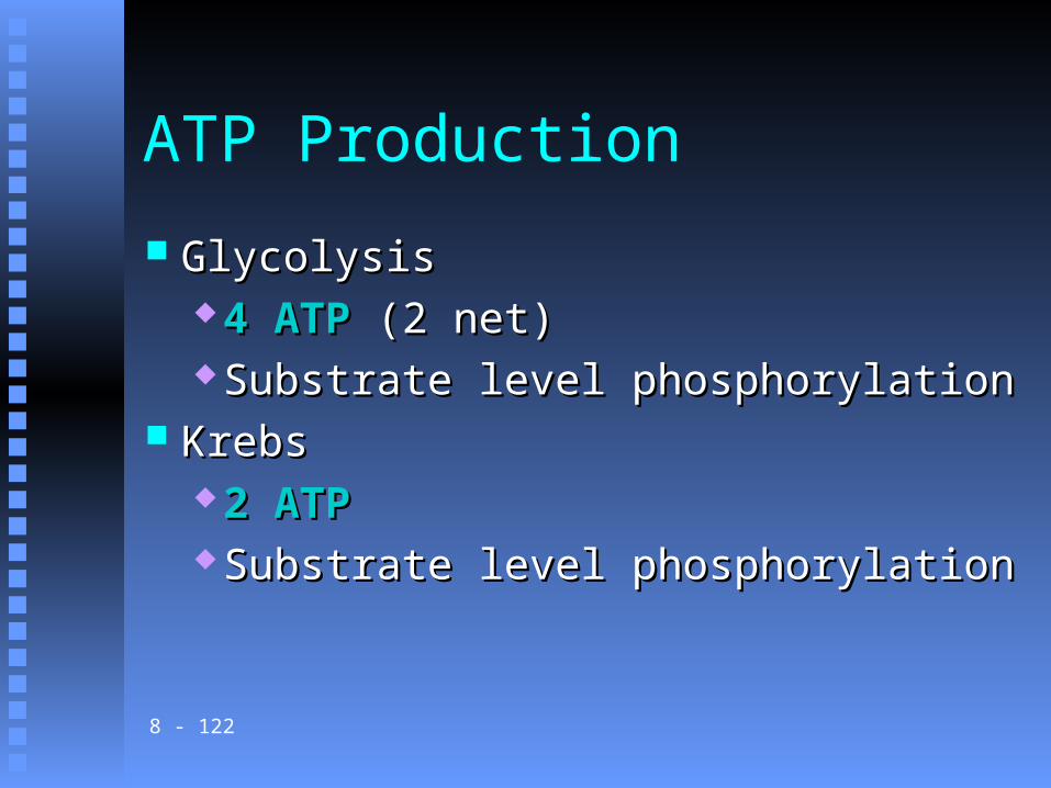
ATP Production
GlycolysisGlycolysis 4 ATP4 ATP (2 net) (2 net) Substrate level phosphorylationSubstrate level phosphorylation
KrebsKrebs 2 ATP2 ATP Substrate level phosphorylationSubstrate level phosphorylation
8 - 123
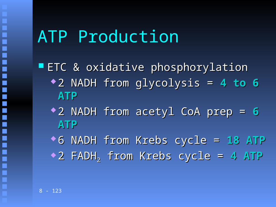
ATP Production
ETC & oxidative phosphorylationETC & oxidative phosphorylation 2 NADH from glycolysis = 2 NADH from glycolysis = 4 to 6 ATP4 to 6 ATP 2 NADH from acetyl CoA prep = 2 NADH from acetyl CoA prep = 6 ATP6 ATP 6 NADH from Krebs cycle = 6 NADH from Krebs cycle = 18 ATP18 ATP 2 FADH2 FADH22 from Krebs cycle = from Krebs cycle = 4 ATP4 ATP
8 - 124
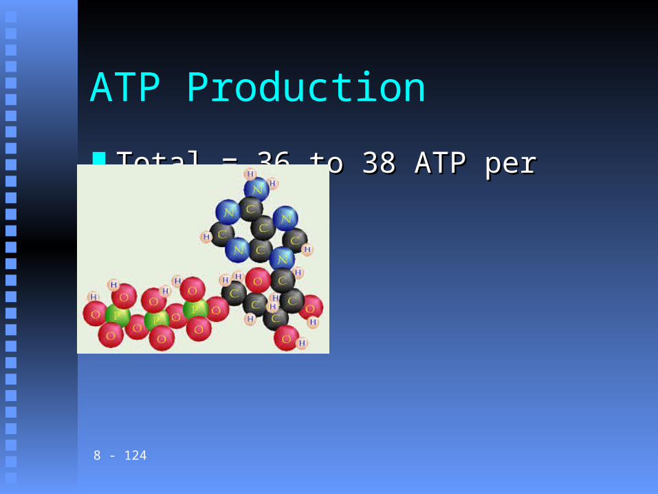
ATP Production
Total = 36 to 38 ATP per glucoseTotal = 36 to 38 ATP per glucose
8 - 125
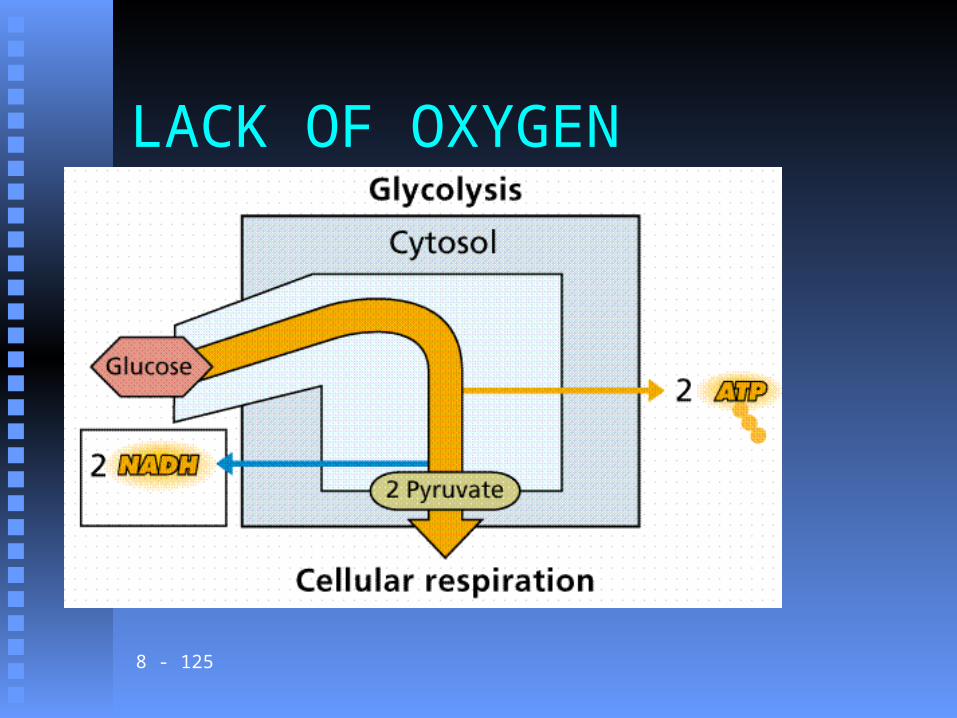
LACK OF OXYGEN

8 - 126
Pyruvate becomes LACTIC ACID
Pyruvate

8 - 127


![Muscular Hypertrophy.pptx [Read-Only] · 2018. 8. 29. · Title: Microsoft PowerPoint - Muscular Hypertrophy.pptx [Read-Only] Author: pwarren Created Date: 8/29/2018 9:25:45 AM](https://static.fdocuments.net/doc/165x107/60fef9acee3f2d51f52382b5/muscular-read-only-2018-8-29-title-microsoft-powerpoint-muscular-read-only.jpg)