6b VESCICOLE EXTRACELLULARI 2016 Casi particolari · una via di maggiore efficienza per lo scambio...
26
30/03/2016 1 Microvescicole Seminari http://biomarkerinsights.qiagen.com/category/liquid‐biopsy/exosomes‐microvesicles/ Yáñez‐Mó M. Biological properties of extracellular vesicles and their physiological functions. J Extracell Vesicles. 2015 May 14;4:27066.. Vescicole extracellulari isolate dal fluidi corporei
-
Upload
truongdang -
Category
Documents
-
view
212 -
download
0
Transcript of 6b VESCICOLE EXTRACELLULARI 2016 Casi particolari · una via di maggiore efficienza per lo scambio...

30/03/2016
1
MicrovescicoleSeminari
http://biomarkerinsights.qiagen.com/category/liquid‐biopsy/exosomes‐microvesicles/
Yáñez‐MóM. Biological properties of extracellular vesicles and their physiological functions. J Extracell Vesicles. 2015 May 14;4:27066..
Vescicole extracellulari isolate dal fluidi corporei

30/03/2016
2
Microambiente tumoralehttp://www.jci.org/articles/view/57099/figure/1
Baginska J, Viry E, Paggetti J, Medves S, Berchem G, Moussay E, Janji B. The critical role of the tumor microenvironment in shaping natural killer cell‐mediated anti‐tumor immunity. Front Immunol. 2013 Dec 25;4:490.

30/03/2016
3
Kucharzewska P, Belting M. Emerging roles of extracellular vesicles in the adaptive response of tumour cells to microenvironmental stress. J Extracell Vesicles. 2013 Mar 5;2. doi: 10.3402/jev.v2i0.20304. eCollection 2013.
Heterotypic cellular interactions in the tumourmicroenvironment.
The tumour microenvironment is a complex scaffold of an extracellular matrix (ECM) and various cell types. In addition to malignant cells, vascular cells, stromal cellsand immune cells are common cellular residents of the tumour niche. Tumour cellsmould this environment for their own needs via intercellular communicationpathways, such as direct cell‐to‐cell contacts and the release of growth factors, matrix metalloproteases, ECM proteins and extracellular vesicles (EVs). Tumour cell‐mediated stromal modifications include: suppression of anti‐tumoural immune responses, deposition and degradation of ECM components, induction of vascularnetwork formation and recruitment of stromal cells and tumour‐promoting immune cells. In turn, heterogeneoustumour microenvironmental components create a favourable environment for tumour growth and dissemination. Various tumour microenvironmentalstressors are inherent features of solid tumours thatprofoundly modify the tumour milieu and accelerate tumour progression towards malignancy.
Kucharzewska P, Belting M. Emerging roles of extracellularvesicles in the adaptive response of tumour cells to microenvironmental stress. J Extracell Vesicles. 2013 Mar 5;2. doi: 10.3402/jev.v2i0.20304. eCollection 2013.
Extracellular vesicles (EVs) are potential conveyors of stress‐mediated tumourprogression.
EVs are shed from various cellular components of the tumour milieu to mediate exchange of signalling proteins and genetic material, which altogether may support tumour growth and progression. Diverse tumour microenvironmentalstress conditions augment tumour‐promoting activities of EVs by modulating their secretion and trafficking in the extracellular space, as well as altering their molecular content and functional activity. Upon release, EVs may also enter the circulation and mediate long‐range exchange of EV‐associated cargo that may support the process of pre‐metastatic niche formation. In addition, circulating EVs carrying multifaceted, molecular stress signatures may offer unique, non‐invasive biomarkers that can be used in the management of cancer patients.

30/03/2016
4
Baginska J, Viry E, Paggetti J, Medves S, Berchem G, Moussay E, Janji B. The critical role of the tumor microenvironment in shaping natural killer cell‐mediated anti‐tumor immunity. Front Immunol. 2013 Dec 25;4:490.
Baginska J, Viry E, Paggetti J, Medves S, Berchem G, Moussay E, Janji B. The critical role of the tumor microenvironment in shaping natural killer cell‐mediated anti‐tumor immunity. Front Immunol.
2013 Dec 25;4:490.
Impairment of NK cell function by tumor‐derived exosomes. Tumor cells secrete extracellular vesicles called exosomes. Tumor‐derived exosomes contain numerous factors able to modulate the function of NK cells such as MICA/B, ULBP3, TGF‐β, PI‐9, and different microRNAs. Exosome‐derived MICA/B, ULBP3, TGF‐β, and miR‐1245 can decrease NKG2D on the surface of NK cells, while PI‐9 degrades granzyme B. Tumor‐derived exosomes can also decrease the level of perforin in NK cells by a still‐unknown mechanism.

30/03/2016
5
Gai C, Carpanetto A, Deregibus MC, Camussi G. Extracellular vesicle‐mediated modulation of angiogenesis. Histol Histopathol. 2016 Apr;31(4):379‐91.
angiogenic
Le vescicole extracellulari (Evs) derivate dai tumori (tEVs) influenzano diversi aspetti della progressione tumorale.
A: Le EVs versate dai tumori contengonometalloproteinasi che sono responsabili dalladegradazione della matrice facilitando l’invasionetumorale;
B: le tEVs permettono alle cellule tumorali di sopravvivere alla chemioterapia e all’apoptosimediante efflusso dei farmaci e della caspasi 3, rispettivamente;
C: Le tEVs stimolano la secrezione di fattori pro‐angiogenici dalle cellule stromali e facilitano la proliferazione delle cellule endoteliali con ciòpromuovendo l’angiogenesi e permettendo la crescita tumorale. L’angiogenesi è inoltre influenzatadal rilascio di mRNA e miRNA mediante tEVs.
D: Le tEVs rilasciate da molte cellule tumoraliespongono Fas ligand, che induce l’apoptosi dellecellule T e inibisce la funzione delle cellule dellarisposta immunitaria adattativa perciòpermettendo alle cellule tumorali di evaderel’immunosorveglianza.
Turturici G, Tinnirello R, Sconzo G, Geraci F. Extracellular membrane vesicles as a mechanism of cell‐to‐cell communication: advantages and disadvantages. Am J Physiol Cell Physiol. 2014 Apr 1;306(7):C621‐33.

30/03/2016
6
Tumor‐derived EVs promote tumor growth via multiple routes
Tumor‐derived EVs suppress anti‐tumor immune responses by inhibiting T‐cell activation and proliferation and stimulating their apoptosis. EVs produced by tumor cells also induce regulatory T cells and MDSCs and inhibit the cytotoxicity of NK and CD8+ T cells Tumor‐derived EVs are taken up by endothelial cells promoting angiogenesis and tumor invasion; the expression of CD147, D6.1A, tissue factor and EGFR in EVs, the transfer of pro‐angiogenic components and the induction of MMPs play a role in the pro‐angiogenic effects of tumor EVs Tumor EVs also contribute to tumor growth by stimulating tumor proliferation and inducing metastatic behavior in bone‐marrow progenitors
Gutiérrez‐Vázquez C, Villarroya‐Beltri C, MittelbrunnM, Sánchez‐Madrid F. Transfer of extracellular vesicles during immune cell‐cell interactions. Immunol Rev. 2013 Jan;251(1):125‐42.
Robbins PD, Morelli AE. Regulation of immune responses by extracellular vesicles. Nat Rev Immunol. 2014 Mar;14(3):195‐208.
RUOLO DELLE VESCICOLE EXTRACELLULARI NELLA REGOLAZIONE DELL’IMMUNITA’ DEI TUMORI E DEI MICROORGANISMI CHE PUO’ ESSERE MODIFICATA PER APPLICAZIONI TERAPEUTICHE

30/03/2016
7
Camussi G, Deregibus MC, Tetta C. Tumor‐derived microvesicles and the cancer microenvironment. Curr Mol Med. 2013 Jan;13(1):58‐67.
Contenuto e funzioni di vescicole extracellulari rilasciateda differenti tipi di cellule tumorali – 1
Contenuto e funzioni di vescicole extracellulari rilasciateda differenti tipi di cellule tumorali – 2
Camussi G, Deregibus MC, Tetta C. Tumor‐derived microvesicles and the cancer microenvironment. Curr Mol Med. 2013 Jan;13(1):58‐67.

30/03/2016
8
Camussi G, Deregibus MC, Tetta C. Tumor‐derived microvesicles and the cancer microenvironment. Curr Mol Med. 2013 Jan;13(1):58‐67.
Contenuto e funzioni di vescicole extracellulari rilasciateda differenti tipi di cellule tumorali – 3
Azmi AS, Bao B, Sarkar FH. Exosomes in cancer development, metastasis, and drug resistance: a comprehensive review. Cancer Metastasis Rev. 2013 Dec;32(3‐4):623‐42.
L’esportazione, mediata dagli exosomi, di materiale biologico può indurre un microambiente favorevole allaresistenza.
I fattori rilasciati dagli exosomi possonopromuovere: (a) morfologia cellulare tipoEpithelial‐to‐Mesenchymal Transition (EMT), che dà origine a staminalità; b) promuovere la formazione di cellule tipofibroblastico che provocano la reazionedesmoplastica (reazione stromale); (c) promuoveremeccanismi di fugaimmunitaria; e (d) promuovereangiogenesi e metastasi. I miRNAs espulsidagli exosomi possono regolare molteplicivie di segnalamento che promuovonocumulativamente un fenotipo resistentenella maggior parte dei tumori.
Ruolo degli exosomi nel sostenere le reti di resistenza tumorale

30/03/2016
9
Azmi AS, Bao B, Sarkar FH. Exosomes in cancer development, metastasis, and drug resistance: a comprehensive review. Cancer Metastasis Rev. 2013 Dec;32(3‐4):623‐42.
Il diagramma illustra un “impacchettamento” intracellulare di farmaci chimici e/o dei loroprodotti di degradazione (forme attive). Tali farmaci residenti negli exosomi possono essereespulsi dalle cellule provocando una minore efficacia del farmaco; questo è un processo diversodagli altri meccanismi di trasporto dei farmaci.
Meccanismi di estrusione dei farmaci ‐ Ipotesi
Microvescicole
Risposta immunitaria
http://www.gene‐quantification.de/exosomes.html

30/03/2016
10
Yáñez‐MóM. Biological properties of extracellular vesicles and their physiological functions. J Extracell Vesicles. 2015 May 14;4:27066.
Ruolo fisiologico delle EVs derivate da celluledel Sistema immunitario innato
Yáñez‐MóM. Biological properties of extracellular vesicles and their physiologicalfunctions. J Extracell Vesicles. 2015 May 14;4:27066. doi: 10.3402/jev.v4.27066. eCollection
2015.
Le EVs nel Sistema Immunitario: presentazione di antigene e immunitàacquisita
Le EVs possono giocare un ruolo sianell’origine che nella progressione nellarisposta immunitario acquisita, attuandoa diversi livelli e su cellule diverse.
La figura reassume come le EVs sonocoinvolte in tale processo.
APC: “antigen‐presenting cell”.Treg: cellule T regolatorie.NK: “natural Killer cell”MHC: “major histocompatibility complex”.

30/03/2016
11
Yáñez‐MóM. Biological properties of extracellular vesicles and their physiological functions. J Extracell Vesicles. 2015 May 14;4:27066
Le EVs derivate dalle MSCs possono indurre effetti differenti a seconda della cellulabersaglio, come qui riassunto. DC: cellula dendritica; NK: “natural killer.»
EVs derivate da cellule staminali mesenchimali (MSC)
Théry C, Ostrowski M, Segura E. Membrane vesicles as conveyors of immune responses. Nat Rev Immunol. 2009 Aug;9(8):581‐93.
Vescicole di membrana come vettori di risposte immunitarie
Porzioni della membrana plasmatica di cellule coinvolte nella risposta immunitaria possono essere trasferite fra cellule, sia tramite contatto diretto (mediante i processi recentemente descritti di «nibbling» (rosicchiamento), trogocitosi e nanotubi) che tramite la secrezione di vescicole di membrana.
Le conseguenze funzionali di tali trasferimenti includono l’induzione, amplificazione e/o modulazione delle risposte immunitarie nonché l’acquisizione di nuove proprietà funzionali da parte delle cellule che le ricevono, quali capacità migratorie o metastatiche.
Inoltre, nelle vescicole di membrana secrete sono stati identificati mRNAs e microRNAs, e ciò ha sollevato l’eccitante ipotesi che il trasferimento di materiale genetico potesse influenzare il comportamento delle cellule riceventi.
Complessivamente, tali dati portano all’ipotesi che il trasferimento di membrane sia un modo comune di comunicazione intercellulare.

30/03/2016
12
Glossario (vedi figura Davis)
«Nibbling» (rosicchiamento): Capacità che hanno le cellule dendritiche di strappare fisicamente frammenti di membrana da cellule vicine durante un contatto stretto senza indurre la morte della cellula donatrice.
Trogocitosi: Trasferimento di frammenti della membrana plasmatica da una cellula ad un’altra senza indurre la morte cellulare. Questo processo è mediato da segnalamento mediato da recettore in seguito a contatto cellula‐cellula.
Nanotubi: Canale membranoso di 50‐200 nm di diametro che collega cellule per lunghe distanze.
Vescicole di membrana: Struttura sferica o approssimativamente sferiche limitata da un bilayer lipidico che racchiude un carico solubile.
Théry C, Ostrowski M, Segura E. Membrane vesicles as conveyors of immune responses. Nat Rev Immunol. 2009 Aug;9(8):581‐93.
MittelbrunnM, Sánchez‐Madrid F. Intercellular communication: diverse structures for exchange of genetic information. Nat Rev Mol Cell Biol. 2012 Apr 18;13(5):328‐35.
La sinapsi immunologica funge da piattaforma per facilitare il passaggio di material genetico tra le
cellule Durante la formazione di una sinapsi immunologica, le molecole coinvolte nel riconoscimento dell’antigene (ad es. il ”T Cell Receptor; TCR) e le molecole del “peptide‐loaded major histocompatibility complex; pMHC) si muovono verso un aggregato centrale circondato da un anello periferico arricchito in molecole di adesione (ad es. l’integrina “leukocyte function‐associated antigen 1” (LFA1) e le ”intercellular cell adhesion molecules; (ICAMs) e di citoscheletro di actina.
Il linfocito T orienta il suo “microtubule‐organizing centre (MTOC) e i compartimenti di secrezione (ad es. l’apparato di Golgi e “i multivesicular bodies» (MVBs) verso la “antigen presenting cell” (APC).
Noi proponiamo che la sinapsi immunologica fornisce una via di maggiore efficienza per lo scambio di materiale geneticomediante la combinazione di differenti meccanismi, incluso la secrezione polarizzata di exosomi carichi di microRNA (miRNA), transendocitosie ponti di membrana. I patogeni, incluso batteri e virus, si appropriano delle sinapsi biologiche per propagarsi da cellula a cellula.
30/03/2016
13
Gutiérrez‐Vázquez C, Villarroya‐Beltri C, MittelbrunnM, Sánchez‐Madrid F. Transfer of extracellular vesicles during immune cell‐cell interactions. Immunol Rev. 2013 Jan;251(1):125‐42.
Typical molecular composition of T‐cellexosomes
Membrane and luminal distribution of moleculespredicted to be found in a typical exosomeproduced by a T lymphocyte. TCR: T‐cell receptor; TGFβ: transforming growth factor beta; ICAMs: intercellular adhesion molecule family; CXCR4: C‐X‐C chemokine receptor type 4 or CD184; Tsg101: tumor susceptibility gene 101; TRAIL: TNF‐relatedapoptosis‐inducing ligand; FasL: Fas ligand or CD95L; TfR: transferrin receptor; Mfge8: milk‐fatglobule‐EGF factor 8; ALIX: ALG‐2‐interacting protein X; GAPDH: glyceraldehyde 3‐phosphate dehydrogenase; ERMs: ezrin, radixin and moesinproteins. For more information about exosomecomposition see http://www.exocarta.org.
Gutiérrez‐Vázquez C, Villarroya‐Beltri C, MittelbrunnM, Sánchez‐Madrid F. Transfer of extracellularvesicles during immune cell‐cell interactions. Immunol Rev. 2013 Jan;251(1):125‐42.
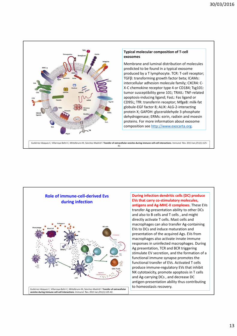
During infection dendritic cells (DC) produce EVs that carry co‐stimulatory molecules, antigens and Ag‐MHC‐II complexes. These EVs transfer Ag‐presentation ability to other DCs and also to B cells and T cells , and might directly activate T cells. Mast cells and macrophages can also transfer Ag‐containing EVs to DCs and induce maturation and presentation of the acquired Ags. EVs from macrophages also activate innate immune responses in uninfected macrophages. During Ag presentation, TCR and BCR triggering stimulate EV secretion, and the formation of a functional immune synapse promotes the functional transfer of EVs. Activated T cells produce immune‐regulatory EVs that inhibit NK cytotoxicity, promote apoptosis in T cells and Ag‐carrying DCs , and decrease DC antigen‐presentation ability thus contributing to homeostasis recovery.
Role of immune‐cell‐derived Evsduring infection

30/03/2016
14
Vescicole extracellulariFEGATO
Masyuk AI, Masyuk TV, Larusso NF. Exosomes in the pathogenesis, diagnostics and therapeutics of liver diseases. J Hepatol. 2013 Sep;59(3):621‐5.
Exosome release
(A) Exosomes containing membrane and cytosolic proteins, mRNAs, and miRNAs, are derived from the multivesicular body (MVB)
sorting pathway. Membrane proteins are oriented in a fashion (extracellular region out) that permits profound biological autocrine
and paracrine effects. (B) Exosomes isolated from rat bile have a cup‐ or “deflated football”‐ shaped morphology by transmission
electron microscopy (TEM), but they have a perfectly round shape by scanning electron microscopy (SEM). (C) In cholangiocytes of
mouse liver, MVBs containing exosomes (arrows) (a) move to the apical plasma membrane (APM) (b), and release exosomes into the
bile duct lumen by exocytosis (c).

30/03/2016
15
Exosomes in intercellular signaling
(A) In the liver, exosomes derived from hepatocytes and cholangiocytes are transported by bile flow to target cholangiocytes with which they may interact via several mechanisms depending on their cargo and biological properties. They can fuse with the plasma membrane and deliver their content into the cytoplasm of a target cell; interact with receptors on the apical plasma and ciliary membrane inducing intracellular signaling; and endocytosed for recycling. (B and C) Biliary exosomes surround and attach to cholangiocytecilia in mouse liver as viewed by TEM (B) and SEM (C), supporting the involvement of exosomes and cilia in mechanisms of intercellular signaling.
Masyuk AI, Masyuk TV, Larusso NF. Exosomes in the pathogenesis, diagnostics and therapeutics of liver
diseases. J Hepatol. 2013 Sep;59(3):621‐5.
Larusso NF, Masyuk TV. The role of cilia in the regulation of bile flow. Dig Dis. 2011;29(1):6‐12.
Gli exosomi sono coinvolti nella funzione
chemosensoriale delle cilia primarie dei
colangiociti (cellule dei dotti biliari). Gli
exosomi sono piccole (30 ‐100 nm di
diametro) vescicole extracellulari rivestite da
membrana. Sono derivati da vescicole
interne di corpi multivescicolari (MVBs) che
si fondono con la membrana plasmatica in
una modalità simile all’esocitosi e rilasciano
il loro contenuto nello spazio extracellulare
(schema). La presenza di vescicole tipo
exosomi di 50‐80 nm di diametro nel lume
dei dotti intraepatici di topi «wild‐type» e
policistici è stata confermata da microscopia
elettronica a trasmissione (destra, panelli di
sopra). Queste vescicole circondono cilia dei
colangiociti ed alcune sembrano attaccarsi
alla membrana ciliare e dei microvilli.
L’immagine del microscopio elettronico a
scansione (SEM) (destra, panello di sotto)
suggerisce che vescicole simili ad exosomi di
fatto si leghino alle cilia.

30/03/2016
16
Masyuk AI, Huang BQ, Ward CJ, Gradilone SA, Banales JM, Masyuk TV, Radtke B, Splinter PL, LaRusso NF. Biliary exosomes influence cholangiocyteregulatory mechanisms and proliferation through interaction with primarycilia. Am J Physiol Gastrointest Liver Physiol. 2010 Oct;299(4):G990‐9.
Exosome‐like vesicles surround and attach to mouse cholangiocyte [primary] cilia in vivo.
By transmission (TEM; A and B) and scanning (SEM; C and D) electron microscopy, exosome‐like vesicles (black and white arrows) are present in the lumen of intrahepatic bile ducts in the wild‐type (A and C) and Pkhd1del2/del2 (B and D) mice. The vesicles surround the cilium (B) and attach to this organelle (A–D) and microvilli (A) of the cholangiocyteapical plasma membrane.
Masyuk AI, Huang BQ, Ward CJ, Gradilone SA, Banales JM, Masyuk TV, Radtke B, Splinter PL, LaRusso NF. Biliary exosomes influence cholangiocyte regulatory mechanisms and proliferation through interaction with primary cilia. Am J Physiol Gastrointest Liver Physiol. 2010 Oct;299(4):G990‐9.
Hepatocyte multivesicular bodies (MVBs) and luminal vesicles are positive for an exosomal marker, CD63
[tetraspanin].
MVBs and intraluminal vesicles positive for an exosomal marker, CD63, (black arrows) were observed in normal rat
hepatocytes. MVBs are seen in a proximity to the hepatic canaliculus (a). CD63‐positive vesicles are also seen in the
canalicular lumen (b), suggesting that hepatocytes release exosomes in vivo.

30/03/2016
17
Lemoinne S, Thabut D, Housset C, Moreau R, Valla D, Boulanger CM, Rautou PE. The emerging roles of microvesicles in liver diseases. Nat Rev GastroenterolHepatol. 2014 Jun;11(6):350‐61.
Lemoinne S, Thabut D, Housset C, Moreau R, Valla D, Boulanger CM, Rautou PE. The emerging roles of microvesicles in liver diseases. Nat Rev GastroenterolHepatol. 2014 Jun;11(6):350‐61.

30/03/2016
18
Lemoinne S, Thabut D, Housset C, Moreau R, Valla D, Boulanger CM, Rautou PE. The emerging roles of microvesicles in liver diseases. Nat Rev GastroenterolHepatol. 2014 Jun;11(6):350‐61.

30/03/2016
19
Vescicole extracellulariIntestino
http://aasarts.com/wp‐content/uploads/2014/06/Enterocyte‐Final.jpg
Mallegol J, van Niel G, Heyman M. Phenotypic and functional characterization of intestinal epithelial exosomes. Blood Cells Mol Dis. 2005 Jul‐Aug;35(1):11‐6.
Intestinal epithelial cells (IEC) secrete exosomes. IEC express accessory molecules (MHC class II, invariant chain, HLA‐DM) and are considered as non‐professional antigen presenting cells. The lack of direct contact between IEC and CD4+ T cells limits direct antigen presentation in vivo. However, IEC secrete exosomes which are small membrane vesicles originating from the MHC class II‐enriched compartment (MIIC) and are released by exocytosis of these compartments in the external medium. Such epithelial exosomes bear class II/peptide complexes and molecules potentially involved in cell– cell or cell –matrix interactions.

30/03/2016
20
van Niel G, Raposo G, Candalh C, Boussac M, Hershberg R, Cerf‐Bensussan N, Heyman M. Intestinal epithelial cells secrete exosome‐like vesicles. Gastroenterology. 2001 Aug;121(2):337‐49.
A model for the molecular structure of epithelial‐
derived exosomes. Ubiquitously expressed
molecules such as enzymes of the intracellular
metabolism (pyruvate kinase M2, creatine kinase, α‐
enolase, phosphoglycerate kinase, glyceraldehyde‐3‐
phosphate dehydrogenase, L‐lactate dehydrogenase)
and cytoskeleton proteins (actin, tubulin), as well as
molecules possibly involved in antigen presentation
(MHC class I, MHC class II, CD63), were found in
both apical and basolateral exosomes. Apical
exosomes also carried molecules involved in apical
addressing of endosomes (syntaxin 3, syntaxin‐
binding protein 2), whereas basolateral exosomes
had molecules that might act as adhesion or
costimulatory molecules (A33 antigen and epithelial
cell surface antigen).
Ménard S, Cerf‐Bensussan N, Heyman M. Multiple facets of intestinal permeability and epithelial handling of dietary antigens. Mucosal Immunol. 2010 May;3(3):247‐59.
Under steady‐state condition, molecules of molecular weight (MW) > 600 Da (such as food antigens, peptides) are sampled by the epithelial cells by endocytosis at the apical membrane and transcytosis toward the lamina propria.
During transcytosis, full‐length peptides or proteins are partly degraded in acidic and lysosomal compartments and released in the form of amino acids (total degradation) or breakdown products (partial degradation) at the basolateral pole of enterocytes. Early endosomes containing partially degraded food antigens meet the major histocompatibility complex (MHC) class II‐enriched compartment (MIIC) where exogenous peptides are loaded on MHC class II molecules. Inward invagination of MIIC compartment lead to the formation of exosomes, which are small membrane vesicles (40 – 90 nm) bearing MHC class II / peptide complexes at their surface. Exosomes can diffuse in the basement membrane and interact with local immune cells. Exosome‐bound peptides are much more potent than free peptides to interact with dendritic cells and stimulate peptide presentation to T cells.
INTESTINOVIE DI TRASPORTO PARACELLULARE

30/03/2016
21
Vescicole extracellulari
Cervello
Colombo E, Borgiani B, Verderio C, Furlan R. Microvesicles: novel biomarkers for neurological disorders. Front Physiol.
2012 Mar 29;3:63.http://renderingedisegno.blogspot.it/2012/04/blog‐
post.html
Dott. Roberto Furlan, Capo UnitàNeuroimmunologia clinicaUnità INSPEOspedale San Raffaele, Milano
http://rstb.royalsocietypublishing.org/content/369/1652/20130516.figures‐only
The role of extracellular vesicles in the healthy CNS.
EVs carry signatures of the cell in question as well as specific EV‐related factors. The impact of such release depends upon the cell type releasing the EVs and the cell type taking up the particles

30/03/2016
22
Postulated roles of microvesicles in neural cell communication. Neural cells release different types of microvesicles with several known or suggested functions. Neurons secrete exosomes which mayinfluence synaptic plasticity. Microgliamodulate neurotransmission via shedding microvesicles. Astrocyte‐derived exosomes carry neuroprotective cargo and could contribute to neuronal survival. Neuronal signals trigger exosome release from oligodendrocytes by raising intracellular Ca2+‐levels. Upon internalization by neurons these exosomes could provide support to axons. Microglia take up and degrade oligodendroglial exosomes without changing their inflammatory properties. Under specificpathological conditions these exosomes may transfer antigens to microglial cells or other APCs and induce inflammatory responses.
Frühbeis C, Fröhlich D, Krämer‐Albers EM. Emerging roles of exosomes in neuron‐glia communication. Front Physiol. 2012 Apr 30;3:119.
Lai CP, Breakefield XO. Role of exosomes/microvesicles in the nervous system and use in emerging therapies. Front Physiol. 2012 Jun 27;3:228.
Extracellular membrane vesicles‐mediated mechanisms in neurons.
(A) A gradient of EMVs in the developing nervous system can serve as a directional guide to axonal growth.
(B) EMVs released from presynaptic nerve terminals and taken up by their postsynaptic partners can carry informational content which can modulate the strength of synaptic activity.
(C) Regeneration of peripheral nerves is enhanced by the EMV transfer of ribosomes and mRNA directly from surrounding Schwann cells into the injured nerve to promote protein synthesis.

30/03/2016
23
http://d1vn86fw4xmcz1.cloudfront.net/content/royptb/369/1652/20130516/F2.large.jpg
Extracellular vesicles in neurological disorders: proposed actions. In neurodegenerative disorders, neurons, and in some cases astrocytes, produce and release aggregated proteins such as α‐synuclein, APP and phosphorylated tau and, in the case of prion disorders, pathogenic PrPSc protein. The EVs released may act as ‘seeds’ that spread the damage throughout the brain. In demyelinating disease, myelin‐stressed oligodendrocytes produce altered myelin proteins and heat shock proteins (hsps) that may (hypothetically) be released in EVs. The ‘disease‐associated’ proteins activate microglia that may augment disease or alternatively affect neurons and axons leading to dysfunction.
Lai CP, Breakefield XO. Role of exosomes/microvesicles in the nervous system and use in emerging therapies. Front Physiol. 2012 Jun 27;3:228.

30/03/2016
24
Sáenz‐Cuesta M, Osorio‐Querejeta I, Otaegui D. Extracellular Vesicles in Multiple Sclerosis: What are They Telling Us? Front Cell Neurosci. 2014 Mar 28;8:100.
Extracellular membrane vesicles‐basedtherapies.
(A) EMV immunotherapy. EMVs containingtumor‐antigen within and/or on the membrane surface are isolated from different sources and introduced in vivo to elicit targeted immuneresponses.
(B) EMV RNAi therapy. EMVs derived from immature dendritic cells (DCs) expressingRabies glycoprotein‐Lamp2b fusion proteinwere electroporated with siRNAs for targeting against neurons, microglia, and oligodendrocytes for subsequent gene silencing.
(C) EMV drug therapy. Therapeuticcompounds can be packaged into/ontoEMVs isolated from donor cells to minimizedegradation and increase delivery to intended sites.
EMV: extracellular membrane vesicles
D'Asti E, Chennakrishnaiah S, Lee TH, Rak J. ExtracellularVesicles in Brain Tumor Progression. Cell Mol Neurobiol. 2016 Mar 18. [Epub ahead of print]

30/03/2016
25
Agosta F, Dalla Libera D, Spinelli EG, Finardi A, Canu E, Bergami A, Bocchio Chiavetto L, Baronio M, Comi G, Martino G, Matteoli M, Magnani G, Verderio C, Furlan R. Myeloid microvesicles in cerebrospinal fluid are associated with myelin damage and neuronal loss in mild cognitive impairment and Alzheimer disease. Ann Neurol. 2014 ec;76(6):813‐25.
Agosta F, Dalla Libera D, Spinelli EG, Finardi A, Canu E, Bergami A, Bocchio Chiavetto L, Baronio M, Comi G, Martino G, Matteoli M, Magnani G, Verderio C, Furlan R. Myeloid microvesicles in cerebrospinal fluid are associated with myelin damage and neuronal loss in mild cognitive impairment and Alzheimer disease. Ann Neurol. 2014 ec;76(6):813‐25.

30/03/2016
26
Carandini T, Colombo F, Finardi A, Casella G, Garzetti L, Verderio C, Furlan R. Microvesicles: What is the Role in Multiple Sclerosis? Front Neurol. 2015 May 26;6:111.
Colombo E, Borgiani B, Verderio C, Furlan R. Microvesicles: novel biomarkers for neurological disorders. Front Physiol. 2012 Mar 29;3:63.