65: Cardioactive Steroids - Children's of Alabama · · 2016-08-1665: Cardioactive Steroids Jason...
25
65: Cardioactive Steroids Jason B. Hack HISTORY AND EPIDEMIOLOGY The Ebers Papyrus provides evidence that the Egyptians used plants containing cardioactive steroids (CASs) at least 3000 years ago. However, it was not until 1785, when William Withering wrote the first systemic account about the effects of the foxglove plant, that the use of CASs was more widely accepted into the Western apothecary. Foxglove, the most common source of plant CAS, was initially used as a diuretic and for the treatment of “dropsy” (edema), and Withering eloquently described its “power over the motion of the heart, to a degree yet unobserved in any other medicine.” 124 Subsequently, CASs became the mainstay of treatment for congestive heart failure and to control the ventricular response rate in atrial tachydysrhythmias. Because of their narrow therapeutic index and widespread use, both acute and chronic toxicities remain important problems. 84 According to the American Association of Poison Control Centers data, between the years 2006 and 2011, there were approximately 8000 exposures to CAS-containing plants with one attributable deaths and about 14,500 exposures to CAS-containing xenobiotics resulting in more than100 deaths (Chap. 136). Pharmaceutically induced CAS toxicity is typically encountered in the United States from digoxin; other internationally available but much less commonly used preparations are digitoxin, ouabain, lanatoside C, deslanoside, and gitalin. Digoxin toxicity most commonly occurs in patients at the extremes of age or those with chronic kidney disease (CKD). In children, most acute overdoses are unintentional by mistakenly ingesting an adult’s medication, or iatrogenic resulting from decimal point dosing errors (digoxin is prescribed in submilligrams, inviting 10-fold dosing calculation errors), or the elderly who are at risk for digoxin toxicity, most commonly from interactions with another medication in their chronic regimen or indirectly as a consequence of an alteration in the absorption or elimination kinetics. These include drug–drug interactions from an adult’s polypharmacy or from additional acute care xenobiotics that change CAS clearance in the liver or kidney, may alter protein binding and may result in increased bioavailability. CAS toxicity may also result from exposure to certain plants or animals, including oleander (Nerium oleander), yellow oleander (Thevetia peruviana), which has been implicated in the suicidal deaths of thousands of patients in Southeast Asia, 26 foxglove (Digitalis spp), lily of the valley (Convallaria majalis), dogbane (Apocynum cannabinum), and red squill (Urginea maritima). CAS poisoning may result from teas containing seeds of these plants and water and herbal products contaminated with plant CASs (Chap. 45). 16,19,52,79,90,97,116 Toxicity has resulted from ingestion, instead of the intended topical application, of a purported aphrodisiac derived from the dried secretion of toads from
Transcript of 65: Cardioactive Steroids - Children's of Alabama · · 2016-08-1665: Cardioactive Steroids Jason...

65: Cardioactive Steroids
Jason B. Hack
HISTORY AND EPIDEMIOLOGY
The Ebers Papyrus provides evidence that the Egyptians used plants containing cardioactive
steroids (CASs) at least 3000 years ago. However, it was not until 1785, when William Withering
wrote the first systemic account about the effects of the foxglove plant, that the use of CASs was
more widely accepted into the Western apothecary. Foxglove, the most common source of plant
CAS, was initially used as a diuretic and for the treatment of “dropsy” (edema), and Withering
eloquently described its “power over the motion of the heart, to a degree yet unobserved in any other
medicine.”124
Subsequently, CASs became the mainstay of treatment for congestive heart failure and to control
the ventricular response rate in atrial tachydysrhythmias. Because of their narrow therapeutic index
and widespread use, both acute and chronic toxicities remain important problems.84 According to the
American Association of Poison Control Centers data, between the years 2006 and 2011, there were
approximately 8000 exposures to CAS-containing plants with one attributable deaths and about
14,500 exposures to CAS-containing xenobiotics resulting in more than100 deaths (Chap. 136).
Pharmaceutically induced CAS toxicity is typically encountered in the United States from digoxin;
other internationally available but much less commonly used preparations are digitoxin, ouabain,
lanatoside C, deslanoside, and gitalin. Digoxin toxicity most commonly occurs in patients at the
extremes of age or those with chronic kidney disease (CKD). In children, most acute overdoses are
unintentional by mistakenly ingesting an adult’s medication, or iatrogenic resulting from decimal point
dosing errors (digoxin is prescribed in submilligrams, inviting 10-fold dosing calculation errors), or the
elderly who are at risk for digoxin toxicity, most commonly from interactions with another medication
in their chronic regimen or indirectly as a consequence of an alteration in the absorption or
elimination kinetics. These include drug–drug interactions from an adult’s polypharmacy or from
additional acute care xenobiotics that change CAS clearance in the liver or kidney, may alter protein
binding and may result in increased bioavailability.
CAS toxicity may also result from exposure to certain plants or animals, including oleander (Nerium
oleander), yellow oleander (Thevetia peruviana), which has been implicated in the suicidal deaths of
thousands of patients in Southeast Asia,26 foxglove (Digitalis spp), lily of the valley (Convallaria
majalis), dogbane (Apocynum cannabinum), and red squill (Urginea maritima). CAS poisoning may
result from teas containing seeds of these plants and water and herbal products contaminated with
plant CASs (Chap. 45).16,19,52,79,90,97,116 Toxicity has resulted from ingestion, instead of the intended
topical application, of a purported aphrodisiac derived from the dried secretion of toads from

theBufo species, which contains a bufadienolide-class CAS.10,12,13 Although there have been no
reported human exposures, fireflies of the Photinusspecies (P. ignitus, P. marginellus, and P.
pyralis) contain the CAS lucibufagin that is structurally a bufadienolides (see Chemistry).30,65
CHEMISTRY
Cardioactive steroids contain an aglycone or “genin” nucleus structure with a steroid core and an
unsaturated lactone ring attached at C-17. Cardioactive glycosides contain additional sugar groups
attached to C-3. The sugar residues confer increased water solubility and enhance the ability of the
molecule to enter cells. Cardenolides are primarily plant-derived aglycons with a five-membered
unsaturated lactone ring. The bufadienolide and lucibufagin groups of CAS molecules are mainly
animal derived and contain a six-membered unsaturated lactone ring (a plant derived exception is
scillaren from red squill). Thus when the aglycone digoxigenin is linked to one or more hydrophilic
sugar (digitoxoses) moieties at C-3, it forms digoxin, a cardiac glycoside. The aglycone of digitoxin
differs from that of digoxin by the absence of a hydroxyl group on C-12, and ouabain differs
from digoxin by both the absence of a hydroxyl group on C-12 and the addition of hydroxyl groups
on C-1, C-5, C-10, and C-11. The cardioactive components in toad secretions are genins and lack
sugar moieties.
PHARMACOKINETICS
The correlation between clinical effects and serum concentrations is based on steady-state
concentrations, which are dependent on absorption, distribution, and elimination (Table 65–1).
Although not proven, other CASs likely follow the absorption and distribution pattern ofdigoxin or
digitoxin such that obtaining a serum concentration before 6 hours after ingestion (the time at which
tissue concentrations plateau) gives a misleadingly high (predistribution) serum concentration. After
therapeutic dosing, the intravascular distribution and elimination ofdigoxin from the plasma are best
described using a two-compartment model that is achieved over approximately 36 to 48 hours in
patients with normal kidney function. The distribution or α-phase represents the decrease in
intravascular drug concentration and is dependent on whether the route of exposure was
intravenous (IV) or oral (PO). Blood concentrations decline exponentially with a distribution half-life
of 30 minutes as the drug moves from the blood to the peripheral tissues. Most of the intravascular
CAS leaves the blood and distributes to the tissues, resulting in a large volume of distribution (Vd)
(eg, the Vd ofdigoxin is 5–7 L/kg with therapeutic use). The β or elimination phase fordigoxin has a
half-life of approximately 36 hours and represents the total-body clearance of the drug, which is
achieved primarily by the kidneys (70% of its clearance in a person with normal kidney function).17,46
TABLE 65–1. Pharmacology of Selected Cardioactive Steroids
View Large |
Favorite Table

After a massive acute digoxin overdose, the apparent half-life may be shortened to as little as 13 to
15 hours because elevated serum concentrations result in greater renal clearance before distribution
to the tissues.51,111 Even with therapeutic administration of CAS, adjustments to the dosing regimen
must be made to avert toxicity caused by the physiologic changes associated with aging, including
hypothyroidism, chronic hypoxemia with alkalosis, and decreased glomerular filtration rate (GFR).
Physiologic changes in CAS kinetics occur with functional decline of the liver, kidney, and heart and
dynamics with electrolyte abnormalities, including hypomagnesemia, hypercalcemia, hypernatremia,
and commonly hypokalemia. Therefore, serum concentrations should be monitored to avoid
inadvertent toxicity. Hypokalemia resulting from a variety of mechanisms, such as the use of loop
diuretics, poor dietary intake, diarrhea, and the administration of potassium-binding resins, enhances
the effects of CASs on the myocardium and is associated with toxicity at lower serum CAS
concentrations. Chronic hypokalemia reduces the number of Na+-K+-adenosine triphosphatase
(ATPase) units in skeletal muscle, which may also alter drug effects.63
Drug interactions between digoxin and quinidine, verapamil, diltiazem, carvedilol, amiodarone,
and spironolactone are common.20,23,45,68,93These interactions occur because of a reduction in the
protein binding of the CAS, increasing availability to the tissues; a reduction in excretion as a
consequence of a decrease in renal perfusion; or, as a result of interference with secretion by the
kidneys and intestines, because of inactivation of P-glycoproteins. Also, in approximately 10% to
15% of patients receiving digoxin, a significant amount of digoxin is inactivated in the gastrointestinal
(GI) tract by enteric bacterium, primarilyEubacterium lentum. Inhibition of this inactivation by the
alteration of the GI flora by many antibiotics, particularly macrolides, results in increased
bioavailability73 and increased serum CAS concentrations.92
MECHANISMS OF ACTION AND PATHOPHYSIOLOGY
Electrophysiologic Effects on Inotropy
It is currently believed that CASs increases the force of contraction of the heart (positive inotropic
effect) by increasing cytosolic Ca2+ during systole. Both Na+ and Ca2+ ions enter and exit cardiac
muscle cells during each cycle of depolarization and contraction–repolarization and relaxation.
Sodium entry heralds the start of the action potential (phase 0) and carries the inward, depolarizing
positive charge. Calcium subsequently enters the cardiac myocyte through L-type calcium channels
during late phase 0 and the plateau phase of the action potential, and this Ca2+ entry triggers the
release of Ca2+ into the cytosol from the sarcoplasmic reticulum. During repolarization and relaxation
(diastole), Ca2+ is both pumped back into the sarcoplasmic reticulum by a local Ca2+-ATPase and is
moved extracellularly by an Na+-Ca2+antiporter (Fig. 65–1; Chap. 17).78
FIGURE 65–1.
Pharmacology and toxicology of the cardioactive steroids (CASs). (A) Normal depolarization. Depolarization occurs
after the opening of fast Na+channels; the increase in intracellular potential opens voltage-dependent Ca2+ channels,
and the influx of Ca2+ induces the massive release of Ca2+from the sarcoplasmic reticulum, producing contraction. (B)
Normal repolarization. Repolarization begins with active expulsion of 3Na+ ions in exchange for 2K+ ions using an
ATPase. This electrogenic (3Na+ for 2K+) pump creates a Na+ gradient used to expel Ca2+ via an antiporter (NCX).

The sarcoplasmic reticulum resequesters its Ca2+ load via a separate ATPase. (C) Pharmacologic CAS. Digitalis
inhibition of the Na+-K+-ATPase raises the intracellular Na+ content, preventing the antiporter from expelling 1Ca2+ in
exchange for 3Na+. The net result is an elevated intracellular Ca2+, resulting in enhanced inotropy through enhanced
SR calcium release. (D) Toxicologic CAS. Excessive elevation of the intracellular Ca2+ elevates the resting potential,
producing myocardial sensitization and predisposing to dysrhythmias.
View Full Size |
Favorite Figure | Download Slide (.ppt)
View Full Size |
Favorite Figure | Download Slide (.ppt)
View Full Size |
Favorite Figure | Download Slide (.ppt)
View Full Size |
Favorite Figure | Download Slide (.ppt)
CASs inhibit active transport of Na+ and K+ across the cell membrane during repolarization by
binding to a specific site on the extracellular face of the α-subunit of the membrane Na+-K+-ATPase.
This inhibits the cellular Na+ pump activity, which decreases Na+ extrusion and increases Na+ in the
cytosol, thereby decreasing the transmembrane Na+ gradient. Because the Na+-Ca2+ antiporter
derives its power not from adenosine triphosphate (ATP) but rather from the Na+ gradient generated
by the Na+-K+ transport mechanism (the antiporter extrudes 1 calcium ion from the cell in exchange
for 3 sodium ions moving into the cell down a concentration gradient),29 the dysfunction of the Na+-K+-
ATPase pump reduces Ca2+ extrusion from the cell. The additional cytoplasmic Ca2+enhances the
Ca2+-induced Ca2+ release from the sarcoplasmic reticulum during systole and by this mechanism
increases the force of contraction of the cardiac muscle. Additional mechanisms of action are being
explored and include creation of transmembrane calcium channels by cardioactive glycosides.1
Effects on Cardiac Electrophysiology
At therapeutic serum concentrations, CASs increase automaticity and shorten the repolarization
intervals of the atria and ventricles (Table 65–2). There is a concurrent decrease in the rate of
depolarization and conduction through the sinoatrial (SA) and atrioventricular (AV) nodes,
respectively. This is mediated both indirectly via an enhancement in vagally mediated
parasympathetic tone and directly by depression of myocardial tissue. These changes in nodal
conduction are reflected on electrocardiography (ECG) by a decrease in ventricular response rate to
suprajunctional rhythms and by PR interval prolongation (the latter is part of digitalis effect). The
effects of CASs on ventricular repolarization are related to the elevated intracellular resting potential
caused by the enhanced availability of Ca2+ that manifests on the ECG as QT interval shortening and

ST segment and T-wave forces opposite in direction to the major QRS forces. The last effect results
in the characteristic scooping of the ST segments (referred to as the digitalis effect; Fig. 65–2).
TABLE 65–2. Electrophysiologic Effects of Cardioactive Steroids on the Myocardium
View Large |
Favorite Table
FIGURE 65–2.
Digitalis effect noted in the lateral precordial lead, V6. Note the prolonged PR interval (long arrow) and the
repolarization abnormality (scooping of the ST segment) (short arrow).
View Full Size |
Favorite Figure | Download Slide (.ppt)
Excessive increases in intracellular Ca2+ caused by CAS toxicity result in delayed after-
depolarizations. These are fluxes in membrane potential caused by spontaneous Ca2+-induced
Ca2+ release, which is caused by the excess intracellular Ca2+ and appear on the ECG as U waves.
Occasionally, these may initiate a cellular depolarization that manifests as a premature ventricular
contraction (Chap. 16).28,60
Hypokalemia inhibits Na+-K+-ATPase activity and contributes to the pump inhibition induced by
CASs, enhances myocardial automaticity, and increases myocardial susceptibility to CAS-related
dysrhythmias. This may be partly a result of decreased competitive inhibition between the CAS and
potassium at the Na+-K+-ATPase exchanger.95 Severe hypokalemia (< 2.5 mEq/L) reduces the
efficacy of sodium-potassium pump function, slowing the pump and exacerbating concomitant Na+–
K+pump inhibition by CASs.60
Effects of Cardioactive Steroids on the Autonomic Nervous System
CASs affect the parasympathetic system by increasing the release of acetylcholine from vagal
fibers,75,114 possibly through augmentation of intracellular Ca2+. CASs affect the sympathetic system by
increasing efferent sympathetic discharge,85,109 which may exacerbate dysrhythmias.
CLINICAL MANIFESTATIONS
Although there are differences in the signs and symptoms of acute versus chronic CAS poisoning,
adults and children have similar manifestations when poisoned.
Noncardiac Manifestations
Acute Toxicity.
An asymptomatic period of several minutes to several hours may follow a single administered toxic
dose of CAS. The first effects are typically nausea, vomiting, or abdominal pain. Central nervous
system effects of acute toxicity may include lethargy, confusion, and weakness that are not caused

by hemodynamic changes.16 The absence of nausea and vomiting within several hours following
exposure makes severe acute CAS poisoning unlikely.
Chronic Toxicity.
Chronic toxicity is often difficult to diagnose as a result of its insidious development and protean
manifestations, including weakness, anhedonia, and loss of appetite. Symptoms may also include
those that occur with acute poisonings; however, they are often less obvious. GI findings include
anorexia, nausea, vomiting, abdominal pain, and weight loss. Neuropsychiatric disorders include
delirium, confusion, drowsiness, headache, hallucinations, and, rarely, seizures.16,38,40 Visual
disturbances include transient amblyopia, photophobia, blurring, scotomata, photopsia, decreased
visual activity, and aberrations of color vision (chromatopsia), such as yellow halos (xanthopsia)
around lights.69,70
Electrolyte Abnormalities.
Elevated serum potassium concentrations frequently occur in patients with acute CAS
poisoning.60,63 Hyperkalemia has important prognostic implications because the serum potassium
concentration is a better predictor of lethality than either the initial ECG changes or the serum CAS
concentration.5,6 In a study of 91 acutely digitoxin poisoned patients conducted before digoxin-
specific Fab was available, approximately 50% of the patients with serum potassium concentrations
of 5.0 to 5.5 mEq/L died. Although a serum potassium concentration lower than 5.0 mEq/L was
associated with no deaths, all 10 patients with serum potassium concentrations above 5.5 mEq/L
died.5 Elevation of the serum potassium concentration after administration of CASs is a result of CAS
inhibition of the Na+-K+-ATPase pump, which results in the inhibition of potassium uptake in
exchange for Na+ by skeletal muscle (the largest potassium reservoir). Hyperkalemia probably
causes further hyperpolarization of myocardial conduction tissue, increasing AV nodal block, thereby
exacerbating CAS-induced bradydysrhythmias and conduction delays.60 However, correction of the
hyperkalemia alone does not increase patient survival5; it is a marker for, but not the cause of, the
morbidity and mortality associated with CAS poisoning. Hyperkalemia may also be marker of
increased morbidity and subsequent mortality with chronic digoxin overdose.126 The interrelationships
between intracellular and extracellular potassium and CAS therapy are complex and incompletely
understood.
Cardiac Manifestations
General.
With therapeutic use, CASs slow tachydysrhythmias without causing hypotension. With poisoning,
the alterations in cardiac rate and rhythm may result in nearly any dysrhythmia with the exception of
a rapidly conducted supraventricular tachydysrhythmia due to the prominent AV nodal depressive
effect of CASs. In 10% to 15% of cases, the first sign of toxicity is the appearance of an ectopic
ventricular rhythm.94 Although no single dysrhythmia is pathognomonic of CAS toxicity, toxicity
should be suspected when there is evidence of increased automaticity in combination with impaired
conduction through the SA and AV nodes.60Bidirectional ventricular tachycardia is nearly diagnostic,
although it may also occur with poisoning by aconitine and other uncommon xenobiotics105 (Fig. 16–

19). Dysrhythmias, including atrial tachycardia with high-degree AV block, result from the complex
electrophysiologic influences on both the myocardium and conduction system of the heart that stem
from direct, vagotonic, and other autonomic actions of the CASs.
The effects of digoxin vary with dose and the type of cardiac tissue involved. The atrial and
ventricular myocardial tissues exhibit increased automaticity and excitability, resulting in
tachydysrhythmias and extrasystoles. In atrial and nodal conducting system tissues, signal velocity
is reduced, resulting in an increased PR interval and AV nodal block. AV junctional blocks of varying
degrees associated with increased ventricular automaticity are the most common cardiac
manifestations, occurring in 30% to 40% of patients with CAS toxicity.76 AV dissociation may result
from suppression of the dominant pacemaker with escape of a secondary pacemaker or from
inappropriate acceleration of a ventricular pacemaker. Hypotension, shock, and cardiovascular
collapse may ensue. Table 65–3 summarizes these findings.
TABLE 65–3. Cardiac Dysrhythmias Associated with Cardioactive Steroid Poisoning
View Large |
Favorite Table
Acute Toxicity.
Many cardiac dysrhythmias are associated with CAS toxicity. These dysrhythmias are unified by a
sensitized myocardium and a depressed AV node (Table 65–3). The initial bradydysrhythmia results
from increased vagal tone at the SA and AV nodes and is often responsive toatropine.
Chronic Toxicity.
Bradydysrhythmias that appear later in acute poisonings and with chronic CAS toxicity occur by
direct actions on the heart and often are minimally responsive to atropine, if at all. Ventricular
tachydysrhythmias are more common in patients with chronic or late acute poisoning.
DIAGNOSTIC TESTING
Properly obtained and interpreted serum digoxin concentrations aid significantly in the management
of patients with suspected digoxintoxicity, as well as in the management of patients poisoned by
several other CAS. Although most institutions report a therapeutic range for
serum digoxin concentration from 0.5 to 2.0 ng/mL (SI units, 1.0–2.6 nmol/mL), current
understanding suggests lowering the upper limit to 1.0 ng/mL maintains benefit while decreasing the
risk of toxicity.98,107 In addition to determining a serum CAS concentration, care must be taken to
interpret the concentration as a correlate with the clinical condition of the patient; the interval
between the last dose and the time the blood sample was taken; and the presence of other
metabolic abnormalities, including hypokalemia, hypomagnesemia, hypercalcemia, hypernatremia,
alkalosis, hypothyroidism, and hypoxemia, and the use of xenobiotics such as amiodarone, calcium
channel blockers, catecholamines, quinidine, and diuretics.

CAS poisoning is multifactorial and using the upper limit of the therapeutic range of digoxin as the
sole indicator of toxicity may be misleading,101 as there is an overlap in serum digoxin concentrations
between toxic and nontoxic patients. In general, a patient’s clinical condition and serum
concentration correlate well; the significance of a serum concentration depends on when the value is
obtained after an acute ingestion to account for the distribution phase of the drug. Asymptomatic
patients with CAS concentrations obtained prior to completion of the α distribution, found to be
above the therapeutic range, are less often toxic but require close observation and retesting.
Patients with mean pharmaceutical CAS serum concentrations above 2 ng/mL for digoxin and above
40 ng/mL for digitoxin measured 6 hours after the last dose often are clinically toxic.59 A patient with
a markedly elevated CAS concentration at any point after ingestion(eg, ≥ 15 mg/mL) requires
definitive therapy.
In most hospitals, “digoxin levels” are the only estimation available to physicians in the acute setting
when evaluating a patient for presumed non-digoxin CAS poisoning. The assays typically used in
most institutions frequently, but unpredictably, cross-react with other plant- or animal-derived CASs.
Although a monoclonal digoxin immunoassay accurately quantifies the serum digoxin concentration,
an elevateddigoxin concentration in the correct clinical setting may qualitatively assist in making a
presumptive diagnosis of nondigoxin CAS exposure (Chaps. 45 and 121).14,88 For example, using
various techniques, including high-performance liquid chromatography and monoclonal and
polyclonal antibody analysis, “digoxin” concentrations were determined from serum to which
oleandrin and oleandrigenin from Nerium oleanderwas added or from patients exposed to Thevetia
peruviana (yellow oleander) or toad-secreted bufadienolides.10,26,54 Patients with CAS poisoning from
plant- or animal-derived CASs may have a positive detection of CAS when using a
polyclonal digoxin assay and a low or negative finding when using a monoclonal assay (Chaps.
45 and 121).
Serum concentrations of digoxin are measured in one of two ways: freedigoxin and total digoxin. The
most common method of quantifying totaldigoxin in the serum is by fluorescence polarization
immunoassay. Under normal circumstances, measuring total digoxin in the serum is sufficient
because serum concentrations are predictive of cardiac concentrations.24 However, after the use of
digoxin-specific Fab (which remains almost entirely within the intravascular space {Vd, 0.40 L/kg}),
there is a large elevation in total CAS concentrations because the CAS is drawn from the tissues
and complexes with the antibody fragment, thus trapping the CAS in the intravascular space. When
this bulk movement is achieved by binding with Fab fragments, a tremendous increase in total
serum digoxin concentrations occurs—representing free plus bound (inactivated) CAS. In this
situation, requesting a “free digoxin level” will avoid this spurious increase and reflect clinically
relevant unbounddigoxin concentration. Paradoxically, excess digoxin Fab may cause a false
elevation in digoxin concentration (Chap. 6).
Endogenous DigoxinLike Immunoreactive Substance
Some patients have a positive digoxin assay resulting from an endogenous digoxinlike
immunoreactive substance (EDLIS) that is structurally and functionally similar to prescribed
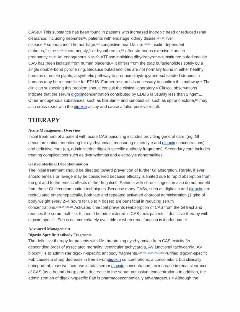
CASs.45 This substance has been found in patients with increased inotropic need or reduced renal
clearance, including neonates117, patients with endstage kidney disase,11,41,53 liver
disease,81 subarachnoid hemorrhage,123 congestive heart failure,39,102 insulin-dependent
diabetes,35 stress,40,118acromegaly,26 or hypothermia,117 after strenuous exercise118 and in
pregnancy.32,42,50 An endogenous Na+-K+-ATPase inhibiting dihydropyrone-substituted bufadienolide
CAS has been isolated from human placenta.33 It differs from the toad bufadienolides solely by a
single double-bond pyrone ring. Because bufadienolides are not normally found in either healthy
humans or edible plants, a synthetic pathway to produce dihydropyrone-substituted steroids in
humans may be responsible for EDLIS. Further research is necessary to confirm this pathway.50 The
clinician suspecting this problem should consult the clinical laboratory.34 Clinical observations
indicate that the serum digoxinconcentration contributed by EDLIS is usually less than 2 ng/mL.
Other endogenous substances, such as bilirubin,81 and xenobiotics, such as spironolactone,103 may
also cross-react with the digoxin assay and cause a false-positive result.
THERAPY
Acute Management Overview
Initial treatment of a patient with acute CAS poisoning includes providing general care, (eg, GI
decontamination, monitoring for dysrhythmias, measuring electrolyte and digoxin concentrations)
and definitive care (eg, administering digoxin-specific antibody fragments). Secondary care includes
treating complications such as dysrhythmias and electrolyte abnormalities.
Gastrointestinal Decontamination
The initial treatment should be directed toward prevention of further GI absorption. Rarely, if ever,
should emesis or lavage may be considered because efficacy is limited due to rapid absorption from
the gut and to the emetic effects of the drug itself. Patients with chronic ingestion also do not benefit
from these GI decontamination techniques. Because many CASs, such as digitoxin and digoxin, are
recirculated enterohepatically, both late and repeated activated charcoal administration (1 g/kg of
body weight every 2–4 hours for up to 4 doses) are beneficial in reducing serum
concentrations.17,21,67,71,86,121 Activated charcoal prevents reabsorption of CAS from the GI tract and
reduces the serum half-life. It should be administered in CAS toxic patients if definitive therapy with
digoxin-specific Fab is not immediately available or when renal function is inadequate.21,
Advanced Management
Digoxin-Specific Antibody Fragments.
The definitive therapy for patients with life-threatening dysrhythmias from CAS toxicity (in
descending order of associated mortality: ventricular tachycardia, AV junctional tachycardia, AV
block127) is to administer digoxin-specific antibody fragments.2,34,36,87,90,97,106,112,125Purified digoxin-specific
Fab causes a sharp decrease in free serumdigoxin concentrations; a concomitant, but clinically
unimportant, massive increase in total serum digoxin concentration; an increase in renal clearance
of CAS (as a bound drug); and a decrease in the serum potassium concentration.2 In addition, the
administration of digoxin-specific Fab is pharmacoeconomically advantageous.22 Although the

antidote itself is relatively expensive, its expense is far outweighed by obviating the need, risk, and
expense of long-term intensive care unit stays and of repetitive evaluation of potassium
and digoxinconcentrations. Table 65–4 lists the indications for administering digoxin-specific Fab.
Extensive discussion is found in Antidotes in Depth: A19.
TABLE 65–4. Indications for Administration of Digoxin-Specific Antibody Fragments (DSFab)
View Large |
Favorite Table
Other Cardiac Therapeutics.
Secondary treatments used in patients with symptomatic CAS exposures include the use
of atropine for supraventricular bradydysrhythmias or high degrees of AV block. Atropine dosing is
0.5 mg administered IV to an adult or 0.02 mg/kg with a minimum of 0.1 mg to a
child. Atropineshould be titrated to block the vagotonic effects of CASs. The dose may be repeated
at 5-minute intervals if necessary. Therapeutic success is unpredictable because the depressant
actions of CASs are mediated only partly through the vagus nerve.
Phenytoin and lidocaine are rarely used (secondary to Fab fragments obviating their utility) for the
management of CAS-induced ventricular tachydysrhythmias and ventricular irritability. These
xenobiotics depress the enhanced ventricular automaticity without significantly slowing, and perhaps
enhancing, AV nodal conduction.96 In fact, phenytoin may reverse digitalis-induced prolongation of
AV nodal conduction while suppressing digitalis-induced ectopic tachydysrhythmia without
diminishing myocardial contractile forces.48 In addition, phenytoin may terminate supraventricular
dysrhythmias induced by digitalis more effectively than lidocaine.96 Underlying atrial fibrillation and
flutter typically do not convert to a normal sinus rhythm with administration of phenytoin or lidocaine.
When used, phenytoin should be slowly IV infused (~ 50 mg/min) or in boluses of 100 mg repeated
every 5 minutes until control of the dysrhythmias is achieved or a maximum of 1000 mg has been
given in adults or 15 to 20 mg/kg in children.9,80 Fosphenytoinhas not been evaluated in this setting.
Maintenance PO doses of phenytoin (300–400 mg/day in adults and 6–10 mg/kg/day in children)
should be continued until digoxin toxicity is resolved. Lidocaine is given as a 1- to 1.5-mg/kg IV bolus
followed by continuous infusion at 1 to 4 mg/min in adults, or as a 1- to 1.5-mg/kg IV bolus followed
by 30 to 50 μg/kg/min in children as required to control the rhythm disturbance (Chap. 64).
Class IA antidysrhythmics are contraindicated in the setting of CAS poisoning because they may
induce or worsen AV nodal block and decrease His-Purkinje conduction at slow heart rates and
because their α-adrenergic receptor blockade and vagal inhibition may induce significant
hypotension and tachycardia. Class IA antidysrhythmics are also prodysrhythmogenic, and their
safety in the setting of CAS poisoning is unstudied. Additionally, quinidine reduces renal clearance
ofdigoxin and digitoxin. The use of isoproterenol should be avoided in CAS-induced conduction
disturbances because there may be an increased incidence of ventricular ectopic activity in the
presence of toxic concentrations of CAS.

Pacemakers and Cardioversion
External or transvenous pacemakers have had limited indications in the management of patients
with CAS poisoning. In one retrospective study of 92 digitalis-poisoned patients, 51 patients were
treated with cardiac pacing, digoxin-specific Fab, or both; the overall mortality rate was
13%.113 Prevention of life-threatening dysrhythmias failed in 8% of patients treated with
immunotherapy and 23% of patients treated with internal pacemakers. The main reasons for failure
of digoxin-specific Fab were pacing-induced dysrhythmias and delayed or insufficient doses of
digoxin-specific Fab. Iatrogenic complications of pacing occurred in 36% of patients. Thus, overdrive
suppression with a temporary transvenous pacemaker should not be used in the presence of CAS
poisoning.6,113 In the setting of digoxin poisoning, administration of transthoracic electrical
cardioversion for atrial tachydysrhythmias is associated with the development of potentially lethal
ventricular dysrhythmias. The dysrhythmias were related to the degree of toxicity and the amount of
administered current in cardioversion.99Transthoracic pacing may be attempted
for atropine unresponsive bradydysrhythmias in settings where definitive care (digoxin-specific Fab
fragments) are delayed or unavailable. In CAS-poisoned patients with unstable rhythms, such as
unstable ventricular tachycardia or ventricular fibrillation, cardioversion, and defibrillation,
respectively, are indicated.
Electrolyte Therapy
Potassium.
Hypokalemia and hyperkalemia may exacerbate CAS cardiotoxicity even at
“therapeutic” digoxin concentrations. When hypokalemia is noted in conjunction with
tachydysrhythmias or bradydysrhythmias, potassium replacement should be administered with serial
monitoring of the serum potassium concentration. Digoxin-specific Fab administration generally
should be withheld until the hypokalemia is corrected as the life-threatening manifestations of CAS
cardiotoxicity may resolve.
Hyperkalemia may also exacerbate CAS-induced cardiotoxicity, at
“therapeutic” digoxin concentrations. Reduction in potassium concentrations should be judiciously
initiated with care to avoid hypokalemia. Any exacerbation of CAS cardiotoxicity despite this
correction should be treated immediately with Fab fragments.
In acute CAS toxicity, if potassium is at least 5 mEq/L, digoxin-specific antibody fragments are
indicated. If digoxin-specific Fab is not available immediately, and ECG evidence of a dysrhythmia
suggestions of hyperkalemia is present, an attempt should be made to lower the serum potassium
with IV insulin, dextrose, sodium bicarbonate, and PO administration of the ion-exchange
resin sodium polystyrene sulfonateas indicated. Caution should be applied to the subsequent
administration of digoxin-specific Fab because of concern for profound hypokalemia.
Although calcium is beneficial in most hyperkalemic patients, in the setting of CAS poisoning,
administration of calcium salts is considered to be potentially dangerous. A number of experimental
studies cite the additive or synergistic actions of calcium and CAS on the heart (because intracellular
hypercalcemia is already present), resulting in dysrhythmias,37,83,104 cardiac dysfunction61 (eg,

hypercontractility, so-called “stone heart,” hypocontractility), and cardiac arrest.72,104,119Although a 2004
study was unable to show an adverse effect,43 there exist three case reports8,64 of CAS-poisoned
patients who died at various intervals after calcium administration, which supports the withholding of
calcium administration in the setting of hyperkalemia induced by CAS poisoning.
The purported mechanism is augmented intracellular cytoplasmic Ca2+, which results from an
increased transmembrane concentration gradient that further inhibits calcium extrusion through the
Na+-Ca2+ exchange or increased intracytoplasmic stores.59 This additional cytoplasmic calcium may
result in altered contraction of myofibril organelles,61 less negative intracellular resting potential that
allows delayed afterdepolarizations to reach firing threshold,47,59,83 altered function of the sarcoplasmic
reticulum,61,95 or increased calcium interfering with myocardial mitochondrial function (Chaps.
16 and 17).61 Although some investigators suspect that the rate of administration of the calcium may
be a factor in the subsequent cardiac toxicity,72,83 calcium administration should be avoided because
better, safer, alternative treatments, such as digoxin-specific Fab, insulin, and sodium bicarbonate,
are available for CAS-induced hyperkalemia.8,37,64,83,104
Magnesium.
Hypomagnesemia may also occur in CAS-poisoned patients secondary to the contributory factors
mentioned with hypokalemia, such as long-term diuretic use to treat congestive heart failure. The
theoretical benefits of magnesium therapy in the setting of hypomagnesemia include blockade of the
transient inward calcium current, antagonism of calcium at intracellular binding sites, decreased
CAS-related ventricular irritability, and blockade of potassium egress from CAS-poisoned
cells.4,31,55,89,100,110,122 Although hypomagnesemia increases myocardial digoxin uptake and decreases
cellular Na+-K+-ATPase activity, there is conflicting evidence as whether magnesium “reactivates” the
CAS-bound Na+-K+-ATPase activity.81,100,110
A common regimen uses 2 g of magnesium sulfate IV over 20 minutes in adults (25–50 mg/kg/dose
to a maximum of 2 g in children). After stabilization, adult patients with severe hypomagnesemia
may require a magnesium infusion of 1 to 2 g/h (25–50 mg/kg/h to a maximum of 2 g in children),
with serial monitoring of serum magnesium concentrations, telemetry, respiratory rate (observing for
bradypnea), deep tendon reflexes (observing for hyporeflexia), and monitoring of blood pressure.
Magnesium is contraindicated in the setting of bradycardia or AV block, preexisting
hypermagnesemia, and renal insufficiency or failure.
Extracorporal Removal of Cardioactive Steroids
Forced diuresis,66 hemoperfusion,77,79,120 and hemodialysis120 are ineffective in enhancing the
elimination of digoxin because of its large volume of distribution (4–10 L/kg), which makes it
relatively inaccessible to these techniques. Because of its high affinity for tissue proteins,
approximately 10% of the amount of digoxin is found in the serum than is found at the tissue level,
and of that amount, approximately 20% to 40% is protein bound.57
SUMMARY

Digoxin and digitoxin are the most commonly prescribed members of the drugs classified as
CASs and they have a narrow therapeutic index.
Both cardiac and noncardiac effects occur after CAS poisoning, including nausea, vomiting,
headache, weakness, altered mental status bradycardia, atrial and ventricular ectopy with
block, or hyperkalemia.
CAS overdose mimics include dysrhythmias from electrolyte abnormalities, primarily
hypokalemia, or hypomagnesemia which can be corrected by repletion of potassium or
magnesium.
Definitive therapy for CAS poisoning is the early administration of digoxin-specific Fab
immunotherapy coupled with both decontamination techniques including activated charcoal
and supportive therapy.
Acknowledgment
Neal A. Lewin, MD, contributed to this chapter in previous editions.
References
1.
Arispe N, Diaz JC, Simakova O, Pollard HB: Heart failure drugdigoxin induces calcium uptake into
cells by forming transmembrane calcium channels. Proc Natl Acad Sci. 2008;105:2610–2615.
CrossRef
2.
Banner W, Bach P, Burk B et al.: Influence of assay methods on serum concentrations
of digoxin during Fab fragment treatments. J Toxicol Clin Toxicol. 1992;30:259–267.
CrossRef [PubMed: 1588675]
3.
Bayer MJ: Recognition and management of digitalis intoxication: implications for emergency
medicine. Am J Emerg Med. 1991;9(suppl 1):29–32.
CrossRef [PubMed: 1997019]
4.
Beller GA, Hood WB, Smith TW et al.: Correlation of serum magnesium level and cardiac digitalis
intoxication. Am J Cardiol.1974;33:225–229.
CrossRef [PubMed: 4810019]
5.
Bismuth C, Gaultier M, Conso F, Efthymiou ML: Hyperkalemia in acute digitalis poisoning:
prognostic significance and therapeutic implications. Clin Toxicol. 1973;6:153–162.
CrossRef [PubMed: 4715199]
6.
Bismuth C, Motte G, Conso F, Chauvin M: Acute digitoxin intoxication treated by intracardiac
pacemaker: experience in sixty-eight patients. Clin Toxicol. 1977;10:443–456.
CrossRef [PubMed: 862379]
7.

Blaustein MP: Physiologic effects of endogenous ouabain: control of intracellular Ca2+ stores and
cell responsiveness. Am J Physiol.1993;264:C1367–C1387. [PubMed: 8392793]
8.
Bower JO, Mengle HAK: The additive effect of calcium and digitalis.JAMA. 1936;106:1151–1153.
CrossRef
[JAMA and JAMA Network Journals Full Text]
9.
Bristow MR, Port JD, Kelly RA: Treatment of heart failure: pharmacologic methods. In:
Braunwald E, Zipes D, Libby P, eds:Heart Disease. A Textbook of Cardiovascular Medicine, 6th
ed. New York: WB Saunders; 2001:573–575.
10.
Brubacher JR, Ravikumar PR, Bania T et al.: Treatment of toad venom poisoning with digoxin-
specific Fab fragments. Chest.1996;110:1282–1288.
CrossRef [PubMed: 8915235]
11.
Carver JL, Valdes R: Anomalous serum digoxin concentrations in uremia. Ann Intern
Med. 1983;98:483–484.
CrossRef [PubMed: 6838072]
12.
Centers for Disease Control and Prevention: Deaths associated with a purported aphrodisiac. New
York City, February 1993–May 1995.MMWR Morb Mortal Wkly Rep. 1995;44:853–855. [PubMed:
7476839]
13.
Chern MS, Ray CY, Wu DL: Biological intoxication due to digitalis-like substance after ingestion of
cooked toad soup. Am J Cardiol.1991;67: 443–444.
CrossRef [PubMed: 1994674]
14.
Cheung K, Hinds JA, Duffy P: Detection of poisoning by plant-origin cardiac glycoside with the
Abbot TDx analyzer. Clin Chem.1989;35:295–297. [PubMed: 2914377]
15.
Chillet P, Korach JM, Vincent N et al.: Digoxin poisoning and anuric acute renal failure: efficiency
of the treatment associating digoxin-specific antibodies (Fab) and plasma exchanges. Int J Artif
Organs. 2002;25:538–541. [PubMed: 12117293]
16.
Cooke D: The use of central nervous system manifestations in the early detection of digitalis
toxicity. Heart Lung. 1993;22:477–481. [PubMed: 8288449]
17.
Critchley JA, Critchley LA: Digoxin toxicity in chronic renal failure: treatment by multiple-dose
activated charcoal intestinal dialysis. Hum Exp Toxicol. 1997;16:733–735.
CrossRef [PubMed: 9429088]
18.

Cummins RO, Haulman J, Quan L: Near-fatal yew berry intoxication treated with external cardiac
pacing and digoxin-specific Fab antibody fragments. Ann Emerg Med. 1990;19:38–43.
CrossRef [PubMed: 2297154]
19.
Dasgupta A, Wu S, Actor J et al.: Effect of Asian and Siberian ginseng on
serum digoxin measurement by five digoxinimmunoassays. Significant variation in digoxin-like
immunoreactivity among commercial ginsengs. Am J Clin Pathol. 2003;119:298–303.
CrossRef [PubMed: 12580002]
20.
De-Mey C, Brendel E, Enterling D: Carvedilol increases the systemic bioavailability of
oral digoxin. Br J Clin Pharmacol.1990;29:486–490.
CrossRef [PubMed: 1970265]
21.
de Silva HA, Fonseka MMD, Pathmeswaran A et al.: Multiple-dose activated charcoal for
treatment of yellow oleander poisoning: a single-blind randomised, placebo-controlled
trial. Lancet.2003;361:1935–1938.
CrossRef [PubMed: 12801736]
22.
DiDomenico RJ, Walton SM, Sanoski CA et al.: Analysis of the use of digoxin immune Fab for the
treatment of non-life-threateningdigoxin toxicity. J Cardiovasc Pharmacol Ther. 2000;5:77–85.
CrossRef [PubMed: 11150387]
23.
Doering W: Quinidine-digoxin interaction: pharmacokinetics, underlying mechanism and clinical
implications. N Engl J Med.1979;301:400–404.
CrossRef [PubMed: 460341]
24.
Doherty JE, Perkins WH, Flanigan WJ: The distribution and concentration of tritiated digoxin in
human tissues. Ann Intern Med.1967;66:116–124.
CrossRef [PubMed: 6015582]
25.
Doolittle MH, Lincoln K, Graves SW: Unexplained increase in serum digoxin: a case report. Clin
Chem. 1994;40:487–492. [PubMed: 8131287]
26.
Eddelston M, Ariaratnam CA, Sjostrom L et al.: Acute yellow oleander (Thevetiaperuviana)
poisoning: cardiac arrhythmias, electrolyte disturbances, and serum cardiac glycoside
concentrations on presentation to hospital. Heart. 2000;83:301–306.
27.
Eddleston M, Sheriff MHR, Hawton K: Deliberate self harm in Sri Lanka: an overlooked tragedy in
the developing world. BMJ. 1998; 317:133–135.
CrossRef [PubMed: 9657795]
28.

Eisner DA, Lederer WJ, Vaughan-Jones RD: The quantitative relationship between twitch tension
and intracellular sodium activity in sheep cardiac Purkinje fibers. J Physiol. 1984;355:251–266.
CrossRef [PubMed: 6092625]
29.
Eisner DA, Smith TW: The Na-K pump and its effect in cardiac muscle. In: Fozzard HA, ed.The
Heart and Cardiovascular System, 2nd ed. New York: –Raven Press;1991:863–902.
30.
Eisner T, Wiemer DF, Haynes LW, Meinwald J: Lucibufagins: defensive steroids from the
fireflies Photinus ignitus and P. marginellus (Coleoptera: Lampyridae). Proc Natl Acad Sci U S
A.1978;75:905.
CrossRef [PubMed: 16592501]
31.
French JH, Thomas RG, Siskind AP et al.: Magnesium therapy in
massive digoxin intoxication. Ann Emerg Med. 1984;13:562–566.
CrossRef [PubMed: 6742564]
32.
Friedman HS, Abramowitz I, Nguyen T et al.: Urinary digoxin-like immunoreactive substance in
pregnancy. Am J Med. 1987;83:261–264.
CrossRef [PubMed: 3618628]
33.
Gao S, Chen Z, Xu Y: The source of endogenous digitalis-like substance in normal
pregnancy. Zhonghua Fu Chan Ke Za Zhi.1998; 33:539–41. [PubMed: 10806730]
34.
George S, Brathwaite RA, Hughes EA: Digoxin measurements following plasma ultrafiltration in
two patients with digoxin toxicity treated with specific Fab fragments. Ann Clin
Biochem.1994;31:380–381.
CrossRef [PubMed: 7979107]
35.
Giampietro O, Clerico A, Gregori G et al.: Increased urinary excretion of digoxin-like
immunoreactive substance by insulin-dependent diabetic patients: a linkage with hypertension?Clin
Chem.1988;34:2418–2422. [PubMed: 3197278]
36.
Gibb T, Adams PC, Parnham AJ, Jennings K: Plasma digoxin: assay anomalies in Fab-treated
patients. Br J Clin Pharmacol.1983;16:445–447.
CrossRef [PubMed: 6626440]
37.
Gold H, Edwards DJ: The effects of ouabain on heart in the presence of hypercalcemia. Am Heart
J. 1927;3:45–50.
CrossRef
38.
Gorelick DA, Kussin SZ, Kahn I: Paranoid delusions and auditory hallucinations associated
with digoxin intoxication. J Nerv Ment Dis.1978;166:817–819.
CrossRef [PubMed: 722304]
39.
Graves SW: Endogenous digitalis-like factors. Crit Rev Clin Lab Sci.1986;23:177–200.
CrossRef [PubMed: 3015491]
40.
Graves SW, Adler G, Stuenkel C et al.: Increases in plasma digitalis-induced
hypoglycemia. Neuroendocrinology. 1989;49:586–591.
CrossRef [PubMed: 2671776]
41.
Graves SW, Brown BA, Valdes R: Digoxin-like substances measured in patients with renal
impairment. Ann Intern Med.1983;99:604–608.
CrossRef [PubMed: 6638719]
42.
Graves SW, Valdes R, Brown BA et al.: Endogenous immunoreactive digoxin-like substance in
human pregnancies. J Clin Endocrinol Metab. 1984; 58:748–751.
CrossRef [PubMed: 6699137]
43.
Hack JB, Woody JH, Lewis DE et al.: The effect of calcium chloride in treating hyperkalemia due
to acute digoxin toxicity in a porcine model. J Toxicol Clin Toxicol. 2004;42:337–342.
CrossRef [PubMed: 15461240]
44.
Haddy FJ: Endogenous digitalis-like factor or factors. N Engl J Med.1987;316:621–622.
CrossRef [PubMed: 3027560]
45.
Hager WD, Fenster P, Mayersohn M et al.: Digoxin-quinidine interaction: pharmacokinetic
evaluation. N Engl J Med.1979;300:1238–1241.
CrossRef [PubMed: 431681]
46.
Hastreiter AR, John EG, van der Horst RL: Digitalis, digitalis antibodies, digitalis-like
immunoreactive substances, and sodium homeostasis: a review. Clin Perinatol. 1988;15:491–
522. [PubMed: 3066550]
47.
Hauptman PJ, Kelly RA. Digitalis. Circulation. 1999;99:1265–1270.
CrossRef [PubMed: 10069797]
48.
Helfant RH, Scherlac BJ, Damata AN: Protection from digitalis toxicity with the prophylactic use of
diphenylhydantoin sodium an arrhythmic-inotropic dissociation. Circulation. 1967:36:119–124.
CrossRef [PubMed: 6027207]
49.
Henderson RP, Solomon CP: Use of cholestyramine in the treatment of digoxin intoxication. Arch
Intern Med. 1988;148:745–746.
CrossRef [PubMed: 3341874]
[Archives of Internal Medicine Full Text]
50.

Hilton PJ, White G, Lord A et al.: An inhibitor of the sodium pump obtained from human
placenta. Lancet. 1996;348:303–305.
CrossRef [PubMed: 8709690]
51.
Hobson J, Zettner A: Digoxin serum half-life following
suicidaldigoxin poisoning. JAMA. 1973;223:147–149.
CrossRef [PubMed: 4739453]
[JAMA and JAMA Network Journals Full Text]
52.
Hollman A: Plants and cardiac glycosides. Br Heart J. 1985;54:258–261.
CrossRef [PubMed: 4041297]
53.
Isensee L, Solomon RJ, Weinberg MS et al.: Digoxin levels in dialysis patients. Hosp
Physician. 1988;24:50–52.
54.
Jortani SA, Helm RA, Valdes R: Inhibition of Na, K-ATPase by oleandrin and oleandrigenen, and
their detection by digoxinimmunoassays. Clin Chem. 1996;42:1654–1658. [PubMed: 8855150]
55.
Kaneko T, Kudo M, Okumura T et al.: Successful treatment ofdigoxin intoxication by
hemoperfusion with specific columns for β2-microglobulin adsorption (Lixelle) in a maintenance
haemodialysis patient. Nephrol Dial Transplant. 2001;16;195–196.
CrossRef [PubMed: 11209033]
56.
Karkal SS, Ordog G, Wasserberg J: Digitalis intoxication: dealing rapidly and effectively with a
complex cardiac toxidrome. Emerg Med Rep. 1991;12:29–44.
57.
Katzung BG, Parmley WM: Cardiac glycosides & other drugs used in congestive heart failure. In:
Katzung BG, ed.Basic and Clinical Pharmacology, 7th ed. Stamford, CT: Appleton & Lange;
1998:197–215.
58.
Kelly RA, Smith TW: Endogenous cardiac glycosides. Adv Pharmacol. 1994;25:263–
288. [PubMed: 8204503]
59.
Kelly RA, Smith TW: Pharmacological treatment of heart failure. In: Hardman JG, Limbird LE,
Molinoff PB, Ruddon RW, eds.Goodman and Gilman’s The Pharmacological Basis of Therapeutics,
9th ed. New York: McGraw-Hill; 1996:809–838.
60.
Kelly RA, Smith TW: Recognition and management of digitalis toxicity. Am J Cardiol. 1992;69:108–
109.
CrossRef [PubMed: 1729858]
61.

Khatter JC, Agbanyo M, Navaratnam S et al.: Digitalis cardiotoxicity: cellular calcium overload as
a possible mechanism.Basic Res Cardiol. 1989;84:553–563.
CrossRef [PubMed: 2619695]
62.
Kinlay S, Buckley N: Magnesium sulfate in the treatment of ventricular arrhythmias due
to digoxin toxicity. J Toxicol Clin Toxicol.1995;33:55–59.
CrossRef [PubMed: 7837314]
63.
Klausen T, Kjeldsen K, Norgaard A: Effects of denervation on sodium, potassium and [3H] ouabain
binding in muscles of normal and potassium depleted rats. J Physiol. 1983;345:123–124.
CrossRef [PubMed: 6663495]
64.
Kne T, Brokaw M, Wax P: Fatality from calcium chloride in a chronic digoxin toxic patient
(abstract). J Toxicol Clin Toxicol.1997;5:505.
65.
Knight M, Glor R, Smedley SR et al.: Firefly toxicosis in lizards. J Chem Ecol. 1999;25:1981–
1986.
CrossRef
66.
Koren G, Klein J: Enhancement of digoxin clearance by mannitol diuresis: in vivo studies and their
clinical implications. Vet Hum Toxicol. 1988;30:25–27. [PubMed: 3127989]
67.
Lalonde RL, Deshpande R, Hamilton PP et al.: Acceleration ofdigoxin clearance by activated
charcoal. Clin Pharmacol Ther.1985;37:367–371.
CrossRef [PubMed: 3978996]
68.
Leahy EB Jr, Reiffel JA, Drusin RE et al.: Interaction between quinidine
and digoxin. JAMA. 1978;240:533–534.
CrossRef [PubMed: 671662]
[JAMA and JAMA Network Journals Full Text]
69.
Lee TC: Van Gogh’s vision. JAMA. 1981;245:727–729.
CrossRef [PubMed: 7007674]
[JAMA and JAMA Network Journals Full Text]
70.
Lely AH, van Enter CH: Large-scale digitoxin intoxication. Br Med J.1970; 3:737–740.
CrossRef [PubMed: 5273245]
71.
Levy G: Gastrointestinal clearance of drugs with activated charcoal.N Engl J Med. 1982;307:676–
678.
CrossRef [PubMed: 7110218]
72.

Lieberman AL: Studies on calcium VI: some interrelationships of the cardiac activities of calcium
gluconate and scillaren-B. J Pharmacol Exp Ther. 1933;47:183–192.
73.
Lindenbaum J, Rund DG, Butler VP: Inactivation of digoxin by the gut flora: reversal by antibiotic
therapy. N Engl J Med.1981;305:789–794.
CrossRef [PubMed: 7266632]
74.
Lown B, Byatt NF, Levine HD: Paroxysmal atrial tachycardia with block. Circulation. 1960;21:129–
143.
CrossRef [PubMed: 14418578]
75.
Madan BR, Khanna NK, Soni RK: Effect of some arrhythmogenic agents upon the acetylcholine
content of the rabbit atria. J Pharm Pharmacol. 1970;22:621–622.
CrossRef [PubMed: 4394539]
76.
Mahdyoon H, Battilana G, Rosman H et al.: The evolving pattern of digoxin intoxication:
observations at a large urban hospital from 1980 to 1988. Am Heart J. 1990;120:1189–1194.
CrossRef [PubMed: 2239670]
77.
Marbury T, Mahoney J, Juncos L et al.: Advanced digoxin toxicity in renal failure: treatment with
charcoal hemoperfusion. South Med J. 1979;72: 279–282.
CrossRef [PubMed: 424817]
78.
McGary SJ, Williams AJ: Digoxin activates sarcoplasmic reticulum Ca2+ release channels: a
possible role in cardiac inotropy. Br J Pharmacol. 1993; 108:1043–1050.
CrossRef [PubMed: 8387382]
79.
McRae S: Elevated serum digoxin levels in a patient taking digoxinand Siberian
ginseng. CMAJ. 1996;155:292–295.
80.
Miller JM, Zipes DP: Management of the patient with cardiac arrhythmias. In: Braunwald E,
Zipes D, Libby P, eds. Heart Disease. A Textbook of Cardiovascular Medicine, 6th ed. New York:
WB Saunders; 2001:726–727.
81.
Nanji AA, Greenway DC: Falsely raised plasma digoxinconcentrations in liver disease. Br Med
J. 1985;290:432–433.
CrossRef
82.
Neff MS, Mendelssohn S, Kim KS et al.: Magnesium sulfate in digitalis toxicity. Am J
Cardiol. 1974;62:377–382.
83.

Nola GT, Pope S, Harrison DC: Assessment of the synergistic relationship between serum calcium
and digitalis. Am Heart J.1970;79:499–507.
CrossRef [PubMed: 5418023]
84.
Ordog GJ, Benaron S, Bhasin V et al.: Serum digoxin levels and mortality in 5,100 patients. Ann
Emerg Med. 1987;16:32–39.
CrossRef [PubMed: 3800074]
85.
Pace DG, Gillis RA: Neuroexcitatory effects of digoxin in the cat. J Pharmacol Exp
Ther. 1976;199:583–600. [PubMed: 994017]
86.
Pond S, Jacos M, Marks J et al.: Treatment of digitoxin overdose with oral activated
charcoal. Lancet. 1981;2:1177–1178.
CrossRef [PubMed: 6118621]
87.
Rabetoy GM, Price CA, Findlay JWA et al.: Treatment of digoxinintoxication in a renal failure
patient with digoxin-specific antibody fragments and plasmapheresis. Am J Nephrol. 1990;10:518–
521.
CrossRef [PubMed: 2075910]
88.
Radford DJ, Cheung K, Urech R et al.: Immunologic detection of cardiac glycosides in plants. Aust
Vet J. 1994;71:236–238.
CrossRef [PubMed: 7986184]
89.
Reisdorff EJ, Clark MR, Walter BL: Acute digitalis poisoning: the role of intravenous magnesium
sulfate. J Emerg Med. 1986;4:463–469.
CrossRef [PubMed: 3549866]
90.
Rich SA, Libera JM, Locke RJ: Treatment of foxglove extract poisoning with digoxin-specific Fab
fragments. Ann Emerg Med.1993;22:1904–1907.
CrossRef [PubMed: 8239114]
91.
Roberge RJ: Congestive heart failure and toxic digoxin levels: role of cholestyramine. Vet Hum
Toxicol. 2000;42:172–173. [PubMed: 10839325]
92.
Rodin SM, Johnson BF: Pharmacokinetic interactions with digoxin.Clin
Pharmacokinetic. 1988;15:227–244.
CrossRef
93.
Rose AM, Valdes R: Understanding the sodium pump and its relevance to disease. Clin
Chem. 1994;40:1674–1685. [PubMed: 8070076]
94.

Rosen MR, Wit AL, Hoffman BF: Cardiac antiarrhythmic and toxic effects of digitalis. Am Heart
J. 1975;89:391–399.
CrossRef [PubMed: 1090138]
95.
Rosen MR: Cellular electrophysiology of digitalis toxicity. J Am Coll Cardiol. 1985;2:22A–34A.
CrossRef
96.
Rumack BH, Wolfe RR, Gilfinch H: Diphenylhydantoin treatment of massive digoxin overdose. Br
Heart J. 1974;36:405–408.
CrossRef [PubMed: 4842639]
97.
Safadi R, Levy T, Amitai Y et al.: Beneficial effect of digoxin-specific Fab antibody fragments in
oleander intoxication. Arch Intern Med. 1995;155:2121–2125.
CrossRef [PubMed: 7575073]
[Archives of Internal Medicine Full Text]
98.
Sameri RM, Soberman JE, Finch CK et al.: Lower serum digoxinconcentrations in heart failure
and reassessment of laboratory report forms. Am J Med Sci. 2002;324:10–13.
CrossRef [PubMed: 12120820]
99.
Sarubbi B, Ducceschi V, D’Antonello A et al.: Atrial fibrillation: what are the effects of drug therapy
on the effectiveness and complications of electrical cardioversion?Can J Cardiol.1998;14:1267–
1273. [PubMed: 9852940]
100.
Seller RH: The role of magnesium in digitalis toxicity. Am Heart J.1971;82:551–556.
CrossRef [PubMed: 5111237]
101.
Selzer A: Role of serum digoxin assay in patient management. J Am Coll Cardiol. 1985;5:106A–
110A.
CrossRef [PubMed: 3886746]
102.
Shilo LM, Adawi A, Solomon G, Shenkman L: Endogenous digoxinlike immunoreactivity in
congestive heart failure. Br Med J.1987;295:415–416.
CrossRef
103.
Silber B, Sheiner LB, Powers JL et al.: Spironolactone-associateddigoxin radioimmunoassay
interference. Clin Chem. 1979;25:48–54. [PubMed: 761378]
104.
Smith PK, Winkler AW, Hoff HE: Calcium and digitalis synergism: the toxicity of calcium salts
injected intravenously into digitalized animals. Arch Intern Med. 1939;64:322–328.
CrossRef
[Archives of Internal Medicine Full Text]
105.

Smith SW, Shah RR, Herzog CA: Bidirectional ventricular tachycardia resulting from herbal aconite
poisoning. Ann Emerg Med. 2005;45:100.
CrossRef [PubMed: 15635326]
106.
Smith TW, Haber E, Yeatman L et al.: Reversal of advanceddigoxin intoxication with Fab
fragments of digoxin-specific antibodies. N Engl J Med. 1976;294:797–800.
CrossRef [PubMed: 943040]
107.
Smith TW: Pharmacokinetics, bioavailability and serum levels of cardiac glycosides. J Am Coll
Cardiol. 1985;5:43A–50A.
CrossRef [PubMed: 3921586]
108.
Smith TW: Digitalis. N Engl J Med. 1988;318:358–365.
CrossRef [PubMed: 3277052]
109.
Somberg JC, Bounous H, Levitt B: The antiarrhythmic effects of quinidine and propranolol in the
ouabain-intoxicated spinally transected cat. Eur J Pharmacol. 1979;54:161–166.
CrossRef [PubMed: 421737]
110.
Spechter MJ, Schweizer E, Goldman RH: Studies on magnesium’s mechanism of action in
digitalis-induced arrhythmias.Circulation. 1975;52:1001–1005.
CrossRef [PubMed: 1182944]
111.
Springer M, Olson KR, Feaster W: Acute massive digoxinoverdose: survival without use of
digitalis-specific antibodies. Am J Emerg Med. 1986;4:364–369.
CrossRef [PubMed: 3718631]
112.
Sullivan JB: Immunotherapy in the poisoned patient. Med Toxicol.1986;1:47–60. [PubMed:
3537615]
113.
Taboulet P, Baud FJ, Bismuth C et al.: Acute digitalis intoxication: is pacing still appropriate?J
Toxicol Clin Toxicol. 1993;31:261–273.
CrossRef [PubMed: 8492339]
114.
Torsti P: Acetylcholine content and cholinesterase activities in the rabbit heart in experimental heart
failure and the effect of g-strophanthin treatment on them. Ann Med Exp Biol Fenn.1959;37(suppl
4):4–9.
115.
Tsuruoka S, Osono E, Nishiki K et al.: Removal of digoxin by column for specific adsorption of β2-
microglobulin: a potential use for digoxin intoxication. Clin Pharmacol Ther. 2001;69:422–430.
CrossRef [PubMed: 11406739]
116.

Tuncok Y, Kozan O, Cavdar C et al.: Urginea maritima (squill) toxicity. J Toxicol Clin
Toxicol. 1995;33:83–86.
CrossRef [PubMed: 7837318]
117.
Valdes R, Graves SW, Brown BA et al.: Endogenous substances in newborn infants causing
false-positive digoxin measurements. J Pediatr. 1983;102:947–950.
CrossRef [PubMed: 6854459]
118.
Valdes R, Hagberg JM, Vaughan TE et al.: Endogenous digoxin-like immunoreactivity in blood is
increased during prolonged strenuous exercise. Life Sci. 1988;42:103–110.
CrossRef [PubMed: 3336270]
119.
Wagner J, Salzer WW: Calcium-dependent toxic effects of digoxinin isolated myocardial
preparations. Arch Int Pharmacodyn.1976;223:4–14.
120.
Warren SE, Fanestil DD: Digoxin overdose: limitations of hemoperfusion-hemodialysis
treatment. JAMA. 1979;242:2100–2101.
CrossRef [PubMed: 490791]
[JAMA and JAMA Network Journals Full Text]
121.
Watson WA: Factors influencing the clinical efficacy of activated charcoal. Drug Intell Clin
Pharm. 1987;21:160–166. [PubMed: 3549212]
122.
Whang R, Aikawa J: Magnesium deficiency and refractoriness to potassium repletion. J Chron
Dis. 1977;30:65–68.
CrossRef
123.
Wildicks EFM, Vermeulen M, van Brummelen P et al.: Digoxin-like immunoreactive substance in
patients with aneurysmal subarachnoid hemorrhage. Br Med J. 1987;294:729–732.
CrossRef
124.
Withering W: An account of the foxglove and some of its medical uses: with practical remarks on
dropsy and other diseases. Med Classics. 1937;2:295–443.
125.
Woolf AD, Wenger T, Smith TW et al.: The use of digoxin-specific Fab fragments for severe
digitalis intoxication in children. N Engl J Med. 1992;326:1739–1744.
CrossRef [PubMed: 1594015]
126.
Manini AF, Nelson LS, Hoffman RS: Prognostic utility of serum potassium in
chronic digoxin toxicity: a case-control study. Am J Cardiovasc Drugs. 2011;11(3):173–178.
CrossRef [PubMed: 21619380]
127.

Gaultier M, Fournier E, Efthymiou ML, Frejaville JP, Jouannot P, Dentan M: Acute digitalis
poisoning (70 cases). Bull Mem Soc Med Hop Paris. 1968;119:247–274.