6 Ocular Leprosy
7
112 Proceedings of the Royal Society of Medicine 6 (3) Local treatment of the eye.-So far, there have been no reports of the use of sulphones locally, in the forms of drops, ointment or by subconjunctival injection. This problem is engaging our atten- tion at the present time. Various gold preparations have been used locally, but reports of their effects are conflicting. The antibiotics are of use in restricting any secondary infection, e.g. of an obstructed lacrimal sac. The local treatment of the plastic iritis, the most dreaded ocular complication of leprosy, is of the first importance. Mydriatics, reinforced by early local use of cortisone, represent a great advance in the management of this condition. Colonel Kirwan and I have successfully treated a number of these cases, who still retain a visual acuity of the order of 6/6-6/9, who would otherwise probably be seeing less than 6/60. Leprosy bacilli are noted for their sluggish powers of reproduction and in the eye for the minimal fibroblastic reaction they provoke, which probably explains the fact that we have, so far, seen no evidence of multiplication of the bacilli beneath the cortisone umbrella. PROGNOSIS Twenty to thirty years ago a dissertation on leprosy would have included the statement that the ocular complications progressed remorselessly towards blindness, being unaffected by any known thera- peutic measures. I have personally noted while abroad a certain apathy and want of interest in the treatment of ocular leprosy, engendered, no doubt, by the onward march of the disease. While the word "cure" can still not be applied to leprosy, I would suggest that it is now possible to talk of delaying the progress of ocular leprosy with systemic sulphones and that a proportion of the blindness associated with the disease can be prevented by the timely use of cortisone. BIBLIOGRAPHY BULL, 0. B., and HANSEN, G. A. (1873) The Leprous Diseases of the Eye. Christiania. ELLIoTr, D. C. (1948) Int. J. Leprosy, 16, 347; (1949) Int. J. Leprosy, 17, 229. FUCHS, A. (194-0) Int. J. Leprosy, 8, 247. KING, E. F. (1936) Brit. J. Ophthal., 20, 561. KiRWAN, E. W. O'G. (1948) Trans. R. Soc. trop. Med. Hyg., 41, 583. MONDONCA DE BARROS, J. (1940) Int. J. Leprosy, 8, 353; (1939) Ophthal. ibero-amer., 1, 169. PENDERGAST, J. J. (1940) Arch. Ophthal., Chicago, 23, 112. SHIoNUMA, E. (1940) Int. J. Leprosy, 8, 401. VALLE, S. (1937) Rev. bras. Leprol., 5, 481. Ocular Leprosy By Lt.-Col. E. W. O'G. KIRWAN, C.I.E., M.D., F.R.C.S.I. IN tropical countries leprosy is one of the great causes of blindness and partial loss of sight. There is no disease that has been so greatly dreaded through the ages. Horrible to live with, difficult to die with, slowly destroying the nerves and marrow, causing deformities and mutilations and finally, to complete the holocaust, often causing blindness. Thanks to the researches of a number of specialists in more recent years, we now possess in the improved treatment and better knowledge of the epidemiological conditions under which leprosy infections arise, the means greatly to reduce and eventually, we hope, to eradicate this terrible disease. More than three million cases of leprosy are supposed to exist in the world, the great strongholds being India, China and Central Africa. India has a million or more cases and it is calculated that 15,000 children develop the disease annually. The first fifteen years of life is the period of highest contagion and especially the period 5-15 years. The incidence of the disease is greater in low-lying, humid areas in which the climate is hot and the rainfall heavy, rather than in more elevated dry ones. Great Britain is responsible for a very large number of patients in her colonies and only a small proportion are as yet receiving the benefits of established treatment. It is difficult to determine the extent of leprosy in these islands in the past but at present there are about 150 cases in England and Wales. All these patients acquired the disease outside the country. Many cases seek refuge here and the disease is not easy to detect. The last British leper whose infection could not be traced directly or indirectly to the tropics died in 1897 in the Shetland Islands. All the changes that affect the body in leprosy can affect the eye and its adnexa, and so the ocular manifestations have no mystery of their own. As in the body, the disease is one of relentless chronicity and presents very serious complications. Ocular involvement is common but varies tremendously in different countries and amongst different races. The gravity of this involvement is directly dependent upon the type of the disease. In the lepromatous type the eye complications are serious and caused by actual leprous infiltration both superficial and deep. In the non-lepromatous type, in which the peripheral nerves are involved, there is no actual leprous lesion of the eyeball. The branches of the VII nerve are frequently
Transcript of 6 Ocular Leprosy

112 Proceedings of the Royal Society of Medicine 6
(3) Local treatment of the eye.-So far, there have been no reports of the use of sulphones locally,in the forms of drops, ointment or by subconjunctival injection. This problem is engaging our atten-tion at the present time. Various gold preparations have been used locally, but reports of their effectsare conflicting. The antibiotics are of use in restricting any secondary infection, e.g. of an obstructedlacrimal sac.The local treatment of the plastic iritis, the most dreaded ocular complication of leprosy, is of
the first importance. Mydriatics, reinforced by early local use of cortisone, represent a great advancein the management of this condition. Colonel Kirwan and I have successfully treated a number ofthese cases, who still retain a visual acuity of the order of 6/6-6/9, who would otherwise probably beseeing less than 6/60. Leprosy bacilli are noted for their sluggish powers of reproduction and in theeye for the minimal fibroblastic reaction they provoke, which probably explains the fact that wehave, so far, seen no evidence of multiplication of the bacilli beneath the cortisone umbrella.
PROGNOSISTwenty to thirty years ago a dissertation on leprosy would have included the statement that the
ocular complications progressed remorselessly towards blindness, being unaffected by any known thera-peutic measures. I have personally noted while abroad a certain apathy and want of interest in thetreatment of ocular leprosy, engendered, no doubt, by the onward march of the disease. While theword "cure" can still not be applied to leprosy, I would suggest that it is now possible to talk ofdelaying the progress of ocular leprosy with systemic sulphones and that a proportion of the blindnessassociated with the disease can be prevented by the timely use of cortisone.
BIBLIOGRAPHYBULL, 0. B., and HANSEN, G. A. (1873) The Leprous Diseases of the Eye. Christiania.ELLIoTr, D. C. (1948) Int. J. Leprosy, 16, 347; (1949) Int. J. Leprosy, 17, 229.FUCHS, A. (194-0) Int. J. Leprosy, 8, 247.KING, E. F. (1936) Brit. J. Ophthal., 20, 561.KiRWAN, E. W. O'G. (1948) Trans. R. Soc. trop. Med. Hyg., 41, 583.MONDONCA DE BARROS, J. (1940) Int. J. Leprosy, 8, 353; (1939) Ophthal. ibero-amer., 1, 169.PENDERGAST, J. J. (1940) Arch. Ophthal., Chicago, 23, 112.SHIoNUMA, E. (1940) Int. J. Leprosy, 8, 401.VALLE, S. (1937) Rev. bras. Leprol., 5, 481.
Ocular LeprosyBy Lt.-Col. E. W. O'G. KIRWAN, C.I.E., M.D., F.R.C.S.I.
IN tropical countries leprosy is one of the great causes of blindness and partial loss of sight. Thereis no disease that has been so greatly dreaded through the ages. Horrible to live with, difficult todie with, slowly destroying the nerves and marrow, causing deformities and mutilations and finally,to complete the holocaust, often causing blindness. Thanks to the researches of a number of specialistsin more recent years, we now possess in the improved treatment and better knowledge of theepidemiological conditions under which leprosy infections arise, the means greatly to reduce andeventually, we hope, to eradicate this terrible disease.More than three million cases of leprosy are supposed to exist in the world, the great strongholds
being India, China and Central Africa. India has a million or more cases and it is calculated that15,000 children develop the disease annually. The first fifteen years of life is the period of highestcontagion and especially the period 5-15 years. The incidence of the disease is greater in low-lying,humid areas in which the climate is hot and the rainfall heavy, rather than in more elevated dry ones.Great Britain is responsible for a very large number of patients in her colonies and only a smallproportion are as yet receiving the benefits of established treatment.
It is difficult to determine the extent of leprosy in these islands in the past but at present thereare about 150 cases in England and Wales. All these patients acquired the disease outside thecountry. Many cases seek refuge here and the disease is not easy to detect. The last British leperwhose infection could not be traced directly or indirectly to the tropics died in 1897 in the ShetlandIslands.
All the changes that affect the body in leprosy can affect the eye and its adnexa, and so the ocularmanifestations have no mystery of their own. As in the body, the disease is one of relentless chronicityand presents very serious complications.
Ocular involvement is common but varies tremendously in different countries and amongstdifferent races. The gravity of this involvement is directly dependent upon the type of the disease.
In the lepromatous type the eye complications are serious and caused by actual leprous infiltrationboth superficial and deep. In the non-lepromatous type, in which the peripheral nerves are involved,there is no actual leprous lesion of the eyeball. The branches of the VII nerve are frequently
7 Section of Ophthalmology 113
involved, giving rise to lagophthalmos from myo-atrophy of the orbicularis oculi muscle. In thetuberculoid type the eyes are not involved except in those cases in which tuberculoid lesions aresituated in the vicinity of the eyes. In these cases, madarosis, mild conjunctival congestion,hypo-aesthesia and anesthesia of the conjunctiva occur.
PROPORTION OF OCULAR INVOLVEMENTWhile all writers agree as to the great frequency with which the eye is affected, there is a wide
difference in the statistics of various countries and climates. Ocular involvement varies from 10% to100% when the different forms of the disease are taken into account and if the adnexa are includedit may be assumed that in the predominantly lepromatous type all but those in the first few yearsof the disease have involvement about the eyes.The longer the duration of the disease, the commoner are the ocular complications, and it is
probably correct to say that if the patient lives long enough and the disease persists, some form ofocular leprosy will eventually occur.The wide difference in the occurrence of ocular complications can partly be explained by the fact
that the surveys are not carried out by ophthalmologists. Early ocular involvement can often onlybe diagnosed by examination of the eye with the slit lamp and corneal microscope. The factor oftime must also be taken into account as eye complications, as a rule, do not occur in the first fewyears of the disease. Time alone will decide whether the good results of sulphone treatment willlower the percentage of ocular complications.
DISTRIBUTION OF OCULAR LESIONSThe Superciliary Region and the Eyelids
Alterations of the superciliary region and the eyelids comprise madarosis, lepromas, infiltrations,macules, anaesthesia and lagophthalmos. They are found in all the clinical forms of leprosy, are ofvery great importance not only for the general diagnosis but also for the diagnosis of the type ofthe disease.
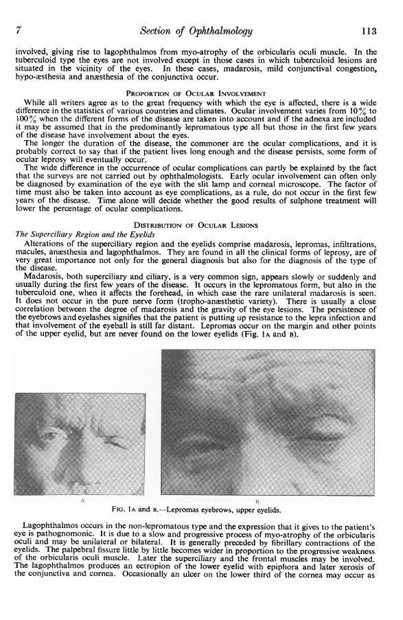
Madarosis, both superciliary and ciliary, is a very common sign, appears slowly or suddenly andusually during the first few years of the disease. It occurs in the lepromatous form, but also in thetuberculoid one, when it affects the forehead, in which case the rare unilateral madarosis is seen.It does not occur in the pure nerve form (tropho-anxsthetic variety). There is usually a closecorrelation between the degree of madarosis and the gravity of the eye lesions. The persistence ofthe eyebrows and eyelashes signifies that the patient is putting up resistance to the lepra infection andthat involvement of the eyeball is still far distant. Lepromas occur on the margin and other pointsof the upper eyelid, but are never found on the lower eyelids (Fig. 1A and B).
..... .... ...
~~~~~~~~~~~~~~~~~~~~~~~~~~~~~~~.:. .̂.. ........... ................... .lIIl^is~~~~~~~~~~~~~~~~~~~~~~~~...... '':':.......;''''.
A BFIG. 1A and B.-Lepromas eyebrows, upper eyelids
Lagophthalmos occurs in the non-lepromatous type and the expression that it gives to the patient'seye is pathognomonic. it is due to a slow and progressive process of myo-atrophy of the orbicularisoculi and may be unilateral or bilateral. It is generally preceded by fibrillary contractions of theeyelids. The palpebral fissure little by little becomes wider in proportion to the progressive weaknessof the orbicularis oculi muscle. Later the superciliary and the frontal muscles may be involved.The lagophthalmos produces an ectropion of the lower eyelid with epiphora and later xerosis ofthe conJunctiva and cornea. Occasionally an ulcer on the lower third of the cornea may occur as
Proceedings of the Royal Society of Medicine
a result of exposure. Unless the lagophthalmos is accompanied by an anesthesia in the region ofthe V nerve, diminution or loss of sensibility of the conjunctiva and cornea does not occur. Theabsence of reflex winking is due to loss of function of the orbicularis oculi and not to the loss ofcorneal sensibility. Once leprous lagophthalmos is established, it is usually permanent and will notrecede, but despite the cornea being so much exposed, it is remarkable how so many of these casesescape corneal ulceration over a very prolonged period often extending into many years.
There is no disease of the lacrimal gland sac or duct peculiar to leprosy. Chronic dacrocystitisis, however, rather common, is secondary to the septic process present in the nose and frequentlygives rise to blepharitis and purulent conjunctivitis. The lacrimal gland may be hypertrophied increas-ing the flow of tears.
The Conjunctiva, Episclera and ScleraMycobacterium lepra has been found in the conjunctival secretion and in biopsies from the
conjunctiva in clinically healthy eyes. Hyperemia and congestion of the conjunctiva frequentlyoccur. These may be diffuse or limited to a triangle on either side of the cornea. The conjunctivaplays a very important role in the transportation of the lepra bacilli. Being an exposed tissue like theskin, it can be infected directly, or more commonly. the infection spreads from the surroundingskin lesions.
Leprous nodules are never found on the conjunctiva as the bacilli do not proliferate in theconjunctiva but in the episcleral tissue and upon the surface of the sclera close to the sclero-cornealjunction where the anterior ciliary nerves penetrate. They appear to have a marked preference forthis area on account of its peculiar structure and position. Yellowish, gelatinous nodules appear inthis area more usually on the temporal side. They extend around the limbus producing a lowrampart of granulation tissue and eventually obliterate the whole contour of the limbus.The infiltration spreads into the superficial layers of the cornea, destroys Bowman's membrane,
and produces a sclerosing keratitis. An anterior staphyloma of the cornea may result. Hypo-esthesia or anaesthesia of the conjunctiva occurs in the lepromatous type and is of great value in
diagnosis, as if it is found present it is a warning of impending serious ocular lesions.Invasion of the conjunctiva, episclera and sclera are ordinarily not accompanied by great pain
or acute reactions so that the patient delays in seeking medical advice.The CorneaThe cornea is the most vulnerable of the ocular tissues and is very commonly involved. All varieties
of keratitis are seen and can best be classified into primary and secondary. The primary comprisefive groups-pannus, sclerosing keratitis, superficial punctate keratitis, deep or interstitial keratitisand leproma of the cornea. Lesions of the sclera and limbus precede or accompany the keratitis.The secondary ones comprise the ulcerative group which may occur in lagophthalmos and when thereis loss of sensibility of the cornea. Pannus in leprosy is very common and presents itself in the formof a net, the meshes of which are composed of uniform branching blood vessels, in contradistinctionto the pannus of trachoma in which the new blood vessels are terminal and arranged in the shape ofbundles. It is seen in all stages from early vascularization in the upper third of the cornea to thesevere forms in which there is deep vascularization as well. Grave forms are seen causing a partialor even complete hyperplastic keratitis.Leprous sclerosing keratitis is a common occurrence, in which the sclerotic cdat of the eye appears
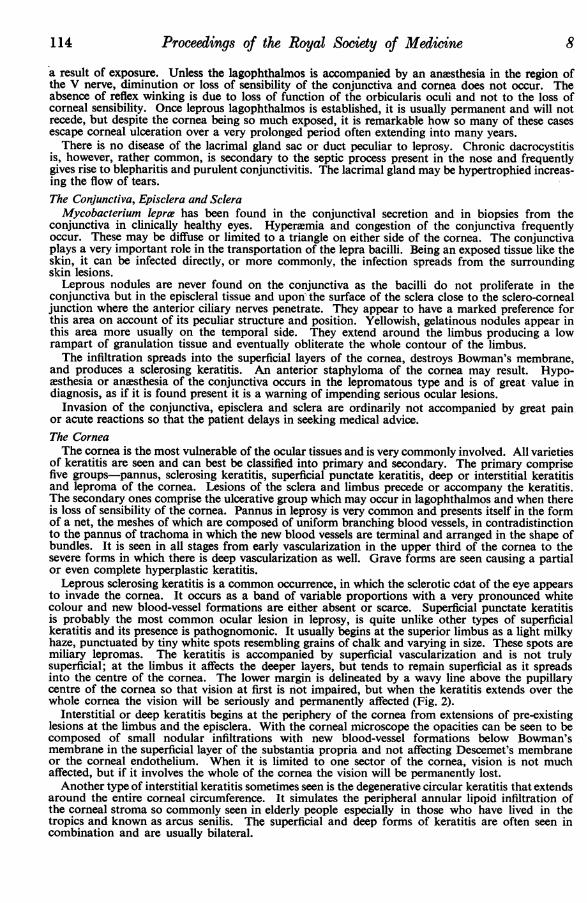
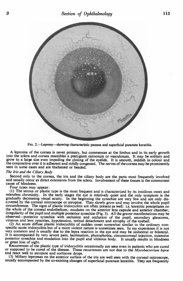
to invade the cornea. It occurs as a band of variable proportions with a very pronounced whitecolour and new blood-vessel formations are either absent or scarce. Superficial punctate keratitisis probably the most common ocular lesion in leprosy, is quite unlike other types of superficialkeratitis and its presence is pathognomonic. It usually begins at the superior limbus as a light milkyhaze, punctuated by tiny white spots resembling grains of chalk and varying in size. These spots aremiliary lepromas. The keratitis is accompanied by superficial vascularization and is not trulysuperficial; at the limbus it affects the deeper layers, but tends to remain superficial as it spreadsinto the centre of the cornea. The lower margin is delineated by a wavy line above the pupillarycentre of the cornea so that vision at first is not impaired, but when the keratitis extends over thewhole cornea the vision will be seriously and permanently affected (Fig. 2).
Interstitial or deep keratitis begins at the periphery of the cornea from extensions of pre-existinglesions at the limbus and the episclera. With the corneal microscope the opacities can be seen to becomposed of small nodular infiltrations with new blood-vessel formations below Bowman'smembrane in the superficial layer of the substantia propria and not affecting Descemet's membraneor the corneal endothelium. When it is limited to one sector of the cornea, vision is not muchaffected, but if it involves the whole of the cornea the vision will be permanently lost.Another type of interstitial keratitis sometimes seen is the degenerative circular keratitis that extends
around the entire corneal circumference. It simulates the peripheral annular lipoid infiltration ofthe corneal stroma so commonly seen in elderly people especially in those who have lived in thetropics and known as arcus senilis. The superficial and deep forms of keratitis are often seen incombination and are usually bilateral.
114 8
Section of Ophthalmology
FIG. 2.-Leprosy-showing characteristic pannus and superficial punctate keratitis.
A leproma of the cornea is never primary, but commences at the limbus and in its early growthinto the sclera and cornea resembles a pterygium carnosum or vasculosum. It may be solitary andgrow to a large size even impeding the closing of the eyelids. It is smooth, reddish in colour andthe conjunctiva over it is adherent and mildly congested. The nerves of the cornea may be prominentlyseen in some cases and are thickened or beaded.The Iris and the Ciliary BodySecond only to the cornea, the iris and the ciliary body are the parts most frequently involved
and usually occur as direct extensions from the sclera. Involvement of these tissues is the commonestcause of blindness.Four types may appear:(1) The serous or plastic type is the most frequent and is characterized by its insidious onset and
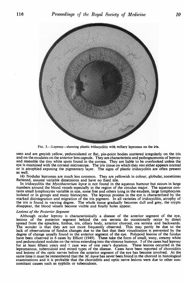
relentless chronicity. In the early stages the eye is relatively quiet and the only symptom is thegradually decreasing visual acuity. In the beginning the synechixe are very few and are only dis-covered by the corneal microscope or atropine. They slowly grow and may involve the whole pupilcircumference. The signs of plastic iridocyclitis are often present as well: i.e. keratitic precipitates onthe whole of the corneal endothelium; exudates on the anterior lens capsule and anterior chamber;irregularity of the pupil and multiple posterior synechiae (Fig. 3). All the graver manifestations may beobserved-posterior synechiEe with seclusion and occlusion of the pupil, secondary glaucoma,vitreous and lens opacities, hypotension, retinal detachment and atrophy of the eyeball.
(2) An acute diffuse plastic iridocyclitis of sudden onset somewhat similar to the ordinary non-specific acute iridocyclitis but of a more violent nature is sometimes seen. In my experience it is notvery common and is usually due to the lepra reaction in the eye and may be unilateral or bilateral.It is accompanied by very severe pain, lacrimation, photophobia, circumcorneal injection, extensiveposterior synechiae and exudation into the pupil and vitreous body. It usually results in blindnessor great loss of sight.
Recurrences of the plastic type of iridocyclitis occasionally are seen even in patients who are curedor supposed to be cured of the disease. These recurrences are not due to the Mycobacterium lepraeand react well to atropine and cortisone drops.
(3) Miliary lepromas on the anterior surface of the iris are well seen with the corneal microscope,usually accompanied by the co-existing changes of superficial punctate keratitis. They'are frequently
115
116 Proceedings of the Royal Society of Medicine 10
FIG. 3.-Leprosy-showing plastic iridocyclitis with miliary lepromas on the iris.
seen and are greyish yellow, pedunculated or flat, pin-point bodies scattered irregularly on the irisand on the exudates on the anterior lens capsule. They are characteristic and pathognomonic of leprosyand resemble the tiny white spots found in the cornea. They are liable to be overlooked unless theeye is examined with the corneal microscope. The iris tissue on which they rest either appears normalor is atrophied exposing the pigmentary layer. The signs of plastic iridocyclitis are often presentas well.
(4) Nodular lepromas are much less common. They are yellowish in colour, globular, sometimesflattened, assume variable dimensions and have no fixed site.
In iridocyclitis the Mycobacterium lepra? is not found in the aqueous humour but occurs in largenumbers around the blood vessels especially in the region of the circulus major. The aqueous con-tains small lymphocytes variable in size, some free and others lying in the exudate, large lymphocytesisolated or in groups and many histiocytes. The leprous process in the eye is characterized by themarked disintegration and migration of the iris pigment. In all varieties of iridocyclitis, atrophy ofthe iris is found in varying degree. The whole tissue gradually becomes dull and grey, the cryptsdisappear, the blood vessels become visible and finally holes appear.Lesions of the Posterior Segment
Although ocular leprosy is characteristically a disease of the anterior segment of the eye,lesions of the posterior segment behind the ora serrata do occasionally occur by directspread from the episclera, limbus, iris, ciliary body, anterior choroid, ora serrata and backwards.The wonder is that they are not more frequently observed. This may partly be due to thelack of observations of fundus changes due to the fact that their visualization is prevented by thedegree of change usually found in the anterior segment of the eye. Polypoid lesions of the fundushave been observed in 6 cases by Elliott (1949). These take the form of small, waxy, creamy whiteand pedunculated nodules on the retina extending into the vitreous humour. 5 of the cases had leprosyfor at least fifteen years and I case was of one year's duration. These lesions occurred in thelepromatous, tuberculoid and mixed types of the disease. Cases have been reported of choroiditisand lesions of the optic nerves before the anterior segment of the eye has become involved. At thesame time it must be remembered that the M. Ieprw has never been found in the choroid in histologicalexaminations and it is probable that the choroiditis and optic nerve lesions were due to other con-comitant causes such as syphilis or tuberculosis.
11 Section of Ophthalmology 117
PATHOGENESIS OF EYE LESIONSLeprosy is a systemic infection, and Hansen's bacilli are found in the peripheral blood. The
lepromatous infiltration of the eye is part of the generalized infection. As lesions in the posteriorsegment of the eye do not occur-or at all events occur rarely-in marked contrast to syphiliticand tuberculous ones, so the portal of entry for ocular invasion in the lepromatous type is bestexplained by the transportation of the bacilli by the blood vessels or lymphatics from the lepromataon the face and eyelids to the conjunctiva, episclera and the anterior segment of the eyeball. Thereare, however, points in favour of the endogenous passage in which the uveal tract is first affected,beginning in the angle of the iris, spreading in front of the iris, inwards to the ciliary body, backwardsto the choroid and outwards to the sclero-corneal limbus.
In tuberculoid leprosy, the conjunctiva being an exposed tissue may be involved when the skinaround the eyes or on the face is affected.
TREATMENT OF EYE LESIONSIn leprosy, loss of vision is caused by changes in the cornea and plastic iridocyclitis. Up to the
advent of the sulphones, treatment was fraught with disappointment, hydnocarpus oil, the ancientEastern remedy, was up to that time the most reliable treatment and had retained its place for a verylong time. In my experience the benefit to the eyes from sulphone therapy is remarkable but theearly diagnosis of eye lesions and early treatment both local and general are of great importance.In incipient cases the specific lesions of the cornea and iris very slowly recede but not in advancedcases in which profound changes in the ocular tissues have already occurred, although even in thegrave cases much can be done to prevent blindness. It takes three to five years for a lepromatouscase to become negative. With sulphone therapy relapses do not occur or at all events it may beyears before a relapse is detected. The case which tends to relapse is the tuberculoid one. This isdetermined by the ability or inability of the tissues to respond to the M. lepra? by a vigorous reaction.In the last ten years in which sulphone therapy has been employed for the treatment of leprosy casesin England, no case of blindness has occurred. In one case sight was seriously affected by acuteiridocyclitis caused by the lepra reaction. No longer does one see the same amount of mucopurulentconjunctivitis, septic inflammation of the eyelids and chronic dacrocystitis which are so common inuntreated cases of leprosy. Unfortunately with sulphone therapy there is a tendency to precipitatethe lepra reaction in the early months of treatment and until it is controlled the eyes must be carefullywatched in case the lepra reaction occurs in the eye. This is the most terrible ocular complicationof leprosy. Great care therefore should be taken against injudicious or inadequate treatment.ACTH and cortisone, in spite of their immediate striking effects in the lepra reactions, have provokeda divided opinion. Some workers have got good results with small doses and others have found thatcessation was followed by acute manifestations of leprosy and by increase in the underlying disease.The local application to the eye of cortisone in 1 per cent drops or ointment is most valuable in theacute eye conditions in which an unchecked inflammatory reaction will cause occlusion andseclusion of the pupil with secondary cataract and secondary glaucoma. It should be used frequentlyand continued till the eye is white. Cortisone, however, should be regarded only as an adjuvant tothe sulphone therapy to lessen the danger of residual damage from an excessive inflammation. Inlepromatous cases, the pupils should be frequently observed and if early signs of iritis are observed,the pupils should be kept well dilated by atropine. These early signs are often only discovered bythe corneal microscope and one should always be available in hospitals where leprosy is treated.Unfortunately many of the patients one sees show the iris bound down by posterior synechik, thepupil small and filled with exudate so that atropine in these cases is of little use. If the pupil refusesto dilate sufficiently or if secondary glaucoma occurs, an iridectomy should be done. The leprouseye, despite the chronic inflammation, stands surgery very well.Lagophthalmos is usually permanent if it has persisted for any length of time. I have seen one
early case clear up with sulphone therapy. In view of the success obtained with cortisone in acutecases of Bell's palsy, it would perhaps be of value in accelerating recovery in early cases. Liquidparaffin drops should be instilled at bed-time to protect the cornea and if corneal ulceration occursa tarsorrhaphy must not be delayed.
SUMMARYThe ocular complications of leprosy are the most serious lesions in the disease. Leprosy has been
one of the great causes of blindness and partial loss of sight. With the advent of sulphone andcortisone therapy, most of this could be prevented if only patients suffering from this disease receivedthe benefits of established treatment in the early stages. There are thousands and thousands ofpeople in backward countries suffering from leprosy who receive no care.
BIBLIOGRAPHYDUKE-ELDER, S. (1938) Textbook of Ophthalmology. London; 2, 1638, 1946; and 3, 2320.ELLIOTT, D. C. (1949) Int. J. Leprosy, 17, 229.ELLIOTT, R. H. (1920) Tropical Ophthalmology. London; p. 429.HARLEY, R. D. (1946) Amer. J. Ophthal., 29, 295.KIRWAN, E. W. O'G. (1948) Trans. R. Soc. trop. Med. Hyg., 41, 583.NAPIER, L. (1946) Principles and Practice of Tropical Medicine. New York; p. 481.SPYRATOS, S. (1954) Ann. Oculist., Paris, 187, 538.VALE, S. (1946) Subsidiary Studies to Leprosy of the Eyes. Rio de Janeiro.
118 Proceedings of the Royal Society of Medicine 12
Mr. E. F. King said that he could only speak from limited experience of ocular leprosy.He had, however, before the war seen a few cases at the Homes of St. Giles in Essex, and twoyears ago he had made a tour of East Africa and had visited a number of leper colonies in Uganda,Kenya, Tanganyika, and Zanzibar.
In visiting these leper colonies in East Africa he had been impressed by the relatively few casesshowing ocular involvement. Moreover, when the eyes were affected, the lesions were usually grossin nature resulting in opaque cornew, shrunken globes and staphylomata. He had discussed thesecases with Dr. J. Ross Innes, the Leprologist to the East African territories, who had emphasized thepoint made by Mr. Choyce, that there was a great variation in the incidence of ocular involvement inleprosy in different parts of the world, and also in the type of eye lesion.Mr. King had been told that at the commencement of sulphone therapy severe generalized
reactions and exacerbation of iritis might occur, on the lines of the Herxheimer reaction. Theseeffects could generally be controlled by systemic and local cortisone.
Dr. Robert G. Cochrane stated that, with the introduction of sulphones, a great deal could bedone for ocular leprosy and the whole outlook had been changed for the better, particularly iftreatment was commenced at an early stage. It should now be possible to prevent blindness, eventhough, upder the older treatments if the leprosy of the eye were adequately treated, blindness couldbe staved off for a very long time. It was interesting to note that there appeared to be a differencein the incidence of eye lesions in the various races of the world. This difference seemed to be in someway connected with pigment, for if one divided the races of the world into lightly pigmented racesand darkly pigmented ones, generally speaking, one would find that in the darkly pigmented raceocular leprosy seemed far less common. This was illustrated in his own institution in India, wherethere were a large number of Anglo-Indians and they showed a high incidence of ocular leprosy ascompared to that seen in the Indian races. This statement also applied to the African races, where,by and large, ocular leprosy was not so serious, neither was the incidence so high as in the lesspigmented peoples.With the introduction of cortisone, serious effects of ocular leprosy could now be very largely
prevented, and if cortisone were used very carefully then specific therapy could be continued. This,in the past, was always a great difficulty, because specific therapy had to be stopped when certaineye reactions showed themselves. While cortisone would benefit any eye reaction condition in leprosy,it was of most value in the so-called erythema nodosum reaction of leprosy, if, at the same time, carefulsulphone therapy was given as well. Several of the cases seen at the meeting had passed through aperiod of very severe erythema nodosum. This phase eventually passed off and when it did theprogress of the patient under treatment was smoother, and there was seldom a recurrence.
Dr. Cochrane confirmed the statement that it was the anterior part of the eye which was involved.He had never seen the posterior chamber affected. He did not think the changes described by Elliotthad been confirmed by others.The speaker added that he had been a little surprised to learn that Mr. Choyce was so relatively
optimistic about operating. Personally, the speaker felt that if the eye was involved there was graverisk in operation. He did not advise this until the eye was quiescent unless the condition happenedto be extremely serious, because so often interference set up changes in the eye and often when theeye was involved and the cornea also one tended to get opacities in the line of the incision. However,he was not an ophthalmologist, and he did not operate unless absolutely forced to do so. One hadto be very careful before operating on an active lepromatous eye.
Dr. A. V. Clemmey spoke as one who had lived for twenty years in East Africa during which timehe had seen little intra-ocular trouble; generally, it was a matter of keratitis and some anaesthesia.He had happened to see in Liverpool the case (Mr. A. D. W.), and in that case iritis had respondedto atropine and cortisone.