6. ANALYTICAL METHODS - atsdr.cdc.gov · PDF file6. ANALYTICAL METHODS ... Sensitivity is...
22
CDDs 507 6. ANALYTICAL METHODS The purpose of this chapter is to describe the analytical methods that are available for detecting, and/or measuring, and/or monitoring CDDs, its metabolites, and other biomarkers of exposure and effect to CDDs. The intent is not to provide an exhaustive list of analytical methods. Rather, the intention is to identify well-established methods that are used as the standard methods of analysis. Many of the analytical methods used for environmental samples are the methods approved by federal agencies and organizations such as EPA and the National Institute for Occupational Safety and Health (NIOSH). Other methods presented in this chapter are those that are approved by groups such as the Association of Official Analytical Chemists (AOAC) and the American Public Health Association (APHA). Additionally, analytical methods are included that modify previously used methods to obtain lower detection limits, and/or to improve accuracy and precision. 6.1 BIOLOGICAL SAMPLES The primary method of determining CDDs in biological samples is gas chromatography (GC) with mass spectrometry (MS). Sample preparation is critical, and extensive extraction and sample clean-up are required to separate the CDD homologues/congeners from fatty material and other organic contaminants. Extreme care must also be used to ensure that all reagents and equipment used in analysis are free of CDD contamination. Losses of CDDs can occur as a result of adsorption onto the surfaces of glassware used in sample preparation (EPA 1994c). The routine baking of glassware as a part of the cleaning process should be avoided because this may cause active sites on the glass that will irreversibly adsorb CDDs. The lack of interferences must be demonstrated under the conditions of analysis. Analysts should avoid polyvinyl chloride (PVC) gloves (EPA 1994c). The basic steps of sample preparation include extraction of the sample with a lipophilic organic solvent (e.g., hexane) followed by several evaporation and column chromatography steps to concentrate, clean up, and fractionate the CDDs. Methods of measuring CDDs in biological samples are very sensitive, generally having method (sample matrix) detection limits in the low- or sub-parts per trillion (ppt) level. If rigorous sample preparation methods are meticulously followed, sensitivity, accuracy, selectivity, and precision can be good. These parameters will vary with the analytical method used, the experience level of the technician, the nature of the sample matrix, the concentrations of the analyte(s) and possible interfering substances, and the specific
Transcript of 6. ANALYTICAL METHODS - atsdr.cdc.gov · PDF file6. ANALYTICAL METHODS ... Sensitivity is...

CDDs 507
6. ANALYTICAL METHODS
The purpose of this chapter is to describe the analytical methods that are available for detecting, and/or
measuring, and/or monitoring CDDs, its metabolites, and other biomarkers of exposure and effect to
CDDs. The intent is not to provide an exhaustive list of analytical methods. Rather, the intention is to
identify well-established methods that are used as the standard methods of analysis. Many of the
analytical methods used for environmental samples are the methods approved by federal agencies and
organizations such as EPA and the National Institute for Occupational Safety and Health (NIOSH). Other
methods presented in this chapter are those that are approved by groups such as the Association of Official
Analytical Chemists (AOAC) and the American Public Health Association (APHA). Additionally,
analytical methods are included that modify previously used methods to obtain lower detection limits,
and/or to improve accuracy and precision.
6.1 BIOLOGICAL SAMPLES
The primary method of determining CDDs in biological samples is gas chromatography (GC) with mass
spectrometry (MS). Sample preparation is critical, and extensive extraction and sample clean-up are
required to separate the CDD homologues/congeners from fatty material and other organic contaminants.
Extreme care must also be used to ensure that all reagents and equipment used in analysis are free of CDD
contamination. Losses of CDDs can occur as a result of adsorption onto the surfaces of glassware used in
sample preparation (EPA 1994c). The routine baking of glassware as a part of the cleaning process should
be avoided because this may cause active sites on the glass that will irreversibly adsorb CDDs. The lack of
interferences must be demonstrated under the conditions of analysis. Analysts should avoid polyvinyl
chloride (PVC) gloves (EPA 1994c). The basic steps of sample preparation include extraction of the
sample with a lipophilic organic solvent (e.g., hexane) followed by several evaporation and column
chromatography steps to concentrate, clean up, and fractionate the CDDs.
Methods of measuring CDDs in biological samples are very sensitive, generally having method (sample
matrix) detection limits in the low- or sub-parts per trillion (ppt) level. If rigorous sample preparation
methods are meticulously followed, sensitivity, accuracy, selectivity, and precision can be good. These
parameters will vary with the analytical method used, the experience level of the technician, the nature of
the sample matrix, the concentrations of the analyte(s) and possible interfering substances, and the specific
CDDs 508
6. ANALYTICAL METHODS
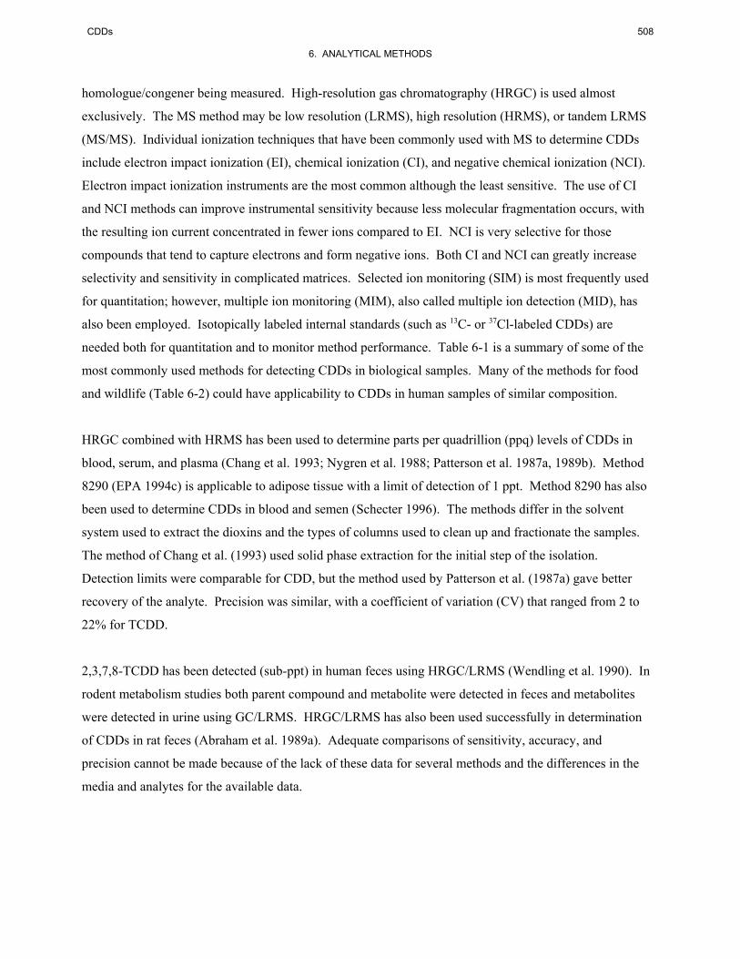
homologue/congener being measured. High-resolution gas chromatography (HRGC) is used almost
exclusively. The MS method may be low resolution (LRMS), high resolution (HRMS), or tandem LRMS
(MS/MS). Individual ionization techniques that have been commonly used with MS to determine CDDs
include electron impact ionization (EI), chemical ionization (CI), and negative chemical ionization (NCI).
Electron impact ionization instruments are the most common although the least sensitive. The use of CI
and NCI methods can improve instrumental sensitivity because less molecular fragmentation occurs, with
the resulting ion current concentrated in fewer ions compared to EI. NCI is very selective for those
compounds that tend to capture electrons and form negative ions. Both CI and NCI can greatly increase
selectivity and sensitivity in complicated matrices. Selected ion monitoring (SIM) is most frequently used
for quantitation; however, multiple ion monitoring (MIM), also called multiple ion detection (MID), has
also been employed. Isotopically labeled internal standards (such as 13C- or 37Cl-labeled CDDs) are
needed both for quantitation and to monitor method performance. Table 6-1 is a summary of some of the
most commonly used methods for detecting CDDs in biological samples. Many of the methods for food
and wildlife (Table 6-2) could have applicability to CDDs in human samples of similar composition.
HRGC combined with HRMS has been used to determine parts per quadrillion (ppq) levels of CDDs in
blood, serum, and plasma (Chang et al. 1993; Nygren et al. 1988; Patterson et al. 1987a, 1989b). Method
8290 (EPA 1994c) is applicable to adipose tissue with a limit of detection of 1 ppt. Method 8290 has also
been used to determine CDDs in blood and semen (Schecter 1996). The methods differ in the solvent
system used to extract the dioxins and the types of columns used to clean up and fractionate the samples.
The method of Chang et al. (1993) used solid phase extraction for the initial step of the isolation.
Detection limits were comparable for CDD, but the method used by Patterson et al. (1987a) gave better
recovery of the analyte. Precision was similar, with a coefficient of variation (CV) that ranged from 2 to
22% for TCDD.
2,3,7,8-TCDD has been detected (sub-ppt) in human feces using HRGC/LRMS (Wendling et al. 1990). In
rodent metabolism studies both parent compound and metabolite were detected in feces and metabolites
were detected in urine using GC/LRMS. HRGC/LRMS has also been used successfully in determination
of CDDs in rat feces (Abraham et al. 1989a). Adequate comparisons of sensitivity, accuracy, and
precision cannot be made because of the lack of these data for several methods and the differences in the
media and analytes for the available data.


CDDs 522
6. ANALYTICAL METHODS
HRGC has been combined with LRMS, HRMS, and MS/MS for the detection of CDDs in tissues.
Sensitivity is generally in the ppt range with the best sensitivity (2 ppt) reported with MS/MS using CI
(Ryan et al. 1987a). The limit of detection was higher for MS than for MS/MS (Schecter et al. 1985b;
Stanley 1986; Takizawa and Muto 1987). No recovery data were given for HRMS (Nygren et al. 1988).
Precision for these methods is usually <20% (Takizawa and Muto 1987; Van den Berg et al. 1989).
CDDs have been measured in breast milk using HRGC/MS in the SIM mode. Reported detection limits
are in the low- to sub-ppt (Van den Berg et al. 1986b), and recovery (75–89%) is good (Noren and Sjoevall
1987).
An additional screening test for TCDD-like (aryl hydrocarbon receptor, AhR, active) chemicals has been
developed (Garrison et al. 1996) and is available commercially (Anonymous 1997). Dubbed the CALUX
(for chemically activated luciferase gene expression) system, the assay is based on recombinant cell lines
into which researchers have inserted a firefly luciferase gene. When exposed to dioxin-like compounds,
the recombinant cells luminesce. The method is sensitive to ppt levels of 2,3,7,8-TCDD equivalents in
blood, serum, and milk (Anonymous 1997). Samples testing positive can be subjected to more definitive
and specific analytical testing.
6.2 ENVIRONMENTAL SAMPLES
As with biological samples, the most common method of determining CDDs in environmental samples is
HRGC/HRMS. Other methods, including enzyme bioassays, and monoclonal antibody-based enzyme-
linked immunosorbent assays (ELISAs) have also been used or are under development. Even in relatively
simple matrices, such as air and water, detection and quantitation of CDDs require rigorous sample
preparation procedures. Methods used to prepare environmental samples are similar to those used for
biological samples: organic solvent extraction of CDDs from the sample and concentration, clean up, and
fractionation of the dioxins using evaporative and column chromatography techniques. The same MS
techniques described for biological samples are available for environmental samples, with essentially the
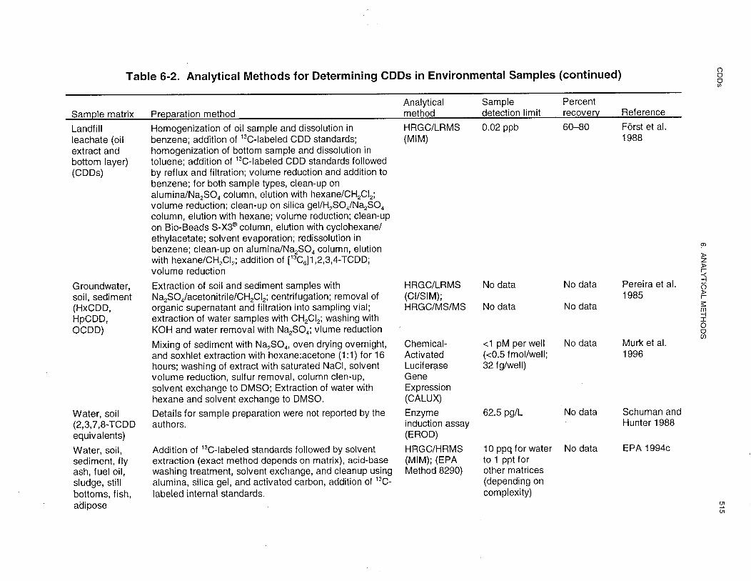
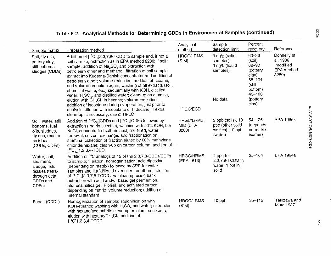
same results and limitations. Table 6-2 describes some of the most common methods that have been used
to determine CDDs in environmental samples, with specific MS techniques listed when known. The
following section describes the methods available for the different types of environmental samples.
CDDs 523
6. ANALYTICAL METHODS
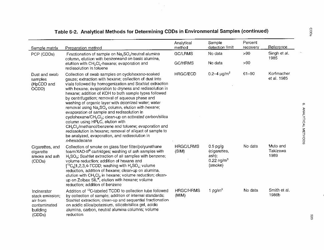
HRGC/LRMS and HRGC/HRMS have been used to analyze for CDDs in ambient and hazardous waste
site air, cigarette smoke, car exhaust, and gaseous waste emissions. Sample preparation steps for gaseous
samples are very similar for these two analytical methods. The steps consist of collection of sample
contaminants on a filter/trapping cartridge apparatus, organic solvent extraction of the cartridge, and clean
up and fractionation of the extract using column chromatography (Bingham et al. 1989; Cooke et al. 1988;
Fairless et al. 1987; Harless et al. 1992; Muto and Takizawa 1989; Oehme et al. 1986; Rappe et al. 1988;
Smith et al. 1986). A quartz fiber filter and polyurethane foam plug are commonly used to collect air
samples (EPA 1988g; Harless et al. 1992; Kuwata et al. 1993), although XAD-2 has also been used
(Hippelein et al. 1993). The sensitivity of these methods is in the low- to sub-pg/m3 range. Reported
recovery and precision were generally good for measurements in air and gaseous waste emissions (Cooke
et al. 1988; Fairless et al. 1987; Oehme et al. 1986), but severe sample loss can occur (Bingham et al.
1989; Rappe et al. 1988). Electron capture, negative ionization, low resolution MS has also been used to
quantify CDDs in ambient air; however, 2,3,7,8-TCDD is difficult to detect using this method and results
must be confirmed with HRGC (Koester et al. 1992).
Methods have been developed for detecting CDDs in liquid samples including drinking water (McCurvin
et al. 1989; O'Keefe et al. 1986), groundwater (EPA 1986k, 1994a, 1994c; Pereira et al. 1985), fog
(Czuczwa et al. 1989), liquid waste effluents (Cooke et al. 1988), an oil extract of landfill leachate (Först et
al. 1988), pentachlorophenol (Singh et al. 1985), fuel oils, still bottoms, and reactor residues (EPA 1986k,
1994a), and pyrolyzed transformer oil (Hardin et al. 1989). HRGC was combined with either LRMS,
HRMS, or MS/MS in these methods. Not all methods reported on recovery, precision, and sensitivity, so
it is difficult to compare these parameters. Based on the data available, sensitivities range from sub-ppq
(O'Keefe et al. 1986) to low-ppt levels (Först et al. 1988). Recoveries were usually >60% (Först et al.
1988; O'Keefe et al. 1986), although some lower values were reported (Hardin et al. 1989).
HRGC/LRMS, HRGC/HRMS, HRGC/MS/MS, and HRGC/ECD have been used to analyze for CDDs in
soils and/or sediments (Bobbie et al. 1989; Creaser and Al-Haddad 1989; Donnelly et al. 1986; EPA
1986k, 1994a, 1994c; Eschenroeder et al. 1986; Jasinski 1989; Pereira et al. 1985; Simon et al. 1989;
Stalling et al. 1986), solid wastes (Donnelly et al. 1986; Först et al. 1988; Popp et al. 1997), and other
solid materials (Donnelly et al. 1986; Hardin et al. 1989; Korfmacher et al. 1985; Muto and Takizawa
1989). Detection limits for the MS methods range from low-ppt to low-ppb levels. The sensitivity cannot
be compared to ECD because no detection limits were reported for the ECD methods. For soil/sediments,
recovery seemed to be better for GC/ECD (92–100%) (Jasinski 1989) than for the HRGC/MS methods

CDDs 524
6. ANALYTICAL METHODS
(40–102%) (Creaser and Al-Haddad 1989; Donnelly et al. 1986; Simon et al. 1989). Polychlorinated
biphenyls, polychlorinated diphenyl ethers, polychlorinated naphthalenes, and polychlorinated
alkydibenzofurans may be found at concentrations several orders of magnitude higher than the analytes of
interest (EPA 1994a) and could thus interfere with the CDDs. Retention times must be verified using
reference standards.
A method for determining CDDs in municipal incinerator fly ash has been reported (Alexandrou and
Pawliszyn 1990). The method uses supercritical fluid extraction (SFE) to recover CDDs from fly ash
samples prior to GC. Supercritical fluid extraction is faster and less expensive than the typically used
Soxhlet extraction and gives quantitative removal of CDDs and CDFs from fly ash. Extracts obtained
using SFE will still require additional clean-up steps prior to analysis. Supercritical CO2 has also been
used to assist solvent-based extraction of CDDs from soils (Friedrich and Kleiböhmer 1997). In this case,
the supercritical fluid was combined with accelerated solvent extraction (liquid extractions conducted
under elevated temperature and pressure) to provide good recoveries relative to Soxhlet extractions.
TCDD and other CDDs have been measured in foods (Jasinski 1989; Schecter et al. 1994; Takizawa and
Muto 1987) and wildlife (birds and bird eggs, fish, and seals) (Bobbie et al. 1989; Buser et al. 1985; EPA
1994a; Stalling et al. 1986) using HRGC/ECD or HRGC/LRMS. Schecter et al. (1994) reported data as
TCDD toxic equivalents with detection limits of approximately 0.01 ppt. Ferrario et al. (1996) reported a
new modification of EPA Method 1613 (EPA 1994a) for use in measuring CDDs and CDFs in beef fat; an
LOD of 0.05 ppt was shown. A comparison of HRGC/LRMS methods conducted using samples from
fish, birds, and seals showed that NCI was substantially more sensitive than EI for some, but not all,
congeners (Buser et al. 1985). A within-lab comparison of fish tissue analysis using HRGC combined
with either LRMS, HRMS, or MS/MS showed HRMS to be the most sensitive of the three methods
(Bobbie et al. 1989). However, the large variations in recovery obtained with these methods also
demonstrated the significance of the problems of sample loss and sample contamination that can occur in
the analyses of CDDs. The data were not sufficient to permit a comparison of methods among different
laboratories.
Bioassays using induction of the enzymes ethoxyresorufin o-deethylase (EROD) and/or arylhydrocarbon
hydroxylase (AHH) in rat hepatoma H-4-IIE cells (Zacharewski et al. 1989) and modified mouse liver
cells (Schuman and Hunter 1988) have been developed and tested on water, soil, and fish samples. The
bioassays are based on induction of AHH or EROD enzymatic activity in the cell cultures. Since the cells
used in the bioassays are most sensitive to induction by 2,3,7,8-TCDD, this dioxin is used to generate a

CDDs 525
6. ANALYTICAL METHODS
standard curve for the bioassays, and induction of activity is expressed as TCDD equivalents. These
bioassays are highly sensitive to concentrations of Ah receptor-mediated cytochrome P-450 inducers
(Holcomb et al. 1988; Zacharewski et al. 1989), and could be used to rapidly pre-screen environmental
samples for 2,3,7,8-TCDD toxicity equivalents. A major drawback to these assays is that they are not
highly selective. A number of halogenated aromatics other than CDDs can induce AHH and EROD
activity (e.g., chlorinated dibenzofurans, polychlorinated biphenyls, and polychlorinated phenols),
although none to the extent of TCDD induction. There is also a question about the possible effects of
chemical mixtures, such as might be found in contaminated soil or fish, on the assay results (Zacharewski
et al. 1989). An ELISA based on derivation of monoclonal antibodies specific to CDDs has also been
investigated as a means of screening environmental samples for chlorinated dioxins (Stanker et al. 1987).
Monoclonal antibodies (MAbs) developed using 1-amino-substituted 3,7,8-TrCDD derivatives could
detect sub-ng levels of TCDD standards. The derived antibodies had a stronger affinity for CDDs
substituted at the 1 position and for CDFs substituted at the 2, 3, 7, and 8 positions than for other CDDs
including 2,3,7,8-TCDD. However, development of MAbs more specific for CDDs, especially
2,3,7,8-TCDD would provide a rapid, inexpensive, sensitive, and reasonably selective method for
screening samples for CDD contamination. Sugawara and coworkers (Sugawara et al. 1998) have recently
described an ELISA-based method for polychlorinated dibenzo-p-dioxins that can detect as little as 0.5
pg/well of 2,3,7,8-TCDD and shows great promise as a screening tool. The cross reactivity for octachloro
dibenzo-p-dioxin is very low (<0.1%), but it is much higher for compounds with three, four, or five
chlorine atoms in a substitution pattern similar to the of 2,3,7,8-TCDD. As with all screening approaches,
more accurate chemical analysis would be needed to confirm the compounds present.
The CALUX assay described in Section 6.1 has been applied to Ah receptor-active compounds (not
limited to dioxins) in sediments and pore waters (Murk et al. 1996) and to blood with mixed results.
Sensitivities as low as 0.5 fmol of 2,3,7,8-TCDD were reported. Two polychlorinated terphenyl mixtures,
the PCB-substituted Ugilec 141, polybrominated diphenyl ethers, and the PCB mixture Clophen 150 were
tested in the CALUX assay and had induction potencies that were 10-4 to 10-7 compared to TCDD. Thus,
this assay is more selective than earlier, induction-based assays, although clearly not as selective as
GC/MS.
6.3 ADEQUACY OF THE DATABASE
Section 104(i)(5) of CERCLA, as amended, directs the Administrator of ATSDR (in consultation with the
Administrator of EPA and agencies and programs of the Public Health Service) to assess whether adequate

CDDs 526
6. ANALYTICAL METHODS
information on the health effects of CDDs is available. Where adequate information is not available,
ATSDR, in conjunction with the NTP, is required to assure the initiation of a program of research designed
to determine the health effects (and techniques for developing methods to determine such health effects) of
CDDs.
The following categories of possible data needs have been identified by a joint team of scientists from
ATSDR, NTP, and EPA. They are defined as substance-specific informational needs that if met would
reduce the uncertainties of human health assessment. This definition should not be interpreted to mean
that all data needs discussed in this section must be filled. In the future, the identified data needs will be
evaluated and prioritized, and a substance-specific research agenda will be proposed.
6.3.1 Identification of Data Needs
Methods for Determining Biomarkers of Exposure and Effect. Methods exist for determining
CDDs in human serum and plasma, feces, biological tissues, and milk (Abraham et al. 1989a; Anonymous
1997; Chang et al. 1993; EPA 1994a, 1994c; Noren and Sjoevall 1987; Nygren et al. 1988; Patterson et al.
1987a, 1987b; Ryan et al. 1987a; Schecter et al. 1985b; Stanley 1986; Takizawa and Muto 1987; Van den
Berg et al. 1989; Wendling et al. 1990). These methods have been used to determine ppq to ppt levels of
CDDs in biological samples. The commonly used methods are sensitive enough to detect background
levels of CDDs in most media, especially adipose tissue. The background concentration for non
occupationally-exposed people has been reported to be on the order of 4 ppt in lipid (Michalek et al. 1998).
Improved clean-up and instrument sensitivity could make blood a more useful monitoring medium,
although it is usually reagent and background contamination that is most problematic; CDD concentrations
in blood tend be quite low. Improvements in current methods or development of new methods to increase
sensitivity and selectivity would help to decrease the time involved in sample preparation, and would
reduce the high cost ($800–$1,000 per sample) and possible errors associated with current methods of
determining exposure to CDDs.
Several effects such as chloracne and alterations in hepatic metabolism have been associated with exposure
to 2,3,7,8-TCDD in humans. However, these effects are not specific for 2,3,7,8-TCDD or other CDDs, but
may be induced by numerous other chlorinated hydrocarbons. Determination of specific biomarkers of
effect for CDD and development of reliable methods to quantify these effects would be useful in assessing
the effects associated with exposure to CDDs.
CDDs 527
6. ANALYTICAL METHODS
Methods for Determining Parent Compounds and Degradation Products in Environmental Media. Methods exist for measuring CDDs in a variety of environmental media, including air, water,
sediment, soil, chemical waste, foods, fish, and other solid matrices (Bingham et al. 1989; Bobbie et al.
1989; Buser et al. 1985; Cai et al. 1994; Cooke et al. 1988; Creaser and Al-Haddad 1989; Donnelly et al.
1986; EPA 1986k, 1988g, 1994a, 1994c; Fairless et al. 1987; Jasinski 1989; Marquis et al. 1994;
McCurvin et al. 1989; Muto and Takizawa 1989; Oehme et al. 1986; O'Keefe et al. 1986; Pereira et al.
1985; Rappe et al. 1988; Smith et al. 1986a). Of the EPA methods, Method 8280 (EPA 1986k) and 8290
(EPA 1994a) are both commonly used; Method 8290 is approximately three orders of magnitude more
sensitive. Assuming an acute oral MRL of 20 pg/kg/day, an intermediate oral MRL of 7 pg/kg/day, and a
70-kg individual, the limit of detection needed for water (2 L/day consumption) is 770 ppq for acute and
245 ppq for intermediate exposure. The methods of O'Keefe et al. (1986) (LOD reported to be
0.5–1.1 ppq) and EPA (1994a, 1994c) (LODs reported to be 4 ppq to 10 ppq) are adequate for detecting
CDDs in drinking water. If a 2 kg/day consumption of food is assumed, the needed method LODs will be
700 ppq for acute and 245 ppq for intermediate exposure. Of those method reporting LODs in foods, the
methods of Bobbie et al. (1989) and of Ferrario et al. (1996) have the required LODs. Since CDDs are
typically determined on a fat weight basis, the method of Ferrario et al. (1996) should be suitable for most
food types once the fat is extracted. The sensitivity of the HRGC/MS methods is excellent, but because of
the very low levels of these chemicals in the environment, increased sensitivity may be desirable in order
to obtain detectable values. Increased accuracy and selectivity would help make analyses more reliable
and possibly reduce the costly and time-consuming sample preparation steps that are currently required.
Additional development of bioassays to detect CDDs could provide screening methods with sufficient
sensitivity to detect the very low concentrations of toxicological importance.
6.3.2 Ongoing Studies
A collaborative study was identified in which researchers at CDC, NIEHS, University of Mainz in
Germany and the German Cancer research Center in Heidelberg are studying biochemical markers of
exposure and susceptibility to dioxin in human peripheral blood lymphocytes (Yang et al. 1997).
The following information was obtained from a search of Federal Research in Progress (FEDRIP 1998).
Under an SBIR (Small Business Innovative Research) grant, Xeonobiotic Detection Systems, Inc. of
Durham, NC, is marketing the CALUX assay (Anonymous 1997) described in Section 6.1. Hybrizyme

CDDs 528
6. ANALYTICAL METHODS
Corp, of Raleigh, NC, is working on a new test method for dioxins in human and animal samples. This
work is also being performed under an SBIR. No other details were available. Antibody-based methods
for 2,3,7,8-TCDD analysis is the subject of a project lead by R. Carlson of Ecochem Research, Inc.
(another SBIR) during which methods for gases will be developed. Finally, G. Wheelock, Paracelsian,
Inc., Ithaca, NY, is using SBIR funding to develop an Ah receptor-based assay for the determination of
toxic equivalency factors.


![7. ANALYTICAL METHODS · 7. ANALYTICAL METHODS ... [SRM] 4200, 60,000 Bq [1.6 µCi] CESIUM 164 7. ANALYTICAL METHODS . and SRM 4207, 300,000 Bq …](https://static.fdocuments.net/doc/165x107/5ae8481b7f8b9aee078f554f/7-analytical-methods-analytical-methods-srm-4200-60000-bq-16-ci-cesium.jpg)