document
8
1997, 179(20):6488. J. Bacteriol. T Iwabuchi and S Harayama by Nocardioides sp. strain KP7. enzyme involved in phenanthrene degradation 2-carboxybenzaldehyde dehydrogenase, an Biochemical and genetic characterization of http://jb.asm.org/content/179/20/6488 Updated information and services can be found at: These include: CONTENT ALERTS more» cite this article), Receive: RSS Feeds, eTOCs, free email alerts (when new articles http://journals.asm.org/site/misc/reprints.xhtml Information about commercial reprint orders: http://journals.asm.org/site/subscriptions/ To subscribe to to another ASM Journal go to: on June 2, 2014 by Hunter College http://jb.asm.org/ Downloaded from on June 2, 2014 by Hunter College http://jb.asm.org/ Downloaded from
-
Upload
trannguyet -
Category
Documents
-
view
213 -
download
1
Transcript of document

1997, 179(20):6488. J. Bacteriol.
T Iwabuchi and S Harayama by Nocardioides sp. strain KP7.enzyme involved in phenanthrene degradation2-carboxybenzaldehyde dehydrogenase, an Biochemical and genetic characterization of
http://jb.asm.org/content/179/20/6488Updated information and services can be found at:
These include:
CONTENT ALERTS more»cite this article),
Receive: RSS Feeds, eTOCs, free email alerts (when new articles
http://journals.asm.org/site/misc/reprints.xhtmlInformation about commercial reprint orders: http://journals.asm.org/site/subscriptions/To subscribe to to another ASM Journal go to:
on June 2, 2014 by Hunter C
ollegehttp://jb.asm
.org/D
ownloaded from
on June 2, 2014 by H
unter College
http://jb.asm.org/
Dow
nloaded from

JOURNAL OF BACTERIOLOGY,0021-9193/97/$04.0010
Oct. 1997, p. 6488–6494 Vol. 179, No. 20
Copyright © 1997, American Society for Microbiology
Biochemical and Genetic Characterization of 2-CarboxybenzaldehydeDehydrogenase, an Enzyme Involved in Phenanthrene
Degradation by Nocardioides sp. Strain KP7TOKURO IWABUCHI† AND SHIGEAKI HARAYAMA*
Marine Biotechnology Institute, Kamaishi Laboratories,3-75-1 Heita, Kamaishi, Iwate 026, Japan
Received 16 April 1997/Accepted 12 August 1997
2-Carboxybenzaldehyde dehydrogenase from the phenanthrene-degrading bacterium Nocardioides sp. strainKP7 was purified and characterized. The purified enzyme had a molecular mass of 53 kDa by sodium dodecylsulfate-polyacrylamide gel electrophoresis and 205 kDa by gel filtration chromatography. Thus, the homotet-ramer of the 53-kDa subunit constituted an active enzyme. The apparent Km and kcat values of this enzyme for2-carboxybenzaldehyde were 100 mM and 39 s21, respectively, and those for NAD1 were 83 mM and 32 s21,respectively. The structural gene for this enzyme was cloned and sequenced. The length of the gene was 1,455bp. The nucleotide sequence of the 10,279 bp of DNA around the gene for 2-carboxybenzaldehyde dehydroge-nase was also determined, and seven open reading frames were found in this DNA region. These were the genesfor 1-hydroxy-2-naphthoate dioxygenase (phdI) and trans-2*-carboxybenzalpyruvate aldolase (phdJ), orf1, thegene for 2-carboxybenzaldehyde dehydrogenase (phdK), orf2/orf3, and orf4. The amino acid sequence of theorf1 product was similar to that of the aromatic hydrocarbon transporter gene (pcaK) in Pseudomonas putidaPRS2000. The amino acid sequence of the orf4 product revealed a similarity to cytochrome P-450 proteins. Theregion between phdK and orf4 encoded orf2 and orf3 on different strands. The amino acid sequences of the orf2and orf3 products exhibited no significant similarity to the reported sequences in protein databases.
Microbial biodegradation of polynuclear aromatic hydrocar-bons (PAHs), especially simple ones such as naphthalene andphenanthrene, has been extensively studied over the last de-cade (4, 14). In bacteria, biodegradation of PAHs is initiated bythe introduction of both atoms of molecular oxygen into thearomatic nucleus, the reaction being catalyzed by a multicom-ponent dioxygenase (15). Further reactions lead to the forma-tion of precursors of tricarboxylic acid cycle intermediates.
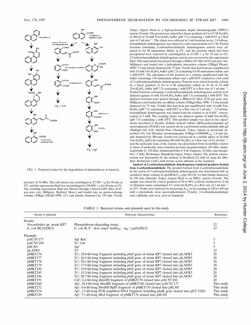
Phenanthrene is degraded by bacteria through one of twodifferent routes via a common intermediate, 1-hydroxy-2-naph-thoate (Fig. 1). In one route, 1-hydroxy-2-naphthoate is oxi-dized to 1,2-dihydroxynaphthalene, which is further degradedvia salicylate, while in the other route, the ring of 1-hydroxy-2-naphthoate is cleaved and further metabolized via o-phtha-late (10, 20–22, 24). The enzymatic steps for the decompositionof the first and second rings of phenanthrene in the salicylateroute resemble those of the first ring of naphthalene. It hasbeen demonstrated that a common set of enzymes was respon-sible for the decomposition of the first and second rings ofphenanthrene, as well as for the first ring of naphthalene (26,34, 36). The PAH-degrading pathway via salicylate found inBurkholderia cepacia F297 seems to exhibit quite broad sub-strate specificity, as this pathway degrades a wide variety ofpolycyclic aromatic compounds, including fluorene, (methyl-)naphthalene, phenanthrene, anthracene, and dibenzothio-phene (12).
The enzymes and genes for the degradation of phenan-threne, naphthalene, and dibenzothiophene via salicylate inmany strains have been characterized (8, 9, 24, 30, 31, 35). Theamino acid sequences of isofunctional catabolic enzymes in
different strains are 90% identical to each other. Less homol-ogous groups of genes for the degradation of PAHs have beenfound in Comamonas testosteroni (11).
The genes and enzymes for phenanthrene degradation viathe o-phthalate route have recently started to be clarified.1-Hydroxy-2-naphthoate dioxygenase, which converts 1-hy-droxy-2-naphthoate to trans-29-carboxybenzalpyruvate, andtrans-29-carboxybenzalpyruvate aldolase, which converts trans-29-carboxybenzalpyruvate to 2-carboxybenzaldehyde, were pu-rified from phenanthrene-degrading, gram-positive Nocardio-ides sp. strain KP7 (20–22). In this bacterium, the genes for1-hydroxy-2-naphthoate dioxygenase (phdI) and trans-29-car-boxybenzalpyruvate aldolase (phdJ) were located close to eachother. In this study, 2-carboxybenzaldehyde dehydrogenase,which catalyzes the step next to that mediated by trans-29-carboxybenzalpyruvate aldolase (phdK), was studied in thesame organism. The nucleotide sequence of the 10,279 bp ofDNA around the gene for 2-carboxybenzaldehyde dehydroge-nase was also determined.
MATERIALS AND METHODS
Bacterial strains, culture conditions, and plasmids. The phenanthrene-de-grading bacterium used in this study, Nocardioides sp. strain KP7, has beendescribed previously (22). This strain was cultivated in Marine broth (DifcoLaboratories, Detroit, Mich.) in the presence or absence of 0.1% (wt/vol)phenanthrene at 30°C. The other bacterial strain and the plasmids used in thisstudy are listed in Table 1.
Enzyme assays. Under standard conditions, the activity of 2-carboxybenzalde-hyde dehydrogenase was determined spectrophotometrically by measuring theformation of NADH at 340 nm at 25°C in 100 mM glycine-NaOH (pH 9.4)containing 0.51 mM 2-carboxybenzaldehyde and 1 mM NAD1.
Purification and analysis of 2-carboxybenzaldehyde dehydrogenase. 2-Car-boxybenzaldehyde dehydrogenase was purified from cells of strain KP7 grown for48 h at 30°C in 28 liters of Marine broth (Difco) containing 0.1% (wt/vol)phenanthrene. The cells (88 g [wet weight]) were pelleted by centrifugation at10,000 3 g for 20 min at 4°C, resuspended in 200 ml of 10 mM Tris-H2SO4 buffer(pH 7.5) containing 1 mM dithiothreitol (DTT), and disrupted by passagethrough a precooled French pressure cell (Ohtake Works, Tokyo, Japan) at a
* Corresponding author. Phone: 81-193-26-6544. Fax: 81-193-26-6592. E-mail: [email protected].
† Present address: Pharmaco Science Laboratories, Shiseido Re-search Center, Kouhoku-ku, Yokohama, Kanagawa 223, Japan.
6488
on June 2, 2014 by Hunter C
ollegehttp://jb.asm
.org/D
ownloaded from
pressure of 76 MPa. The cell extract was centrifuged at 27,700 3 g for 30 min at4°C, and the supernatant fluid was recentrifuged at 250,000 3 g for 60 min at 4°C.The resulting supernatant fluid was filtered through a Sterivex-HV filter (0.45-mm pore size; Millipore, Bedford, Mass.) and loaded into an anion-exchangecolumn (TSKgel DEAE-5PW; 21.5 mm [inside diameter] by 150 mm; Tosoh,
Tokyo, Japan) fitted to a high-performance liquid chromatography (HPLC)system (Tosoh). The protein was eluted by a linear gradient of 0 to 0.5 M Na2SO4
in 300 ml of 10 mM Tris-H2SO4 buffer (pH 7.5) containing 1 mM DTT at a flowrate of 5 ml min21. The eluate was collected in 5-ml fractions on ice. 2-Carboxy-benzaldehyde dehydrogenase was eluted at a salt concentration of 0.1 M. Pooledfractions containing 2-carboxybenzaldehyde dehydrogenase activity were ad-justed to 0.6 M ammonium sulfate at 4°C, and the proteins which had beenprecipitated were removed by centrifugation at 27,700 3 g for 30 min at 4°C.2-Carboxybenzaldehyde dehydrogenase activity was recovered in the supernatantfluid. This supernatant was passed through a Millex-GV filter (0.45-mm pore size;Millipore) and loaded into a hydrophobic interaction column (TSKgel Phenyl-5PW; 7.5 mm [inside diameter] by 75 mm; Tosoh) that had been pre-equilibratedwith 10 mM Tris-H2SO4 buffer (pH 7.5) containing 0.6 M ammonium sulfate and1 mM DTT. The adsorption of the proteins to a column equilibrated with thebuffer containing 1 M ammonium sulfate and 1 mM DTT resulted in a low yieldof 2-carboxybenzaldehyde dehydrogenase. Proteins were eluted from the columnby a linear gradient of 0.6 to 0 M ammonium sulfate in 30 ml of 10 mMTris-H2SO4 buffer (pH 7.5) containing 1 mM DTT at a flow rate of 1 ml min21.Pooled fractions containing 2-carboxybenzaldehyde dehydrogenase activity weredialyzed against 10 mM Tris-H2SO4 buffer (pH 7.5) containing 1 mM DTT. Thedialyzed fractions were passed through a Millex-GV filter (0.45-mm pore size;Millipore) and loaded into an affinity column (TSKgel Blue-5PW; 7.5 mm [insidediameter] by 75 mm; Tosoh) that had been pre-equilibrated with 10 mM Tris-H2SO4 (pH 7.5) containing 1 mM DTT at a flow rate of 1 ml min21. 2-Carboxy-benzaldehyde dehydrogenase was eluted from the column at an NAD1 concen-tration of 5 mM. The resulting eluate was dialyzed against 10 mM Tris-H2SO4
(pH 7.5) containing 1 mM DTT. This purified sample was used in the experi-ments described in Results. Sodium dodecyl sulfate (SDS)-polyacrylamide gelelectrophoresis (PAGE) was carried out in a preformed polyacrylamide slab gel(Multigel Gel 4/20; Daiichi Pure Chemicals, Tokyo, Japan) as previously de-scribed (19). Gel filtration chromatography (TSKgel G3000SWXL; 7.8 mm [in-side diameter] by 300 mm; Tosoh) was carried out in a mobile phase of 20 mMTris-H2SO4 (pH 6.8) containing 100 mM Na2SO4 at a flow rate of 0.5 ml min21,and the molecular mass of the enzyme was determined from its mobility relativeto those of molecular mass standard proteins (b-galactosidase, 465 kDa; immu-noglobulin G, 150 kDa; immunoglobulin G Fab fragment, 50 kDa; and myoglo-bin, 17 kDa; Boehringer Mannheim Japan, Tokyo, Japan). The protein concen-tration was determined by the method of Bradford (2) with an assay kit (Bio-Rad, Richmond, Calif.) with bovine serum albumin as the standard.
Analysis of 2-carboxybenzaldehyde dehydrogenase-catalyzed products formedfrom 2-carboxybenzaldehyde. The product formed from 2-carboxybenzaldehydeby the action of 2-carboxybenzaldehyde dehydrogenase was determined with anoctyldecyl silane column (CapcellPak-C18 type SG120; 4.6 mm [inside diameter]by 250 mm; Shiseido, Tokyo, Japan) fitted to an HPLC system (Tosoh). Thesample was eluted by a linear gradient of 50 to 90% (vol/vol) methanol in 15 mlof ultrapure water containing 0.1% (vol/vol) H3PO4 at a flow rate of 1 ml min21
at 25°C. Peaks were detected by measuring A254 or by scanning at 220 to 400 nmwith a photodiode array spectrophotometer (Tosoh). 2-Carboxybenzaldehydeand o-phthalic acid were used as standards.
FIG. 1. Proposed routes for the degradation of phenanthrene in bacteria.
TABLE 1. Bacterial strains and plasmids used in this study
Strain or plasmid Relevant characteristics Reference
StrainsNocardioides sp. strain KP7 Phenanthrene-degrading strain 22E. coli BL21(DE3) E. coli B; F2 dcm ompT hsdS(rB
2 mB2) gall(DE3) 13
PlasmidspACYC177 Apr Kmr 5pACYC184 Tcr Cmr 5pSL301 Apr 3pLAFR3 Tcr 32pMKT176 Tcr; 18.0-kb-long fragment including phdI gene of strain KP7 cloned into pLAFR3 20pMKT177 Tcr; 26.5-kb-long fragment including phdI gene of strain KP7 cloned into pLAFR3 20pMKT178 Tcr; 20.1-kb-long fragment including phdI gene of strain KP7 cloned into pLAFR3 20pMKT179 Tcr; 17.9-kb-long fragment including phdI gene of strain KP7 cloned into pLAFR3 20pMKT184 Tcr; 19.0-kb-long fragment including phdI gene of strain KP7 cloned into pLAFR3 20pMKT185 Tcr; 19.3-kb-long fragment including phdI gene of strain KP7 cloned into pLAFR3 20pMKT186 Tcr; 20.7-kb-long fragment including phdI gene of strain KP7 cloned into pLAFR3 20pMKT191 Cmr; 6.2-kb-long BamHI fragment of pMKT176 cloned into pACYC184 20pMKT260 Apr; 10.4-kb-long HindIII fragment of pMKT186 cloned into pACYC177 This studypMKT272 Apr; 6.6-kb-long HindIII-BglII fragment of pMKT176 cloned into pSL301 This studypMKT314 Apr; 1.5-kb-long PCR-amplified DNA fragment including phdK gene cloned into pET-22(b) This studypMKT329 Apr; 7.1-kb-long MluI fragment of pMKT176 cloned into pSL301 This study
VOL. 179, 1997 PHENANTHRENE DEGRADATION BY NOCARDIOIDES SP. STRAIN KP7 6489
on June 2, 2014 by Hunter C
ollegehttp://jb.asm
.org/D
ownloaded from

Amino-terminal sequencing. The amino-terminal sequence of the purifiedenzyme was determined by Edman degradation with an automated proteinsequencer (model 477; Perkin-Elmer Applied Biosystems, Branchburg, N.J.).
Gene cloning and sequencing. Two degenerated PCR primers (N and cC)which would collaboratively amplify a 71-bp-long DNA fragment encoding theamino-terminal 29 amino acid residues of 2-carboxybenzaldehyde dehydroge-nase were designed. Their sequences were 59-TT(CT)GA(CT)GA(AG)TA(CT)(AC)G(AGCT)TGGAACGT-39 for the N primer and 59-AT(AGT)AT(AGCT)GG(AG)TA(AGCT)C(GT)(AGCT)GACTC-39 for the cC primer. Cloning ofthe amplified PCR product was carried out by using the pCRII plasmid vector(TA cloning kit; Invitrogen Co., San Diego, Calif.), and the nucleotide sequenceof the cloned PCR product was determined by using a Taq DyeDeoxy terminatorcycle sequencing kit and a 373A DNA sequencer (Perkin-Elmer Applied Bio-systems). The gene library based on cosmid pLAFR3 (32) and clones containingthe gene for 1-hydroxy-2-naphthoate dioxygenase have been described previous-y (20). DNA fragments of these clones were subcloned into pACYC177(5), pACYC184 (5), and pSL301 (3) to construct pMKT260, pMKT191, andpMKT329, respectively. These three subclones were used as templates for nu-cleotide sequencing.
Expression of 2-carboxybenzaldehyde dehydrogenase in Escherichia coli. TheDNA fragment carrying a putative gene for 2-carboxybenzaldehyde dehydroge-nase was amplified by Vent DNA polymerase (New England BioLabs, Beverly,Mass.) with a set of two PCR primers. One primer, 59-CAGGAGCAACATATGACGACACC-39, was designed to add an NdeI site in the 212 to 111 regionof the gene. This primer also changed the initiation codon of the gene from GTGto ATG. The other PCR primer, 59-TGAACTAAAGGATCCTGGCCG-39, wasdesigned to add a BamHI site after the termination codon of the gene. Theamplified DNA fragment was digested with NdeI and BamHI and cloned intopET-22b(1) (33) to construct pMKT314. The cloned DNA in pMKT314 wassequenced to confirm the absence of any mutation in the amplified fragment. E.coli BL21(DE3) (13) containing pMKT314 was cultivated in 100 ml of L brothcontaining 50 mg of ampicillin ml21 until an A660 of 0.6 was reached. Afteraddition of 0.1 ml of 100 mM isopropyl-b-D-thiogalactopyranoside (IPTG), thecells were further cultured for 2 h at 37°C. The resulting cells were pelleted bycentrifugation at 10,000 3 g for 20 min at 4°C, resuspended in 5 ml of 10 mMTris-H2SO4 buffer (pH 7.5), and disrupted by passage through a precooledFrench pressure cell (Ohtake Works) at 76 MPa. The cell extract was centrifugedat 27,700 3 g for 30 min at 4°C, and the supernatant fluid was recentrifuged at250,000 3 g for 60 min at 4°C. This supernatant fluid was filtered through aSterivex-HV filter (0.45-mm pore size; Millipore). 2-Carboxybenzaldehyde dehy-drogenase activity for 2-carboxybenzaldehyde in the cell extract was measured asalready described.
Nucleotide sequence accession number. The nucleotide sequence of 10,279 bpof DNA that includes phdI, -J, and -K has been deposited in the DDBJ, Gen-Bank, and EMBL DNA databases under accession no. AB000735.
RESULTS
Expression of 2-carboxybenzaldehyde dehydrogenase instrain KP7. The expression of 2-carboxybenzaldehyde dehy-drogenase was examined in strain KP7 grown on marine brothin the presence or absence of phenanthrene. The specific ac-tivity of 2-carboxybenzaldehyde dehydrogenase was 0.03 mmolmin21 mg of protein21 in an extract of strain KP7 grown in theabsence of phenanthrene, while it was 0.12 mmol min21 mg ofprotein21 in an extract of the cells grown in the presence ofphenanthrene. This result indicates that 2-carboxybenzalde-
hyde dehydrogenase of strain KP7 was induced by phenan-threne or its metabolite(s).
Purification and characterization of 2-carboxybenzaldehydedehydrogenase. The purification of 2-carboxybenzaldehyde de-hydrogenase from strain KP7 grown on marine broth in thepresence of phenanthrene is summarized in Table 2. The en-zyme activity was eluted from an anion-exchange column at anNa2SO4 concentration of 0.1 M. By hydrophobic interactionchromatography, the enzyme was adsorbed to the column at anammonium sulfate concentration of 0.6 M and eluted at anammonium sulfate concentration of 0.24 M. The enzyme wasadsorbed to a dye-ligand affinity column and eluted from it atan NAD1 concentration of 5 mM. The affinity chromatogra-phy-purified sample gave a single protein band by SDS-PAGE.The molecular mass of 2-carboxybenzaldehyde dehydrogenaseevaluated by gel filtration chromatography was 205 kDa (datanot shown), and that determined by SDS-PAGE was 53 kDa(Fig. 2). Thus, the homotetramer of the 53-kDa subunit con-stituted an active enzyme. The UV absorption spectrum of thisenzyme between 210 and 400 nm did not indicate any enzyme-associated chromophores.
The Michaelis-Menten kinetic constants of this enzyme for2-carboxybenzaldehyde, its related compounds, and NAD1
were determined. 2-Carboxybenzaldehyde dehydrogenase oxi-dized 2-carboxybenzaldehyde and 2-nitrobenzaldehyde but didnot oxidize the other compounds tested. The Km and catalyticconstant (kcat) values for 2-carboxybenzaldehyde were 100 mMand 39 s21, respectively; those for 2-nitrobenzaldehyde were2.8 mM and 48 s21, respectively; and those for NAD1 were 83mM and 32 s21, respectively. No activity was detected for thecompounds 3-carboxybenzaldehyde, 4-carboxybenzaldehyde,benzaldehyde, salicylaldehyde, 2-methylbenzaldehyde, 2-chlo-robenzaldehyde, 2-fluorobenzaldehyde, 2-benzaldehydesulfon-ate, 2-methoxybenzaldehyde, 1-hydroxy-2-naphthoaldehyde,and n-hexylaldehyde.
Determination of the 2-carboxybenzaldehyde dehydroge-nase activity in different buffers at different pH values indi-cated the optimum for this enzyme to be pH 9. The optimumtemperature of this enzyme was 40°C. The effects of metals andchelators on the activity of 2-carboxybenzaldehyde dehydroge-nase were examined. Three metals (Mn21, Mg21, and Ca21 at10 mM) revealed weak inhibitory effects (27 to 32% inhibition),while Zn21, Co21, and Cu21 at 10 mM did not (less than 15%inhibition). The inhibitory effects of 10 mM EDTA (28% inhibi-
FIG. 2. SDS-PAGE of 2-carboxybenzaldehyde dehydrogenase. Lanes: 1, mo-lecular size markers; 2, pooled active fractions after affinity chromatography (0.5mg).
TABLE 2. Purification of 2-carboxybenzaldehyde dehydrogenasefrom Nocardioides sp. strain KP7
Step Vol(ml)
Proteinconcn
(mg ml21)
Totalprotein
(mg)Sp acta
Totalactivity(mmolmin21)
Yield(%)
Cell extract 180 5.02 904 0.12 108.5 100Anion exchange 20 6.06 121 0.42 50.8 46.8Hydrophobic inter-
action5.0 0.88 4.4 7.38 32.5 30.0
Dye-ligand affinity 2.0 0.29 0.6 31.0 18.6 17.1
a 2-Carboxybenzaldehyde dehydrogenase was assayed in 100 mM glycine-NaOH buffer (pH 9.4) containing 1 mM NAD1 and 0.51 mM 2-carboxybenzal-dehyde, and its activity is expressed as micromoles of NADH formed per minuteper milligram of protein.
6490 IWABUCHI AND HARAYAMA J. BACTERIOL.
on June 2, 2014 by Hunter C
ollegehttp://jb.asm
.org/D
ownloaded from

tion) and 10 mM ethylenedioxybis(ethylamine)-N,N,N9,N9-tet-raacetic acid (14% inhibition) were also weak.
The reaction product from 2-carboxybenzaldehyde catalyzedby this enzyme was analyzed by HPLC, and the product hadthe same retention time as o-phthalate (data not shown).
Amino-terminal sequence of 2-carboxybenzaldehyde dehy-drogenase. The 29-residue amino-terminal sequence of puri-fied 2-carboxybenzaldehyde dehydrogenase was determined byautomated Edman degradation to be Thr-Thr-Pro-Arg-Lys-Phe-Asp-Glu-Tyr-Arg-Trp-Asn-Val-Leu-Val-Asp-Glu-Val-Pro-Leu-Asn-Val-Glu-Ser-Arg-Tyr-Pro-Ile-Ile.
Nucleotide sequence of the gene for 2-carboxybenzaldehydedehydrogenase. Based on the amino acid sequence, two degen-erated PCR primers (N and cC) which would allow amplifica-tion of the 71-bp-long product were designed as described inMaterials and Methods. By using this primer set, a PCR prod-uct corresponding to the predicted size was amplified whentotal DNA from strain KP7 was used as the template. The71-bp-long PCR product was cloned in the pCRII plasmid(Invitrogen), and the nucleotide sequence of this fragment wasdetermined by cycle sequencing. The amino acid sequencededuced from the DNA sequence of this PCR product was100% identical to the amino-terminal sequence just described.This result indicates that the amplified product correspondedto the partial sequence of the 2-carboxybenzaldehyde dehydro-genase gene. Hereafter, the gene for 2-carboxybenzaldehydedehydrogenase is called phdK (polyaromatic hydrocarbon deg-radation).
Seven cosmid clones carrying the 1-hydroxy-2-naphthoatedioxygenase gene (phdI) have previously been screened fromthe gene library of strain KP7 (20). The 71-bp-long PCR prod-ucts were also amplified by using these primers and the cosmid
clones pMKT176, pMKT177, pMKT184, pMKT185, andpMKT186 as templates. However, no amplification was ob-served when pMKT178 and pMKT179 were used as templates(Fig. 3). These results mapped the 59 region of phdK betweenthe ends of pMKT179 and pMKT184.
The 10.4-kb-long HindIII fragment from pMKT186 was sub-cloned into pACYC177 to construct pMKT260. It is knownthat the 39 region of phdI is located on the plasmid (20). Tomore accurately localize the 71-bp-long region on the 10.4-kbHindIII fragment, three PCR primers were used. Two ofthem were the N and cC primers used to amplify the 71-bp-long fragment, while the third primer was designed from theDNA sequence of the 39 region of phdI (do primer). Long-fragment PCR amplification with combinations of the cCand do primers amplified a 4.2-kb-long fragment. Thus, thelocation of the 59 region of phdK was determined on a mapof the 10.2-kb HindIII fragment. It was also indicated thatthe orientation of phdK was the same as that of phdI andthat the distance between phdK and phdI was approximately4.2 kb.
The nucleotide sequencing of phdK was initiated by using aprimer designed from the sequences of the 71-bp fragment andfurther extended by using oligonucleotides designed from thedetermined sequence. The length of phdK was 1,455 bp, andthe deduced amino acid sequence of the enzyme was 485amino acids (Fig. 4). The deduced amino acid sequence in theamino-terminal region was a perfect match with the sequencedetermined by Edman degradation, while the molecular mass(52 kDa) deduced from the amino acid sequence of this en-zyme was in agreement with the size evaluated by SDS-PAGE(53 kDa). The amino acid sequence of this enzyme exhibitedsignificant (33%) similarity to those of XylC and XylG (Fig. 5).
FIG. 3. Mapping of phdK by PCR. The structures of the cosmid clones used for amplification are shown. The results of PCR amplification with the N and cC primers(E, positive; 3, negative) are indicated on the right, while the locations of the sequences corresponding to the N, cC, and do primers are indicated at the center. Atthe bottom, the results of PCR amplification with the primer sets (do and N primers, negative; do and cC primers, positive) are indicated. V, EcoRV; Bm, BamHI;M, MluI; Bg, BglII; H, HindIII.
VOL. 179, 1997 PHENANTHRENE DEGRADATION BY NOCARDIOIDES SP. STRAIN KP7 6491
on June 2, 2014 by Hunter C
ollegehttp://jb.asm
.org/D
ownloaded from

XylC is a benzaldehyde dehydrogenase and is encoded by thexylC gene on toluene-xylene catabolic plasmid TOL (pWW0)of P. putida (19). XylG is a 2-hydroxymuconic semialdehydedehydrogenase and is also encoded on TOL (pWW0) (18).Amino acids 227 to 234 (FIGSTDTG) of 2-carboxybenzalde-hyde dehydrogenase fitted to the consensus sequence for al-dehyde dehydrogenase superfamily (F/Y)(I/T)G(S/E)(T/P)XX(G/F), and this sequence is possibly a part of the NAD1 bind-ing fold (18).
Expression of phdK in E. coli. A set of PCR primers wasdesigned to amplify phdK as described in Materials and Meth-ods, and a 1,520-bp-long DNA fragment amplified by VentDNA polymerase was cloned in pET-22b(1) to constructpMKT314. The nucleotide sequence of this amplified fragmentwas identical to that of the original phdK sequence, except forthe primer regions.
E. coli BL21(DE3) containing pMKT314 was cultivated inthe presence or absence of 0.1 mM IPTG in L broth at 37°C,and the 2-carboxybenzaldehyde dehydrogenase activity in theextracts of cells was measured. The specific activity of thisenzyme in noninduced cells was less than 1 nmol min21 mg of
protein21, while it was 170 nmol min21 mg of protein21 in cellsinduced by 0.1 mM IPTG. The results indicate that the1,455-bp open reading frame (ORF) is the structural gene for2-carboxybenzaldehyde dehydrogenase.
Organization of the phenanthrene catabolic genes of strainKP7. The nucleotide sequence of 10,279 bp of DNA whichincludes phdI, -J, and -K was determined. In this region, fiveORFs, in addition to phdI, -J, and -K, were found in the orderphdI-phdJ-orf1-phdK-(orf2/orf3)-orf4 (Fig. 6). The orientationof phdI, phdJ, orf1, phdK, and orf2 was the same, i.e., theopposite of that of orf3 and orf4. A protein encoded by orf1(1,419 bp) exhibited significant (27%) similarity to PcaK, atransport protein for 4-hydroxybenzoate encoded by pcaK in P.putida PRS2000 (16) (Fig. 7). Amino acids 22 to 26 (RRQRI)fitted the consensus sequence for the superfamily of transport-ers, (R/K)XXX(R/K). Furthermore, amino acids 86 to 91(DRWGRK) matched perfectly the consensus sequence (N/D)(R/K)XGR(R/K) for the transporter superfamily (16).Thus, orf1 probably encodes a transporter for some aromaticcompound(s). orf2 (768 bp), orf3 (1,191 bp), and orf4 (1,248bp) were found downstream of phdK. The deduced amino acidsequences of the orf2 and orf3 products had no significantsimilarity to the amino acid sequences reported in the Swiss-Prot database. The deduced amino acid sequence of orf4showed similarity to those of bacterial cytochrome P-450 pro-teins: 26, 31, 29, 29, and 26% identity to CPXC_AGRT6(cytochrome P-450-pinf1 from Agrobacterium tumefaciens;23), CPXJ_SACER (6-deoxyerythronolide B hydroxylasefrom Saccharopolyspora erhthraea; 17), CPXL_PSESP (cyto-chrome P-450-terp from Pseudomonas sp.; 29), CPXM_BACSU (cytochrome P-450-109 from Bacillus subtilis; 1), andFAS1_RHOFA (cytochrome P-450 fas1 from Rhodococcusfascians; 6), respectively. The amino acid sequence of orf4
FIG. 4. Nucleotide sequence of the gene for 2-carboxybenzaldehyde dehy-drogenase (phdK). The deduced amino acid sequence is also presented. Theputative Shine-Dalgarno sequence is underlined, while the stop codon is indi-cated by an asterisk.
FIG. 5. Alignment of the deduced amino acid sequences of 2-carboxybenz-aldehyde dehydrogenase from strain KP7, benzaldehyde dehydrogenase encodedby xylC on TOL plasmid pWW0, and 2-hydroxymuconic semialdehyde dehydro-genase encoded by the xylG gene on TOL plasmid pWW0. PhdK, 2-carboxy-benzaldehyde dehydrogenase; XylC, benzaldehyde dehydrogenase; XylG, 2-hydroxymuconic semialdehyde dehydrogenase. The asterisks indicateidentical amino acids, and the dashes indicate gaps introduced to optimizethe alignment.
6492 IWABUCHI AND HARAYAMA J. BACTERIOL.
on June 2, 2014 by Hunter C
ollegehttp://jb.asm
.org/D
ownloaded from

matched the consensus sequence for cytochrome P-450 pro-teins, FGXGXHXCXG (7).
DISCUSSION
Two enzymes in the phenanthrene-degrading pathway viao-phthalate, namely, 1-hydroxy-2-naphthoate dioxygenase andtrans-29-carboxybenzalpyruvate aldolase, and their structuralgenes have previously been characterized (20, 21). In thisstudy, we characterized 2-carboxybenzaldehyde dehydroge-nase, which catalyzes the next step to trans-29-carboxybenzal-pyruvate aldolase.
The biochemical properties of 2-carboxybenzaldehyde dehy-drogenase from strain KP7 were different from those of theenzyme from Alcaligenes faecalis AFK2, which also degradesphenanthrene via the o-phthalate route (25). The activeform of 2-carboxybenzaldehyde dehydrogenase from strainKP7 was the homotetramer of the 53-kDa subunit; however,that from strain AFK2 has been reported to be the homotet-ramer of the 40-kDa subunit. The Km values for 2-carboxy-benzaldehyde of the enzymes from strains KP7 and AFK2were 100 and 54 mM, and the optimum temperatures ofthese enzymes were 40 and 30°C, while their optimum pHvalues were 9 and 8, respectively. 2-Carboxybenzaldehydedehydrogenase from strain KP7 did not oxidize 4-carboxy-benzaldehyde, salicylaldehyde, and benzaldehyde; however,that from strain AFK2 did oxidize these compounds. Twometals, Mn21 and Mg21, had inhibitory effects on the ac-tivity of the KP7 enzyme but not on the activity of the AFK2enzyme, while Co21 and Cu21, conversely, revealed inhibi-tory effects on the activity of the AFK2 enzyme but not onthe activity of the KP7 enzyme.
The amino acid sequence of 2-carboxybenzaldehyde dehy-drogenase deduced from the DNA sequence of phdK exhibitedsignificant similarity to that of many proteins belonging to thealdehyde dehydrogenase superfamily. The sizes of enzymesbelonging to this superfamily range between 50 and 58 kDa,with the exception of two: human aldehyde dehydrogenase 8(DHA8_HUMAN) has a molecular mass of 43 kDa, while ahypothetical aldehyde dehydrogenase from yeast (YHJ9-YEAST) has a molecular mass of 71 kDa. The size of theAFK2 enzyme, 40 kDa, strongly suggests that this enzyme doesnot belong to the aldehyde dehydrogenase superfamily andtherefore is evolutionarily different from the KP7 enzyme. Inthis context, it would be very interesting to clone the gene for2-carboxybenzaldehyde dehydrogenase from strain AFK2 anddetermine its nucleotide sequence.
We determined the sequence of the 10,279-bp-long DNAfragment which includes phdI, -J, and -K. In addition, fourORFs, orf1, orf2, orf3, and orf4, were found. The orf1 producthad similarity to the transporter of 4-hydroxybenzoate in P.putida PRS2000 (16). It is possible that the orf1 product trans-ports 1-hydroxy-2-naphthoate, whose structure partially resem-bles that of 4-hydroxybenzoate. The orf4 product exhibitedsignificant similarity to cytochrome P-450 proteins. The pro-teins are known to exert diverse functions in prokaryotes andeukaryotes (27, 28), including PAH degradation in fungi (4,14). However, it is unclear whether the orf4 product has a rolein phenanthrene degradation or not.
In general, the genes for the degradation of aromatic com-pounds are compacted in operons. The organization of thephenanthrene-degrading genes in strain KP7 was differentfrom that of other genes for the degradation of aromaticcompounds: spacer regions of the phd genes and otherORFs are rather long compared to those in other aromatic-catabolic genes, in which even overlapping genes have beenidentified. Further studies are required to clarify the phys-iological and evolutionary significance of the specific genearrangement.
FIG. 6. Organization of phdI, -J, -K, and other ORFs on a 10,279-bp-long DNA fragment of Nocardioides sp. strain KP7. phdI, the 1-hydroxy-2-naphthoatedioxygenase gene; phdJ, the trans-29-carboxybenzalpyruvate aldolase gene; phdK, the 2-carboxybenzaldehyde dehydrogenase gene. Locations: phdI, 228 to 1391; phdJ,1388 to 2386; orf1, 2875 to 4294; phdK, 4987 to 6444; orf2, 7191 to 7961; orf3, 9350 to 8157; orf4, 7978 to 6728. Abbreviations: Bg, BglII; Bm, BamHI; EV, EcoRV;H, HindIII; M, MluI. The nucleotide sequence of the DNA fragment, apart from that of the phkK region, is not shown in this report but is available from theDDBJ/GenBank/EMBL DNA databases (accession no. AB000735).
FIG. 7. Alignment of the deduced amino acid sequence of the orf1 productwith that of pcaK from P. putida PRS2000. Orf1, deduced amino acid sequenceof the orf1 product; PcaK, deduced amino acid sequence of the pcaK gene.Asterisks indicate identical amino acids, periods indicated similar amino acidsbetween two sequences, and dashes indicate gaps introduced to maximize thealignment.
VOL. 179, 1997 PHENANTHRENE DEGRADATION BY NOCARDIOIDES SP. STRAIN KP7 6493
on June 2, 2014 by Hunter C
ollegehttp://jb.asm
.org/D
ownloaded from

ACKNOWLEDGMENTS
We are grateful to S. Miyachi, A. Saitoh, and J. Inoue for theirsupport and valuable advice. We thank Y. Itazawa and Y. Sasaki fortheir technical assistance.
This work was supported by the New Energy and Industrial Tech-nology Development Organization.
REFERENCES
1. Ahn, K. S., and R. G. Wake. 1991. Variations and coding features of thesequence spanning the replication terminus of Bacillus subtilis 168 and W23chromosomes. Gene 98:107–112.
2. Bradford, M. M. 1976. A rapid and sensitive method for the quantitation ofmicrogram quantities of protein utilizing the principle of protein-dye bind-ing. Anal. Biochem. 72:248–254.
3. Brosius, J. 1989. Superpolylinkers in cloning and expression vectors. DNA8:759–777.
4. Cerniglia, C. E., and M. A. Heitkamp. 1989. Microbial degradation of poly-cyclic aromatic hydrocarbons (PAH) in the aquatic environment, p. 41–68. InU. Varanasi (ed.), Metabolism of polycyclic aromatic hydrocarbons in theaquatic environment. CRC Press Inc., Boca Raton, Fla.
5. Chang, A. C. Y., and S. N. Cohen. 1978. Construction and characterization ofamplifiable multicopy DNA cloning vehicles derived from the P15A crypticminiplasmid. J. Bacteriol. 134:1141–1156.
6. Crespi, M., D. Vereecke, W. Temmerman, M. van Montagu, and J. Desomer.1994. The fas operon of Rhodococcus fascians encodes new genes requiredfor efficient fasciation of host plants. J. Bacteriol. 176:2492–2501.
7. Degtyarenko, K. N. 1995. Structural domains of P450-containing monooxy-genase systems. Protein Eng. 8:737–747.
8. Denome, S. A., D. C. Stanley, E. S. Olson, and K. D. Young. 1993. Metab-olism of dibenzothiophene and naphthalene in Pseudomonas strains: com-plete DNA sequence of an upper naphthalene catabolic pathway. J. Bacte-riol. 175:6890–6901.
9. Eaton, R. W. 1994. Organization and evolution of naphthalene catabolicpathways: sequence of the DNA encoding 2-hydroxychromene-2-carboxylateisomerase and trans-o-hydroxybenzylidenepyruvate hydratase-aldolase fromthe NAH7 plasmid. J. Bacteriol. 176:7757–7762.
10. Evans, W. C., H. N. Fernley, and E. Griffiths. 1965. Oxidative metabolism ofphenanthrene and anthracene by soil pseudomonads. Biochem. J. 95:819–831.
11. Goyal, A. K., and G. J. Zylstra. 1996. Molecular cloning of novel genes forpolycyclic aromatic hydrocarbon degradation from Comamonas testosteroniGZ39. Appl. Environ. Microbiol. 62:230–236.
12. Grifoll, M., S. A. Selifonov, C. V. Gatlin, and P. J. Chapman. 1995. Actionsof a versatile fluorene-degrading bacterial isolate on polycyclic aromaticcompounds. Appl. Environ. Microbiol. 61:3711–3723.
13. Grodberg, J., and J. J. Dunn. 1988. ompT encodes the Escherichia coli outermembrane protease that cleaves T7 RNA polymerase during purification.J. Bacteriol. 170:1245–1253.
14. Harayama, S. 1997. Polycyclic aromatic hydrocarbon bioremediation design.Curr. Opin. Biotechnol. 8:268–278.
15. Harayama, S., M. Kok, and E. L. Neidle. 1992. Functional and evolutionaryrelationships among diverse oxygenases. Annu. Rev. Microbiol. 46:565–601.
16. Harwood, C. S., N. N. Nichols, M.-K. Kim, J. L. Ditty, and R. E. Parales.1994. Identification of the pcaRKF gene cluster from Pseudomonas putida:involvement in chemotaxis, biodegradation, and transport of 4-hydroxyben-zoate. J. Bacteriol. 176:6479–6488.
17. Haydock, S. F., J. A. Dowson, N. Dhillon, G. A. Roberts, J. Cortes, and P. F.Leadlay. 1991. Cloning and sequence analysis of genes involved in erythro-mycin biosynthesis in Saccharopolyspora erythraea: sequence similarities be-tween EryG and a family of S-adenosylmethionine-dependent methyltrans-ferases. Mol. Gen. Genet. 230:120–128.
18. Horn, J. M., S. Harayama, and K. N. Timmis. 1991. DNA sequence deter-
mination of the TOL plasmid (pWW0) xylGFJ genes of Pseudomonas putida:implications for the evolution of aromatic catabolism. Mol. Microbiol. 5:2459–2474.
19. Inoue, J., J. P. Shaw, M. Rekik, and S. Harayama. 1995. Overlapping sub-strate specificities of benzaldehyde dehydrogenase (the xylC gene product)and 2-hydroxymuconic semialdehyde dehydrogenase (the xylG gene product)encoded by TOL plasmid pWW0 of Pseudomonas putida. J. Bacteriol. 177:1196–1201.
20. Iwabuchi, T., and S. Harayama. Biochemical and molecular characterizationof 1-hydroxy-2-naphthoate dioxygenase from Nocardioides sp. KP7. Submit-ted for publication.
21. Iwabuchi, T., and S. Harayama. Biochemical and genetic characterization oftrans-29-carboxybenzalpyruvate aldolase from a phenanthrene-degradingNocardioides strain. Submitted for publication.
22. Iwabuchi, T., Y. Yamauchi-Inomata, A. Katsuta, and S. Harayama. Isolationand characterization of marine Nocardioides capable of growing and degrad-ing phenanthrene at 42°C. J. Mar. Biotechnol., in press.
23. Kanemoto, R. H., A. T. Powell, D. E. Akiyoshi, D. A. Regier, R. A. Kerstetter,E. W. Nester, M. C. Hawes, and M. P. Gordon. 1989. Nucleotide sequenceand analysis of the plant-inducible locus pinF from Agrobacterium tumefa-ciens. J. Bacteriol. 171:2506–2512.
24. Kiyohara, H., K. Nagao, and R. Nomi. 1976. Degradation of phenanthrenethrough o-phthalate by an Aeromonas sp. Agric. Biol. Chem. 40:1075–1082.
25. Kiyohara, H., K. Nagao, and K. Yano. 1981. Isolation and some properties ofNAD-linked 2-carboxybenzaldehyde dehydrogenase in Alcaligenes faecalisAFK2 grown on phenanthrene. J. Gen. Appl. Microbiol. 27:443–455.
26. Kiyohara, H., S. Torigoe, N. Kaida, T. Asaki, T. Iida, H. Hayashi, and N.Takizawa. 1994. Cloning and characterization of a chromosomal gene clus-ter, pah, that encodes the upper pathway for phenanthrene and naphthaleneutilization by Pseudomonas putida OUS82. J. Bacteriol. 176:2439–2443.
27. Munro, A. W., and J. G. Lindsay. 1996. Bacterial cytochromes P-450. Mol.Microbiol. 20:1115–1125.
28. Nebert, D. W., and F. J. Gonzalez. 1987. P450 genes: structure, evolution,and regulation. Annu. Rev. Biochem. 56:945–993.
29. Peterson, J. A., J. Y. Lu, J. Geisselsoder, S. Graham-Lorence, C. Carmona,F. Witney, and M. C. Lorence. 1992. Cytochrome P-450terp. Isolation andpurification of the protein and cloning and sequencing of its operon. J. Biol.Chem. 267:14193–14203.
30. Platt, A., V. Shingler, S. C. Taylor, and P. A. Williams. 1995. The 4-hydroxy-2-oxovalerate aldolase and acetaldehyde dehydrogenase (acylating) encodedby the nahM and nahO genes of the naphthalene catabolic plasmidpWW60-22 provide further evidence of conservation of meta-cleavage path-way gene sequences. Microbiology 141:2223–2233.
31. Simon, M. J., T. D. Osslund, R. Saunders, B. D. Ensley, S. Suggs, A.Harcourt, W. C. Suen, D. L. Cruden, D. T. Gibson, and G. J. Zylstra. 1993.Sequences of genes encoding naphthalene dioxygenase in Pseudomonasputida strains G7 and NCIB 9816-4. Gene 127:31–37.
32. Staskawicz, B., D. Dahlbeck, N. Keen, and C. Napoli. 1987. Molecularcharacterization of cloned avirulence genes from race 0 and race 1 of Pseudo-monas syringae pv. glycinea. J. Bacteriol. 169:5789–5794.
33. Studier, F. W., A. H. Rosenberg, J. J. Dunn, and J. W. Dubendorff. 1990. Useof T7 RNA polymerase to direct expression of cloned genes. Methods En-zymol. 186:60–89.
34. Takizawa, N., N. Kaida, S. Torigoe, T. Moritani, T. Sawada, S. Satoh, and H.Kiyohara. 1994. Identification and characterization of genes encoding poly-cyclic aromatic hydrocarbon dioxygenase and polycyclic aromatic hydrocar-bon dihydrodiol dehydrogenase in Pseudomonas putida OUS82. J. Bacteriol.176:2444–2449.
35. Williams, P. A., and J. R. Sayers. 1994. The evolution of pathways foraromatic hydrocarbon oxidation in Pseudomonas. Biodegradation 5:195–217.
36. Yang, Y., R. F. Chen, and M. P. Shiaris. 1994. Metabolism of naphthalene,fluorene, and phenanthrene: preliminary characterization of a cloned genecluster from Pseudomonas putida NCIB 9816. J. Bacteriol. 176:2158–2164.
6494 IWABUCHI AND HARAYAMA J. BACTERIOL.
on June 2, 2014 by Hunter C
ollegehttp://jb.asm
.org/D
ownloaded from