document
3
news and views RNA helicases are found in all cellular organisms and in many viral genomes. They perform essential functions in RNA metabolism and are most likely to be involved in all processes involving RNA, as shown by extensive genetic analyses in the yeast Saccharomyces cerevisiae. Neverthe- less, despite the wide interest in the study of viral and cellular RNA helicases, little is known about their biochemical character- istics. Moreover, their designation as RNA helicases has been questioned, since unlike DNA helicases, RNA helicases may not be required for unwinding long stretches of double stranded RNA, but could be involved in disrupting secondary struc- tures or short RNA–RNA interactions of a few base pairs. To address the mode of action of RNA helicases, Jankowsky et al. 1 have now analyzed in detail the vaccinia nucleoside-triphosphate phosphohydro- lase-II (NPH-II) protein. They show that NPH-II that is a highly processive 3'->5' RNA helicase, provided the supply of ATP is plentiful. In fact, this is the first demon- stration of a processive RNA helicase. They found the step size to be ∼6 base pairs (bp), which is similar to the step size of a processive DNA helicase, UvrD 2 . Thus, similarities between RNA and DNA heli- cases are becoming apparent, suggesting that differences between RNA and DNA helicases may essentially reside in the other proteins with which they interact and their particular substrates. The RNA helicase families are charac- terized according to eight conserved sequence motifs 3–5 . Two of the motifs (I and II) are characteristic of NTP hydrolyzing proteins, whereas others have been shown to be involved in the coupling of ATP hydrolysis and unwinding (III) or are required for the nucleic acid depen- dent NTP hydrolysis (VI) (Fig. 1). The remaining motifs await an analysis to understand their roles in the function of these proteins. Motif variations allow the RNA helicases to be grouped into distinct families, such as DEAD, DEAH or DExH families, named according to the conserved motif II. In addition to the conserved motifs representing the core domain of RNA helicases, these proteins possess N- and C-terminal extensions that are required for cellular localization and/or interactions with RNAs or other proteins. The best overview of cellular processes involving RNA helicases can be obtained from the study of DEAD-box and related proteins in the yeast Saccharomyces cerevisi- ae 6 . Genetic analyses have shown that RNA helicases are involved in transcription, pre- mRNA processing, ribosome biogenesis, RNA export, translational initiation, mito- chondrial gene expression and RNA degra- dation (Fig. 2). Although the translation initiation factor eIF4A has served as the prototype for the DEAD-box proteins, its in vivo function is not known. The first demonstration of an essential biological role for RNA helicases came from the pre-mRNA splicing field. The spliceo- some is a transient and highly dynamic structure that assembles in a highly ordered and stepwise manner and undergoes several conformational changes. The spliceosome is made up of five small nuclear RNAs (snRNAs: U1, U2, U4, U5 and U6) and many different polypeptides. Several of the assembly steps that lead to the formation of a catalytically active spliceosome involve well-described changes in RNA–RNA inter- actions 7,8 . Indeed, the two transesterifica- tion steps in the splicing reaction do not require energy per se, but the splicing process is dependent on ATP hydrolysis. It is generally believed that the ATP-depen- dent RNA helicases are primarily responsi- ble for the energy requiring rearrangements within the splicing process to allow the for- mation of new, sometimes mutually exclu- sive interactions to occur. Several of these RNA rearrangements have been attributed to specific RNA helicases, such as the unwinding of duplexes formed between U4 and U6 snRNAs by Brr2p (ref. 9). Similarly, other studies show that a majority of the DEAD-box proteins are involved in ribosome biogenesis. This can be explained by the dynamic process of ribosome formation which involves numer- ous sequential steps of pre-ribosomal RNA (pre-rRNA) maturation and assembly with ribosomal proteins. Nevertheless, in the case of ribosome biogenesis, a correlation between in vivo requirements and defined biochemical steps, as in the case of the spliceosome rearrangements, are largely missing due to the absence of a reconstitut- ed in vitro system. Finally, studies on putative RNA helicases without clear homologs in S. cerevisiae have been reported, suggesting that these pro- teins could be involved in other processes not present in this lower eukaryote 6,10 . RNA helicases are expected to melt dou- ble stranded RNA molecules. This activity Are DEAD-box proteins becoming respectable helicases? Patrick Linder and Marie-Claire Daugeron The vaccinia NPH-II RNA helicase, a member of the DEAD/DExH-box protein family, has been shown to be a processive, unidirectional RNA helicase with a step size of about one half turn of a helix. This finding demonstrates that RNA helicases can function as molecular motors. Fig. 1 Sequence comparison groups the putative RNA helicases in the DEAD, DEAH, DExH, and Upf1p families. Conserved residues are given, but variations within the family exist. Therefore, a clear assignment to a particular family can be made in most cases, but a few proteins show similar- ities to more than one family. While many members of these families have been analyzed, it is not clear whether members of different families use the same mode of action. The N- and C-terminal extensions, as well as the spacing between the motifs, vary and are only schematically represented here. Proteins of the Snf2p family involved in chromatin remodeling and transcription are not included since they are most likely not bona fide helicases 28 . nature structural biology • volume 7 number 2 • february 2000 97 © 2000 Nature America Inc. • http://structbio.nature.com © 2000 Nature America Inc. • http://structbio.nature.com
-
Upload
marie-claire -
Category
Documents
-
view
212 -
download
0
Transcript of document

news and views
RNA helicases are found in all cellularorganisms and in many viral genomes.They perform essential functions in RNAmetabolism and are most likely to beinvolved in all processes involving RNA, asshown by extensive genetic analyses in theyeast Saccharomyces cerevisiae. Neverthe-less, despite the wide interest in the studyof viral and cellular RNA helicases, little isknown about their biochemical character-istics. Moreover, their designation as RNAhelicases has been questioned, since unlikeDNA helicases, RNA helicases may not berequired for unwinding long stretches ofdouble stranded RNA, but could beinvolved in disrupting secondary struc-tures or short RNA–RNA interactions of afew base pairs. To address the mode ofaction of RNA helicases, Jankowsky et al.1
have now analyzed in detail the vaccinianucleoside-triphosphate phosphohydro-lase-II (NPH-II) protein. They show thatNPH-II that is a highly processive 3'->5'RNA helicase, provided the supply of ATPis plentiful. In fact, this is the first demon-stration of a processive RNA helicase.They found the step size to be ∼ 6 base pairs(bp), which is similar to the step size of aprocessive DNA helicase, UvrD2. Thus,similarities between RNA and DNA heli-cases are becoming apparent, suggestingthat differences between RNA and DNAhelicases may essentially reside in theother proteins with which they interactand their particular substrates.
The RNA helicase families are charac-terized according to eight conservedsequence motifs3–5. Two of the motifs(I and II) are characteristic of NTPhydrolyzing proteins, whereas others havebeen shown to be involved in the couplingof ATP hydrolysis and unwinding (III) orare required for the nucleic acid depen-dent NTP hydrolysis (VI) (Fig. 1). Theremaining motifs await an analysis tounderstand their roles in the function ofthese proteins. Motif variations allow theRNA helicases to be grouped into distinctfamilies, such as DEAD, DEAH or DExH
families, named according to the conservedmotif II. In addition to the conservedmotifs representing the core domain ofRNA helicases, these proteins possess N-and C-terminal extensions that arerequired for cellular localization and/orinteractions with RNAs or other proteins.
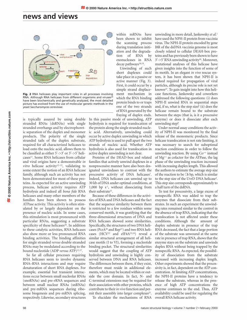
The best overview of cellular processesinvolving RNA helicases can be obtainedfrom the study of DEAD-box and relatedproteins in the yeast Saccharomyces cerevisi-ae6. Genetic analyses have shown that RNAhelicases are involved in transcription, pre-mRNA processing, ribosome biogenesis,RNA export, translational initiation, mito-chondrial gene expression and RNA degra-dation (Fig. 2). Although the translationinitiation factor eIF4A has served as theprototype for the DEAD-box proteins, itsin vivo function is not known.
The first demonstration of an essentialbiological role for RNA helicases came fromthe pre-mRNA splicing field. The spliceo-some is a transient and highly dynamicstructure that assembles in a highly orderedand stepwise manner and undergoes severalconformational changes. The spliceosomeis made up of five small nuclear RNAs(snRNAs: U1, U2, U4, U5 and U6) andmany different polypeptides. Several of theassembly steps that lead to the formation ofa catalytically active spliceosome involvewell-described changes in RNA–RNA inter-actions7,8. Indeed, the two transesterifica-
tion steps in the splicing reaction do notrequire energy per se, but the splicingprocess is dependent on ATP hydrolysis. Itis generally believed that the ATP-depen-dent RNA helicases are primarily responsi-ble for the energy requiring rearrangementswithin the splicing process to allow the for-mation of new, sometimes mutually exclu-sive interactions to occur. Several of theseRNA rearrangements have been attributedto specific RNA helicases, such as theunwinding of duplexes formed between U4and U6 snRNAs by Brr2p (ref. 9).
Similarly, other studies show that amajority of the DEAD-box proteins areinvolved in ribosome biogenesis. This canbe explained by the dynamic process ofribosome formation which involves numer-ous sequential steps of pre-ribosomal RNA(pre-rRNA) maturation and assembly withribosomal proteins. Nevertheless, in thecase of ribosome biogenesis, a correlationbetween in vivo requirements and definedbiochemical steps, as in the case of thespliceosome rearrangements, are largelymissing due to the absence of a reconstitut-ed in vitro system.
Finally, studies on putative RNA helicaseswithout clear homologs in S. cerevisiae havebeen reported, suggesting that these pro-teins could be involved in other processesnot present in this lower eukaryote6,10.
RNA helicases are expected to melt dou-ble stranded RNA molecules. This activity
Are DEAD-box proteins becomingrespectable helicases?Patrick Linder and Marie-Claire Daugeron
The vaccinia NPH-II RNA helicase, a member of the DEAD/DExH-box protein family, has been shown to be aprocessive, unidirectional RNA helicase with a step size of about one half turn of a helix. This findingdemonstrates that RNA helicases can function as molecular motors.
Fig. 1 Sequence comparison groups the putative RNA helicases in the DEAD, DEAH, DExH, andUpf1p families. Conserved residues are given, but variations within the family exist. Therefore, aclear assignment to a particular family can be made in most cases, but a few proteins show similar-ities to more than one family. While many members of these families have been analyzed, it is notclear whether members of different families use the same mode of action. The N- and C-terminalextensions, as well as the spacing between the motifs, vary and are only schematically representedhere. Proteins of the Snf2p family involved in chromatin remodeling and transcription are notincluded since they are most likely not bona fide helicases28.
nature structural biology • volume 7 number 2 • february 2000 97
© 2000 Nature America Inc. • http://structbio.nature.com©
200
0 N
atu
re A
mer
ica
Inc.
• h
ttp
://s
tru
ctb
io.n
atu
re.c
om
news and views
is typically assayed by using doublestranded RNAs (dsRNAs) with singlestranded overhangs and by electrophoret-ic separation of the duplex and monomerproducts. The polarity of the singlestranded tails of the duplex substrate,required for all characterized helicases toload onto the nucleic acid, allows them tobe classified as either 5'->3' or 3'->5' heli-cases11. Some RNA helicases from cellularand viral origins have a demonstrable invitro helicase activity12–15, validating tosome extent the notion of an RNA helicasefamily, although such an activity has notbeen demonstrated for most of these pro-teins. As expected for an active unwindingprocess, helicase activity requires ATPhydrolysis and indeed all bona fide RNAhelicases and many other members of thefamilies have been shown to possessATPase activity. This activity is often stim-ulated by or largely dependent on thepresence of nucleic acids. In some cases,this stimulation is most pronounced withparticular RNAs, suggesting a substratespecificity of these helicases16. In additionto these catalytic activities, RNA helicasesalso show more or less pronounced RNAbinding activities. The binding affinitiesfor single stranded versus double strandedRNAs may be modulated according to thebound nucleotide (ADP versus ATP)12,17.
So far all cellular processes requiringRNA helicases seem to involve dynamicRNA–RNA interactions and may requiredenaturation of short RNA duplexes. Forexample, essential but transient interac-tions occur between small nucleolar RNAs(snoRNAs) and pre-rRNA sequences andbetween small nuclear RNAs (snRNAs)and pre-mRNA sequences during ribo-some biogenesis and pre-mRNA splicing,respectively. Likewise, secondary structures
within mRNAs havebeen shown to inhibitthe scanning processduring translation initi-ation and the degrada-tion of RNA byexonucleases in RNAdecay pathways18,19.
Unwinding of suchshort duplexes couldtake place in a passive oractive manner (Fig. 3).First, it could occur by asimple strand displace-ment mechanism inwhich the RNA bindingprotein binds to or trapsone of the two strandsof RNA generated by thefraying of duplex ends.
In this passive mode of unwinding, ATPhydrolysis is required for translocation ofthe protein along the single stranded nucle-ic acid. Alternatively, unwinding couldoccur by active duplex unwinding in whichATP hydrolysis is used to pull apart the twostrands of nucleic acid. Whether ATPhydrolysis is also used for translocation inactive duplex unwinding is not clear.
Proteins of the DEAD-box and relatedfamilies that actively unwind duplexes in anonprocessive manner have also been des-ignated unwindases to contrast with theprocessive activity of DNA helicases7.Indeed, DNA helicases can unwind up to30 kb of DNA under optimal conditions, at1,000 bp s-1, without dissociating fromtheir substrate11.
Because of these differences in the activi-ties of RNA and DNA helicases and the factthat the sequence similarity between themis restricted to a few residues within someconserved motifs, it was gratifying that thethree-dimensional structures of DNA andRNA helicases showed some similarities.Specifically, the structures of two DNA heli-cases (PcrA20 and Rep21) and two RNA heli-cases (HCV22 and eIF4A23,24) reveal asimilar structural arrangement of all heli-case motifs (I to VI), forming a nucleotidebinding pocket. The structural similaritieswould suggest that the coupling of ATPhydrolysis and unwinding is highly con-served between DNA and RNA helicases.The differences between them, if they exist,therefore must reside in additional ele-ments, which may be located within or out-side the core domain. In fact, N- andC-terminal extensions may be required fortheir association with other proteins, whichcontribute to their in vivo function and per-mit their assembly into larger complexes25.
To elucidate the mechanisms of RNA
98 nature structural biology • volume 7 number 2 • february 2000
unwinding in more detail, Jankowsky et al.1
have used the NPH-II protein from vacciniavirus. The NPH-II protein encoded by geneI8R of the dsDNA vaccinia genome is mostclosely related to cellular DEAH-box pro-teins and has previously been shown to have3'->5' RNA unwinding activity26. Moreover,mutational analyses of this helicase havegiven insights into the function of some ofits motifs. In an elegant in vivo rescue sys-tem, it has been shown that NPH-II isindeed required for propagation of viralparticles, although its precise role is not yetknown27. To gain insight into how this heli-case functions, Jankowsky and coworkersaddressed the following questions: (i) doesNPH-II unwind RNA in sequential stepsand, if so, what is the step size? (ii) does thehelicase remain bound to the substratebetween the steps (that is, is it a processiveenzyme) or does it dissociate after eachunwinding step?
Under normal assay conditions the activ-ity of NPH-II was monitored by the finalrelease of the monomeric products. Sincehelicase translocation is not rate limiting, itwas necessary to search for suboptimalreaction conditions in order to follow theunwinding reaction. By using Co++ insteadof Mg++ as cofactor for the ATPase, the lagphase of the unwinding reaction increasedwith increasing duplex length. This allowedthe authors to estimate the average step sizeof the reaction to be ∼ 6 bp, which is similarto the step size of the UvrD DNA helicase2.This step size corresponds approximately toa half turn of the dsRNA.
To test for processivity, a large excess ofnonspecific RNA was added to trap theenzymes that dissociate from their sub-strates. In such an experiment the unwind-ing rates remained similar to the control inthe absence of trap RNA, indicating that thetranslocation is not affected under theseconditions. Although the amount ofduplex substrate in presence of the trapRNA decreased, the fact that a large portionof the substrate was unwound at the samerate in presence of trap RNA, shows that theenzyme stays on the substrate and unwindsduplex RNA without being trapped by thenonspecific RNA. As expected, the possibil-ity of dissociation from the substrateincreased with increasing duplex length.Other experiments showed that the proces-sivity is, in fact, dependent on the ATP con-centration. At limiting ATP concentrations,the NPH-II proteins have a tendency torelease the substrate, whereas in the pres-ence of high ATP concentrations theenzyme continues to the end. Thus, ATPavailability could be used for regulating theoverall RNA helicase activity.
Fig. 2 RNA helicases play important roles in all processes involvingRNA. Although RNA helicases from different organisms and viruses29
have been biochemically and genetically analyzed, the most detailedpicture has evolved from the use of molecular genetic methods in theyeast Saccharomyces cerevisiae.
© 2000 Nature America Inc. • http://structbio.nature.com©
200
0 N
atu
re A
mer
ica
Inc.
• h
ttp
://s
tru
ctb
io.n
atu
re.c
om

news and views
In conclusion, genetic experiments havebeen able to assign individual RNA helicas-es to specific cellular processes. Bio-chemical experiments have shown that inthe presence of ATP and Mg++ some ofthese proteins are able to dissociate shortRNA duplexes. With the report byJankowsky et al.1 our view of RNA helicaseshas expanded. RNA helicase have now beenshown to migrate in a directional fashion,in distinct steps along the substrate RNAwithout dissociating from it.
Despite this important step forward, weremain at the beginning of our under-standing of RNA helicase function. Indeed,we do not know how helicases unwindduplex molecules. Do they act as an activesnow-plough, as a rolling oligomer tearingthe substrate apart or by following in anATPase-dependent manner the sponta-neous denaturation of the duplex (Fig. 3)?In the case of active DNA helicases it hasbeen shown that they exist in oligomericcomplexes. So far there is little informationon this concerning RNA helicases, and even
in the case of‘monomeric’ RNA heli-cases it cannot beexcluded that oligomer-ization is induced bycontact with the RNA.
Thus, it may turn out— depending on the molecular environ-ment or their tertiary structure — thatsome RNA helicases unwind duplex sub-strates in a processive fashion, whereas oth-ers do not. Moreover, the RNA helicases arehighly specific and cannot be freely inter-changed. Thus, they most certainly possessspecificity determinants and/or interactwith other components that let them workin a controlled manner on the right sub-strate and at the right time.
Patrick Linder is in the Dépt. de Biochimiemédicale CMU, 1, rue Michel Servet,Genève 4, 1211, Switzerland and Marie-Claire Daugeron is in the Inst. de Génétiqueet Microbiologie, Bâtiment 400 URA 2225,Université Paris-Sud, 91405 Orsay Cedex,France. Correspondence should beaddressed to P.L. email: [email protected]
1. Jankowsky, E., Gross, C.H., Shumann, S. & Pyle, A.M.Nature (in the press) (2000).
2. Ali, J.A. & Lohman, T.M. Science 275, 377-380(1997).
3. Gorbalenya, A.E., Koonin, E.V., Donchenko, A.P. &Blinov, V.M. Nucl. Acids Res. 17, 4713-4730 (1989).
4. Koonin, E.V. Nature 352, 290 (1991).5. Linder, P., et al. Nature 337, 121–122 (1989).6. de la Cruz, J., Kressler, D. & Linder, P. Trends
Biochem. Sci. 24, 192–198 (1999).7. Staley, J.P. & Guthrie, C. Cell 92, 315–326 (1998).8. Nilsen, T.W. In: RNA Structure and Function,
R.Simons and M. Grunberg-Manago, eds. ColdSpring Harbor NY, Cold Spring Harbor laboratorypress, pp 279–307 (1998)
9. Raghunathan, P.L., & Guthrie, C. Curr. Biol. 8,847–855 (1998)
10. Lee, C.G., da Costa Soares, V., Newberger, C.,Manova, K. Lacy, E. & Hurwitz, J. Proc. Natl. Acad.Sci. USA 95,13709–13713 (1998).
11. Lohman, T.M. & Bjornson, K.P. Annu. Rev. Biochem.65, 169–214 (1996).
12. Iost, I., Dreyfus, M. & Linder, P. J. Biol. Chem. 274,17677–17683 (1999).
13. Hirling, H., Scheffner, M., Restle, T. & Stahl, H.Nature 339, 562–564 (1989).
14. Liang, L., Diehl-Jones, W. & Lasko, P. Development120, 1201–1211 (1994).
15. Schwer, B., & Gross, C.H. EMBO J. 17, 2086–2094 (1998).16. Fuller-Pace, F.V., Nicol, S.M., Reid, A.D. & Lane, D.P.
EMBO J. 12, 3619–3626 (1993).17. Lorsch, J.R. & Herschlag, D. Biochemistry 37,
2194–2206 (1998).18. Pelletier, J., & Sonenberg, N. Cell 40, 515–526 (1985)19. Jacobs-Anderson, J.S. & Parker, R. EMBO J. 17,
1497–1506 (1998).20. Subramanya, H.S., Bird, L.E., Brannigan, J.A. &
Wigley, D.B. Nature 384, 379–383 (1996)21. Korolev, S., Hsieh, J., Gauss, G.H., Lohman, T.M. &
Waksman, G. Cell 90, 635–647 (1997)22. Yao, N., et al. Nature Struct. Biol. 4, 463–467 (1997).23. Benz, J., Trachsel, H. & Baumann, U. 1999. Structure
Fold Des 7, 671–679 (1999)24. Johnson, E.R. & McKay, D.B. RNA 5:1526–1534
(1999).25. West, S.C. Cell 86, 177–180 (1996).26. Shuman, S. J. Biol. Chem. 268, 11798–11802 (1993).27. Gross, C.H. & Shuman, S. J. Virol. 72, 4729–4736 (1998).28. Muchardt, C. & Yaniv, M. J. Mol. Biol. 293, 187–198
(1999).29. Kadare, G. & Haenni, A.L. J. Virol. 71, 2583–2590
(1997).
Fig. 3 Proposed models for the action of helicases. In the rolling model, anoligomeric helicase binds alternating to single and double stranded nucleicacid, where the ATP-bound proteins have a high affinity for double strandednucleic acid. In the snow-plough model, the RNA helicase is moving along thefork and uses energy from NTP hydrolysis to melt hydrogen bonding betweenthe two nucleic acid strands. By contrast, in the passive unwinding model, thelocal denaturation by thermal fluctuation could be fixed by a single strandedRNA (ssRNA) binding protein. The movement of the ssRNA binding proteinalong the single stranded nucleic acid would be an ATP-dependent reaction.
Active duplex unwinding
Rolling mode mechanism
Passive duplex unwinding
Snow-plough or inchworm mechanism
The servant with the scissors
history
In 1978, Werner Arber (Biozentrum derUniversität, Basel, Switzerland), DanNathans and Hamilton Smith (both atJohns Hopkins University School ofMedicine, Baltimore, Maryland, USA) wereawarded the Nobel Prize in Physiology orMedicine for the discovery of “restrictionenzymes and their application to problemsof molecular genetics”. Almost immediate-ly, the application of these enzymes togenetics led to “new and far reachingresults”. In fact, it is hard to imagine what
the biological sciences would look liketoday without restriction maps, cloningand the ability to alter genes at will, to namejust a few everyday tools of the trade. Buthow did this crucial discovery come about?
While studying a phenomenon knownas ‘host controlled restriction of bacterio-phages’, Arber and Dussoix1–3 found that itprovided bacteria with a defense mecha-nism against invading foreign DNA, suchas viral DNA. This process, which wasshown to be a property of the recipient
bacteria, could be divided into two parts:restriction and modification. Restrictioninvolved the endonucleolytic cleavage ofDNA at specific DNA sequences. Becausethis would restrict viral growth, theseenzymes came to be known as restrictionenzymes. Modification involved nucleo-tide methylation at these same specificDNA sequences in the genome. In this way,the bacteria’s own DNA was protectedfrom cleavage because it was methylatedwhile the inappropriately or unmodified
nature structural biology • volume 7 number 2 • february 2000 99
© 2000 Nature America Inc. • http://structbio.nature.com©
200
0 N
atu
re A
mer
ica
Inc.
• h
ttp
://s
tru
ctb
io.n
atu
re.c
om