4 Surgical Treatment of special Tumours
42
Surgical Treatment of special Tumours Winnie Achilles Tierklinik Hollabrunn Lastenstrasse 2 2020 Hollabrunn [email protected]
Transcript of 4 Surgical Treatment of special Tumours
Surgical
Treatment of
special Tumours
Winnie Achilles
Tierklinik Hollabrunn
Lastenstrasse 2
2020 Hollabrunn
Hepatocellular Tumours
Hepatocellular Carcinoma, hepatocellular adenoma, and hepatoblastoma
•HCC is the most common primary liver tumor in dogs, with 50% of cases
•hepatocellular adenoma is usually an incidental finding and rarely causes
clinical signs
•hepatocellular adenoma is the most common in cats,
HCC the 2nd most common

Clinical Symptoms
in 50% of cats and 75% of dogs
inappetence, weight loss, lethargy, vomiting, polydipsia-polyuria, ascites
•seizures caused by hepatic encephalopathy are uncommon
•icterus is more commonly seen in dogs with extrahepatic bile duct
carcinomas and diffuse neuroendocrine tumors
•cranial abdominal mass is palpable in up to 75% of cats and dogs with
liver tumors

Laboratory changes
Usually non-specific
Hematology
•leukocytosis, anemia, and thrombocytosis
•leukocytosis is probably caused by inflammation and necrosis associated
with large liver masses
•prolonged coagulation times and clotting factor abnormalities are rarely
clinically relevant
� BMBT

Serum Biochemistry
liver enzymes are commonly elevated in dogs with hepatobiliary tumours
�this may provide an indication of the type of tumour, and differentiate
primary and metastatic liver tumors
•ALP and ALT are commonly increased in dogs with primary hepatic tumours
•AST and bilirubin are more consistently elevated in dogs with metastatic
liver tumors
Unspecific changes
hypoglycemia, hypoalbuminemia, hyperglobulinemia, and increased pre-
and post-prandial bile acids
•azotemia might be the abnormality in cats
ALT, AST and total bilirubin are also common and are significantly higher in
cats with malignant tumors


Abdominal CT scan

Laparoscopy


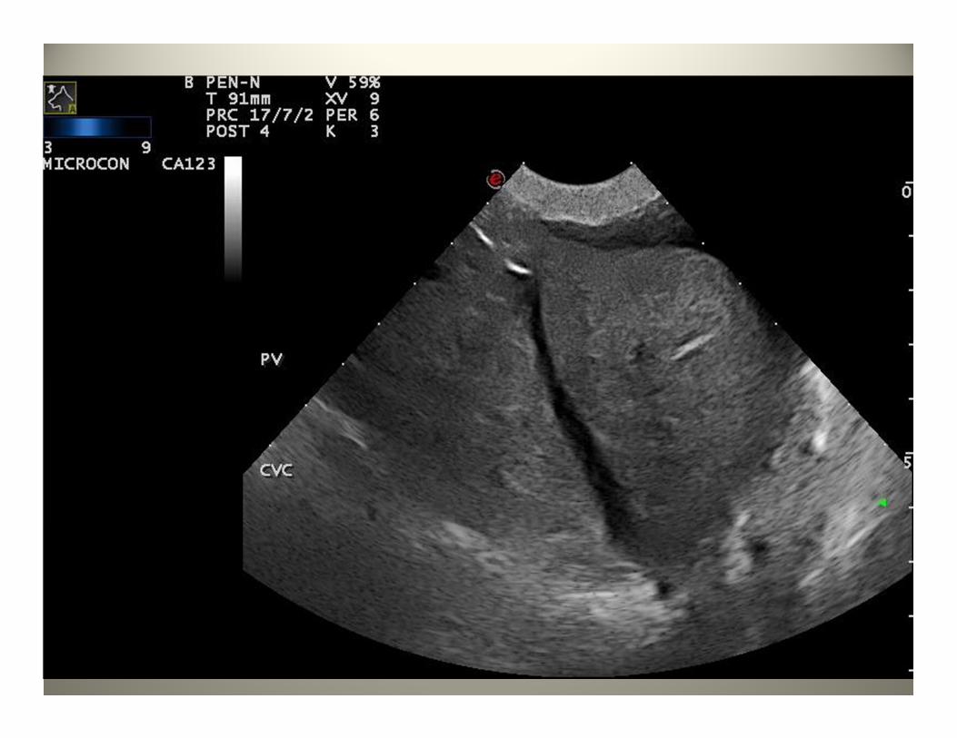
Hepatocellular Carcinoma
More commen in Miniature Schnauzers and male dogs
53%-83% of HCC are massive
16%-25% are nodular
up to 19% are diffuse
left liver lobes are involved in > 67% of dogs with massive HCC
metastatic rate varies from 0%-37% for dogs with massive HCC and
93%-100% with nodular and diffuse HCC
•metastasis to regional lymph nodes, peritoneum, and lungs
(heart, kidneys, adrenal gland, pancreas, intestines, spleen, and urinary
bladder)

Treatment
Liver Lobectomy for massive HCC
complications: hemorrhage
vascular compromise to adjacent liver lobes, hypoglycemia, reduced
hepatic function
no effective treatment for nodular and diffuse HCC
HCC is considered chemoresistant
embolization and chemoembolization have been reported with moderate
success in the palliation of 4 dogs with HCC
Prognosis
good for dogs with massive HCC
MST > 1,460 days, 0%-6% local tumor recurrence rate, and 0%-37%
distant metastatic rate
poor for dogs with nodular and diffuse HCC is poor

Bile duct Adenoma and Carcinoma
accounting for > 50% of all feline hepatobiliary tumors in cats
single and multiple lesions
aggressive biologic behavior
metastasis in 67%-80% of cats, up to 88% in dogs
surgical resection is recommended for cats and dogs with massive bile
duct carcinoma
MST < 6 months due to local tumor recurrence and metastatic disease
Myelolipoma benign hepatobiliary tumour in cats
with excellent survival times after resection

Sarcomas
primary hepatic sarcomas (leiomyosarcoma, HSA, and FSA) are rare
only 4%-6% HSA are of primary hepatic origin in dogs
liver is a common site for metastatic HSA
metastasis to the spleen and lungs in 86%-100% cases
liver lobectomy can be attempted for solitary and massive sarcomas
prognosis is poor as metastatic disease is often present at the time of
surgery
chemotherapy response rates are likely to be poor

Transitional cell carcinoma
of the dog

Clinical Presentation
hematuria, pollakiuria, and dysuria
urinary obstruction or incontinence
sex predisposition in dogs: female
TCC should occur in the
•proximal 3rd of the urethra in females
•in the entire urethra in males
first metastasis to the regional lymph node

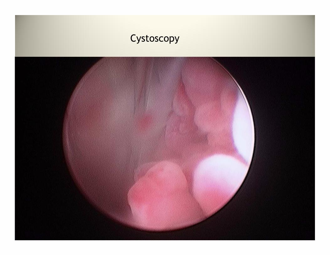
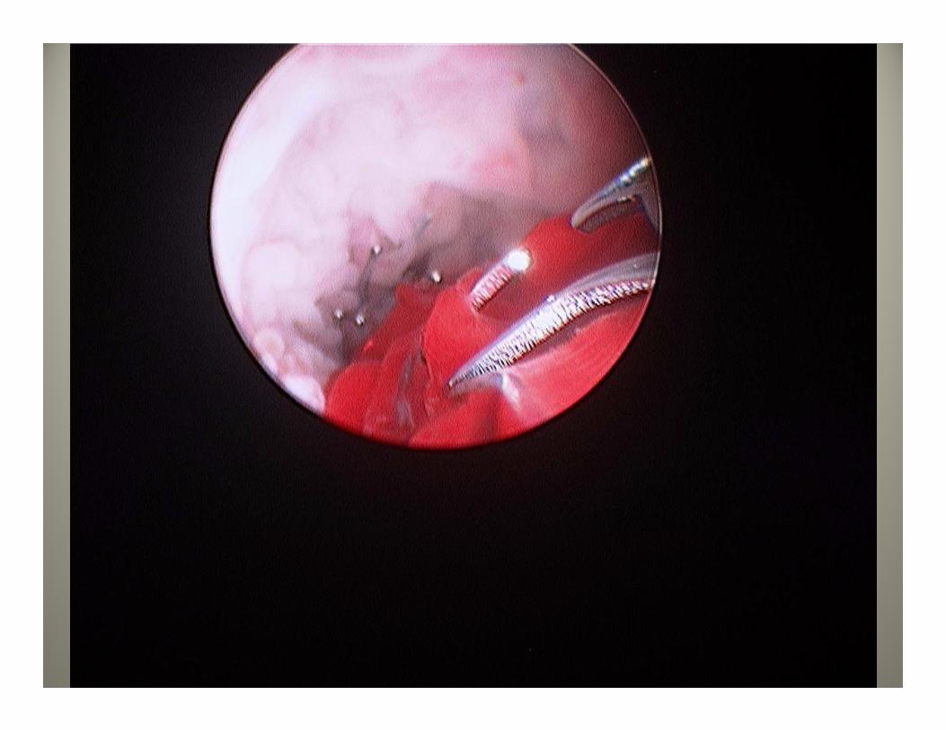
Cystoscopy

Chemotherapy
•MST 181 days with piroxicam alone
•MST 220 days with cisplatin alone

Surgical Treatment
small, localized and benign lesions can be excised
end-to-end anastomosis, healing over an urethral catheter
urinary diversion techniques for lesions in the proximal urethra
ureterocolonic or trigonal-colonic anastomosis
� permanent tube cystostomy
survival times range from 2-22 months

CystostomyTube

Permanent Prescrotal Fistula

Interventional Radiology


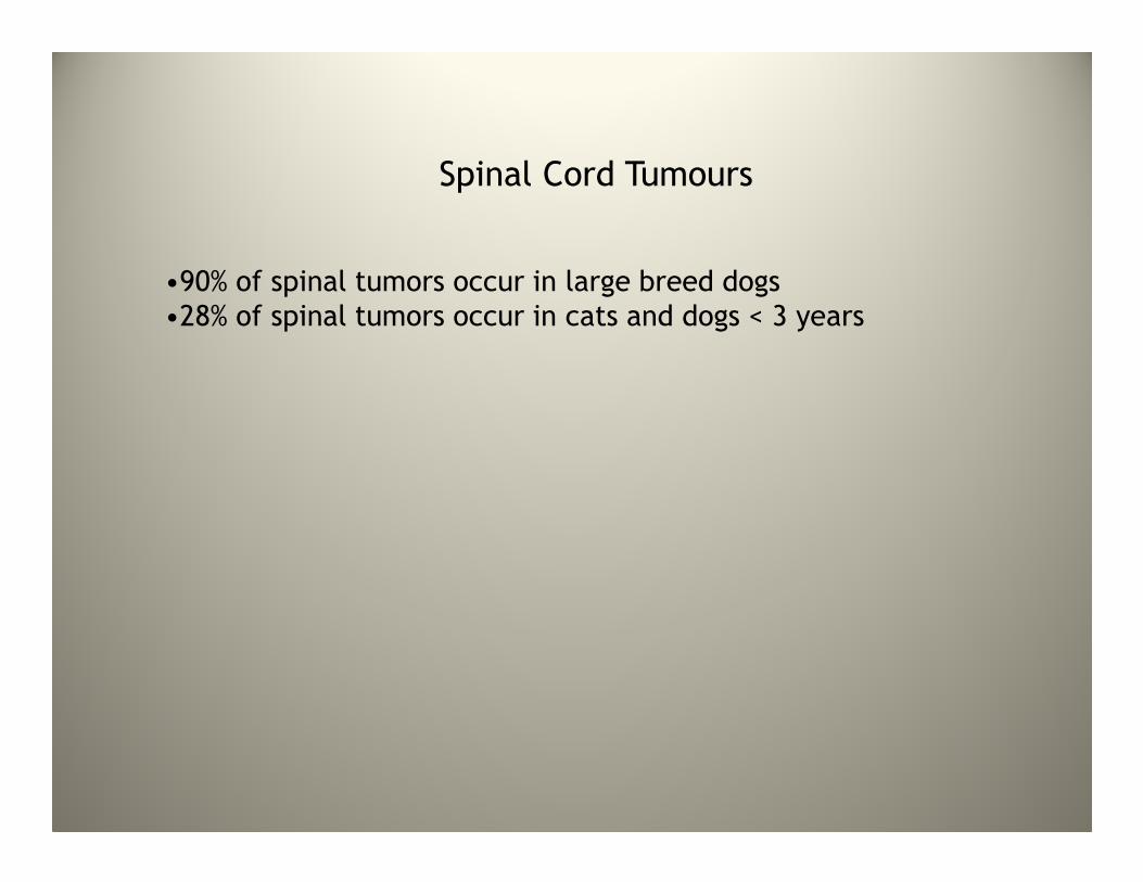
Spinal Cord Tumours
•90% of spinal tumors occur in large breed dogs
•28% of spinal tumors occur in cats and dogs < 3 years

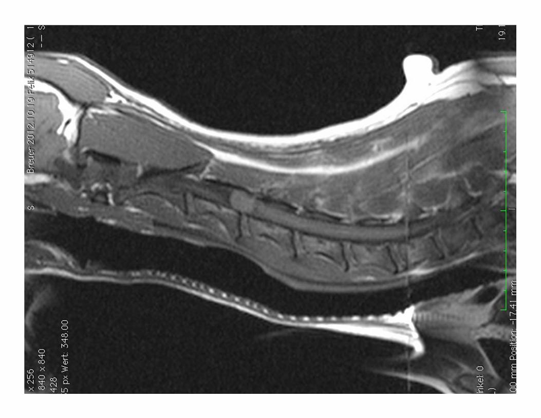
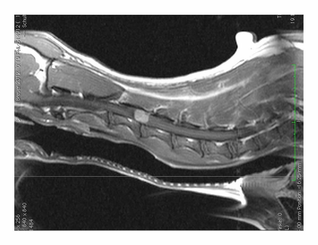
Extradural Spinal Cord Tumours
meningioma and peripheral nerve sheath tumors are the most common
spinal meningioma has a predilection for the cervical spinal cord:
•40%-77% cervical spinal cord
•0%-32% thoracic spinal cord
•23%-28% lumbar spinal cord
56% dogs with spinal meningioma alive > 6 months
peripheral nerve sheath tumors involve the spinal cord in 65% cases
compleate resection is usually curative
further: primary vertebral tumors and multiple myeloma

Intradural-Extramedullary Spinal Cord Tumour of Young Dogs
•synonyms:
ependymoma, neuroepithelioma, spinal cord blastoma,
medulloepithelioma, hamartoma, and nephroblastoma
•age: 6 months to 3 years
•breed predisposition: GSD, Labrador Retriever, and Golden Retriever
•clinical signs: lateralized with vast majority of lesions between T10-L2
•treatment
Surgical removal associated with long-term survival 4 months and > 3 years
± radiation therapy for incompletely excised tumours

Intramedullary Spinal Cord Tumours
are rare and mostly of glial cell origin
astrocytoma, oligodendroglioma, ependymoma, choroid plexus papilloma
most commonly located between C6-T2
often already with intramedullary spinal cord metastasis
before evidence of the primary tumour
metastasis HSA and LSA ± mammary ADC and malignant melanoma


Questions?