394 Supratentorial and infratentorial cavernous malformation
Upload
muhammad-bin-zulfiqarCategory
view
67download
0
4 Hypodense Supratentorial Masses on Computed
Tomography
CLINICAL IMAGAGINGAN ATLAS OF DIFFERENTIAL DAIGNOSIS
EISENBERG
DR. Muhammad Bin Zulfiqar PGR-FCPS III SIMS/SHL
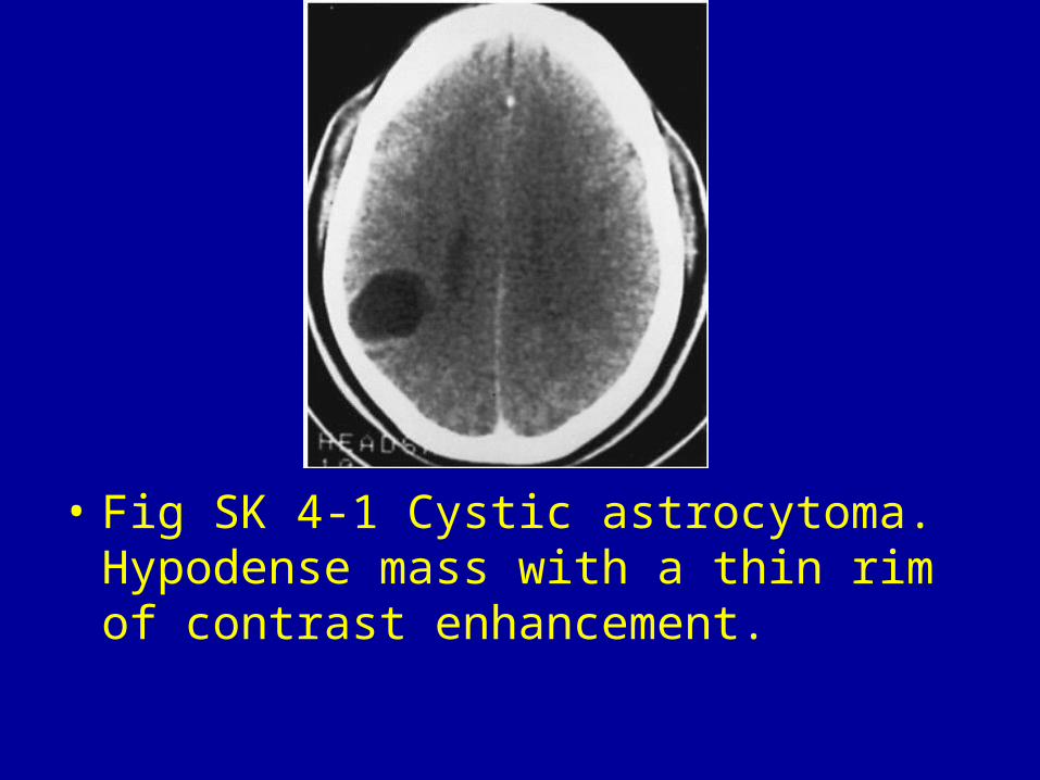
• Fig SK 4-1 Cystic astrocytoma. Hypodense mass with a thin rim of contrast enhancement.
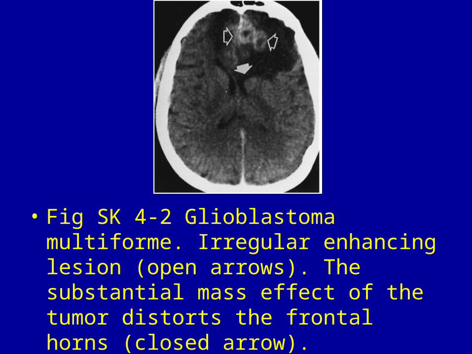
• Fig SK 4-2 Glioblastoma multiforme. Irregular enhancing lesion (open arrows). The substantial mass effect of the tumor distorts the frontal horns (closed arrow).
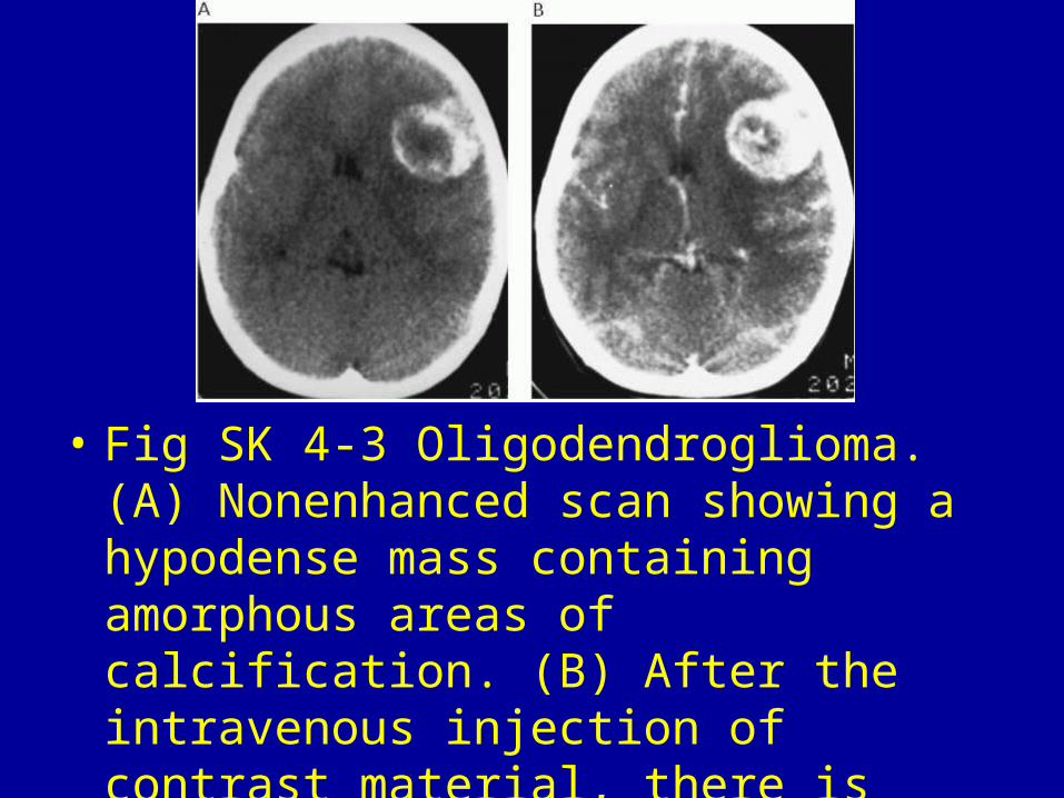
• Fig SK 4-3 Oligodendroglioma. (A) Nonenhanced scan showing a hypodense mass containing amorphous areas of calcification. (B) After the intravenous injection of contrast material, there is marked contrast enhancement.
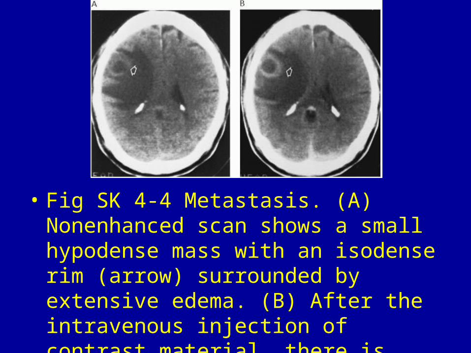
• Fig SK 4-4 Metastasis. (A) Nonenhanced scan shows a small hypodense mass with an isodense rim (arrow) surrounded by extensive edema. (B) After the intravenous injection of contrast material, there is prominent ring enhancement (arrow) about the metastasis.
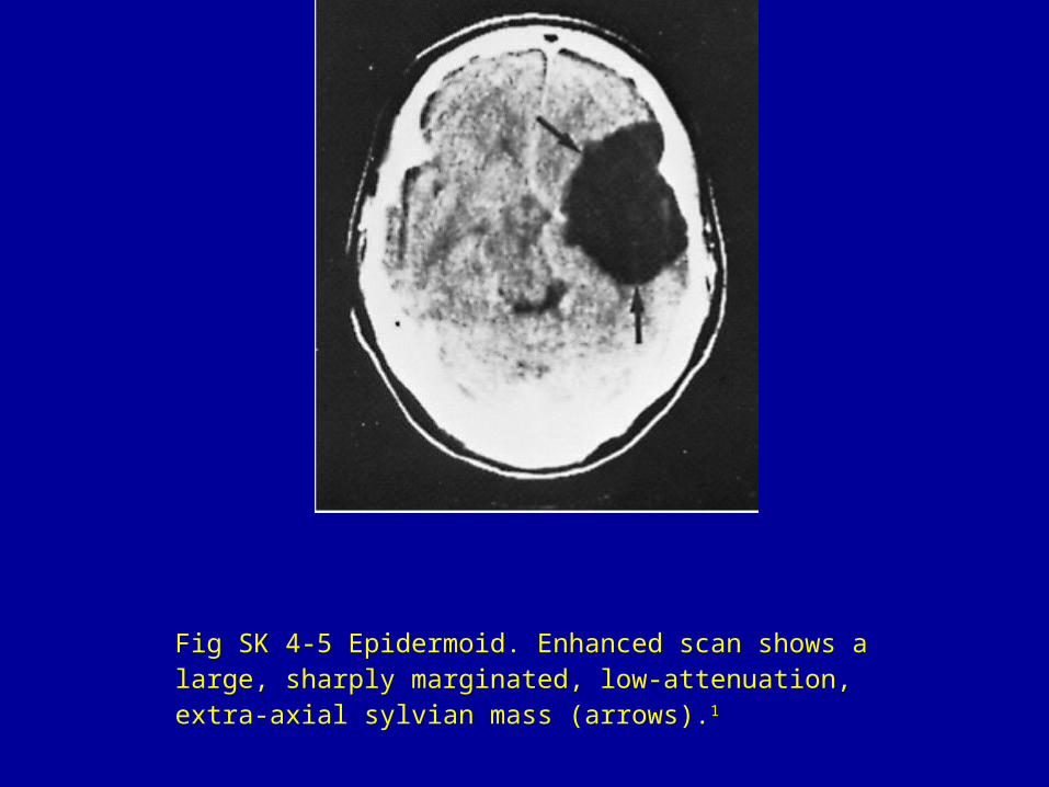
Fig SK 4-5 Epidermoid. Enhanced scan shows a large, sharply marginated, low-attenuation, extra-axial sylvian mass (arrows).1
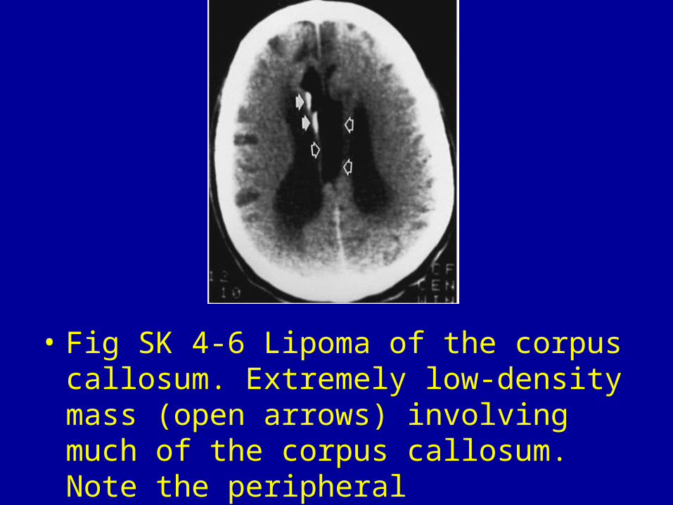
• Fig SK 4-6 Lipoma of the corpus callosum. Extremely low-density mass (open arrows) involving much of the corpus callosum. Note the peripheral calcifications (closed arrows).
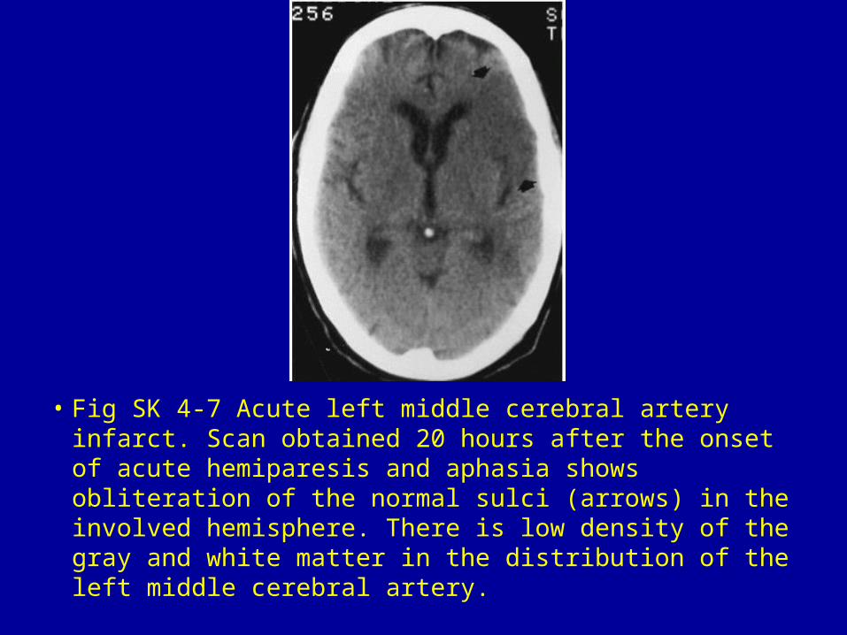
• Fig SK 4-7 Acute left middle cerebral artery infarct. Scan obtained 20 hours after the onset of acute hemiparesis and aphasia shows obliteration of the normal sulci (arrows) in the involved hemisphere. There is low density of the gray and white matter in the distribution of the left middle cerebral artery.
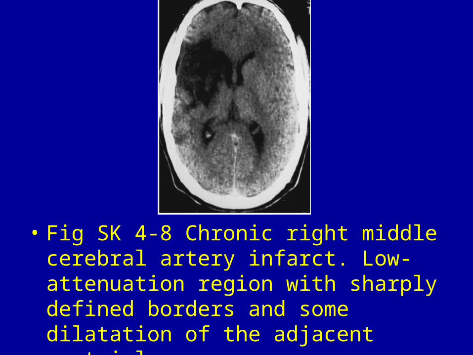
• Fig SK 4-8 Chronic right middle cerebral artery infarct. Low-attenuation region with sharply defined borders and some dilatation of the adjacent ventricle.
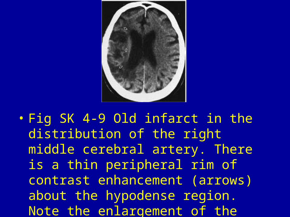
• Fig SK 4-9 Old infarct in the distribution of the right middle cerebral artery. There is a thin peripheral rim of contrast enhancement (arrows) about the hypodense region. Note the enlargement of the right lateral ventricle.
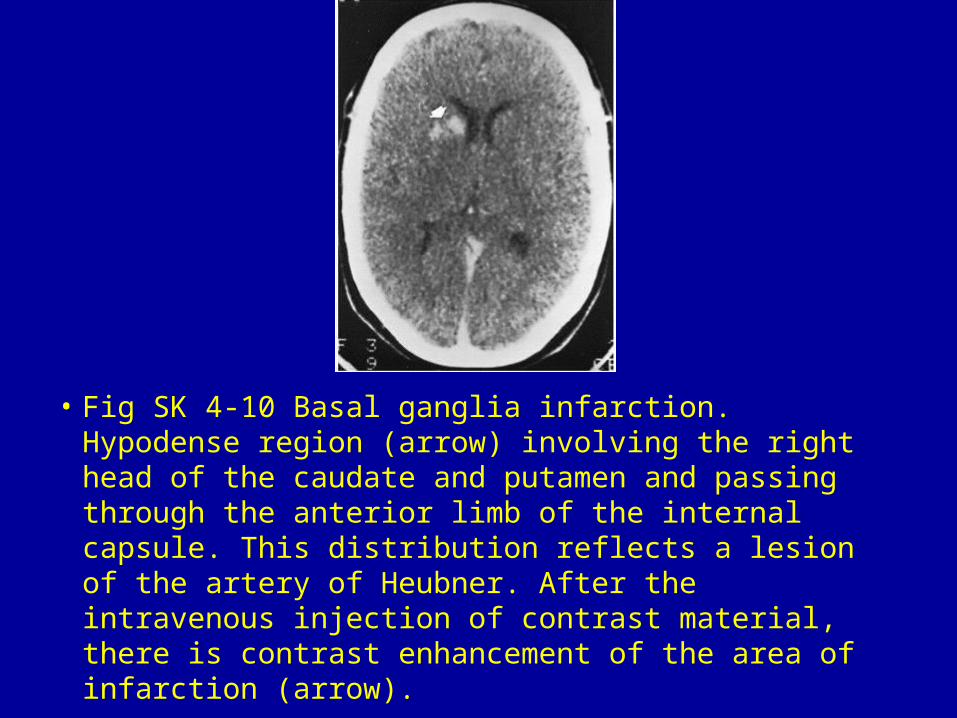
• Fig SK 4-10 Basal ganglia infarction. Hypodense region (arrow) involving the right head of the caudate and putamen and passing through the anterior limb of the internal capsule. This distribution reflects a lesion of the artery of Heubner. After the intravenous injection of contrast material, there is contrast enhancement of the area of infarction (arrow).
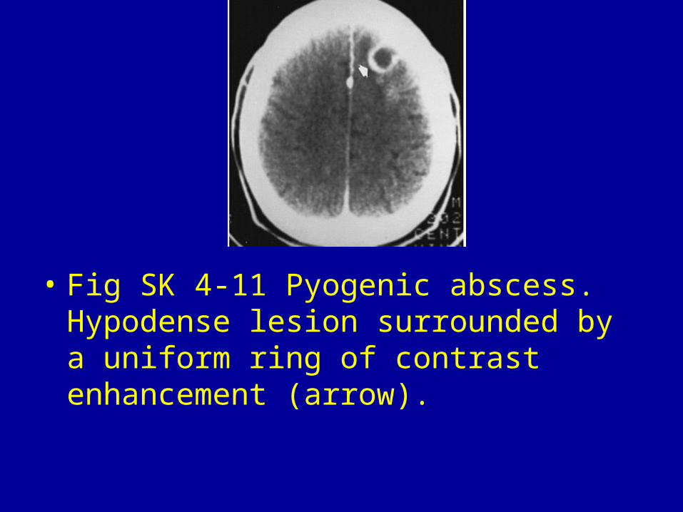
• Fig SK 4-11 Pyogenic abscess. Hypodense lesion surrounded by a uniform ring of contrast enhancement (arrow).
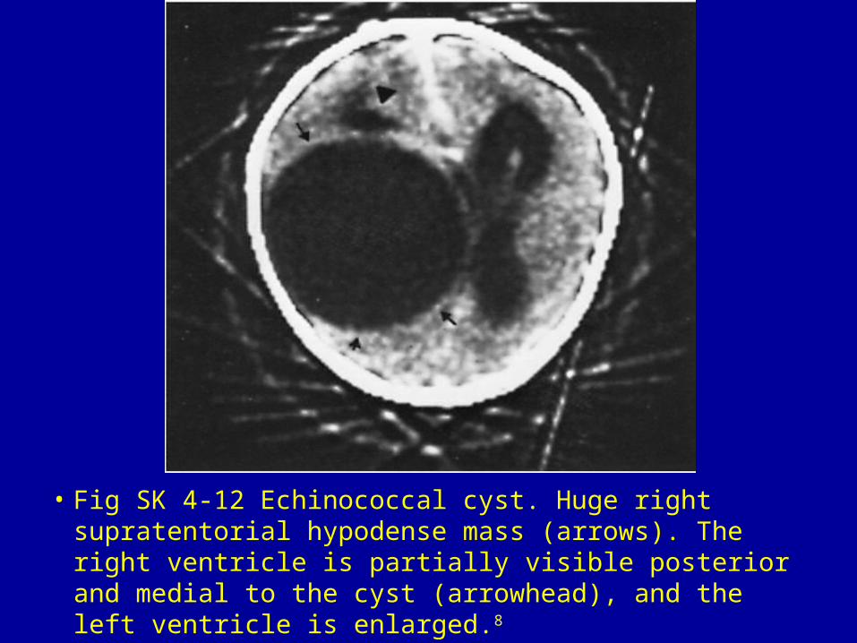
• Fig SK 4-12 Echinococcal cyst. Huge right supratentorial hypodense mass (arrows). The right ventricle is partially visible posterior and medial to the cyst (arrowhead), and the left ventricle is enlarged.8
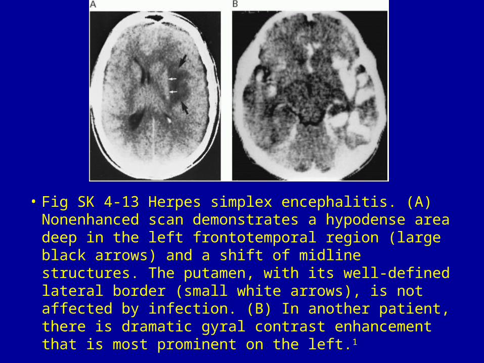
• Fig SK 4-13 Herpes simplex encephalitis. (A) Nonenhanced scan demonstrates a hypodense area deep in the left frontotemporal region (large black arrows) and a shift of midline structures. The putamen, with its well-defined lateral border (small white arrows), is not affected by infection. (B) In another patient, there is dramatic gyral contrast enhancement that is most prominent on the left.1
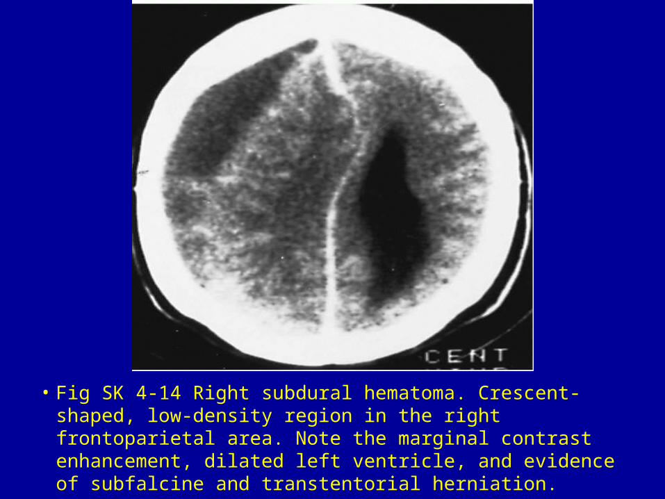
• Fig SK 4-14 Right subdural hematoma. Crescent-shaped, low-density region in the right frontoparietal area. Note the marginal contrast enhancement, dilated left ventricle, and evidence of subfalcine and transtentorial herniation.
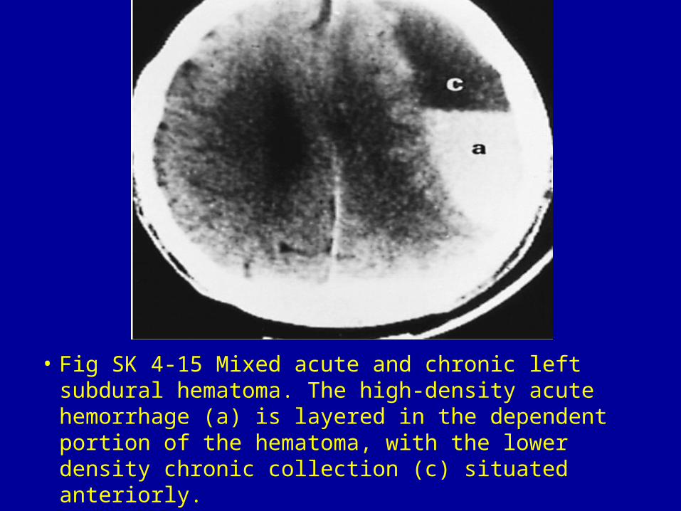
• Fig SK 4-15 Mixed acute and chronic left subdural hematoma. The high-density acute hemorrhage (a) is layered in the dependent portion of the hematoma, with the lower density chronic collection (c) situated anteriorly.
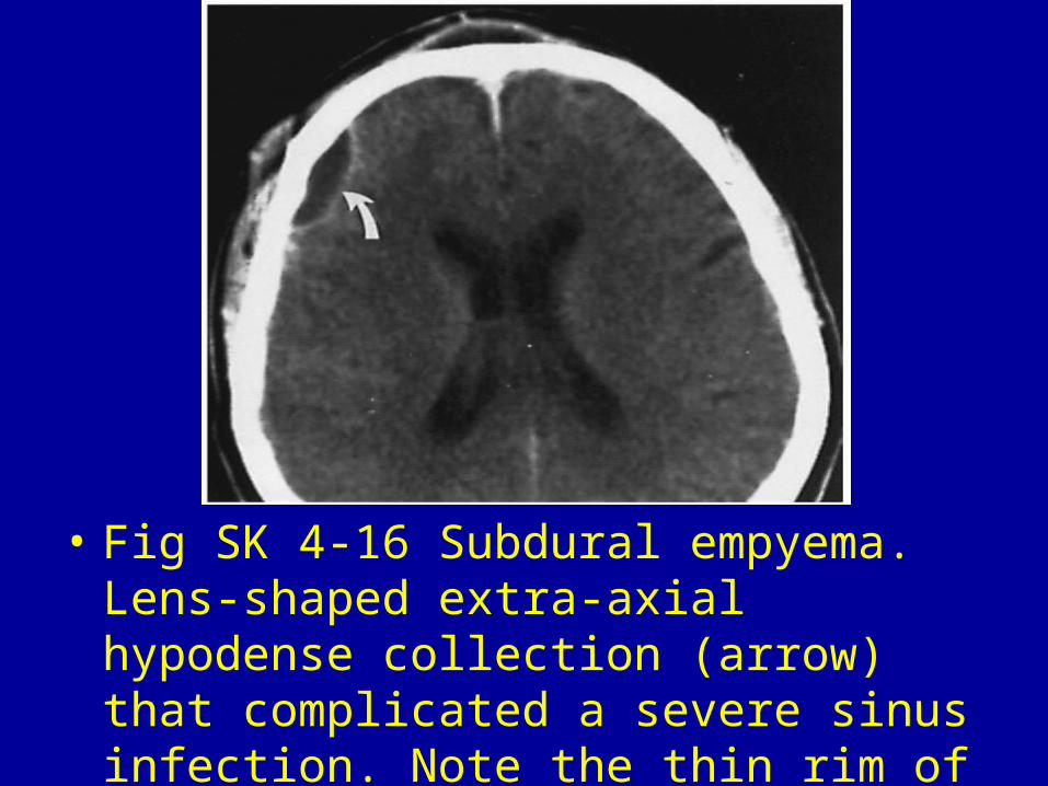
• Fig SK 4-16 Subdural empyema. Lens-shaped extra-axial hypodense collection (arrow) that complicated a severe sinus infection. Note the thin rim of peripheral contrast enhancement.
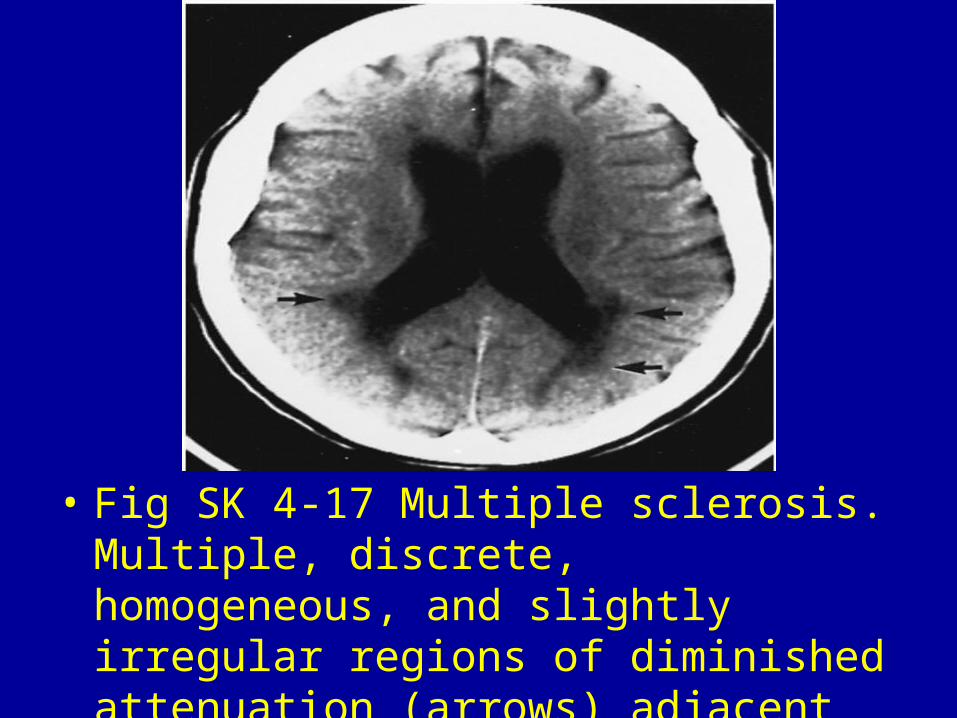
• Fig SK 4-17 Multiple sclerosis. Multiple, discrete, homogeneous, and slightly irregular regions of diminished attenuation (arrows) adjacent to the slightly enlarged ventricles.1