4. Bones of the upper limb...Which bones are most likely to be damaged in a fall on the outstretched...
8
SECTION THREE: Fleshed out 32 The pectoral girdle, which attaches the upper limbs to the trunk, consists of the scapulae and clavicles. It does not form a complete bony ring like the pelvis because there is no joint with the vertebral column (Fig. 3.4.1). Posteriorly, the limbs are suspended by muscles that are attached to the vertebrae. Clavicle (collar bone) The clavicle acts as a strut that increases the range of abduction at the glenohumeral (GH) joint by supporting the limb in a lateral position (Fig. 3.4.2). It articulates medially with the sternum at the sternoclavicular joint, which is the only bony attachment of the upper limb to the trunk and is very effectively stabilized by strong ligaments and an intra-articular disc. Laterally, the clavicle articulates with the acromion of the scapula at the acromioclavicular (AC) joint. The clavicle transmits 4. Bones of the upper limb Questions ■ Why is the clavicle more likely to fracture than to dislocate? ■ Which bones are most likely to be damaged in a fall on the outstretched hand? Thorax Acromioclavicular joint Scapula Clavicle Sternoclavicular joint P A Fig. 3.4.1 Cross-section of the thorax showing the attachment of the pectoral girdle. Bicipital groove Greater tubercle Lesser tubercle Greater tubercle Spine of scapula Scapula Acromion Clavicle Coracoid process Glenohumeral joint Surgical neck Radial groove Olecranon process Capitulum Clavicle Scapula costal surface Acromioclavicular joint Trochlea Head of radius Radius Coranoid process Radial tuberosity Medial epicondyle Ulna Head of ulna Ulna Radius Carpals Metacarpophalangeal joint Phalanges Metacarpals Radiocarpal joint Humerus Right anterior view A Right posterior view B Fig. 3.4.2 Bones of the right upper limb. (A) Anterior view; (B) posterior view.
Transcript of 4. Bones of the upper limb...Which bones are most likely to be damaged in a fall on the outstretched...

SECTION THREE: Fleshed out32
The pectoral girdle, which attaches the upper limbs to the trunk,
consists of the scapulae and clavicles. It does not form a complete
bony ring like the pelvis because there is no joint with the
vertebral column (Fig. 3.4.1). Posteriorly, the limbs are suspended
by muscles that are attached to the vertebrae.
Clavicle (collar bone)The clavicle acts as a strut that increases the range of abduction
at the glenohumeral (GH) joint by supporting the limb in a
lateral position (Fig. 3.4.2). It articulates medially with the
sternum at the sternoclavicular joint, which is the only bony
attachment of the upper limb to the trunk and is very effectively
stabilized by strong ligaments and an intra-articular disc.
Laterally, the clavicle articulates with the acromion of the
scapula at the acromioclavicular (AC) joint. The clavicle transmits
4. Bones of the upper limb
Questions■ Why is the clavicle more likely to fracture than to dislocate?■ Which bones are most likely to be damaged in a fall on the
outstretched hand?
ThoraxAcromioclavicularjoint
Scapula
ClavicleSternoclavicularjoint
P
A
Fig. 3.4.1 Cross-section of the thorax showing the attachment ofthe pectoral girdle.
Bicipital groove
Greater tubercle
Lesser tubercle Greater tubercle
Spine of scapula
Scapula
Acromion
Clavicle
Coracoid process
Glenohumeraljoint
Surgical neck Radial groove
Olecranonprocess
Capitulum
Clavicle
Scapulacostal surface
Acromioclavicularjoint
Trochlea
Head of radius
Radius
Coranoid process
Radial tuberosity
Medial epicondyle
Ulna
Head of ulna
Ulna Radius
Carpals
Metacarpophalangeal joint
Phalanges
Metacarpals
Radiocarpal joint
Humerus
Right anterior viewA Right posterior view BFig. 3.4.2 Bones of the right upper limb. (A) Anterior view; (B) posterior view.
M3354-Section 03.04.qxd 4/15/06 10:25 AM Page 32

forces from the arm to the sternum; because it is supported at
both ends by strong ligaments, it is more likely to fracture than
to dislocate.
Scapula (shoulder blade)The scapula is a flat triangular bone that provides attachment
for a number of powerful muscles. The clavicle articulates with
the acromion at the AC joint and is the most lateral bony point
of the shoulder region. The AC joint is stabilized by strong liga-
ments that fix the coracoid process to the inferior surface of the
clavicle (see Fig. 3.9.1, p. 42). Laterally, the glenoid fossa
provides a shallow socket for the head of the humerus to form
the GH joint. The spine of the scapula normally lies at the level
of the spinous process of T3 and the inferior angle lies at T8 (or
the seventh rib posteriorly).
HumerusThe head is hemispherical, but at any time only one-third of its
surface can articulate with the glenoid fossa at the GH joint
(p. 42). The head is connected to the shaft by the anatomical
neck, which is thickened by the greater and lesser tubercles to
provide attachment for the rotator cuff muscles (p. 42). Between
the tubercles is the bicipital (intertubercular) groove for the
tendon of the long head of biceps. Inferior to the tubercles, the
shaft narrows to form the surgical neck, a common site of
fracture. The axillary nerve lies in direct contact with the bone
of the surgical neck and is vulnerable to damage in a fracture or
dislocation of the GH joint.
Posteriorly, there is a shallow spiral groove, the radial groove,
where the radial nerve lies in contact with the bone. Distally, the
capitulum articulates with the head of the radius and the
trochlea with the ulna.
Radius and ulnaThe complex articulations between the humerus, radius and
ulna permit flexion and extension at the elbow and pronation
and supination of the forearm (Fig. 3.4.3). In the supinated
position, the palm of the hand faces anteriorly and the radius
and ulna lie parallel. During pronation, the radius (carrying the
hand) pivots around the ulna and the forearm bones cross. The
olecranon process of the ulna is the bony point of the elbow (the
funny bone). The ulnar nerve lies between the olecranon and the
medial epicondyle of the humerus (Fig. 3.4.2); it is stimulation
of this nerve that causes the unpleasant tingling sensation
(paraesthesia) when you ‘bang your elbow’. Distally, the radius
articulates with the scaphoid and lunate to form the radiocarpal
joint. The ulna does not articulate directly with the carpus; it is
separated by a fibrocartilagenous disc that binds the radius and
ulna together.
Carpus, metacarpals and phalangesSee p. 48.
Upper limb 33
PronationSupination
Fig. 3.4.3 Relative position of the forearm bones in pronationand supination.
COMMON FRACTURES TO THE UPPER LIMB
When we trip, we automatically stretch out an arm to protectourselves. The upper limb will often, therefore, take the fullforce of a fall. Depending on the nature of the fall, the forcemay result in fractures to:■ first metacarpal (Bennett’s fracture, which may also involve
the trapezium)■ scaphoid (tenderness in the ‘anatomical snuff box’)■ radius (Colles’ fracture causes a ‘dinner fork’ deformity, which
is a typical injury in osteoporotic patients)■ shaft of the humerus (at the surgical neck or spiral groove)■ middle third of clavicle (most commonly fractured bone).
M3354-Section 03.04.qxd 4/15/06 10:25 AM Page 33
SECTION THREE: Fleshed out34
The only bony attachment of the upper limb to the trunk is
through the sternoclavicular joint; the powerful muscles of the
chest wall attach the limb to the trunk. These can be divided into
the muscles of the anterior chest wall (Fig. 3.5.1) and those of the
back and shoulder (Fig. 3.5.2).
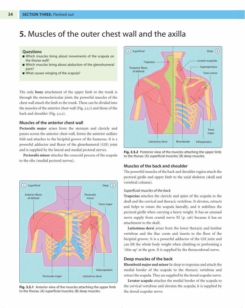
Muscles of the anterior chest wallPectoralis major arises from the sternum and clavicle and
passes across the anterior chest wall, forms the anterior axillary
fold and attaches to the bicipital groove of the humerus. It is a
powerful adductor and flexor of the glenohumeral (GH) joint
and is supplied by the lateral and medial pectoral nerves.
Pectoralis minor attaches the coracoid process of the scapula
to the ribs (medial pectoral nerves).
Muscles of the back and shoulderThe powerful muscles of the back and shoulder region attach the
pectoral girdle and upper limb to the axial skeleton (skull and
vertebral column).
Superficial muscles of the back
Trapezius attaches the clavicle and spine of the scapula to the
skull and the cervical and thoracic vertebrae. It elevates, retracts
and helps to rotate the scapula laterally, and it stabilizes the
pectoral girdle when carrying a heavy weight. It has an unusual
nerve supply from cranial nerve XI (p. 136) because it has an
attachment to the skull.
Latissimus dorsi arises from the lower thoracic and lumbar
vertebrae and the iliac crests and inserts to the floor of the
bicipital groove. It is a powerful adductor of the GH joint and
can lift the whole body weight when climbing or performing a
‘chin up’ at the gym. It is supplied by the thoracodorsal nerve.
Deep muscles of the backRhomboid major and minor lie deep to trapezius and attach the
medial border of the scapula to the thoracic vertebrae and
retract the scapula. They are supplied by the dorsal scapular nerve.
Levator scapula attaches the medial border of the scapula to
the cervical vertebrae and elevates the scapula; it is supplied by
the dorsal scapular nerve.
5. Muscles of the outer chest wall and the axilla
Questions■ Which muscles bring about movements of the scapula on
the thorax wall?■ Which muscles bring about abduction of the glenohumeral
joint?■ What causes winging of the scapula?
Pectoralis major Latissimus dorsi
Subscapularis
Pectoralisminor
Teres major
Anterior fibresof deltoid
SuperficialA Deep B
Fig. 3.5.1 Anterior view of the muscles attaching the upper limbto the thorax: (A) superficial muscles; (B) deep muscles.
Trapezius
Posterior fibresof deltoid
Teres minor
Levator scapulae
Supraspinatus
Latissimus dorsi Rhomboids Infraspinatus
Teresmajor
SuperficialA Deep B
Fig. 3.5.2 Posterior view of the muscles attaching the upper limbto the thorax: (A) superficial muscles; (B) deep muscles.
M3354-Section 03.05.qxd 4/15/06 10:26 AM Page 34

Serratus anterior (sometimes called the boxer’s muscle) is
attached laterally to the upper eight ribs and to the medial
border of the scapula. It pulls the scapula anteriorly, rotates it
when the arm is raised above the head and also holds the scapula
flat on the thorax wall when pushing forwards (protraction) or
punching. It is innervated by the long thoracic nerve, which may
be damaged during surgery. Loss of innervation allows the
scapula to stick out when the patient pushes against something
(winging of the scapula).
Short muscles of the shoulder
The short muscles of the shoulder attach the scapula to the
humerus. They are important in stabilizing the GH joint.
Deltoid is attached proximally to the clavicle and spine of the
scapula and inserts on to the lateral side of the humerus, giving the
rounded shape to the shoulder. It is a powerful abductor of the
humerus and stabilizes the GH joint when carrying a heavy load in
the hand. Its anterior fibres flex, middle fibres abduct and posterior
fibres extend the GH joint. It is supplied by the axillary nerve.
Teres major adducts and medially rotates the scapula; it is
supplied by the lower subscapular nerve.
The four rotator cuff muscles are particularly important in
stabilizing the GH joint (p. 43):
■ subscapularis: arises from the anterior surface of the
scapula and attaches to the lesser tubercle of the humerus
■ supraspinatus: arises from the scapula above the spine and
attaches to the top of the greater tubercle (assists abduction)
■ infraspinatus: arises from the scapula below the spine and
attaches to the greater tubercle
■ teres minor: arises from the posterior aspect of the scapula
and attaches to the greater tubercle.
AxillaThe pyramidal space between the thoracic wall and the upper
part of the humerus is known as the axilla. Table 3.5.1 gives the
boundaries of the axilla. Pectoralis major forms the anterior
axillary fold and the muscles of the posterior wall form the
posterior axillary fold. It contains the axillary lymph nodes and
all the neurovascular structures passing between the neck and
the limb; the nerves in particular are vulnerable to traction
injuries of the neck (p. 53).
The axilla contains:
■ structures lying between the outer border of the first rib and
the inferior border of teres major, posterior to pectoralis
major and pectoralis minor
■ axillary artery and its branches
■ axillary vein, lying medial to the artery
■ cords and terminal branches of the brachial plexus arranged
around the artery
■ axillary lymph nodes.
MovementThe flexibility and range of movements of the arm are greatly
increased by muscles that alter the position of the scapula on the
thorax wall. These scapulothoracic movements (lateral and
medial rotation, retraction and protraction, elevation and
depression) are separate from the movements of the GH joint.
Upper limb 35
RANGE OF MOVEMENT OF THE SHOULDER INARTHRITIS
The flexibility and range of movements of the shoulder aregreatly increased by muscles that alter the position of thescapula on the thorax wall. These scapulothoracic move-ments (lateral and medial rotation, retraction and protraction,elevation and depression) are separate from the movementsof the GH joint. Surprisingly, patients with very little abductionat the GH joint, as a result of severe arthritis, can still reach upto shelves by elevating their scapula.
Table 3.5.1 BOUNDARIES OF THE AXILLA
Area Boundaries
Apex Clavicle, scapula, first ribAnterior wall Pectoralis major, pectoralis minor,
clavipectoral fasciaPosterior wall Subscapularis, teres major, latissimus dorsi Medial wall Serratus anterior, upper ribsLateral wall HumerusBase Skin of the arm pit
M3354-Section 03.05.qxd 4/15/06 10:26 AM Page 35

SECTION THREE: Fleshed out36
The subclavian artery is the only arterial supply to the upper limb.
The left subclavian artery arises as a direct branch of the arch of the
aorta. The first branch from the arch is the brachiocephalic trunk,
which divides posterior to the sternoclavicular joint to form the
right subclavian and right common carotid arteries. The
subclavian artery enters the axilla by crossing the first rib posterior
to the subclavian vein and scalenus anterior muscle. In the neck, it
gives a number of branches including the thyrocervical trunk,
which, in turn, branches to give the suprascapular and transverse
cervical arteries. These pass posteriorly to supply the scapular
region (Figs 3.6.1–3.6.3).
6. Arterial supply
Questions■ Where are the arteries of the upper limb most vulnerable to
damage?■ Where can the pulse be felt in the upper limb?■ Which arteries contribute to the blood supply of the hand?
Collateral branches
Circumflex humeralaccompanies axillary nerve
Anterior branchessupply anterior wall
of the axilla
Supply scapular region
Right subclavian artery
LSA
LCCA
Right commoncarotid artery
Aorta
1
2
3
4
Profunda brachiiaccompanies radial nerve
in spiral groove
Superficial palmar archsupplies medial 31/2 digits
Deep palmar archsupplies lateral 11/2 digits
Ulnar arteryaccompanied byulnar nerve
Radial arteryaccompanied by
superficial branchof radial nerve
Brachial arteryaccompanied bymedian nerve
Subscapularsupplies posterior wall of axilla and connects with the scapular anastomosis
Lateralthoracicsuppliesbreast
Pulse points
Fig. 3.6.1 Anterior view of the major arteries of the right upperlimb.
Thoracodorsal
Subscapular
Circumflexscapular
3rd partaxillary
Posteriorcircumflex humeral
Thoracoacrimial
Thyrocervical trunk
SuprascapularTransverse cervical
Anastomoses
Rightsubclavian
Fig. 3.6.2 Posterior view of right scapula to show the major areasof anastomosis between the subclavian and axillary arteries.
Fig. 3.6.3 Posterior view of right scapula to show the position ofthe major neurovascular structures.
Rhomboids
Teres major
Circumflexscapular passingthrough uppertriangular space
Triceps
Teres minor
Profunda brachiiartery and radialnerve passing through lowertriangular space
Posteriorcircumflexhumeral arteryand axillary nervepassing throughquadrilateral space
Axillary artery
Subclavian artery
Suprascapular arteryTransverse cervical artery
Levator scapulae
Anastomosis
M3354-Section 03.06.qxd 4/15/06 10:27 AM Page 36

Axillary arteryBeginning at the outer border of the first rib, the axillary artery
passes posterior to pectoralis minor and is closely related to the
cords of the brachial plexus (see Fig. 3.8.2, p. 40). Its branches
supply the superior part of the thoracic wall, the muscular walls
of the axilla and the lateral half of the breast. Its posterior
branches connect with the branches of the subclavian artery
supplying the scapular region, to form the scapular
anastomosis (Figs 3.6.2 and 3.6.3). The axillary artery is closely
related to the cords of the brachial plexus.
Brachial arteryThe brachial artery begins at the inferior border of teres major
as the continuation of the axillary artery. It lies in the anterior
compartment of the arm accompanied by the median nerve and
a pair of deep veins. It gives rise to the profunda brachii artery,
supplying triceps, and collateral branches that anastomose
around the elbow with branches of the ulnar artery. Anterior to
the elbow joint, deep to the bicipital aponeurosis, the brachial
artery terminates by dividing to form the radial and ulnar
arteries (p. 44). The pulse of the brachial artery can be felt in the
cubital fossa, medial to the biceps tendon and is located during
the measurement of blood pressure.
Ulnar arteryThe ulnar artery passes deep to the superficial flexor muscles of
the forearm and gives the following branches:
■ collateral branches, which contribute to the anastomosis
around the elbow
■ anterior interosseous, supplying the deep flexor muscles
■ posterior interosseous, supplying the extensor compartment.
At the wrist, lateral to the pisiform bone, the artery passes super-
ficial to the flexor retinaculum with the ulnar nerve. Just deep to
the palmar aponeurosis and level with the distal border of the
extended thumb, the ulnar artery forms the superficial palmar
arch, which is usually completed by a small superficial palmar
branch from the radial artery. The branches of the superficial
arch supply the medial 31⁄2 fingers (p. 49).
Radial arteryThe radial artery passes distally with the superficial branch of the
radial nerve. The pulse can be felt over the distal radius. Here it
gives a small branch to the superficial palmar arch and continues
laterally around the base of the thumb, deep to the tendons of
the anatomical snuff box. It passes into the palm through the
muscles that lie between the first and second metacarpals, to
form the deep palmar arch and gives branches to the lateral
11⁄2 digits (p. 49). The deep palmar arch is completed by the deep
branch of the ulnar artery and lies at the level of the proximal
border of the extended thumb. Metacarpal branches link the
deep and superficial arches, ensuring a rich supply to the digits
in any position of the hand.
Upper limb 37
ROLE OF ANASTOMOSES IN PROTECTING THEBLOOD SUPPLY OF THE ARM FOLLOWINGTRAUMATIC INJURIES
Arteries are most vulnerable where they are relatively fixed orvery superficial. In the upper limb, for example, the subclavianartery is restricted by scalenus anterior muscle as it crosses thefirst rib. Trauma to the upper part of the thorax may damagethe subclavian or the axillary arteries. However, the scapularanastomosis provides alternative routes for blood flow and sothe limb is rarely completely ischaemic. Likewise, the richanastomoses between the superficial and deep palmar archesin the hand protect the supply to the digits when a lacerationat the wrist severs either the radial or the ulnar artery.
M3354-Section 03.06.qxd 4/15/06 10:27 AM Page 37

SECTION THREE: Fleshed out38
Superficial veinsBecause they are very commonly used for venepuncture, it is
important to know the position of the superficial veins of the
upper limb (Fig. 3.7.1).
The cephalic vein begins on the lateral side of the dorsal
venous arch of the hand, crosses the tendons of the anatomical
snuff box, remains superficial and eventually pierces deep fascia
just inferior to the clavicle to join the axillary vein.
The basilic vein begins on the medial side of the dorsum of
the hand and is usually connected to the cephalic vein anterior
to the elbow by the median cubital vein (most common site for
venepuncture). Just above the elbow, the basilic vein pierces
deep fascia medially and joins with the deep veins to form the
axillary vein.
The axillary vein lies within the curve of the axillary artery,
receiving tributaries from the axillary and scapular regions. It lies
anterior to scalenus anterior as it crosses the first rib and continues
as the subclavian vein in the neck, before joining with the internal
jugular vein to form the brachiocephalic vein (Fig. 3.7.1).
Deep veinsThe deep veins are the venae comitantes that accompany major
arteries.
Lymphatic drainageAll lymph from the upper limb and adjacent trunk above
the umbilicus drains to the axillary nodes (Fig. 3.7.2). There
7. Venous and lymphatic drainage of the upper limb andthe breast
Questions■ Why is it important to know the position of the superficial
veins?■ Where are breast tumours most likely to spread?
Dorsal venous arch
Axillary vein
BCV
Cephalic veinpierces deep fascia
Subclavian vein
Deep veinsaccompanyingbrachial artery
Internal jugular vein
Cephalic vein(important site for
venepuncture)
Median cubital vein(important sitefor venepuncture)
Basilic veinpierces deep fascia
Deep veinSuperficial vein
Fig. 3.7.1 Anterior view of the major veins of the right upperlimb.
From breast
From arm
Central
drain to+
drain to
drains to thoracic duct on left or right lymphatic duct
Infraclavicular Apical
Posterior(subscapular)nodes
Anterior(pectoral) nodes
Lateral(brachial) nodes
M
S
2
1
4
3
5
6
4321
654
6
Fig. 3.7.2 Diagram to show the arrangement of lymph nodes inthe right axilla.
M3354-Section 03.07.qxd 4/15/06 10:28 AM Page 38

are six groups, totalling approximately 25–30 nodes in all
(Table 3.7.1).
The axillary nodes drain via the apical group to the thoracic
duct on the left and the right subclavian lymph trunk on the
right. These lymph vessels then drain into the large veins at the
root of the neck (usually at the point where the internal jugular
and the subclavian veins meet).
■ THE BREASTThe anatomy of the breast and its lymphatic and venous drainage
is important because in Western cultures 1 in 12 women will
suffer from breast cancer (Fig. 3.7.3). The initial development of
breast tissue in the superficial fascia of the chest wall is the same
in both girls and boys. In girls, under the influence of oestrogen
at puberty, glandular tissue differentiates and becomes
embedded in fat. The breasts undergo further glandular develop-
ment during pregnancy and secrete milk after the birth when
stimulated by the hormone prolactin.
The medial part of the breast receives its arterial supply from
the second, third and fourth perforating branches of the internal
thoracic artery. The lateral half is supplied by thoracic branches
of the axillary artery and there are deep branches from adjacent
intercostal arteries. The venous drainage passes either anteriorly
to the venae comitantes of the internal thoracic artery or to the
posterior intercostal veins, which eventually drain to the azygos
system (p. 69).
Upper limb 39
Position of breaston pectoralis major
75% lymphto pectoral
lymph nodes
Rib 2
Rib 6
Lactiferous duct
Intercostalmuscles
Rib 4
Connectivetissue strands
Fatty tissue
Areola
Nipple
Pectoralismajor
Glandulartissue
Rib 1
ClavicleA B
Fig. 3.7.3 The breast and its lymphaticdrainage. (A) Anterior view; (B) sagittalsection of the breast.
SPREAD OF BREAST CANCER
Approximately 75% of the lymph from the breast drains to thepectoral (anterior) group of axillary lymph nodes. Malignantcells may be carried in lymph vessels and undergo metastasisin the nodes of the axilla. Malignant cells can also be carried viaintercostal veins to the azygos system and the vertebralvenous plexus, causing secondary tumours in the thoracicvertebral bodies.
Table 3.7.1 AXILLARY LYMPH NODES
Group Location Drains
1 Anterior/pectoral Inferior border of Breast, thoracic andpectoralis minor abdominal wallsand the lateral above the umbilicusthoracic vessels
2 Lateral Medial to axillary Upper limbvein
3 Posterior/ Posterior axillary Scapular regionsubscapular wall and the and neck
subscapular vessels
4 Central Embedded in Groups 1, 2, 3axillary fat
5 Infraclavicular Terminal part of the Lateral part upper cephalic vein limb
6 Apical Apex of the axilla Groups 5 and 6
M3354-Section 03.07.qxd 4/15/06 10:28 AM Page 39