3.incisors
21
INCISORS 15 Maxillary Incisors Principal Identifying Features The maxillary central incisor is the tooth in each maxillary quadrant of the permanent dentition which is on each side of the midline. Normally, the max central incisors are the most prominent teeth in the oral cavity and so they are most noticeable in the dental arch, hence they contribute a focal point for the eye of the observer. In general, the labial outline of the crown conform to the general outline of the face. The max central incisor (MCI) is the widest tooth mesio-distally (MD), the labial surface is less convex than lateral incisor and canine that give the tooth a rectangular appearance. It is the first tooth from the midline. It has a slightly straight incisal edge. Central Incisor Chronology Appearance of enamel organ 5 m.i.u First evidence of calcification 3 - 4 months. Crown completed 4 - 5 years. Eruption 7-8 y Root completion 10-11 y
Transcript of 3.incisors
INCISORS
15
Maxillary Incisors Principal Identifying Features
The maxillary central incisor is the tooth in each maxillary
quadrant of the permanent dentition which is on each side of the
midline.
Normally, the max central incisors are the most prominent teeth in
the oral cavity and so they are most noticeable in the dental arch,
hence they contribute a focal point for the eye of the observer. In
general, the labial outline of the crown conform to the general
outline of the face.
The max central incisor (MCI) is the widest tooth mesio-distally
(MD), the labial surface is less convex than lateral incisor and
canine that give the tooth a rectangular appearance. It is the first
tooth from the midline. It has a slightly straight incisal edge.
Central Incisor Chronology
Appearance of enamel organ 5 m.i.u
First evidence of calcification 3 - 4 months.
Crown completed 4 - 5 years.
Eruption 7-8 y
Root completion 10-11 y
INCISORS
16
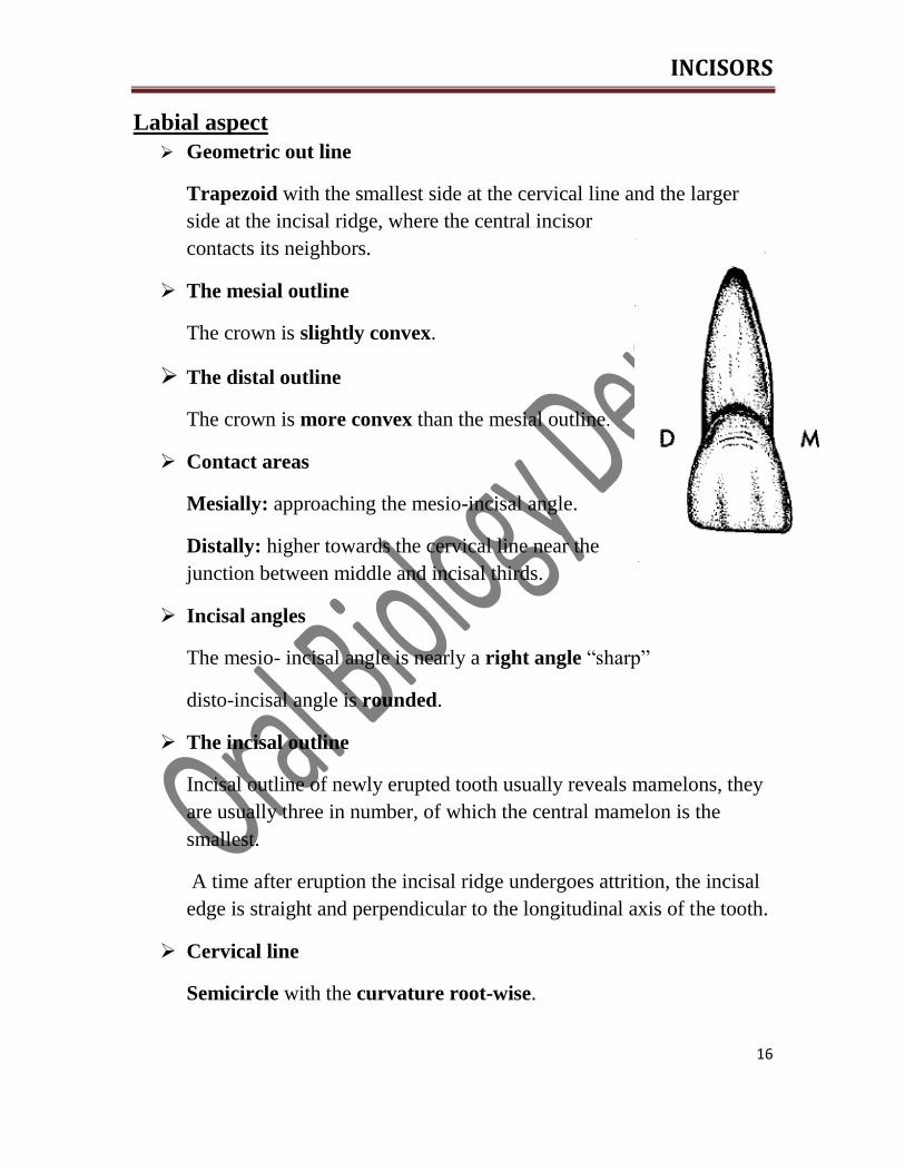
Labial aspect
Geometric out line
Trapezoid with the smallest side at the cervical line and the larger
side at the incisal ridge, where the central incisor
contacts its neighbors.
The mesial outline
The crown is slightly convex.
The distal outline
The crown is more convex than the mesial outline.
Contact areas
Mesially: approaching the mesio-incisal angle.
Distally: higher towards the cervical line near the
junction between middle and incisal thirds.
Incisal angles
The mesio- incisal angle is nearly a right angle “sharp”
disto-incisal angle is rounded.
The incisal outline
Incisal outline of newly erupted tooth usually reveals mamelons, they
are usually three in number, of which the central mamelon is the
smallest.
A time after eruption the incisal ridge undergoes attrition, the incisal
edge is straight and perpendicular to the longitudinal axis of the tooth.
Cervical line
Semicircle with the curvature root-wise.
INCISORS
17
Surface description
The labial surface of this tooth is smoothly convex with the maximum
convexity at the cervical third, referred to as cervical ridge. The labial
surface is marked by 2 faint or shallow vertical grooves which divide
the labial surface into 3 portions or lobes.
The root
Root is cone shaped with blunt apex. A line drawn through the center
of the root and crown of the maxillary central incisor tends to parallel
the mesial outline of the crown and root.
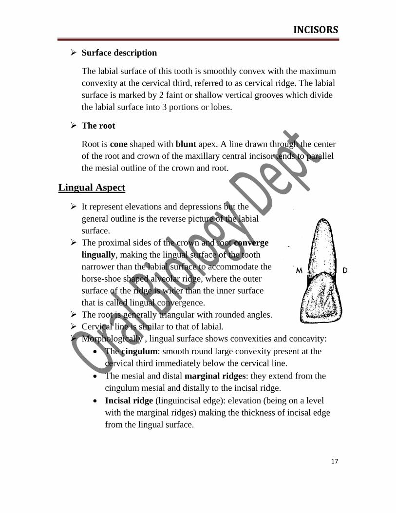
Lingual Aspect
It represent elevations and depressions but the
general outline is the reverse picture of the labial
surface.
The proximal sides of the crown and root converge
lingually, making the lingual surface of the tooth
narrower than the labial surface to accommodate the
horse-shoe shaped alveolar ridge, where the outer
surface of the ridge is wider than the inner surface
that is called lingual convergence.
The root is generally triangular with rounded angles.
Cervical line is similar to that of labial.
Morphologically , lingual surface shows convexities and concavity:
The cingulum: smooth round large convexity present at the
cervical third immediately below the cervical line.
The mesial and distal marginal ridges: they extend from the
cingulum mesial and distally to the incisal ridge.
Incisal ridge (linguincisal edge): elevation (being on a level
with the marginal ridges) making the thickness of incisal edge
from the lingual surface.
INCISORS
18
Lingual fossa: lingual depression between the marginal ridges
and extend from the cingulum to the incisal ridge. It has M-
shape and occupying the incisal two third.
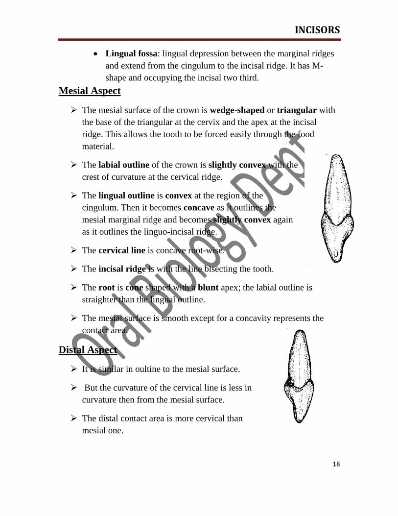
Mesial Aspect
The mesial surface of the crown is wedge-shaped or triangular with
the base of the triangular at the cervix and the apex at the incisal
ridge. This allows the tooth to be forced easily through the food
material.
The labial outline of the crown is slightly convex with the
crest of curvature at the cervical ridge.
The lingual outline is convex at the region of the
cingulum. Then it becomes concave as it outlines the
mesial marginal ridge and becomes slightly convex again
as it outlines the linguo-incisal ridge.
The cervical line is concave root-wise.
The incisal ridge is with the line bisecting the tooth.
The root is cone shaped with a blunt apex; the labial outline is
straighter than the lingual outline.
The mesial surface is smooth except for a concavity represents the
contact area.
Distal Aspect
It is similar in oultine to the mesial surface.
But the curvature of the cervical line is less in
curvature then from the mesial surface.
The distal contact area is more cervical than
mesial one.

INCISORS
19
Incisal edge distally is broader than mesially so the crown appear
thicker.
Incisal Aspect
The incisal ridge is seen to be centered over the root.
The crown superposes over the root entirely so that nothing of the root
is visible i.e. the crown and the root base on the same long axis.
The crown outline is roughly triangular with somewhat curved labial
outline that form the base of the triangular while the proximal sides
converge toward the cingulum.
Crown is wider MD than LL. Labial
outline is broad, flat compared with the
lingual surface. Cingulum is located off
center toward the distal side. So, MMR is
longer than DMR. Fossa is seen as a
concavity between the two MR and cingulum.
Note that
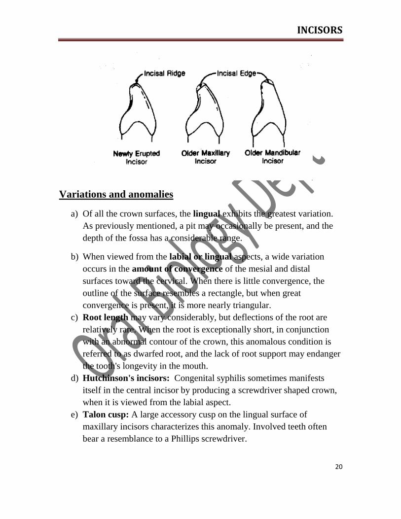
The incisal ridge is that portion of the crown which makes up the
complete incisal portion. When an incisor is newly erupted, the incisal
portion is rounded and merges with the mesioincisal and distoincisal
angles and the labial and lingual surfaces. This ridge portion of the crown
is called the incisal ridge. The term edge implies an angle formed by the
merging of two flat surfaces. Therefore, an incisal edge does not exist on
an incisor until occlusal wear has created a flattened surface linguo-
incisally, which surface forms an angle with the labial surface. The
incisal edge is formed by the junction of the linguoincisal surface,
(sometimes called the incisal surface), and the labial surface.
INCISORS
20
Variations and anomalies
a) Of all the crown surfaces, the lingual exhibits the greatest variation.
As previously mentioned, a pit may occasionally be present, and the
depth of the fossa has a considerable range.
b) When viewed from the labial or lingual aspects, a wide variation
occurs in the amount of convergence of the mesial and distal
surfaces toward the cervical. When there is little convergence, the
outline of the surface resembles a rectangle, but when great
convergence is present, it is more nearly triangular.
c) Root length may vary considerably, but deflections of the root are
relatively rare. When the root is exceptionally short, in conjunction
with an abnormal contour of the crown, this anomalous condition is
referred to as dwarfed root, and the lack of root support may endanger
the tooth's longevity in the mouth.
d) Hutchinson's incisors: Congenital syphilis sometimes manifests
itself in the central incisor by producing a screwdriver shaped crown,
when it is viewed from the labial aspect.
e) Talon cusp: A large accessory cusp on the lingual surface of
maxillary incisors characterizes this anomaly. Involved teeth often
bear a resemblance to a Phillips screwdriver.
INCISORS
21
f) The alveolar bone between the roots of the two central incisors is
occasionally the site of supernumerary teeth or extra teeth, known
as mesiodens. Cysts may also be found in this area.
Lateral Incisor
Principal Identifying Features
The maxillary lateral incisor is the tooth in each maxillary quadrant
of the permanent dentition which is second from the midline.
Contact is shared mesially with the permanent central incisor;
distally with the deciduous canine until its exfoliation at about 12
years, and then with the permanent canine.
General form and function
o The lateral incisor supplements the central incisor in function.
o It resembles the central incisor in all aspects, but on a smaller
scale. In fact, it is smaller in all measurements, except root
length.
o Its relative crown dimensions, differ slightly from the central,
however. It is relatively longer inciso-cervically and narrower
mesio-distally.
o It also is generally a more round tooth than the central incisor.
o The upper lateral incisors display greater variation in form than
any other permanent tooth, except the third molars.
o There are two known forms:
Square form → similar to that of the central.
Rounded form → similar to that of the canine.
INCISORS
22
Chronology
Appearance of enamel organ 1 y.
First evidence of calcification 4 - 5 y.
Crown completed y.
Eruption 8 - 9 y.
Root completion 1 1 y.
Labial aspect
Geometric outline
Trapezoid similar to labial aspect of maxillary permanent central
incisor.
Mesial Outline
Similar to maxillary permanent central incisor.
Distal Outline
The distal outline is always more rounded than the mesial outline and
distal outline of maxillary central incisor.
Contact areas
Mesially: at the junction of the middle and incisal thirds.
Distally: more cervical, usually in the center of the middle third.
Incisal angles
The mesio-incisal angle is more rounded.Occasionally, in the
square forms, the mesio-incisal angle is almost as sharp as that
found on most maxillary central incisors. However, a more
rounded mesio-incisal angle is seen more often.
The distoincisal angle is more rounded.
Incisal Outline
It is formed by the incisal ridge.

INCISORS
23
Mesial half of incisal outline is relatively straight and distal half
is more rounded curving towards cervical line to join the distal
outline.
The number and prominence of mamelons is variable, but two are
the most common finding.
Cervical Outline
The cervical line curves in a regular arc apically, but less deep
than that of the central incisor.
Surface description
Labial surface is about 2 mm narrower and 2 to 3
mm shorter than the maxillary permanent central
incisor.
It is more convex compared to maxillary
permanent central incisor, both mesiodistally and
incisogingivally.
Labial developmental depressions and imbrication
lines are similar to those of the central incisor.
Root
Usually as long, if not somewhat longer, than that of the central
incisor.
The root is often about 1.5 times the length of the crown.
The root tapers evenly from the cervical line to a point
approximately two thirds of its length apically. In most cases, it
curves sharply from this location in a distal direction and ends
in a pointed apex.
The apex is relatively sharper than that of the central.
INCISORS
24
Lingual aspect:
It is the reverse of the labial aspect. There is lingual convergence of
proximal walls as seen in maxillary permanent central incisor.
Cervical outline: The CEJ curves toward the apical, but is offset to the
distal.
M
Morphologically, lingual surface shows convexities and
concavity: Marginal ridges are more prominent and
stronger than those found on central incisor.
The linguoincisal ridge is well developed.
Lingual fossa (v shape) is deeper and well-
circumscribed.
Cingulum is more prominent.
Palatogingival or palatoradicular groove: a deep
developmental groove originates from the lingual pit, crosses
the distal side of the cingulum and extends on the root for part
or all of its length.
Faults in the enamel of the crown are often found in the deep
portions of these developmental grooves.
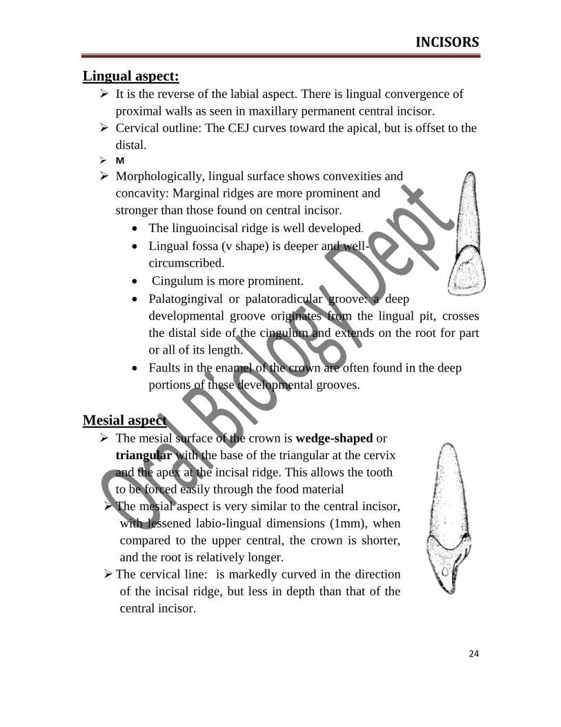
Mesial aspect
The mesial surface of the crown is wedge-shaped or
triangular with the base of the triangular at the cervix
and the apex at the incisal ridge. This allows the tooth
to be forced easily through the food material
The mesial aspect is very similar to the central incisor,
with lessened labio-lingual dimensions (1mm), when
compared to the upper central, the crown is shorter,
and the root is relatively longer.
The cervical line: is markedly curved in the direction
of the incisal ridge, but less in depth than that of the
central incisor.

INCISORS
25
The incisal ridge is well developed, making the incisal portion and
appears somewhat thicker than that of the central incisor. The mesial
surface is smooth except for a concavity represents the contact area.
The root
Appears as a tapered cone from this aspect, with a bluntly rounded
apical end. This varies in individuals, with the apical end sometimes
being quite blunt, while at other times, it is pointed.
A line drawn through the center of the root tends to bisect the incisal
ridge of the crown.
Distal aspect:
The distal surface is smaller and more convex in all dimensions
than the mesial surface. The width of the crown
distally appears thicker than it does on the mesial
aspect from marginal ridge to labial face, because
of the placement of the crown on the root.
The contact area is shorter and not as incisally
placed, when compared to the mesial contact. It is
normally located at center of the middle third.
The cervical line shows less curvature incisally
than on the mesial surface.
A developmental groove may be found on the
crown extending on the root for part or all of its
length.
Incisal aspect:
All maxillary lateral incisors exhibit more convexity labially and
lingually from the incisal aspect than do the maxillary central incisors.
The incisal aspect shows 2 forms
Resembles the central incisor except
in size (triangular in shape)
Resembles a small canine
(rhomboidal or oval) if :
1-The cingulum is large, as well
INCISORS
26
as the incisal ridge.
2-The labiolingual dimension is greater than the
mesiodistal dimension.
Developmental anomalies
The maxillary lateral incisor varies in form more than any other tooth
in the mouth except the third molar.
If the variation is too great, it is considered a developmental anomaly.
For example:
Congenitally missing laterals, i.e.: tooth buds do not form
(agenesis).
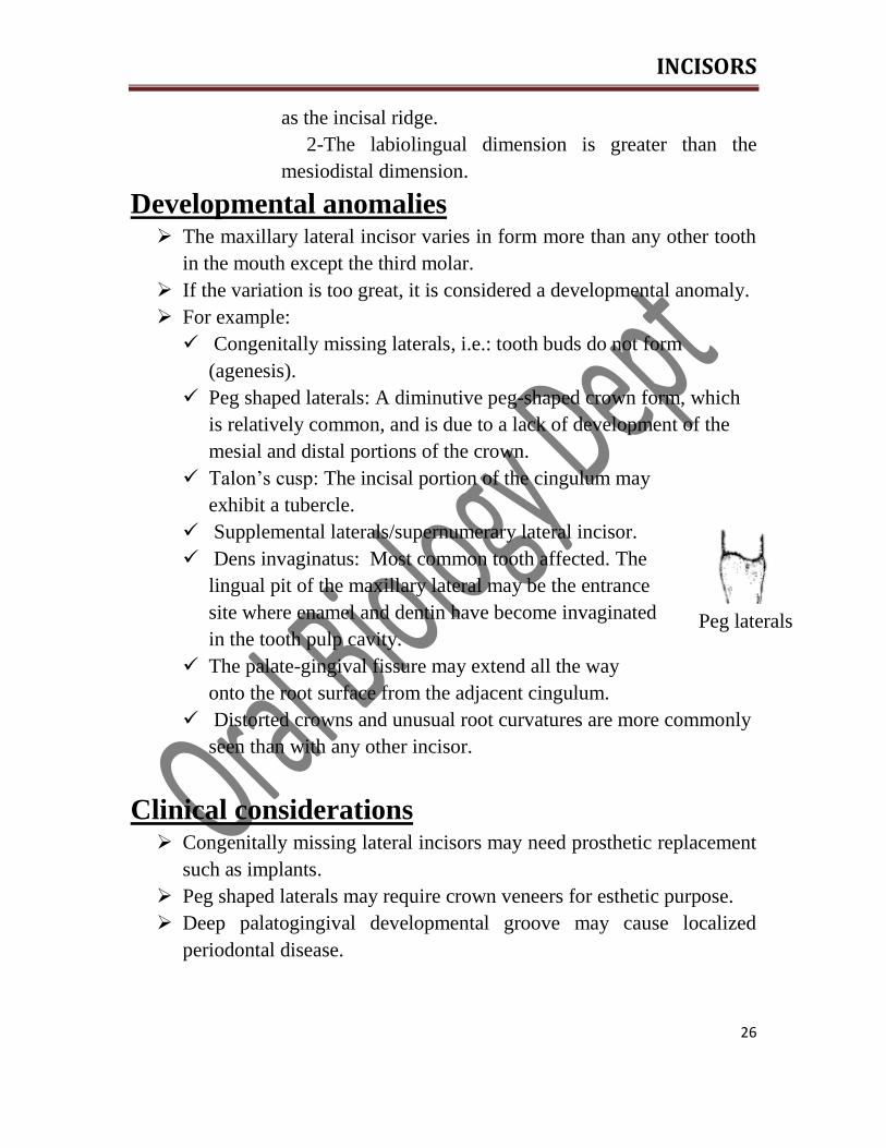
Peg shaped laterals: A diminutive peg-shaped crown form, which
is relatively common, and is due to a lack of development of the
mesial and distal portions of the crown.
Talon’s cusp: The incisal portion of the cingulum may
exhibit a tubercle.
Supplemental laterals/supernumerary lateral incisor.
Dens invaginatus: Most common tooth affected. The
lingual pit of the maxillary lateral may be the entrance
site where enamel and dentin have become invaginated
in the tooth pulp cavity.
The palate-gingival fissure may extend all the way
onto the root surface from the adjacent cingulum.
Distorted crowns and unusual root curvatures are more commonly
seen than with any other incisor.
Clinical considerations Congenitally missing lateral incisors may need prosthetic replacement
such as implants.
Peg shaped laterals may require crown veneers for esthetic purpose.
Deep palatogingival developmental groove may cause localized
periodontal disease.
Peg laterals
INCISORS
27
Mandibular Incisors
Introduction
Mandibular incisors are four in number, two on each side from the
central line.
These are the first permanent teeth to erupt, replacing deciduous
incisors, and are the smallest teeth in either arch.
The mandibular central incisors are centered in the mandible, with
the mesial surface of each one in contact with the mesial surface of
the other.
The mandibular lateral incisors are distal to the central incisors which
make contact mesially with the central incisor and distally with the
canine.
Mandibular central incisor and lateral are similar in anatomy and
complement each other in function but the central is somewhat
smaller than the lateral incisor.
They have smaller mesiodistal dimensions than any of the other
teeth.
The anatomic form of these teeth differs from that of the maxillary
incisors in that the labial surface is inclined lingually so that the
incisal ridges are lingual to a line bisecting the root.
Chronology
Central incisor Lateral incisor
Appearance of enamel organ 5 m.i.u
First evidence of calcification 3 - 4 months.
Crown completed 4 - 5 years.
Eruption 6 – 7 Ys. 7 – 8 Ys.
Root completion 9 Ys. 10 Ys.

INCISORS
28
Central Incisor
It is the narrowest and the smallest tooth mesiodistally of all the
permanent teeth.
These teeth function in biting, and incising, just as do their maxillary
counterparts.
Labial aspect
Labial surface of mandibular central incisor is regular and
very small.
It is bilaterally symmetrical.
Geometric outline.
Trapezoidal with the smallest side cervically.
Mesial and distal outlines.
The mesial and distal outlines of the crown make a straight
drop from the incisal angles to the contact areas. The mesial
and distal sides taper evenly from the contact areas to the
narrow cervix (neck), giving the crown fan shaped
appearance.
Although these two surfaces are nearly parallel at the incisal
edge, they converge toward the cervical margin.
Contact areas: Mesial and distal contacts at equal heights in incisal
third incisal to the junction of the incisal and middle thirds of the
crown near the mesioincisal and distoincisal angles.
Incisal outline.
The incisal edge is straight and is at nearly a right angle to the
long axis of the tooth.
Broadest portion of tooth in incisal third.
Mamelons most always 3 in number occasionally present on newly
erupted teeth.
It is the only incisor where both mesioincisal and distoincisal
angles are sharp and at right angles.
Distoincisal angle is barely more rounded than the mesioincisal
angle.

INCISORS
29
Cervical outline.
Is symmetrically and evenly convex toward the root.
Surface description.
The labial surface is smooth and tends to be more convex,
particularly in the cervical third, and flattens out toward the
incisal third.
The developmental grooves may or may not be present. When
present, they appear as very faint furrows in newly erupted
teeth.
The root
The root is single, slender and straight.
The root length is as great, if not greater, than that of the maxillary
central incisor.
The mesial and distal root outlines are straight with the mesial
and distal outlines of the crown down to the apical portion.
The root surface is regular and convex and extremely flattened on
its mesial and distal surfaces.
The apical third of the root terminates in a small pointed taper, in
most cases curving distally. Sometimes the roots are straight.
Lingual aspect
The crown is narrower on the lingual than on the labial
surface (lingual convergence).
The mesial, distal and incisal outlines closely resemble
those of the labial aspect and they are regular and
symmetrical.
The lingual aspect is shallow, with slightly concavity at
the incisal third between the less prominent mesial and
distal marginal ridges then it becomes flat and then
convex as progression from the incisal third to the
cervical third.

INCISORS
30
Less prominent smooth, small and centered cingulum and
marginal ridges than maxillary incisors.
The incisal ridge is narrow and rounded or worn flat.
The lingual surface is smooth and devoid of any neither grooves
nor fissures. No other tooth in the mouth, except the mandibular
lateral incisor, shows so few developmental lines and grooves.
Shallow lingual fossa. No lingual pit.
The cervical line is convex toward the root.
The root is slightly narrower on the lingual side than on the labial
side.
Mesial aspect
The geometric outline is triangular. So, the crown is wedge-
shaped but the curvature labially and lingually is less than that
found on maxillary incisors.
The labial outline is almost straight, except near cervical
third where it is convex (cervical ridge). The labial surface is
inclined lingually.
Height of contour (crest of curvature):
o Labial→→ Junction of cervical and middle thirds.
o Lingual→→ Middle of cervical third.
The lingual outline is ‘S’ shaped. It is convex over the
cingulum, in the cervical third. In the middle and incisal thirds
the lingual outline is slightly concave.
The incisal outline. The lingual outline of the narrow incisal ridge is
convex (rounded) or flat because of attrition. Incisal ridge center is
slightly lingual to the root axis.
The cervical line on the mesial and distal surface is marked and
convex incisally about one third the length of the crown.
INCISORS
31
The mesial surface is convex and smooth at the incisal third till the
contact point, below the contact area it is flattened and then it
becomes quit flat and even concave till the cervical line.
The contact area is located half way from labial to lingual and in the
incisal third, very close to the incisal edge. It has an ovoid shape.
The root
The labial and lingual outlines of the root are nearly straight
from the cervical line to the middle third; then tapers to the
rounded or pointed apex. The root tapering is rapid in the
apical third. The root apex is on the axis line.
The mesial surface of the root is flat. Usually there is a broad
and deep longitudinal developmental depression on the
mesial surface of the root for most of its length that is deeper
at the junction of the middle and apical thirds.
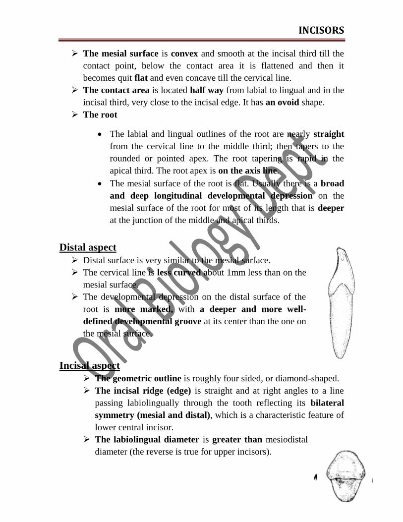
Distal aspect
Distal surface is very similar to the mesial surface.
The cervical line is less curved about 1mm less than on the
mesial surface.
The developmental depression on the distal surface of the
root is more marked, with a deeper and more well-
defined developmental groove at its center than the one on
the mesial surface.
Incisal aspect
The geometric outline is roughly four sided, or diamond-shaped.
The incisal ridge (edge) is straight and at right angles to a line
passing labiolingually through the tooth reflecting its bilateral
symmetry (mesial and distal), which is a characteristic feature of
lower central incisor.
The labiolingual diameter is greater than mesiodistal
diameter (the reverse is true for upper incisors).
INCISORS
32
The crown tapers lingually, therefore, the labial outline is wider
than the lingual outline.
The labial surface of the crown is slightly convex at incisal third.
The lingual surface of the crown is slightly concave at the incisal
third with centered, smooth, and convex cingulum.
Newly erupted teeth show mamelons which wear off upon
mastication.
Variations and Anomalies:
This tooth is rarely absent. There is great variability in the
lingual inclination of the labial surface of the mandibular
central incisor.
Anomalies are very rare. Occasionally a bifurcated root is
found.
In some Mongoloid peoples, cingulum has a short deep groove
running cervicoincisally. It is a site of dental caries.
INCISORS
33
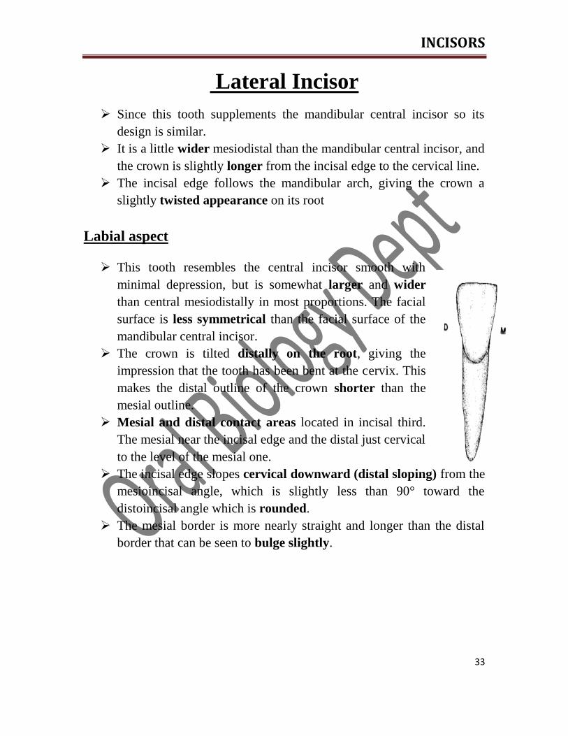
Lateral Incisor
Since this tooth supplements the mandibular central incisor so its
design is similar.
It is a little wider mesiodistal than the mandibular central incisor, and
the crown is slightly longer from the incisal edge to the cervical line.
The incisal edge follows the mandibular arch, giving the crown a
slightly twisted appearance on its root
Labial aspect
This tooth resembles the central incisor smooth with
minimal depression, but is somewhat larger and wider
than central mesiodistally in most proportions. The facial
surface is less symmetrical than the facial surface of the
mandibular central incisor.
The crown is tilted distally on the root, giving the
impression that the tooth has been bent at the cervix. This
makes the distal outline of the crown shorter than the
mesial outline.
Mesial and distal contact areas located in incisal third.
The mesial near the incisal edge and the distal just cervical
to the level of the mesial one.
The incisal edge slopes cervical downward (distal sloping) from the
mesioincisal angle, which is slightly less than 90° toward the
distoincisal angle which is rounded.
The mesial border is more nearly straight and longer than the distal
border that can be seen to bulge slightly.

INCISORS
34
Lingual aspect
The lingual surface is similar in outline to the labial
surface. The incisal portion of the lingual surface is
concave.
The cervical portion of the lingual aspect is narrower
while the incisal portion is wider. This gives the
crown a more or less a fan shaped appearance.
The cingulum is quite large but blends in smoothly
with the rest of the surface so the cingulum and
marginal ridges more prominent on lateral than
central.
The cingulum is more offset to the distal, and as a
result, the curvature of the cervical line is also offset
distally.
No lingual pit.
The concavity in the lingual aspect is slightly more when
compared to mandibular central incisor.
Proximal aspect (mesial and distal aspects)
Like the central, the crown presents a triangular
outline. When viewed critically, the rotation of the
incisal edge can be seen.
The incisal edge is on or lingual to the mid-root
axis. From the mesial side, the distolingual twist
of the incisal ridge places the distal portion at the
ridge somewhat more lingual than on the mesial.
The distal surface is shorter than the mesial.
The cervical line curvature in both mesially and
distally is slightly less than the central incisors.
The distal contact area is more cervically located
than on the mesial.

INCISORS
35
Incisal aspect
As with the mandibular central incisor, but unlike the maxillary
incisors, the crown is broader labiolingually than mesiodistally.
It not bilaterally symmetrical.
The incisal edge is not straight and it curves toward the
lingual in its distal portion (distolingual twist) giving
the crown the appearance of being slightly twisted on
its root. This twist corresponds to the curvature of the
mandibular dental arch.
The distal half of the incisal edge is bent lingually, so that the
distoincisal angle is more lingual in position than the mesioincisal
angle.
The cingulum is slightly off-center to the distal.
The root
Lateral incisor has slightly longer, thicker and wider root in all
dimensions than the central incisor.
The root is single and extremely flattened on its mesial and distal
surfaces.
The root appears very narrow mesiodistally, and tapers gradually
from cervical line toward pointed apex which may slightly to the
distal.
Lateral more likely to have longitudinal developmental grooves that
the distal more pronounced than mesial.
Variations and Anomalies
This tooth is stable, but variations in root length and direction are
occasionally seen also bifurcated root may be found.
Fused mandibular incisors, a missing central, and lateral emerged
distally to the canine may be found.