3D Protein Structure Visualization3D Protein Structure Search Search for 3D protein structure files...
2
• Search and visualize experimentally determined protein structures in 3D • Display influenza virus sequence conservation heat map on the structure • Highlight sequence features (regions of influenza proteins with specific functional, structural, or immune epitope properties, etc.) • Download highlighted protein structure image or 3D protein structure movie SEARCH TYPE Basic Influenza A Structures Search for protein structure files Search for proteins with structure files Enter PDB ID VIRUS TYPE A B C SUBTYPE Example: H1N1, H1N5 Use comma to separate multiple entries. STRAIN NAME Example: A/Viet Nam/1203/2004 Use % as wildcard, separate multiple strain names by a comma All 1 PB2 2 PB1 2 PB1-F2 3 PA 4 HA 5 NP 6 NA 7 M1 7 M2 8 NS1 8 NS2 SELECT PROTEINS TO SEARCH ADVANCED OPTIONS Select Advanced Option Select An Advanced Option PROTEIN STRUCTURE HIGHLIGHT FEATURE Filter to only include protein structures that allow for highlighting of the following data types. Sequence Conservation Experimentally Determined Epitopes Active Sites Sequence Features (SOP) Remove Select Advanced Option Select An Advanced Option GenBank Protein GI PUBLIC DATABASE IDENTIFIER 408677 Example: ABH05852 Separate multiple IDs by a comma and specify "All" if different types of identifiers are entered. Select Advanced Option Display Fields Select/Deselect All Protein UniProt ID Description Strain Subtype Protein Accession CHOOSE WHICH DATA FIELDS TO DISPLAY IN RESULT DISPLAY FIELDS Remove Select Advanced Option Author Tip: To select multiple or deselect, Ctrl-click (Windows) or Cmd-click (MacOS) Show All 3D Protein Structure Search Search for 3D protein structure files obtained from the Protein Data Bank (PDB). Influenza protein(s) are associated with each PDB file based on assignments in the UniProt database or manual curation by the IRD Team. One protein structure can be assigned to multiple GenBank protein records. 6 matching results Home 3D Protein Structure Search SEARCH DATA ANALYZE & VISUALIZE WORKBENCH SUBMIT DATA AUTHOR Webster Separate multiple author(s) by a comma Remove Select Advanced Option Description Keyword DESCRIPTION KEYWORD Example: neuraminidase, antibody Separate multiple keywords by a comma Search Clear Add Another Advanced Option http://www.fludb.org/ Freely available Integrated datasets Bioinformatics tool suite Platform for influenza data submission 1. Mouse-over “Search Data” and click “3D Protein Structures”. 2. Search for protein structures of interest using one of the Search Types: • Basic influenza A structures: This will retrieve a list of representative structures for each influenza A virus protein; • Search for protein structure files: This will retrieve a list of protein structures matching selected search criteria; • Search for proteins with structure files: This will retrieve protein structures grouped by protein UniProt ID; • Enter PDB ID: Search protein structures by PDB ID. Select or enter in search criteria and click “Search” to run your search. Note that IRD shows the instant count of search results at the top-right corner of the page to help you search quickly and efficiently. When you select search criteria on search pages, you will instantly know how many records match your search criteria without clicking the “Search” button and actually running the search. 1 Click “Advanced Options” to display additional search fields IRD is funded by the National Institute of Allergy and Infectious Diseases (NIH/DHHS) under Contract No. HHSN266200400041C and is a collaboration between Northrop Grumman Health IT, J. Craig Venter Institute, Vecna Technologies, SAGE Analytica and Los Alamos National Laboratory. Comments, questions, suggestions? Contact us at [email protected] 3D Protein Structure Visualization Retrieve all Influenza protein structures with a specific public database identifier. Ex., GenBank Protein GI 408677 Will retrieve protein structures grouped by protein UniProt ID Search structures by PDB ID Will retrieve a list of protein structures matching search criteria 2 Options to limit search results to protein structures with certain data types Search by structure authors Influenza virus hemagglutinin complexed with a neutralizing antibody (PDB ID: 1EO8) Search for 3D protein structures
Transcript of 3D Protein Structure Visualization3D Protein Structure Search Search for 3D protein structure files...
• Search and visualize experimentally determined protein structures in 3D
• Display influenza virus sequence conservation heat map on the structure
• Highlight sequence features (regions of influenza proteins with specific functional,
structural, or immune epitope properties, etc.)
• Download highlighted protein structure image or 3D protein structure movie
SEARCH TYPE Basic Influenza A Structures
Search for protein structure files
Search for proteins with structure files
Enter PDB ID
VIRUS TYPE A
B
C
SUBTYPE
Example: H1N1, H1N5
Use comma to separate multiple entries.
STRAIN NAME
Example: A/Viet Nam/1203/2004
Use % as wildcard, separate multiple strain namesby a comma
All1 PB22 PB12 PB1-F23 PA4 HA5 NP6 NA7 M17 M28 NS18 NS2
SELECT PROTEINS TO SEARCH
ADVANCED OPTIONS
RemoveSelect Advanced Option
Select An Advanced OptionPROTEIN STRUCTURE HIGHLIGHT FEATUREFilter to only include protein structures that allow for highlighting of the following data types.
Sequence Conservation
Experimentally Determined Epitopes
Active Sites
Sequence Features (SOP)
RemoveSelect Advanced Option
Select An Advanced Option GenBank Protein GI
PUBLIC DATABASE IDENTIFIER408677
Example: ABH05852
Separate multiple IDs by a comma and specify "All" if different types of identifiers are entered.
RemoveSelect Advanced Option
Display Fields
Select/Deselect All
Protein
UniProt ID
Description
Strain
Subtype
Protein Accession
CHOOSE WHICH DATA FIELDS TO DISPLAY IN RESULT
DISPLAY FIELDS
RemoveSelect Advanced Option
Author
Tip: To select multiple or deselect, Ctrl-click (Windows) or Cmd-click (MacOS) Show All
3D Protein Structure Search Search for 3D protein structure files obtained from the Protein Data Bank (PDB). Influenza protein(s) are associated with each PDB file based on assignments in the UniProt
database or manual curation by the IRD Team. One protein structure can be assigned to multiple GenBank protein records.
6 matching results
Home 3D Protein Structure Search
SEARCH DATA ANALYZE & VISUALIZE WORKBENCH SUBMIT DATA
About Us Community Announcements Links Resources Support Workbench Sign In
Influenza Research Database - PDB Protein Structure Search http://www.fludb.org/brc/influenza_proteinStructure_search.do...
1 of 2 6/21/12 2:21 PM
Cite IRD Tutorials Glossary of Terms Report a Bug Request Web Training Contact Us Release Date: Jun 3, 2012
This project is funded by the National Institute of Allergy and Infectious Diseases (NIH / DHHS) under Contract No. HHSN266200400041C and is a collaboration between NorthropGrumman Health IT, University of Texas Southwestern Medical Center , Vecna Technologies, SAGE Analytica and Los Alamos National Laboratory.
AUTHORWebster
Separate multiple author(s) by a comma
RemoveSelect Advanced Option
Description KeywordDESCRIPTION KEYWORD
Example: neuraminidase, antibodySeparate multiple keywords by a comma
SearchClear
Add Another Advanced Option
Influenza Research Database - PDB Protein Structure Search http://www.fludb.org/brc/influenza_proteinStructure_search.do...
2 of 2 6/21/12 2:21 PM
http://www.fludb.org/
Freely available Integrated datasets Bioinformatics tool suite Platform for influenza data submission
1. Mouse-over “Search Data” and click “3D Protein Structures”.
2. Search for protein structures of interest using one of the Search Types: • Basic influenza A structures: This will retrieve a list of
representative structures for each influenza A virus protein;
• Search for protein structure files: This will retrieve a list of protein structures matching selected search criteria;
• Search for proteins with structure files: This will retrieve protein structures grouped by protein UniProt ID;
• Enter PDB ID: Search protein structures by PDB ID. Select or enter in search criteria and click “Search” to run your search. Note that IRD shows the instant count of search results at the top-right corner of the page to help you search quickly and efficiently. When you select search criteria on search pages, you will instantly know how many records match your search criteria without clicking the “Search” button and actually running the search.
1
Click “Advanced Options” to display
additional search fields
IRD is funded by the National Institute of Allergy and Infectious Diseases (NIH/DHHS) under Contract No. HHSN266200400041C and is a collaboration between Northrop Grumman Health IT, J. Craig Venter Institute, Vecna Technologies, SAGE Analytica and Los Alamos National Laboratory. Comments, questions, suggestions? Contact us at [email protected]
3D Protein Structure Visualization
Retrieve all Influenza protein structures with a specific public database identifier. Ex., GenBank Protein GI 408677
Will retrieve protein structures grouped
by protein UniProt ID
Search structures by
PDB ID
Will retrieve a list of protein structures
matching search criteria
2
Options to limit search results to protein structures with certain data types
Search by structure authors
Influenza virus hemagglutinin complexed with a neutralizing antibody (PDB ID: 1EO8)
Search for 3D protein structures
2
Reset View Save View As Image
Type JMol command here. Run
Save View:
Description: INFLUENZA VIRUS HEMAGGLUTININ COMPLEXED WITH A
NEUTRALIZING
PDB Link: 1EO8
JMOL COMMAND LINE:
Advanced users can enter a JMol command directly using JMol Interactive Script .
SAVE/RESTORE
The state of the viewer (highlighting, zooming, etc) can be saved at any time and then
retrieved later. Choose the view you want to save, then restore it later when you are
ready.
Display Type: Ball & Stick
Details
Show:
Sequence conservation computed using all sequences
- B Cell Epitopes Clear
Highlight Clear
DISPLAY OPTIONS:
These options control the general appearance of the protein structure in
the viewer.
HIGHLIGHT SEQUENCE CONSERVATION
Overlay the structure with a sequence conservation "heat map" (see SOP
for details). Blue represents conserved regions and red represents
non-conserved regions
HIGHLIGHT LIGANDS
Highlight Ligands in
HIGHLIGHT EPITOPES
Highlight epitopes on the structure in . First, select an epitope type
from the list. Then check epitopes to highlight.
Peptide sequence Range IEDB ID
EGFTWTGVTQNGGSNA 139-154 12167KRGPGSG 156-162 33143WTGVTQN 143-149 73132
HIGHLIGHT BY SWISS-PROT POSITION
Highlight in in this structure corresponding to defined SwissProt
positions. With the dropdown, select a chain and view the positions present
in this file. Enter one or more comma-delimited positions (15,30) or a range
(15-30) then click Highlight. If no chain is selected, these positions will be
highlighted on all chains which include them.
Select Chain:
A - P03437 (25 - 343) 25
PDB Sequence/Structure DetailsRestore View:
Zoom: 100%
Spin:
Home Protein Structure Viewer (1EO8)
SEARCH DATA ANALYZE & VISUALIZE WORKBENCH SUBMIT DATA
About Us Community Announcements Links Resources Support Workbench Sign In
Your search returned 93 protein structures. Search Criteria Displaying 50 per page , sorted by PDB ID ascendinglyDisplay Settings
Download Save Search
1 2 Next > Page: 1 of 2
3D Protein Structure Search Results
Your Selected Items: 50 items selected | Deselect All
Select all 93 protein structures
PDB ID Protein UniProt ID Description Strain Subtype ProteinAccession
ViewStructure
1EO8 HA P03437 influenza virus hemagglutinin complexed with a neutralizing A/Aichi/2/1968 H3N2 CAA24269.1AAA43178.1BAF37221.1
More...
ViewStructure
1FRG HA P03438 crystal structure, sequence, and epitope mapping of a peptidecomplex of an anti-influenza ha peptide antibody fab 26(slash)9:
fine-tuning antibody specificity
A/Puerto Rico/8/34(lab reassortant)
H3N2 ABH05852.1
ViewStructure
1HA0 HA P03437 hemagglutinin precursor ha0 A/Aichi/2/1968 H3N2 CAA24269.1AAA43178.1BAF37221.1
More...
ViewStructure
1HGD HA P03438 binding of influenza virus hemagglutinin to analogs of its c surfacereceptor, sialic acid: analysis by proton nuclear m resonance
spectroscopy and x-ray crystallography
A/Puerto Rico/8/34(lab reassortant)
H3N2 ABH05852.1
ViewStructure
1HGE HA P03438 binding of influenza virus hemagglutinin to analogs of its c surfacereceptor, sialic acid: analysis by proton nuclear m resonance
spectroscopy and x-ray crystallography
A/Puerto Rico/8/34(lab reassortant)
H3N2 ABH05852.1
ViewStructure
1HGF HA P03438 binding of influenza virus hemagglutinin to analogs of its c surfacereceptor, sialic acid: analysis by proton nuclear m resonance
spectroscopy and x-ray crystallography
A/Puerto Rico/8/34(lab reassortant)
H3N2 ABH05852.1
ViewStructure
1HGG HA P03437 binding of influenza virus hemagglutinin to analogs of its c surfacereceptor, sialic acid: analysis by proton nuclear m resonance
spectroscopy and x-ray crystallography
A/Aichi/2/1968 H3N2 CAA24269.1AAA43178.1BAF37221.1
More...
ViewStructure
1HGH HA P03438 binding of influenza virus hemagglutinin to analogs of its c surfacereceptor, sialic acid: analysis by proton nuclear m resonance
spectroscopy and x-ray crystallography
A/Puerto Rico/8/34(lab reassortant)
H3N2 ABH05852.1
ViewStructure
1HGI HA P03438 binding of influenza virus hemagglutinin to analogs of its c surfacereceptor, sialic acid: analysis by proton nuclear m resonance
spectroscopy and x-ray crystallography
A/Puerto Rico/8/34(lab reassortant)
H3N2 ABH05852.1
ViewStructure
1HGJ HA P03438 binding of influenza virus hemagglutinin to analogs of its c surfacereceptor, sialic acid: analysis by proton nuclear m resonance
spectroscopy and x-ray crystallography
A/Puerto Rico/8/34(lab reassortant)
H3N2 ABH05852.1
ViewStructure
1HTM HA P03437 structure of influenza haemagglutinin at the ph of membrane fusion A/Aichi/2/1968 H3N2 CAA24269.1AAA43178.1BAF37221.1
More...
ViewStructure
1IBN HA P03442 nmr structure of hemagglutinin fusion peptide in dpc micelles at ph 5 A/duck/Ukraine/1/1963
H3N8 CAA24271.1
ViewStructure
1IBO HA P03442 nmr structure of hemagglutinin fusion peptide in dpc micelles at ph 7.4 A/duck/Ukraine/1/1963
H3N8 CAA24271.1
ViewStructure
1JSD HA Q91CD4 crystal structure of swine h9 haemagglutinin A/Swine/HongKong/9/98
H9N2 AAL14080.1
ViewStructure
1JSH HA Q91CD4 crystal structure of h9 haemagglutinin complexed with lsta r analog A/Swine/HongKong/9/98
H9N2 AAL14080.1
ViewStructure
1JSI HA Q91CD4 crystal structure of h9 haemagglutinin bound to lstc recepto A/Swine/HongKong/9/98
H9N2 AAL14080.1
ViewStructure
1JSM HA A5Z226 structure of h5 avian haemagglutinin A/duck/Singapore/3/1997
H5N3 ABQ58920.1
Home 3D Protein Structure Search Results
SEARCH DATA ANALYZE & VISUALIZE WORKBENCH SUBMIT DATA
About Us Community Announcements Links Resources Support Workbench Sign In
Influenza Research Database - 3D Protein Structure Search Results http://www.fludb.org/brc/influenza_proteinStructure_search.do
1 of 3 6/21/12 3:04 PM
3
http://www.fludb.org/
Freely available Integrated datasets Bioinformatics tool suite Platform for influenza data submission
IRD is funded by the National Institute of Allergy and Infectious Diseases (NIH/DHHS) under Contract No. HHSN266200400041C and is a collaboration between Northrop Grumman Health IT, J. Craig Venter Institute, Vecna Technologies, SAGE Analytica and Los Alamos National Laboratory. Comments, questions, suggestions? Contact us at [email protected]
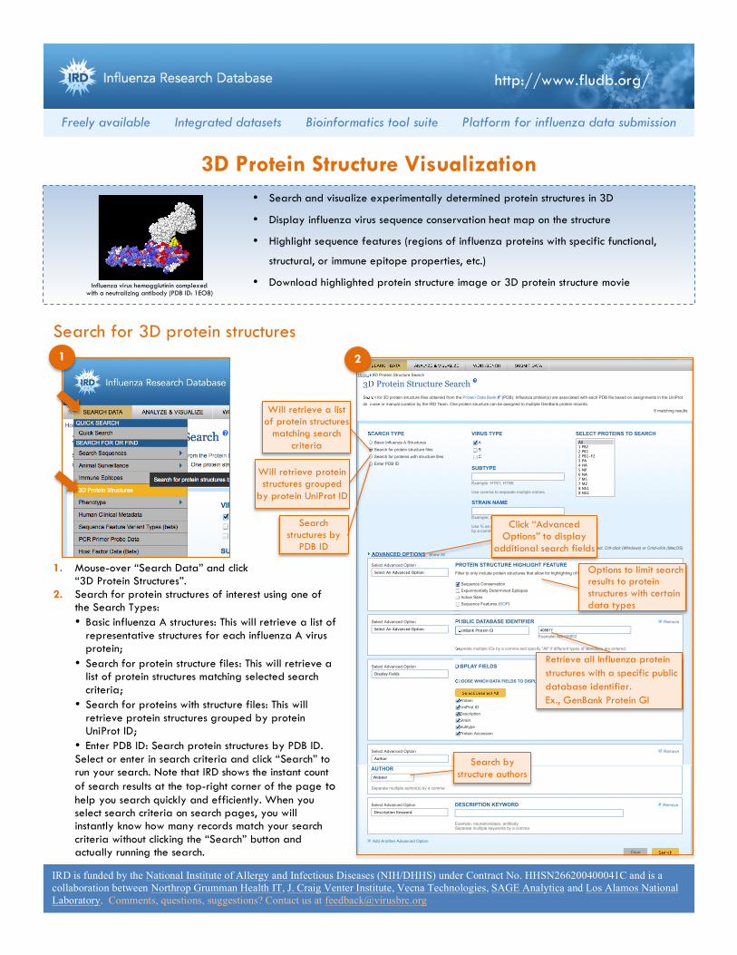
3. On the search result page, you can: • Click “View Structure” next to the structure
name to load the structure viewing window. • Click “Search Criteria” to revise your selected
search criteria and run another search. • Click “Display Settings” to select display
fields, change the number of records per page, or custom-sort records.
• Download structure records by checking the checkboxes next to the desired records and clicking the “Download” button located above the table.
• Save the search criteria to your personal Workbench account, which is a free service provided to IRD users. You will be able to rerun the search later.
Visualize and highlight 3D protein structures
Save a publication quality structure image
with highlighted features
• Select display fields • Custom-sort records
Click “View Structure” to display the
selected structure
Download selected records
Save your search criteria to your Workbench space. You will be able to rerun the search later.
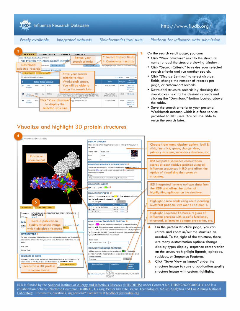
4. On the protein structure page, you can rotate and zoom in/out the structure as needed. To the right of the structure, there are many customization options: change display type; display sequence conservation on the structure; highlight ligands, epitopes, residues, or Sequence Features.
5. Click “Save View as Image” under the structure image to save a publication quality structure image with custom highlights.
Cite IRD Tutorials Glossary of Terms Report a Bug Request Web Training Contact Us Release Date: Jun 3, 2012
This project is funded by the National Institute of Allergy and Infectious Diseases (NIH / DHHS) under Contract No. HHSN266200400041C and is a collaboration between NorthropGrumman Health IT, University of Texas Southwestern Medical Center , Vecna Technologies, SAGE Analytica and Los Alamos National Laboratory.
functional Clear
Color Structure By:
Label:
HIGHLIGHT SEQUENCE FEATURESHighlight sequence features on the structures in . A disabled
checkbox means the mapping between swissprot and pdb position is notcurrently available.
Sequence Feature Feature Name SequenceFeaturePositions
HIGHLIGHT/LABEL FEATURESThese options color, highlight or label certain features of the structure.
Influenza A_H3_SF61
InfluenzaA_H3_sialic-acid-binding-site_98(19)
98,153,190,194,195,183,155,190,194
Revise your search criteria
IRD computed sequence conservation scores at each residue position using all influenza sequences in IRD and offers the option of visualizing the scores on structures.
Choose from many display options: ball & stick, line, stick, space, charge view, primary structure, secondary structure, etc.
IRD integrated immune epitope data from the IEDB and offers the option of highlighting epitopes on the structure.
Highlight amino acids using corresponding SwissProt position, with Met as position 1.
Highlight Sequence Features: regions of influenza proteins with specific functional, structural, or immune epitope properties, etc.
Rotate or zoom in/out
4
5 Reset View Save View As Image
Type JMol command here. Run
Show JMol Console
Generate Video
Description: INFLUENZA VIRUS HEMAGGLUTININ COMPLEXED WITH ANEUTRALIZING
PDB Link: 1EO8
JMOL COMMAND LINE:Advanced users can enter a JMol command directly using JMol Interactive Script .
SAVE/RESTORE STATE You can save the current state of the image (Display Options, Zoom, Highlight Epitopes,
etc.) for later use. Click the Show JMol Console button below, click State, and
copy/paste/save the contents of the upper panel into any text document. When you
reopen this PDB file in a later session, you can restore the image to the saved state.
Click the Show JMol Console button, paste the text into the lower panel, and click
Execute.
GENERATE 3D MOVIEGenerate a mp4/avi movie, starting with the rendering in above window and rotating
around Y axis by 360 deg. It takes about 30 seconds to generate the movie.
Display Type: Ball & Stick
Details
Show:
Sequence conservation computed using all sequences
- B Cell Epitopes Clear
Highlight Clear
DISPLAY OPTIONS:These options control the general appearance of the protein structure in
the viewer.
HIGHLIGHT SEQUENCE CONSERVATION Overlay the structure with a sequence conservation "heat map" (see SOP
for details). Blue represents conserved regions and red represents
non-conserved regions
HIGHLIGHT LIGANDS Highlight Ligands in
HIGHLIGHT EPITOPES Highlight epitopes on the structure in . First, select an epitope type
from the list. Then check epitopes to highlight.
Peptide sequence Range IEDB ID
EGFTWTGVTQNGGSNA 139-154 12167
KRGPGSG 156-162 33143
WTGVTQN 143-149 73132
HIGHLIGHT BY SWISS-PROT POSITION Highlight in in this structure corresponding to defined SwissProt
positions. With the dropdown, select a chain and view the positions present
in this file. Enter one or more comma-delimited positions (15,30) or a range
(15-30) then click Highlight. If no chain is selected, these positions will be
highlighted on all chains which include them.
Select Chain:
A - P03437 (25 - 343) 25
PDB Sequence/Structure Details
Zoom: 100%
Spin:
Home 3D Protein Structure Search Results Protein Structure Viewer (1EO8)
SEARCH DATA ANALYZE & VISUALIZE WORKBENCH SUBMIT DATA
About Us Community Announcements Links Resources Support Workbench Sign In
Generate a 3D protein structure movie