3D Echo Basics: Acquisition, Cropping and Display 14 (Monday)/6. 915am- 3D Ech… · 3D Echo...
14
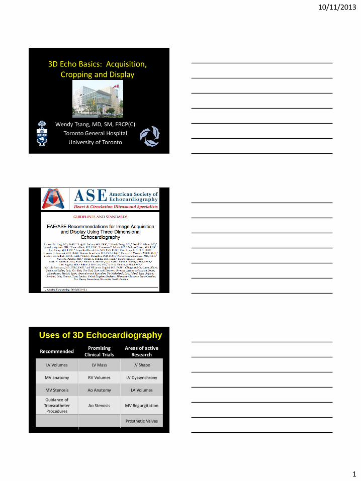
10/11/2013 1 3D Echo Basics: Acquisition, Cropping and Display Wendy Tsang, MD, SM, FRCP(C) Toronto General Hospital University of Toronto Uses of 3D Echocardiography Recommended Promising Clinical Trials Areas of active Research LV Volumes LV Mass LV Shape MV anatomy RV Volumes LV Dyssynchrony MV Stenosis Ao Anatomy LA Volumes Guidance of Transcatheter Procedures Ao Stenosis MV Regurgitation Prosthetic Valves
Transcript of 3D Echo Basics: Acquisition, Cropping and Display 14 (Monday)/6. 915am- 3D Ech… · 3D Echo...
10/11/2013
1
3D Echo Basics: Acquisition, Cropping and Display
Wendy Tsang, MD, SM, FRCP(C)
Toronto General Hospital
University of Toronto
Uses of 3D Echocardiography
Recommended Promising
Clinical Trials Areas of active
Research
LV Volumes LV Mass LV Shape
MV anatomy RV Volumes LV Dyssynchrony
MV Stenosis Ao Anatomy LA Volumes
Guidance of Transcatheter
Procedures Ao Stenosis MV Regurgitation
Prosthetic Valves
10/11/2013
2
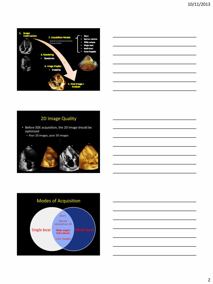
Spatial vs temporal resolution Gating artifacts
2D Image Quality
• Before 3DE acquisition, the 2D image should be optimized – Poor 2D images, poor 3D images
Modes of Acquisition
Single-beat Multi-beat
Color Doppler
Wide angle/ Full volume
Zoom
Narrow volume/Live 3D
10/11/2013
3
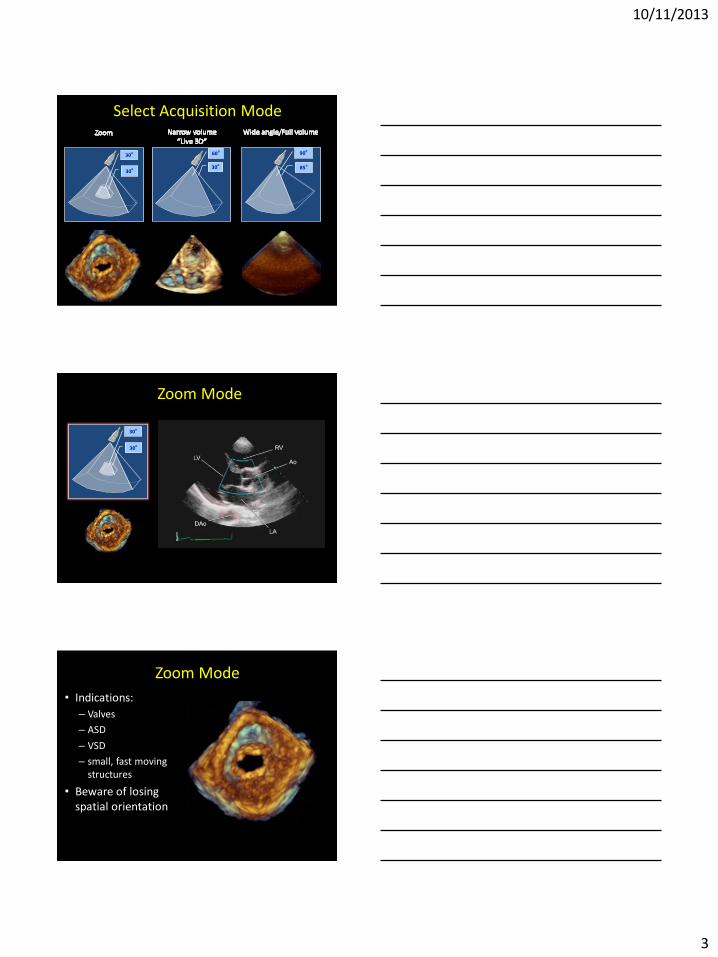
Select Acquisition Mode
30o
60o
85o
90o
30o
30o
Zoom Mode
30o
30o
Zoom Mode
• Indications:
– Valves
– ASD
– VSD
– small, fast moving structures
• Beware of losing spatial orientation
10/11/2013
4
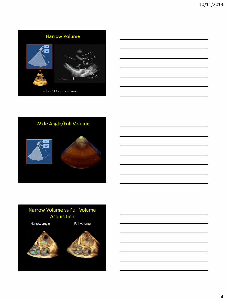
Narrow Volume
30o
60o
• Useful for procedures
Wide Angle/Full Volume
85o
90o
Narrow Volume vs Full Volume Acquisition
Narrow angle Full volume
10/11/2013
5
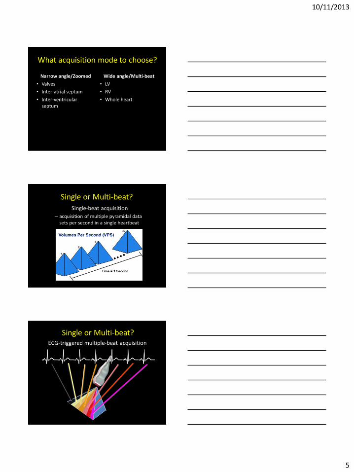
What acquisition mode to choose?
Narrow angle/Zoomed
• Valves
• Inter-atrial septum
• Inter-ventricular septum
Wide angle/Multi-beat
• LV
• RV
• Whole heart
Single or Multi-beat?
Single-beat acquisition
– acquisition of multiple pyramidal data sets per second in a single heartbeat
Single or Multi-beat? ECG-triggered multiple-beat acquisition
10/11/2013
6
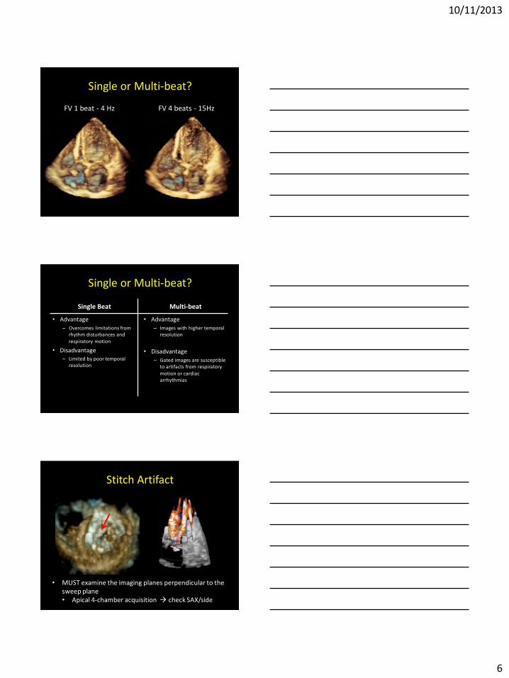
FV 1 beat - 4 Hz FV 4 beats - 15Hz
Single or Multi-beat?
Single or Multi-beat?
Single Beat
• Advantage
– Overcomes limitations from rhythm disturbances and
respiratory motion
• Disadvantage – Limited by poor temporal
resolution
Multi-beat
• Advantage
– Images with higher temporal resolution
• Disadvantage
– Gated images are susceptible to artifacts from respiratory
motion or cardiac arrhythmias
Stitch Artifact
• MUST examine the imaging planes perpendicular to the sweep plane • Apical 4-chamber acquisition check SAX/side
10/11/2013
7
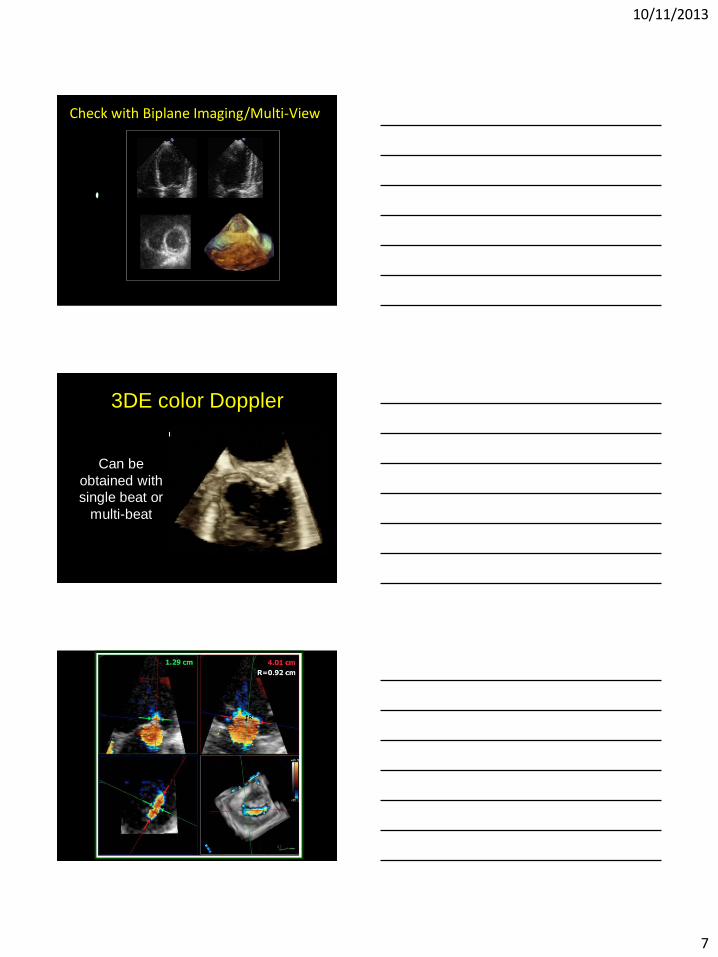
Check with Biplane Imaging/Multi-View
3DE color Doppler
Can be
obtained with
single beat or
multi-beat
10/11/2013
8
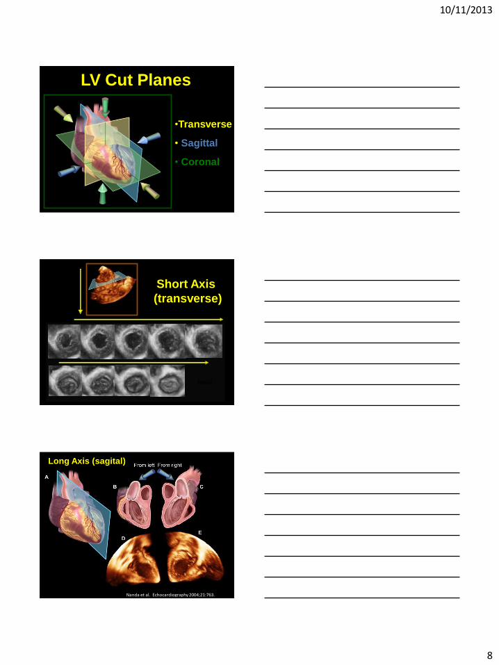
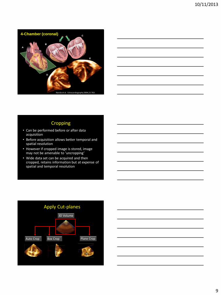
LV Cut Planes
•Transverse
• Sagittal
• Coronal
apical
basal
Short Axis
(transverse)
Long Axis (sagital)
Nanda et al. Echocardiography 2004;21:763.
10/11/2013
9
4-Chamber (coronal)
Nanda et al. Echocardiography 2004;21:763.
Cropping
• Can be performed before or after data acquisition
• Before acquisition allows better temporal and spatial resolution
• However if cropped image is stored, image may not be amenable to ‘uncropping’
• Wide data set can be acquired and then cropped, retains information but at expense of spatial and temporal resolution
Apply Cut-planes
3D Volume
Auto Crop Box Crop Plane Crop
10/11/2013
10
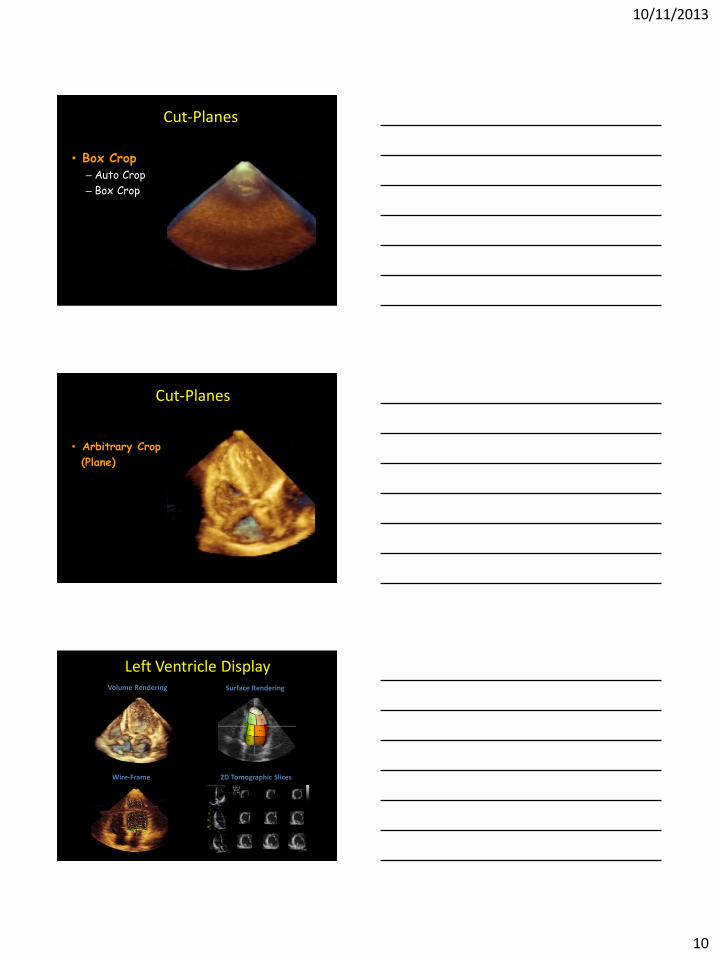
Cut-Planes
• Box Crop – Auto Crop
– Box Crop
Cut-Planes
• Arbitrary Crop
(Plane)
Left Ventricle Display Volume Rendering Surface Rendering
2D Tomographic Slices Wire-Frame
10/11/2013
11
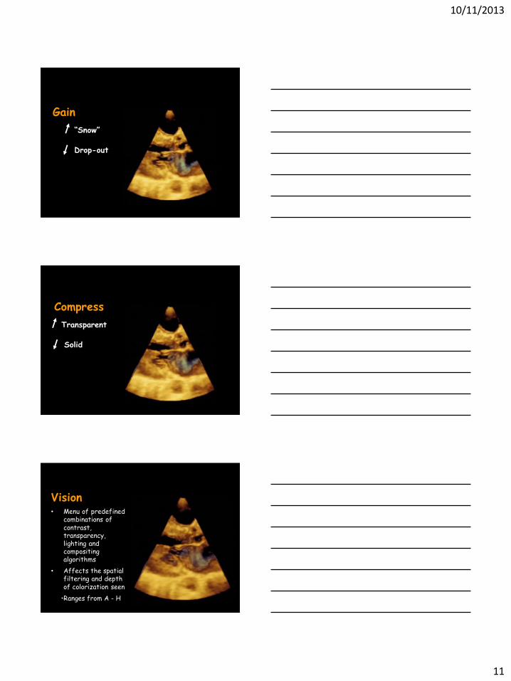
Gain
“Snow”
Drop-out
Compress
Transparent
Solid
Vision • Menu of predefined
combinations of contrast, transparency, lighting and compositing algorithms
• Affects the spatial filtering and depth of colorization seen
•Ranges from A - H
10/11/2013
12
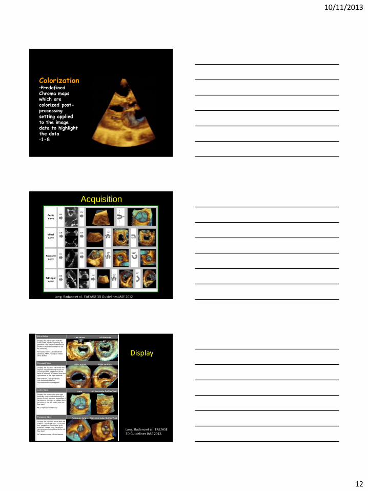
Colorization •Predefined Chroma maps which are colorized post-processing setting applied to the image data to highlight the data •1-8
Acquisition
Lang, Badano et al. EAE/ASE 3D Guidelines JASE 2012
Tricuspid Valve
Display the tricuspid valve with the septum placed inferiorly in the six o’clock position, regardless if the valve is oriented as viewed from the right atrium or the right ventricle.
Ant=anterior, Post=posterior, IAS=interatrial septum, IVS=interventricular septum
Mitral Valve
Display the mitral valve with the aortic valve placed superiorly, re-gardless if the valve is oriented as viewed from the left atrium or the left ventricle.
AV=aortic valve, Lat=lateral left ventricle, PMVL=posterior mitral valve leaflet
Pulmonic Valve
Display the pulmonic valve with the anterior cusp at the 12 o’clock posi-tion, regardless if the valve is ori-ented as viewed from the pulmo-nary artery or the right ventricle out-flow tract.
AC=anterior cusp, LA=left atrium
Aortic Valve
Display the aortic valve with right coronary cusp located inferiorly at the six o’clock position, regardless if the valve is oriented as viewed from the aorta or the left ventricular out-flow tract.
RCC=right coronary cusp RCC RCC
AV AV
PMVL PMVL
Lat Lat
AC
Left Atrium Left Ventricle
Right Atrium Right Ventricle
Aorta Left Ventricular Outflow Tract
Right Ventricular Outflow Tract Pulmonary Artery
Ant Post
IVS
IAS
Post Ant
AC AC
LA#
Display
Lang, Badano et al. EAE/ASE 3D Guidelines JASE 2012.
10/11/2013
13
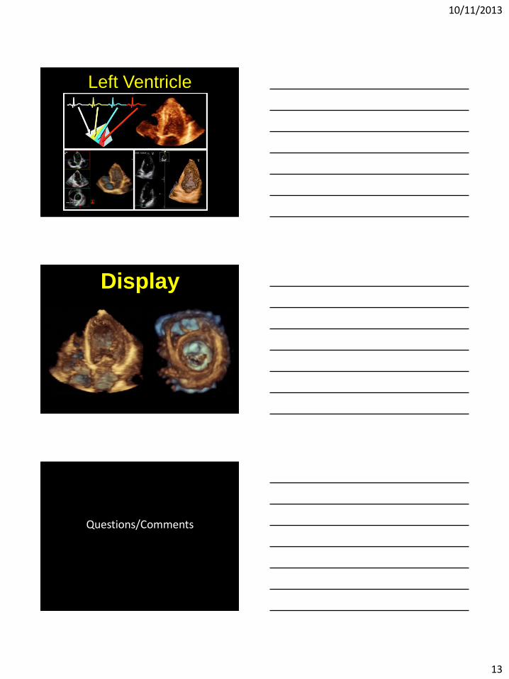
Left Ventricle
Display
Questions/Comments
10/11/2013
14
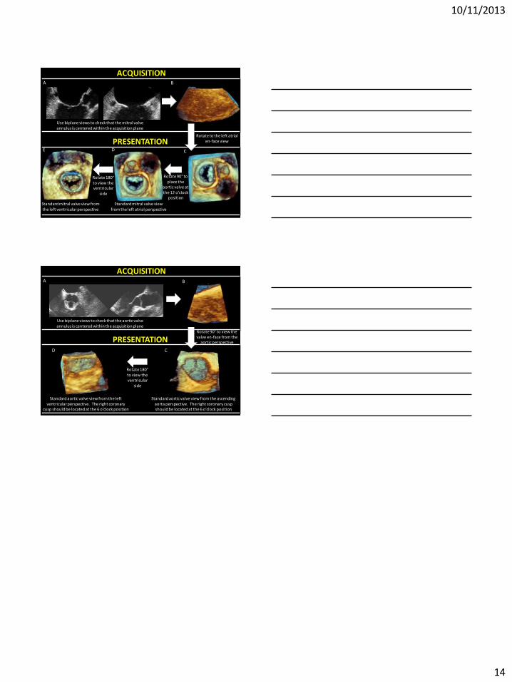
B
C
Rotate to the left atrial en-face view
Rotate 90° to place the
aortic valve at the 12 o’clock
position
Standard mitral valve view from the left atrial perspective
E D
Rotate 180° to view the ventricular
side
Standard mitral valve view from the left ventricular perspective
A
Use biplane views to check that the mitral valve annulus is centered within the acquisition plane
PRESENTATION
ACQUISITION
B
C
Rotate 90° to view the valve en-face from the
aortic perspective
Standard aortic valve view from the ascending aorta perspective. The right coronary cusp should be located at the 6 o’clock position
D
Rotate 180° to view the ventricular
side
Standard aortic valve view from the left ventricular perspective. The right coronary
cusp should be located at the 6 o’clock position
A
Use biplane views to check that the aortic valve annulus is centered within the acquisition plane
PRESENTATION
ACQUISITION