
Scaffolds for bone repair using computer aided design and ...

lable at ScienceDirect
Bioactive Materials xxx (2017) 1e37
Contents lists avai
Bioactive Materialsjournal homepage: http: / /www.keaipubl ishing.com/en/ journals /
bioact ive-mater ia ls /
3D bioactive composite scaffolds for bone tissue engineering
Gareth Turnbull a, b, Jon Clarke b, Fr�ed�eric Picard a, b, Philip Riches a, Luanluan Jia c,Fengxuan Han c, Bin Li c, Wenmiao Shu a, *
a Department of Biomedical Engineering, Wolfson Building, University of Strathclyde, 106 Rottenrow, Glasgow, G4 0NW, United Kingdomb Department of Orthopaedic Surgery, Golden Jubilee National Hospital, Agamemnon St, Clydebank, G81 4DY, United Kingdomc Orthopaedic Institute, Department of Orthopaedic Surgery, The First Affiliated Hospital, Soochow University, Suzhou, Jiangsu, PR China
a r t i c l e i n f o
Article history:Received 31 August 2017Received in revised form31 October 2017Accepted 31 October 2017Available online xxx
Keywords:Bioactive composites3D scaffold3D printingBioprintingBoneTissue engineering
* Corresponding author.E-mail address: [email protected] (W. Shu).Peer review under responsibility of KeAi Commu
https://doi.org/10.1016/j.bioactmat.2017.10.0012452-199X/© 2017 The Authors. Production and hostlicense (http://creativecommons.org/licenses/by-nc-n
Please cite this article in press as: G. Turnbuhttps://doi.org/10.1016/j.bioactmat.2017.10.0
a b s t r a c t
Bone is the second most commonly transplanted tissue worldwide, with over four million operationsusing bone grafts or bone substitute materials annually to treat bone defects. However, significantlimitations affect current treatment options and clinical demand for bone grafts continues to rise due toconditions such as trauma, cancer, infection and arthritis. Developing bioactive three-dimensional (3D)scaffolds to support bone regeneration has therefore become a key area of focus within bone tissueengineering (BTE). A variety of materials and manufacturing methods including 3D printing have beenused to create novel alternatives to traditional bone grafts. However, individual groups of materialsincluding polymers, ceramics and hydrogels have been unable to fully replicate the properties of bonewhen used alone. Favourable material properties can be combined and bioactivity improved whengroups of materials are used together in composite 3D scaffolds. This review will therefore consider theideal properties of bioactive composite 3D scaffolds and examine recent use of polymers, hydrogels,metals, ceramics and bio-glasses in BTE. Scaffold fabrication methodology, mechanical performance,biocompatibility, bioactivity, and potential clinical translations will be discussed.© 2017 The Authors. Production and hosting by Elsevier B.V. on behalf of KeAi Communications Co., Ltd.This is an open access article under the CC BY-NC-ND license (http://creativecommons.org/licenses/by-
nc-nd/4.0/).
1. Introduction
1.1. Clinical demand for bone grafts, bone substitutes & implants
The capacity for bone to self-regenerate has prompted study andintrigue since the times of Hippocrates and Galen [1]. Despite this,congenital and acquired pathologies including trauma, infection,neoplasm and failed arthroplasty remain capable of leaving patientswith bone defects beyond a critical-size which the body cannotheal. Such patients often require invasive surgical intervention toaid healing. This can involve clinical use of bone grafts, bone sub-stitute materials, growth factors, free fibula vascularized grafts andinsertion of metalwork to aid stability and bone regeneration [2,3].As a result, bone is the second most commonly transplanted tissueworldwide, with at least fourmillion operationsmaking use of bonegrafts and bone substitute materials annually [4e6].
nications Co., Ltd.
ing by Elsevier B.V. on behalf of Ked/4.0/).
ll, et al., 3D bioactive compo01
However, significant limitations affect current treatmentoptions. Bone grafts taken from one part of a patient for use in theirown body (autografts) are considered the gold standard but arerestricted by the size of graft that can be harvested and carry afurther risk of donor site morbidity including infection and ongoingpain following surgery [7,8]. Allograft tissue harvested fromcadaveric and living sources (such as femoral heads removedduring hip replacements) is in relatively greater supply comparedto autograft tissue. However, allografts carry potential risk ofdisease transmission and immune response, whilst lacking acellular component to aid tissue regeneration. Metalwork and bonesubstitutes can also be inserted to aid bone regeneration; limita-tions associated with their use include bone thinning due to stressshielding, wear and failure over time and risk of revision surgery[6]. Non-invasive therapies including ultrasound treatment havealso show promise in aiding bone healing, although use is oftenrestricted to stable, well aligned and well reduced fracturenon-unions in adult patients [9].
To overcome the limitations of current treatment options, sig-nificant research in the field of bone tissue engineering (BTE) hasbeen directed towards creating novel alternatives to traditional
Ai Communications Co., Ltd. This is an open access article under the CC BY-NC-ND
site scaffolds for bone tissue engineering, Bioactive Materials (2017),

G. Turnbull et al. / Bioactive Materials xxx (2017) 1e372
bone grafts. Porous 3D scaffolds fabricated through a variety ofmethods and including a range of biomaterials have been utilised toaid and direct bone regeneration [10e12]. However, the perfectscaffold material has yet to be encountered and clinical translationof 3D scaffolds has been limited as a result [13].
Bone is a heterogenous composite material with constituentsincluding hydroxyapatite mineral (Ca10(PO4)6(OH)2) [14], a mixedorganic component (type I collagen, lipids and non-collagenousproteins) and water [15,16]. During scaffold manufacture it wouldtherefore seem logical to include a combination of materials tocreate a composite scaffold, potentially allowing greater scaffoldbioactivity and structural biomimicry to be achieved.
Scaffold bioactivity is also increased by incorporating materialsthat possess the ability to interact with or bind to living tissues.Increased scaffold bioactivity can in turn lead to improved bone cellingrowth (osteoconduction), stable anchoring of scaffolds to hostbone tissue (osseointegration), stimulation of immature host cellsto develop into osteogenic cells (osteoinduction) and increasedvascularisation [17e20].
This review will therefore examine some of the significantbioactive composites that have been utilised recently in BTE afterreference to the properties of an ideal scaffold and available scaffoldmanufacturing methods. 3D scaffolds that have successfullybridged bone defects whilst actively inducing bone regenerationwill be highlighted.
2. Properties of an ideal scaffold
In general terms, the ideal 3D scaffold is composed of abiocompatible, biodegradable material with similar mechanicalproperties to the tissue which it is to be implanted in. Scaffolds bydesign are not intended to be permanent implants and will ideallyfacilitate host cells to deposit extracellular matrix (ECM) andreplace the scaffold structure over time. The 3D architecture of thescaffold should be highly porous with an interconnected structureto allow cell and nutrient migration. The scaffold surface shouldalso be optimised to facilitate cell attachment, proliferation anddifferentiation (see Table 1). Form a surgical point of view, it is alsodesirable for the scaffold material to be easily manipulated intodifferent shapes and sizes to allow in-situ treatment of individualpatient bone defects [13,21e23].
Scaffolds and their breakdown products must above all bebiocompatible. This requires scaffold materials to be nontoxic tocells, easily eliminated from the body and to elicit negligible
Table 1Summary of desirable scaffold properties.
ScaffoldCharacteristics
Desirable Features
Biocompatibility � Non-toxic breakdown products� Non-inflammatory scaffold components, avoiding immune re
Biodegradability � Controlled scaffold degradation which can complement tissu� Degradable by host enzymatic or biological processes� Allows invading host cells to produce their own extracellular
Bioactivity � Scaffold materials that can interact with and bind to host tiss� Osteoconductive and osteoinductive properties� Inclusion of biological cues and growth factors to stimulate c
Scaffold Architecture � Interconnected pores allowing diffusion and cell migration� Microporosity to present a large surface area for cell-scaffold� Macroporosity to allow cell migration and invasion of vascula� Pore size tailored to target tissue and cells� Sufficient porosity to facilitate cell ingrowth without weaken� Inbuilt vascular channels to enhance angiogenesis in vivo
MechanicalProperties
� Compressive, elastic and fatigue strength comparable to hostin vivo
� Scaffold material that can be readily manipulated in the clini
Please cite this article in press as: G. Turnbull, et al., 3D bioactive compohttps://doi.org/10.1016/j.bioactmat.2017.10.001
immune response through their presence [11,24,25]. Controlledbiodegradability is also an essential characteristic for a scaffold toachieve; if a scaffold degrades too quickly, mechanical failure couldoccur. This is particularly relevant in BTE, as an implanted scaffold islikely to undergo load-bearing and could fracture if unable toprovide mechanical support whilst new bone is forming. Similarly,if a scaffold does not degrade sufficiently quickly an inflammatoryresponse could be triggered towards the foreign material of thescaffold, impairing tissue regeneration [26].
Growth factors also have a significant role to play in successfulbone tissue engineering scaffolds. The processes of new bone for-mation and extracellular matrix deposition are regulated by a rangeof growth factors and biomolecules. Bone morphogenic proteins(BMP) play a critical role in bone and cartilage development, andhave the ability to trigger proliferation and differentiation ofosteoprogenitor cells [27]. There are several examples of scaffoldsthat have successfully incorporated BMPs resulting in improvedbone formation. However, risk of bone formation within softtissues, or heterotopic ossification, is also associated with use ofBMPs [28e32]. Vascular endothelial growth factor (VEGF) has alsofrequently been included in scaffolds, with desirable propertiesincluding the ability to enhance blood vessel formation and boneformation in vivo [33e35]. Transforming growth factor b (TGF b),platelet-derived growth factor (PDGF), insulin-like growth-factor 1(IGF-1), and fibroblast growth factors (FGFs) provide further ex-amples of growth factors that have been utilised in bone andcartilage tissue engineering [36,37].
The microarchitecture of scaffolds is also centrally important inencouraging cell viability and fostering tissue ingrowth. An inter-connected pore structure, in the absence of an engineered bloodsupply, allows inwards diffusion of oxygen and nutrients andoutwards diffusion of waste products from the scaffold. Porosityalso supports cell migration into the scaffold and improves avail-able surface area for cell-scaffold binding and interaction withsurrounding tissues [38e40] (Fig. 1A and B). Individual pore sizewithin the scaffold is also an important consideration. It haspreviously been shown that scaffold pore density and size signifi-cantly impact upon cellular growth and attachment [41,42]. As poresize decreases, the surface area of the scaffold increases. This in-creases the availability of scaffold ligands for cells to bind to andinteract with. However, if pore sizes become too small, cells maystruggle to migrate into the scaffold structure. Scaffolds musttherefore be precisely engineered with parameters favourable tothe cells and tissue that they will be exposed to. For example, it has
jectione ingrowth whilst maintaining sufficient support
matrixue
ell ingrowth, attachment and differentiation
interactionsture
ing mechanical properties
tissue allowing cell mechanoregulation to occur and structural integrity to remain
cal environment to treat individual patient bone defects
site scaffolds for bone tissue engineering, Bioactive Materials (2017),
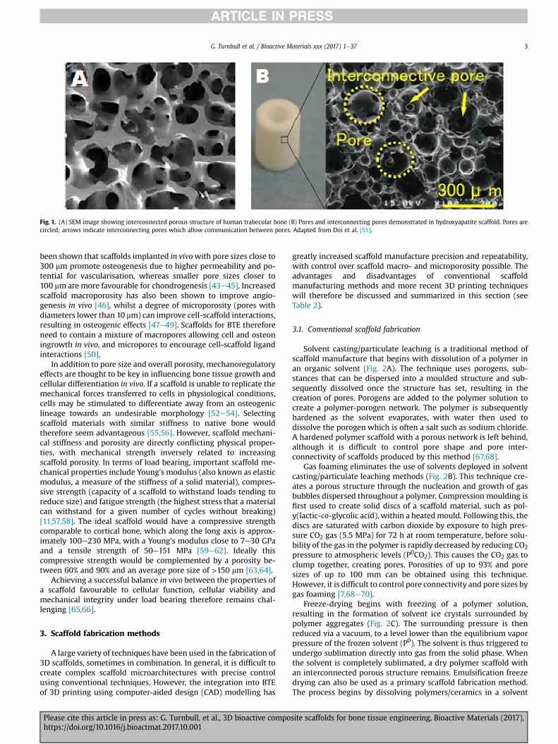
Fig. 1. (A) SEM image showing interconnected porous structure of human trabecular bone (B) Pores and interconnecting pores demonstrated in hydroxyapatite scaffold. Pores arecircled; arrows indicate interconnecting pores which allow communication between pores. Adapted from Doi et al. [51].
G. Turnbull et al. / Bioactive Materials xxx (2017) 1e37 3
been shown that scaffolds implanted in vivowith pore sizes close to300 mm promote osteogenesis due to higher permeability and po-tential for vascularisation, whereas smaller pore sizes closer to100 mm are more favourable for chondrogenesis [43e45]. Increasedscaffold macroporosity has also been shown to improve angio-genesis in vivo [46], whilst a degree of microporosity (pores withdiameters lower than 10 mm) can improve cell-scaffold interactions,resulting in osteogenic effects [47e49]. Scaffolds for BTE thereforeneed to contain a mixture of macropores allowing cell and osteoningrowth in vivo, and micropores to encourage cell-scaffold ligandinteractions [50].
In addition to pore size and overall porosity, mechanoregulatoryeffects are thought to be key in influencing bone tissue growth andcellular differentiation in vivo. If a scaffold is unable to replicate themechanical forces transferred to cells in physiological conditions,cells may be stimulated to differentiate away from an osteogeniclineage towards an undesirable morphology [52e54]. Selectingscaffold materials with similar stiffness to native bone wouldtherefore seem advantageous [55,56]. However, scaffold mechani-cal stiffness and porosity are directly conflicting physical proper-ties, with mechanical strength inversely related to increasingscaffold porosity. In terms of load bearing, important scaffold me-chanical properties include Young's modulus (also known as elasticmodulus, a measure of the stiffness of a solid material), compres-sive strength (capacity of a scaffold to withstand loads tending toreduce size) and fatigue strength (the highest stress that a materialcan withstand for a given number of cycles without breaking)[11,57,58]. The ideal scaffold would have a compressive strengthcomparable to cortical bone, which along the long axis is approx-imately 100e230 MPa, with a Young's modulus close to 7e30 GPaand a tensile strength of 50e151 MPa [59e62]. Ideally thiscompressive strength would be complemented by a porosity be-tween 60% and 90% and an average pore size of >150 mm [63,64].
Achieving a successful balance in vivo between the properties ofa scaffold favourable to cellular function, cellular viability andmechanical integrity under load bearing therefore remains chal-lenging [65,66].
3. Scaffold fabrication methods
A large variety of techniques have been used in the fabrication of3D scaffolds, sometimes in combination. In general, it is difficult tocreate complex scaffold microarchitectures with precise controlusing conventional techniques. However, the integration into BTEof 3D printing using computer-aided design (CAD) modelling has
Please cite this article in press as: G. Turnbull, et al., 3D bioactive compohttps://doi.org/10.1016/j.bioactmat.2017.10.001
greatly increased scaffold manufacture precision and repeatability,with control over scaffold macro- and microporosity possible. Theadvantages and disadvantages of conventional scaffoldmanufacturing methods and more recent 3D printing techniqueswill therefore be discussed and summarized in this section (seeTable 2).
3.1. Conventional scaffold fabrication
Solvent casting/particulate leaching is a traditional method ofscaffold manufacture that begins with dissolution of a polymer inan organic solvent (Fig. 2A). The technique uses porogens, sub-stances that can be dispersed into a moulded structure and sub-sequently dissolved once the structure has set, resulting in thecreation of pores. Porogens are added to the polymer solution tocreate a polymer-porogen network. The polymer is subsequentlyhardened as the solvent evaporates, with water then used todissolve the porogen which is often a salt such as sodium chloride.A hardened polymer scaffold with a porous network is left behind,although it is difficult to control pore shape and pore inter-connectivity of scaffolds produced by this method [67,68].
Gas foaming eliminates the use of solvents deployed in solventcasting/particulate leaching methods (Fig. 2B). This technique cre-ates a porous structure through the nucleation and growth of gasbubbles dispersed throughout a polymer. Compression moulding isfirst used to create solid discs of a scaffold material, such as pol-y(lactic-co-glycolic acid), within a heatedmould. Following this, thediscs are saturated with carbon dioxide by exposure to high pres-sure CO2 gas (5.5 MPa) for 72 h at room temperature, before solu-bility of the gas in the polymer is rapidly decreased by reducing CO2pressure to atmospheric levels (P0CO2). This causes the CO2 gas toclump together, creating pores. Porosities of up to 93% and poresizes of up to 100 mm can be obtained using this technique.However, it is difficult to control pore connectivity and pore sizes bygas foaming [7,68e70].
Freeze-drying begins with freezing of a polymer solution,resulting in the formation of solvent ice crystals surrounded bypolymer aggregates (Fig. 2C). The surrounding pressure is thenreduced via a vacuum, to a level lower than the equilibrium vaporpressure of the frozen solvent (P0). The solvent is thus triggered toundergo sublimation directly into gas from the solid phase. Whenthe solvent is completely sublimated, a dry polymer scaffold withan interconnected porous structure remains. Emulsification freezedrying can also be used as a primary scaffold fabrication method.The process begins by dissolving polymers/ceramics in a solvent
site scaffolds for bone tissue engineering, Bioactive Materials (2017),

Table 2Comparison of scaffold fabrication methods.
ManufacturingMethod
Benefits Potential Limitations
Solvent casting/particulateleaching
� Relatively simple technique that allows creation of scaffolds with regular porosity,controlled composition and pore size.
� Use of organic solvents precludes cells andbiomolecules being included directly inscaffolds
� Can be difficult to control pore shape andinterconnectivity
� Limited thickness of structures andmechanical properties achievable
Gas Foaming � Eliminates use of chemical solvents � High pressures involved prohibits inclusionof cells and bioactive molecules directlyinto scaffolds
� Temperature labile materials may bedenatured during compression mouldingstep
� Difficult to control pore sizes and ensureinterconnectivity
EmulsificationFreeze-Drying
� Does not require use of solid porogen � Requires use of organic solvents� Small pore size and� Porosity often irregular� Long processing time
Phase Separation � Eliminates leaching step of porogen� Can be combined with other techniques easily
� Small pore sizes limit use� Use of organic solvents inhibits use of
bioactive molecules or cells during scaffoldfabrication
Electrospinning � Creates scaffold with large surface area for cell attachment� Simple and inexpensive technique
� Organic solvents may be required, whichcan be harmful to cells
� Limited mechanical properties� Difficult to incorporate precise
microarchitecture into constructs3D Printing� SLA� SLS� FDM� Inkjet� Laser-assisted� Microvalve� Microextrusion
� Complex 3D shapes with high resolution, controlled pore size & morphology and controlledinternal structures can be fabricated. Improved capacity to incorporate vascular structuresinto constructs
� Depending on technique used, cells may be included in high concentration directly inscaffold materials
� Some techniques are limited by printablematerials
� Set up costs can be expensive for machinery
G. Turnbull et al. / Bioactive Materials xxx (2017) 1e374
and then mixing with water, to obtain an emulsion. The mixture ispoured into amould and frozen before the two phases can separate.The frozen emulsion is then freeze-dried to remove the solvent anddispersed water, creating pores in a solidified scaffold [72].
Phase separation relies on changes in thermal energy to inducethe de-mixing of a homogenous polymer/solvent solution. When apolymer such as PLLA is dissolved in a solvent, it can becomethermodynamically unstable at a low temperature and spontane-ously separate into a polymer-rich phase and a solvent-rich phase.Phase separation scaffold manufacture takes advantage of thisphenomenon and begins with dissolution of a polymer in a high-boiling, low molecular weight solvent at an elevated temperature,typically around the melting point of the polymer, allowing for-mation of a homogenous melt-blend. The solution is then cast intoa desired scaffold shape, and cooled in a controlled manner toinduce phase separation and precipitation of the solution into apolymer-rich phase and a solvent-rich phase, creating a nano-fibrous matrix (Fig. 2D). The solvent which is in the solvent-richphase will then be removed through extraction, evaporation, orsublimation. This creates a porous scaffold, as removal of the sol-vent leaves pores behind in the polymer matrix.
Electrospinning is another popular scaffold fabrication tech-nique with the ability to create nanofibrous interconnected porousscaffolds (Fig. 2E). This method uses an externally applied electricfield to draw charged threads of polymer solutions or polymermelts as thin jets from a capillary tube towards a collector plate.Fibres in the micro- and nanometre range can be created anddeposited sequentially to create a scaffold, with potential to includecomposite materials and biomolecules [73e76].
Please cite this article in press as: G. Turnbull, et al., 3D bioactive compohttps://doi.org/10.1016/j.bioactmat.2017.10.001
3.2. 3D printing techniques
The traditional methods of scaffold fabrication that have beendiscussed in brief so far generally offer limited control over poresize, geometry and interconnectivity. Overtime there has been animprovement in the ability to spatially control scaffold micro-architecture and spatial content as technologies such as 3D printinghave emerged. In general, 3D printing fabricates objects via layer-by-layer processing of powder, liquid or solid material substrates.Starting from the bottom and building up, each newly formed layeris triggered to adhere to the previous layer, resulting in the creationof construct of gradually increasing size. The structure of a 3Dprinted object is dictated by a computer-aided design (CAD) modelloaded onto a 3D printer. CADmodels describe 3D objects in a seriesof cross-sectional layers, allowing 3D printers to physically repro-duce models through an additive process.
Patient specific CAD models can be created by convertingcomputed tomography (CT) or magnetic resonance imaging (MRI)of clinical defects into (CAD) models. Further software is then usedto slice CAD models into G-code, which encodes 3D CAD models ina format that can control 3D printers. Parameters such as printspeed, layer height, print head temperature and pressure can all bemodified and optimised through G-code.
Several 3D printing methods have been adapted into BTE, withstereolithography (SLA) representing one of the earliest 3D printingtechniques to have been developed (Fig. 3A).
It relies on the directed use of a laser to polymerize liquid UV-curable photopolymer resin layer-by-layer, resulting in a solidi-fied 3D model. The UV laser can solidify the model's cross-section,
site scaffolds for bone tissue engineering, Bioactive Materials (2017),

Fig. 2. Common scaffold fabrication techniques. (A) Solvent casting-particle leaching process (B) Gas foaming (C) Freeze-drying (D) Phase separation (E) Electrospinning. Adaptedfrom Puppi et al. [71].
G. Turnbull et al. / Bioactive Materials xxx (2017) 1e37 5
Please cite this article in press as: G. Turnbull, et al., 3D bioactive composite scaffolds for bone tissue engineering, Bioactive Materials (2017),https://doi.org/10.1016/j.bioactmat.2017.10.001

Fig. 3. Common 3D Printing Techniques. (A) Stereolithography (B) Fused deposition modelling (C) Selective laser sintering. Adapted from Jaster L [77].
G. Turnbull et al. / Bioactive Materials xxx (2017) 1e376
leaving remaining areas in liquid form. After each cross-section, theprint platform moves down, covering the solid polymer withanother layer of resin for curing. Excess resin that has not beencured is then removed from the 3D structure, allowing rapidfabrication of a structure that can be cured further in an oven.Whilst SLA can quickly produce scaffolds with controlled archi-tecture and micrometre-level resolution, there is a limited numberof materials applicable to this costly technique [78].
Fused deposition modelling (FDM) uses a temperaturecontrolled printhead to deposit thermoplastic material onto aplatform in a layer by layer manner to build up a 3D construct(Fig. 3B). A thermoplastic filament is driven into a heated printhead,causing the filament to melt, allowing thin layers of a semi-moltenpolymer such as polycaprolactone to be precisely depositedsequentially. The molten filament cools in the air of the printenvironment, allowing filaments to fuse together rapidly to create ascaffold. FDM has been successfully adapted into BTE as method ofproducing synthetic scaffolds, although the elevated temperaturesinvolved limit the inclusion of biomolecules and hydrogels [79,80].
Selective laser sintering (SLS) involves the use of a computercontrolled laser beam to fuse layer-upon-layer of a powder, sin-tering the powder material together to build a solid 3D structure(Fig. 3C). Some success with this technique has been demonstrated,through the production of bioactive, composite scaffolds withsimilar mechanical properties to trabecular bone [81,82]. However,the elevated temperatures involved in the process limit the inclu-sion of cells and biomaterials directly into SLS scaffolds.
Please cite this article in press as: G. Turnbull, et al., 3D bioactive compohttps://doi.org/10.1016/j.bioactmat.2017.10.001
3.3. 3D bioprinting
As an emerging technology, 3D bioprinting offers a potentialsolution to help ease the burden of arthritis and other cause of bonedefects within orthopaedics. Bioprinting can be used to depositliving cells, extracellular matrices and other biomaterials in user-defined patterns to build complex tissue constructs “from thebottom up.” The potential to create inherent vascular structures isalso improved by bioprinting, as internal channels containingvascular cells can be printed into constructs, fostering the ingrowthof blood vessels in vivo. By contrast, the conventional tissue engi-neering method of seeding cells onto a pre-fabricated scaffold doesnot allow for precise 3D placement of cells or biological content,limiting capacity to create complex hierarchical tissue constructs[83,84].
The process of bioprinting typically begins with the selection ofcells and biomaterials for inclusion in bioprinted constructs (Fig. 4).Cells for printing can be sourced from tissue biopsies, blood sam-ples and from other sources, and expanded in number throughculture to maximise cell density on bioprinting. The additional stepof 3D cell culture may also be performed to creating aggregates ofcells for printing. Cell aggregates or spheroids have superiorintercellular communication and extracellular matrix developmentwhen compared to cells grown in 2D culture, potentially acceler-ating the growth of printed constructs towards functional tissueafter bioprinting [85]. Mesenchymal stem cell spheroids alsoexhibit enhanced in vitro and in vivo osteoregenerative potentialcompared to MSCs cultured in monolayer [86,87].
site scaffolds for bone tissue engineering, Bioactive Materials (2017),

Fig. 4. Summary of bioprinting process.
G. Turnbull et al. / Bioactive Materials xxx (2017) 1e37 7
Following culture, cells and selected biomaterials such as hy-droxyapatite are encapsulated in a delivery medium, or bioink.Print cartridges containing bioink are then loaded into a 3D bio-printer, which dispenses the bioink in a pre-determined 3D ge-ometry according to a CAD model. Bioprinters often have multipleprint nozzles, allowing combinations of cells and biomaterials to beincludedwithin a printed construct. A high degree of spatial controlcan therefore be achieved over construct architecture and content[88,89]. Following printing the construct can be directly implantedinto a patient, or alternatively matured first in vitro. Biologicallyactive culture environments known as bioreactors are available tohelp direct and support cell growth towards specific tissue types.
3.4. 3D bioprinting techniques
Commonly used bioprinting techniques include inkjet, laser-assisted, microvalve and extrusion bioprinting.
Inkjet bioprinting (or drop-on-demand bioprinting) uses ther-mal or acoustic forces to eject droplets from a pint head nozzle(Fig. 5A). Thermal inkjet printers use heat to generate a pressurepulse within a print head for a brief period, causing ejection of adroplet of bioink. Other systems rely on piezoelectric crystals,which becomemechanically stressed by the application of a voltageand as a result change shape. This generates an acoustic wavewhich in turn creates sufficient pressure to eject droplets from anozzle. As a technology adapted form desktop inkjet printers,benefits include low cost, wide availability and high print speed.However, limitations include frequent nozzle clogging, risk of
Please cite this article in press as: G. Turnbull, et al., 3D bioactive compohttps://doi.org/10.1016/j.bioactmat.2017.10.001
exposing cells and materials to thermal and mechanical stress andnonuniform droplet size. The liquid droplet deposited is also of lowviscosity, relying on further gelation or crosslinking to create a solidstructure [90e92].
Laser-assisted bioprinting (LAB) systems avoid the use of anozzle; instead they rely on a pulsed laser beam to generate a high-pressure bubble, which in turn propels cell-containing materialstoward a collector substrate from an initial print material “ribbon”(Fig. 5B). Recently Keriquel et al. used LAB to print mesenchymalstromal cells, associated with collagen and nano-hydroxyapatite,directly in situ onto a mouse cranial defect to aid bone regenera-tion [93]. Some limitations of LAB include potential heat-induceddamage to cells, difficulty of creating 3D structures, high systemcosts and the time-consuming nature of creating ribbons with highcell and biomaterial concentrations [94].
Microvalve bioprinting is a droplet-based system where fluidsunder a constant pneumatic pressure are dispensed from cartridgetips by opening and closing a small valve (Fig. 5C). The valve inquestion can be controlled mechanically, electrically or magneti-cally. Microvalve systems can print cells including MSCs with highviability and functionality, with deposition of other biomaterialsalso possible such as collagen and bone morphogenic protein[95e97].
Extrusion bioprinters deposit continuous filaments of materialsrather than individual droplets (Fig. 5D). Pneumatic or mechanicalpressure is applied to a syringe to cause bioink extrusion through anozzle. A significant advantage of this approach is the ability todeposit very high cell densities, with some studies manging to
site scaffolds for bone tissue engineering, Bioactive Materials (2017),

Fig. 5. Common bioprinting techniques: (A) Inkjet, (B) Laser-assisted, (C) Microvalve, and (D) Extrusion bioprinting.
G. Turnbull et al. / Bioactive Materials xxx (2017) 1e378
purely print cells, for example as filaments of cartilage [98]. A broadrange of bioinks have been successfully extrusion bioprintined,including tissue spheroids, tissue strands, cell pellets, decellular-ized matrix components and cell-laden hydrogels. Potential limi-tations and challenges include achieving high print resolution,shear stress effect on cells within print nozzles and development ofprintable bioinks [99].
4. Materials used within bone tissue engineering
Materials that have been utilised within bone repair andregeneration include metals, ceramics, polymers, hydrogels andrelated composites. Groups of materials will be reviewed in thefollowing corresponding sections and summarized (see Table 3).
4.1. Metals
Metal alloys such as cobalt-chromium, zirconium, titanium andstainless steel have excellent biocompatibility and strength[21,100]. As such, they are commonly used in joint replacement andfracture fixation implants to offer support for healing bone [101].
Please cite this article in press as: G. Turnbull, et al., 3D bioactive compohttps://doi.org/10.1016/j.bioactmat.2017.10.001
However, a lack of biodegradability makes them less suited to BTEwhere the aim is for native tissue to resorb and replace implantedconstructs. Additional surgery is often required to remove metallicimplants, particularly when they are used in paediatric patientswho have not reached skeletal maturity [102]. The superior elasticmodulus of metals relative to bone can also predispose to stressshielding occurring. In this phenomena, mechanical bypass of loadsoccurs in the bone surrounding implants, leading to bone resorp-tion and increased fracture risk [103]. Despite these limitations,some success has been achieved through creation of compositemetal scaffolds. Strontium (Sr) was combined via freeze-dryingwith hydroxyapatite (HA) and chitosan (CS) by Lei et al. to createcomposite nanohybrid scaffolds. The presence of SrHA nanocrystalsin the scaffolds was found to significantly enhance cell proliferationand osteogenic differentiation of human bone marrow mesen-chymal stem cells (hBMSCs) [104]. Hierarchical structure wasdeveloped by Wu et al. on a microporous nickel-titanium com-posite (NiTi) scaffold by treating the surfacewith sodium hydroxidein a hydrothermal reaction. This led to the creation of a nano-structured, microporous exposed surface on an already micropo-rous NiTi scaffold. Improved surface hydrophilicity, deposition of
site scaffolds for bone tissue engineering, Bioactive Materials (2017),

Table 3Comparison of scaffold materials.
ManufacturingMaterial
Benefits Potential Limitations
Hydrogels � High water content/growth media inclusion allows for cell encapsulationand growth
� Mechanical properties can be modified through crosslinking� Controlled drug/growth factor release possible� Ease of patterning via 3D printing to mimic tissue microarchitectures
� Mechanical properties limit use in load bearing constructs� Optimising printing conditions for individual hydrogels can be time
consuming� Physical manipulation of constructs can be difficult� Loading evenly with cells can be challenging
Polymers � Natural polymers can be derived from extracellular matrix, ensuringhigh biocompatibility and low toxicity
� Biodegradable� Often contain biofunctional molecules on their surface� Synthetic polymers offer improved control over physical properties
� Natural and synthetic polymers generally lack mechanical propertiesfor load bearing
� Pathological impurities such as endotoxin may be present in naturalpolymers
� Synthetic polymers are often hydrophobic and lack cell recognitionsites
Ceramics � Osteoconductive and osteoinductive properties allow strong integrationwith host tissue
� Similar composition to host bone mineral content� Can be delivered as granules, paste or in an injectable format
� Hard and brittle when used alone� May display inappropriate degradation/resorption rates, with decline
in mechanical properties as a result
Bioactive glasses � Osteoconductive, osteoinductive properties� Adapted into clinical prosthesis already
� Inherent brittleness� Difficult to tune resorption rate� Manipulation of constructs into 3D shapes to treat specific defects
challenging� Potential for release of toxic metal ions
Metals � Biocompatible� Superior strength� Superior mechanical properties can be advantageous in situations where
slow bone growth likely
� Superior modulus can lead to stress-shielding� Poor biodegradability may result in further surgery/impairment of
tissue ingrowth� Secondary release of metal ions may cause local and distal toxicity
G. Turnbull et al. / Bioactive Materials xxx (2017) 1e37 9
hydroxyapatite, accelerated cell attachment and proliferation wasseen in vitro as a result [105]. Titanium based scaffolds were alsofabricated by Chen et al., who sintered microporous Ti spheres andTi powder. Maximum porosity of 50% was achieved, with scaffoldcompressive strength reported to be up to 109 MPa. In vitro, themicroporosity of the scaffolds helped promote attachment andgrowth of mesenchymal stem cells (MSCs) [106].
Chou et al. utilised 3D inkjet printing to create iron-magnesium(FeMg) composite scaffolds (Fig. 6). Following 3D printing, theFeMg constructs were found to have an open, porous structure withsimilar tensile mechanical properties to cancellous bone. In vitroanalysis found good cell viability on exposure to the scaffolds, withcell infiltration into pores also seen [107].
Selective laser sintering (SLS) is another 3D printing methodthat has used to successfully produce composite metallic scaffolds.Layer-upon-layer of a titanium powder and silica sol slurry were
Fig. 6. SEM images of MC3T3 cells on the surface of 3D-printed FeeMg scaffold. White arrowpore walls after 3 days [107].
Please cite this article in press as: G. Turnbull, et al., 3D bioactive compohttps://doi.org/10.1016/j.bioactmat.2017.10.001
sintered by Liu et al. to produce composite titanium-silica scaffoldswith complex geometry [108]. Scaffold compressive strength wasincreased by heat treatment post-fabrication, with significant hu-man sarcoma cell (MG63) proliferation seen over 7 days. However,the significant heat involved in manufacturing metallic scaffoldsusing SLS and other methods limits the potential to directly includebiomolecules. Attempts have therefore been made to coat thesurface of metallic scaffolds with bioactive ceramics such as HA andcalcium silicate [75].
Stainless steel, titanium and cobalt chromium alloys have allbeen combined using SLS and secondarily modified using phos-phonic acid. This process results in the creation of a compositescaffold with a biocompatible phosphonic layer on the scaffoldsurface. Biomolecules and drugs including paracetamol and anti-biotics have then been successfully deposited on scaffold phos-phonic acid surfaces, improving bioactivity [109,110].
denotes a cellecell junction after one day; black arrows denote cellular extensions to
site scaffolds for bone tissue engineering, Bioactive Materials (2017),

G. Turnbull et al. / Bioactive Materials xxx (2017) 1e3710
4.2. Bioceramics
Bioceramics, including ceramic composites, amorphous glassesand crystalline ceramics, show great promise within BTE as me-chanically strong materials, with favourable bioactivity [111].Further material properties can include corrosion resistance,resistance to compression, and a weakness to shearing and tensileforces, resulting in brittleness [112].
Perhaps the most frequently utilised crystalline bioceramics inBTE are calcium phosphates (CaPs), partly due to their prevalence innative bone tissue [113]. Hydroxyapatite (HA), tricalcium phos-phate (TCP) and a composite of both substances known as biphasiccalcium phosphate (BCP) have all been adapted in BTE scaffolds.Cell mediated degradation of these ceramics in vivo produces cal-cium and phosphate ions, which promote new bone formationthrough osteoinduction [114,115]. CaPs also share a large degree ofsimilarity in structure and chemical composition to the mineralcontent of native bone. This allows CaP constructs to provide abiocompatible, osteoconductive interface capable of facilitatingintegration with host tissue without formation of scar tissue[116,117].
HA has excellent properties for BTE, including biocompatibility,controlled degradation and lack of cytotoxicity. HA also has a strongcompositional similarity to bone, with proven osteoinductive andosteoconductive properties allowing HA constructs to form a strongbond with surrounding bone [118,119]. HA can also stimulateendogenous expression of osteogenic growth factors such as bonemorphogenetic protein (BMP) and enhance alkaline phosphatase(ALP) activity in mesenchymal stem cells (MSCs). This is
Fig. 7. Photograph of injectable 3D-formed composite of b-TCP beads and alginate capable oof the composite (B). SEM photographs of the composite (C) and surface of the compositeMatsuno et al. [133].
Please cite this article in press as: G. Turnbull, et al., 3D bioactive compohttps://doi.org/10.1016/j.bioactmat.2017.10.001
particularly important as ALP activity is pivotal in the earlymineralization process associated with bone formation [120e122].However, in common with other ceramics, HA has properties ofbeing hard and brittle, which limits HA construct load-bearing andmanipulation into shapes specific to individual bone defects. HAalso has a slow degradation rate in vivo which can predispose tomechanical failure at implant sites [114,119]. To overcome thesemechanical limitations, HA has been combined with several natu-rally occurring and synthetic polymers to create composite scaf-folds, as will be discussed later [17,30,123e129].
Dicalcium phosphate (DCP) has been used much less frequentlythan HAwithin BTE, due to weak, brittle mechanical properties anda high in vivo resorption rate [115]. Although DCP/synthetic poly-mer composites have been produced resulting in improved me-chanical properties, success has been limited in comparison to HAand tricalcium phosphate based scaffolds with degradation ratesdifficult to control [130e132].
Tricalcium phosphate (TCP) is another popular bioceramic thathas been adapted into BTE, with commercial products alreadyavailable. Beta-TCP (b-TCP) beads have been combined with algi-nate gels to create injectable 3D scaffolds, capable of supportingin vivo osteogenic differentiation of MSCs [133] (Fig. 7). The b-TCPallotrope has also been combined with type I collagen to produceporous scaffolds with equivalent clinical performance to autograftsin a spinal fusion model [134]. Nanocomposite TCP/collagen scaf-fold performance has also been compared favourably to HA scaf-folds in vivo. Histologically advanced bone formation was foundafter 45 days in TCP/collagen scaffolds that had been implanted in arabbit segmental femur bone defect, with superior results found
f triggering MSC osteogenic differentiation in vivo. (A) and light microscope photograph(D). The composite was composed of b-TCP beads (*) and alginate (#). Adapted from
site scaffolds for bone tissue engineering, Bioactive Materials (2017),
G. Turnbull et al. / Bioactive Materials xxx (2017) 1e37 11
compared to implanted HA scaffolds [135].Tarafder and Bose 3D printed a TCP scaffold, with poly-
caprolactone (PCL) and alendronic acid coating (AL) of the scaffoldperformed post-fabrication [136]. It was found that in vivo local ALdelivery from PCL-coated TCP scaffolds led to increased early boneformation compared to bare TCP and PCL coated TCP scaffolds.
A further study 3D printed a composite 3D TCP scaffold, withmagnesium oxide and silicon dioxide doping incorporated into thescaffold design [137]. Significantly higher bone and blood vesselformation was seen in Mg and Si containing scaffolds compared tobare TCP controls in vivo. However, the hard material based scaffoldlacked a soft niche to support neo-angiogenesis in vivo. This ulti-mately impaired integration of the construct into host rat tissue.Overall the results suggest that magnesium and silicon incorpo-rated into 3DP TCP scaffolds could have potential for future bonetissue repair and regeneration.
Rakovsky et al. employed salt leaching to create a composite b-TCP and polylactic acid (PLA) scaffold. Composite scaffolds wereproduced with 50% porosity and a large pore size (300e420 mm),potentially favourable to BTE. Further analysis found a compressivestrength of ~5MPa, comparable to trabecular bone, although in vivocellular response was not assessed [138].
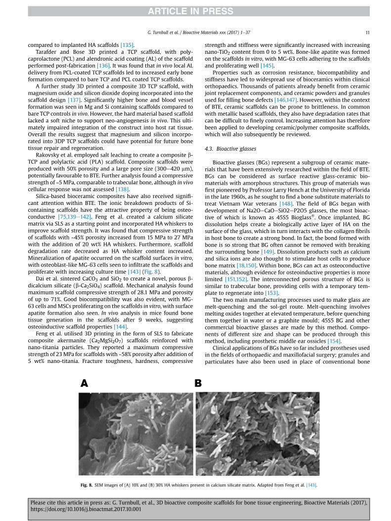
Silica-based bioceramic composites have also received signifi-cant attention within BTE. The ionic breakdown products of Si-containing scaffolds have the attractive property of being osteo-conductive [75,139e142]. Feng et al. created a calcium silicatematrix via SLS as a starting point and incorporated HA whiskers toimprove scaffold strength. It was found that compressive strengthof scaffolds with ~45% porosity increased from 15 MPa to 27 MPawith the addition of 20 wt% HA whiskers. Furthermore, scaffolddegradation rate decreased as HA whisker content increased.Mineralization of apatite occurred on the scaffold surfaces in vitro,with osteoblast-like MG-63 cells seen to infiltrate the scaffolds andproliferate with increasing culture time [143] (Fig. 8).
Dai et al. sintered CaCO3 and SiO2 to create a novel, porous b-dicalcium silicate (b-Ca2SiO4) scaffold. Mechanical analysis foundmaximum scaffold compressive strength of 28.1 MPa and porosityof up to 71%. Good biocompatibility was also evident, with MG-63 cells andMSCs proliferating on the scaffolds in vitro, with surfaceapatite formation also seen. In vivo analysis in mice found bonetissue generation in the scaffolds after 9 weeks, suggestingosteoinductive scaffold properties [144].
Feng et al. utilised 3D printing in the form of SLS to fabricatecomposite akermanite (Ca2MgSi2O7) scaffolds reinforced withnano-titania particles. They reported a maximum compressivestrength of 23MPa for scaffolds with ~58% porosity after addition of5 wt% nano-titania. Fracture toughness, hardness, compressive
Fig. 8. SEM images of (A) 10% and (B) 30% HA whiskers present
Please cite this article in press as: G. Turnbull, et al., 3D bioactive compohttps://doi.org/10.1016/j.bioactmat.2017.10.001
strength and stiffness were significantly increased with increasingnano-TiO2 content from 0 to 5 wt%. Bone-like apatite was formedon the scaffolds in vitro, with MG-63 cells adhering to the scaffoldsand proliferating well [145].
Properties such as corrosion resistance, biocompatibility andstiffness have led to widespread use of bioceramics within clinicalorthopaedics. Thousands of patients already benefit from ceramicjoint replacement components, and ceramic powders and granulesused for filling bone defects [146,147]. However, within the contextof BTE, ceramic scaffolds can be prone to brittleness. In commonwith metallic based scaffolds, they also have degradation rates thatcan be difficult to finely control. Increasing attention has thereforebeen applied to developing ceramic/polymer composite scaffolds,which will also subsequently be reviewed.
4.3. Bioactive glasses
Bioactive glasses (BGs) represent a subgroup of ceramic mate-rials that have been extensively researched within the field of BTE.BGs can be considered as surface reactive glass-ceramic bio-materials with amorphous structures. This group of materials wasfirst pioneered by Professor Larry Hench at the University of Floridain the late 1960s, as he sought to find a bone substitute materials totreat Vietnam War veterans [148]. The field of BGs began withdevelopment of Na2OeCaOeSiO2eP2O5 glasses, the most bioac-tive of which is known as 45S5 Bioglass®. Once implanted, BGdissolution helps create a biologically active layer of HA on thesurface of the glass, which in turn interacts with the collagen fibrilsin host bone to create a strong bond. In fact, the bond formed withbone is so strong that BG often cannot be removed with breakingthe surrounding bone [149]. Dissolution products such as calciumand silica ions are also thought to stimulate host cells to producebone matrix [18,150]. Within bone, BGs can act as osteoconductivematerials, although evidence for osteoinductive properties is morelimited [151,152]. The interconnected porous structure of BGs issimilar to trabecular bone, providing cells with a temporary tem-plate to regenerate into [153].
The two main manufacturing processes used to make glass aremelt-quenching and the sol-gel route. Melt-quenching involvesmelting oxides together at elevated temperature, before quenchingthem together in water or a graphite mould; 45S5 BG and othercommercial bioactive glasses are made by this method. Compo-nents of different size and shape can be produced through thismethod, including prosthetic middle ear ossicles [154].
Clinical applications of BGs have so far included prostheses usedin the fields of orthopaedic and maxillofacial surgery; granules andparticulates have also been used in place of conventional bone
in calcium silicate matrix. Adapted from Feng et al. [143].
site scaffolds for bone tissue engineering, Bioactive Materials (2017),

Fig. 9. Micro-CT and histomorphic analysis showing new bone formation in polymer-coated BG scaffolds implanted in mice for 8 weeks [166].
G. Turnbull et al. / Bioactive Materials xxx (2017) 1e3712
grafting to aid treatment of chronic osteomyelitis, soft tissue de-fects and wounds [18,155,156]. More than a million patients withinorthopaedics and maxillofacial surgery have now had bone defectsrepaired with 45S5 Bioglass® [148]. Within the field of spinal sur-gery, a study comparing 45S5 Bioglass® versus iliac crest autograftfor spinal fusion in adolescent idiopathic scoliosis (AIS) demon-strated positive results in a series of 88 patients. 45S5 BG® wasfound to be as effective as iliac crest graft, the current gold standardfor spinal fusion, in terms of ability to achieve deformity correctionand spinal fusion in AIS patients. Additionally, the morbidity ofharvesting an iliac crest graft was avoided in BG patients, who alsoexperienced fewer complications [157].
Whilst properties such as bioactivity and osteoconductivity areattractive, the inherent brittle nature of bioactive glasses representsa major potential limitation in their clinical application [155,158].Through incorporation of biodegradable polymers to create BGcomposites, properties such as porosity, degradation rate andelastic modulus can be improved [159,160].
Vergnol et al. investigated the potential of a polylactic acid(PLLA)-bioactive glass composite for bone fixation devices. PLLAhas previously been reported to require up to 4 years to fullydegrade in humans and lacks osteointegration ability [161]. Thein vivo behaviour of PLLA-HA composites has already been inves-tigated, with osteointegration found to be significantly improved inPLLA-HA composites compared to pure PLLA structures [162e165].Vergnol et al. therefore attempted to characterise the performanceof 45S5 BG® -PLLA composites in vitro and in vivo. Compositescontaining increasing 45S5 BG® content with PLLA were manu-factured by injection moulding and tested for up to 56 days insimulated body fluid (SBF). Whilst formation of a mineralised or HAlayer was not seen on the surface of PLLA polymer, crystallisation ofHA and calcite was evident on composite 45S5 BG® -PLLA scaffoldson X-ray diffraction analysis. Larger 45S5 BG® content, especially30e50%, led to rapid HA crystallisation on the surface of compositescaffolds. However, degradation of composites containing 50% 45S5BG® occurred rapidly within 7 days in vitro. Therefore, it wasconcluded that composites with 30% 45S5 BG® seemed to exhibitthe best balance between bioactivity and stability at least duringthe first weeks of immersion in contact with SBF. In vivo analysis ofthe 30% 45S5 BG® -PLLA composite within rabbits also found strong
Please cite this article in press as: G. Turnbull, et al., 3D bioactive compohttps://doi.org/10.1016/j.bioactmat.2017.10.001
osseointegration a month after implantation.Westhauser et al. investigated the osteoinductive properties of
different polymer coated 3D-45S5 BG® scaffolds seeded with hu-man MSCs (hMSCs) in vivo [166]. 45S5 BG® scaffolds were dip-coated with either gelatin, cross-linked gelatin, or poly(3-hydroxybutyrate-co-3-hydroxyvalerate). After seeding withhMSCs, the scaffolds were implanted into immunodeficient mice.Histomorphometry and micro-computed tomography (micro-CT)was then performed after 8 weeks (Fig. 9). Although bone forma-tionwas detected in all scaffolds, gelatin-coated 45S5 BG® scaffoldsperformed the best overall with further studies required to fullyevaluate their potential for BTE on a larger scale.
Murphy et al. utilised 3D extrusion bioprinting to create acomposite polycaprolactone (PCL)/BG scaffold containing humanadipose-derived stem cells (ASCs). Borate glass and PCL wereinitially dissolved in organic solvent to create a pastewith printableviscosity. ASCs suspended in Matrigel were then co-printed via asecond syringe as droplets into the PCL-BG scaffold. Degradation ofthe scaffolds in SBF was analysed, with 23.2 ± 4% weight loss due tocontrolled BG dissolution found at 14 days. Cell viability after 24 hwas 70 ± 10% and after 7 days was 58 ± 11%. Scaffold pore sizesranged from 100 to 300 mm, making it ideally suited for BTE.Bioactivity of the BG component was also seen, with formation ofHA crystals witnessed on the scaffold surface. This study thereforedemonstrated the potential for solvent-based 3D bioprinting tofabricate a scaffold containing cells and BG-polymer composites forBTE applications [167].
Baino et al. utilised a sponge template method involving sin-tering to fabricate a silicate-based glass-ceramic scaffold. Theirintention was to create a scaffold capable of repairing large defectsin load-bearing bones. Total scaffold porosity was 56%, with poresizes ranging from 100 to 500 mm on micro-CT analysis. Whentested under compression, the scaffolds had an elastic modulus of380 MPa and a compressive strength of 18 MPa [168].
Eqtesadi et al. utilised the 3D printing technique of robocastingto fabricate 13e93 BG scaffolds with a pore size of ~230 mmand 51%overall porosity. The scaffolds unfortunately showed brittle char-acteristics with a compressive strength of 86 MPa and a modulus ofrupture of 15 MPa. PCL was therefore added to the BG to createcomposite scaffolds, as an attempt to address the brittleness and
site scaffolds for bone tissue engineering, Bioactive Materials (2017),

G. Turnbull et al. / Bioactive Materials xxx (2017) 1e37 13
flexural strength of scaffolds. Whilst compressive strength of thescaffolds was maintained, brittleness was significantly reduced as13e93/PCL composite scaffolds were able to survive large stains[169].
BGs have also been used to improve the surface bioactivity of 3Dprinted b-TCP scaffolds, with promising results found. Zhang et al.spin-coated mesoporous (pores with diameters between 2 and50 nm) bioactive glass (MBG) nanoparticles onto porous b-TCPscaffolds to create a 100 nm layer of MBG on scaffold surfaces [170].This resulted in a hierarchical pore structure with both MBG mes-opores and BGmacropores present in the scaffold. The compressivestrength and mineralization of MBG-b-TCP scaffolds were alsosignificantly enhanced as compared to b-TCP scaffolds without theMBG nanolayer. Culture of human umbilical vein endothelial cells(HUVECs) found increased cell attachment, viability and angiogenicgene expression compared with conventional BG-modified b-TCP(BG-b-TCP) and pure b-TCP scaffolds. Furthermore, MBG-b-TCPscaffolds significantly enhanced the formation of new bone in vivoas compared to BG-b-TCP and b-TCP scaffolds.
The majority of drugs used in clinical practice are measured onthe nanometre scale and can therefore be introduced into meso-porous bioglass structures, potentially improving bioactivity anddrug delivery [171]. Jiang et al. coated mesoporous BG scaffoldswith amides, with subsequent gentamicin loading onto the scaffoldmesoporous surface achieved. As a result, higher gentamicinloading and longer drug release were achieved in vitro compared toBG scaffolds that had not undergone amination. In vitro bioactivitywas also improved, with increased formation of surface hydroxy-apatite found after soaking in simulated body fluid for 3 days(Fig. 10) [172]. MBGs have been utilised by several other studies tocreate composite scaffolds with increased bioactivity[170,173e175].
BG composites have also been used as injectable cements insome studies, with antibiotic [176], natural polymer [177] andsynthetic polymer [178] composites found to be cell friendly andcapable of treating in vivo bone and cartilage defects. Zhang et al.prepared a strontium-doped, borate bioactive glass (BBG)-chitosancomposite cement and evaluated in vitro and in vivo performance[179]. The Sr-BBG cement showed the valuable ability to set in situ(initial setting time ¼ 11.6 ± 1.2 min) and a compressive strength of19 ± 1 MPa. The Sr-BBG cement was also found to enhance theproliferation and osteogenic differentiation of hBMSCs in vitrowhen compared to a similar cement composed of chitosan-bondedBBG particles without Sr. The cement was then injected into rabbit
Fig. 10. SEM images of the amine-coated MBG before (A) and after (B) soaking in SBF. Forma[172].
Please cite this article in press as: G. Turnbull, et al., 3D bioactive compohttps://doi.org/10.1016/j.bioactmat.2017.10.001
femoral condyle defects, with new bone formation supported bythe Sr-BBG cement. It would therefore appear to be a promisingtreatment for treating irregularly shaped bone defects throughminimally invasive surgery.
4.4. Polymers
Use of natural polymers in scaffolds has attracted great interestwithin BTE due to favourable properties including ductility,biocompatibility and biodegradability. As a further benefit, naturalpolymers often contain biofunctional molecules on their surfacethat can aid cell attachment, integration and differentiation onscaffolds. Naturally occurring polymers that have been investigatedin BTE include collagen, silk, alginate, chitosan and hyaluronic acid.However, use of naturally occurring polymers can also be subject tolimitations including presence of pathogenic impurities such asendotoxin [180], lack of tuneability of degradation rates anddegradation related inhibition of local cells. Mechanical propertiesof natural polymers are also suboptimal for BTE, although cross-linking of polymers can enhance structural properties [181e183].
Synthetic polymers that have been deployed in BTE includepoly(lactic acid) (PLA), poly(glycolic acid) (PGA), poly(caprolactone)(PCL) and poly(ethylene glycol) (PEG). Co-polymers including pol-y(lactic-co-glycolic acid) (PLGA) have also been used. Syntheticpolymers can be created with tailored pore size, porosity, degra-dation rate and mechanical strength as required [184e187]. How-ever, they are often hydrophobic and lack cell recognition sites,limiting application without secondary modification to improvebioactivity [188,189]. Synthetic and natural polymers in generalhave relatively poor load bearing capacity when used alone, withlow elastic moduli compared to metallic and ceramic compounds[190,191]. However, the mechanical requirements of BTE scaffoldsare complex, with compressive, tensile and fatigue properties allrequired for load bearing [192].
One approach that has proved popular in addressing theselimitations is the combination of polymers with bioceramics orbioglasses to produce composite scaffolds. Most often, bioceramicsor bioglasses are added as a coating or filler to a polymer matrix toimprove bioactivity in addition to ramifying mechanical properties[180,193,194]. Porosity can also be added to composite polymer-ceramic scaffolds with varying levels of precision using tech-niques including 3D printing [80] and electrospinning [127,195].These techniques are capable of depositing material with gapsbetween fibres to allow interconnectivity. Alternatively, porogens
tion of a crystalline HA layer was confirmed on Fourier transform infrared spectroscopy
site scaffolds for bone tissue engineering, Bioactive Materials (2017),

G. Turnbull et al. / Bioactive Materials xxx (2017) 1e3714
can be incorporated into the structure before being dissolved out,or chemically triggered to release gas, to create pores [196e198].Combining use of porogens with techniques such as 3D printingduring manufacturing can increase micro and macroporosity.Polymer-based scaffold surfaces have also been secondarilychemically treated to increase microporosity, through use oforganic solvents [199,200]. Scaffold performance can also beincreased by surface modification with bioactive substancesincluding growth factors and surface ligands to aid cell adhesionand proliferation [121,193,201e203].
4.4.1. Collagen ebased composite scaffoldsAmongst the natural polymers used in bone tissue engineering,
collagen is perhaps the most frequently adapted into scaffolds.Collagen composes 90% of the total weight of bone extracellularmatrix proteins and is therefore a logical choice for inclusion in acomposite BTE scaffold. Although there are approximately twenty-nine known types of collagen, type I collagen has been used mostfrequently within BTE due to the lack of immune reactivity asso-ciated its use [204]. As part of the normal ECM it is inherentlybiocompatible, biodegradable and can stimulate cell proliferationand differentiation [205,206]. Furthermore, the mechanical anddegradation properties of collagen can be tailored through theprocess of crosslinking [207]. However, in common with othernatural polymers, collagen has mechanical properties that areinsufficient for creating a load-bearing scaffold [208]. It is thereforeoften combined with more robust materials within BTE to createcomposite scaffolds. As the major inorganic component of bone,hydroxyapatite (HA) has frequently been combined with collagenin composite scaffolds.
Villa et al. developed a collagen-HA (Col-HA) scaffold through aco-precipitation and freeze casting process [209]. The scaffoldcreated had a high degree of permeability suitable for cell infil-tration, attachment and osteogenesis with 99% interconnectivity ofpores. Mouse bone marrow derived mesenchymal stem cells(BMSCs) were seeded onto the scaffold and seen to bewell attachedafter 12 h in vitro culture. Subsequently the scaffolds wereimplanted into a mouse calvarial defect. After three weeks in vivo,near complete filling of the calvarial defects with bone on radio-graphic andmineralization analysis was found. After several weeks,host matrix metalloproteinase breakdown of collagen had to led toscaffold degradation occurring. By contrast, Marcacci et al. previ-ously found that a pure HA scaffolds failed to degrade 6 years afterinsertion into 4 patients with long bone defects [210]. However,compliant mechanical properties of the Col-HA scaffold wereobserved making it perhaps best suited for non-load bearing ap-plications such as craniofacial repair [209]. Alternatively, it could beused to aid treatment of a complex fracture in the same way thatbone substitutes or bone grafts are commonly used, in combination
Fig. 11. Photograph of the biomimetic scaffold showing the external appearance and la
Please cite this article in press as: G. Turnbull, et al., 3D bioactive compohttps://doi.org/10.1016/j.bioactmat.2017.10.001
with mechanical fixation [211].Calabrese et al. also demonstrated the osteoinductive potential
of a type 1 collagen (30%) -HA (70%) scaffold. The scaffold in thisinstance was prepared by a freeze casting process, with the addi-tion of a magnesium to create bioactiveMg-doped HA (MHA) nano-crystals. Human MSCs isolated from adipose tissue were seededonto the scaffold and cultured in vitro in the absence of specificosteogenic inducing factors. Analysis through quantitative PCR andimmunohistochemistry at up to 8 weeks demonstrated osteogenicdifferentiation of MSCs. This study therefore showed that thescaffold materials alone could trigger osteogenic differentiation ofMSCs, with extracellular matrix production, gene expression andmineralization analysis all demonstrating the osteoinductive po-tential of the scaffold. Nevertheless, osteogenic differentiation wasfound to be significantly accelerated with the addition of osteo-genic factors to the culture medium [123]. In vivo analysis was thenperformed of the scaffold in mice, with ectopic osteoinductive andangiogenic performance of the Col-MHA composite scaffoldcompared to a pure collagen scaffold. Bone augmentation andangiogenesis were found to spontaneously occur into the com-posite Col-MHA scaffold, with recruitment of host cells into thestructure. The Col-MHA scaffold performed significantly betterthan the collagen alone scaffold, with less fibrotic tissue and moreosteogenic tissue deposited at up to 16 weeks [212].
Grigolo et al. also utilised a scaffold composed of type 1 collagenand Mg doped-HA. The scaffold was designed to be biomimetic,with three distinct layers included to replicate the cartilaginous,tidemark and subchondral layered structure of articulating bone[213,214]. The cartilaginous layer was composed of purely type 1collagen; the intermediate layer type I collagen (60%) and Mg-HA(40%); and the sub-chondral layer type I collagen (30%) and Mg-HA (70%). The scaffold was manufactured by combining a sinter-ing and a freeze-drying technique, to obtain an integrated mono-lithic composite. Human MSCs (hMSCs) were seeded onto thesingle layers of the composite scaffold individually, and onto theintegrated composite scaffold. Cells were then grown in eitherchondrogenic or osteogenic media for comparison. Immunostain-ing confirmed chondrogenic differentiation of hMSCs in thecollagen-only cartilaginous layer when using chondrogenic media.Chondrogenic differentiation did not occur in the Mg-HA bonelayer despite use of chondrogenic media. Immunostaining alsoconfirmed osteogenic differentiation of hMSCs in both layers con-taining Mg-HA, and infiltration into the cartilaginous layer whenosteogenic mediawas used, but this did not occur in the presence ofchondrogenic media. Therefore, the processes of osteogenic andchondrogenic differentiation tended to depend mainly on themedia used for culture, rather than the biomaterial composition inthis study [215] (Fig. 11). Human bone marrow concentrate wasthen used in place of hMSCs on the three-layered scaffold in a
yered structure; SEM images showing the interface between scaffold layers [215].
site scaffolds for bone tissue engineering, Bioactive Materials (2017),

Fig. 12. Osteochondral scaffold, sized and press-fit into a patella defect. Adapted fromPerdisa et al. [223].
G. Turnbull et al. / Bioactive Materials xxx (2017) 1e37 15
further study. Having found a lack of osteogenesis in the cartilagi-nous layer, and a similar lack of chondrogenesis in the bone layer,they decided to induce chondrogenic differentiation only in thecartilaginous layer and osteogenesis in the composite layer (type Icollagen (60%) and Mg-HA (40%)) and bone layer (type I collagen(30%) and Mg-HA (70%)). After 52 days in vitro, cell viabilityremained high and differentiation of cells down both the chon-drogenic and osteogenic pathways was demonstrated on histo-logical and immunohistochemical analysis. This biomimetic, nano-composite material would therefore appear to offer a potentialoption for treating osteochondral lesions. In combination withpatient-derived bone marrow concentrate, chondrogenic andosteogenic areas could be combined into the same scaffold to offer a“one step” transplantation procedure for osteochondral defects[216].
Kane et al. looked to improve upon the mechanical properties ofcollagen-HA scaffolds previously described by modifying thefreeze-drying process most commonly used in their manufacture[217e219]. Attempting to improve scaffold porosity andmechanicalstrength, they used compression moulding to combine HA, paraffinmicro-spheres and concentrated collagen fibrils. The paraffinmicro-spheres were then leached out, acting as porogens, and thecollagenwas chemically cross-linked [126]. Interconnected pores of300e400 mm in size with walls 3e100 mm thick were found onmicro-CT analysis, with overall 85e90% porosity. This is significant,as scaffold pores greater than 300 mm have been shown to befavourable for osteointegration [160]. Mechanical testing foundscaffolds with 60% HA to exhibit fully elastic deformation uponloading to 50% compressive strain, maintained over greater than100 000 cycles. Compared to Col-HA scaffolds created throughfreeze-drying, the compressive modulus of the scaffold created inthis study was a magnitude greater at approximately 1 MPa. Theseproperties make the scaffold well suited to clinical application, aselastic deformation would potentially facilitate surgical handlingand manipulation, whilst the compressive modulus demonstratedwould potentially facilitate a degree of load bearing. In vitrobioactivity was investigated by seeding murine adipose derivedstromal cells (mASCs) onto the scaffolds and culturing in osteogenicmedia. After 14 days, significant increases in ALP activity were seen,with complete infiltration of the scaffold by mASCs. HA containingscaffolds showed vastly superior bioactivity compared to collagen-only scaffolds, in keeping with other studies [180,220,221], withincreased osteogenic differentiation and ALP levels found. How-ever, increasing HA content beyond 40% had no significant benefit.In vivo angiogenesis and osteogenesis in 40% HA scaffolds was thenevaluated by implanting acellular scaffolds subcutaneously in micefor 8 weeks. In vivo the scaffold was shown to be conducive to theinfiltration and differentiation of endogenous cells, with osteo-genesis and angiogenesis observed on histological analysis, sug-gesting the scaffold is osteoinductive. Therefore, the collagen-HAscaffolds in this study would appear to have potential for clinicaluse as a synthetic bone graft replacement, given their superiormechanical properties to scaffolds prepared by freeze-drying andfavourable osteoinductive and angiogenic properties.
Meagher et al. investigated the impact of HA volume fraction onthe in vivo performance of Col-HA scaffolds produced bycompression moulding. Acellular collagen scaffolds containing 0,20, and 40 wt% HA were implanted subcutaneously for up to 12weeks in mice. Endogenous cell infiltration after 6 weeks wasincreased in scaffolds containing HA versus collagen alone. Angio-genesis, remodelling of the original scaffold matrix, mineralizationand osteogenic gene expressionwas evident in scaffolds containingHA, but not observed in pure collagen scaffolds. Increasing scaffoldHA content was found to be directly correlated with improvedvascularity, cell density, matrix deposition and mineralization on
Please cite this article in press as: G. Turnbull, et al., 3D bioactive compohttps://doi.org/10.1016/j.bioactmat.2017.10.001
histological and micro-CT analysis. It would therefore seem that HApromotes the recruitment and differentiation of endogenous cellpopulations, leading to angiogenic and osteogenic activity incollagen scaffolds. Contrastingly, collagen scaffolds exhibited nomatrix deposition, mineralization, osteogenic gene expression anda significantly lower cell infiltration density [222].
Perdisa et al. demonstrated the osteoinductive potential of Col-HA scaffolds further in a prospective clinical study involving pa-tients with patellar osteochondral defects [223] (Fig. 12). Cell-freeCol-HA scaffolds were implanted into knee or patellar osteochon-dral lesions, with MRI imaging performed 24 months followingsurgery. The composite scaffold in this study utilised the samethree-layered approach as used by Grigolo et al. [215]; the carti-laginous layer was made of type I collagen with a smooth surface;the intermediate layer had a combination of type I collagen (60%)and HA (40%); and the lower layer was a mineralised blend of type Icollagen (30%) and HA (70%), mimicking subchondral bonecomposition. Patient functional outcome scores improved signifi-cantly at 12 and 24 months follow up, with MRI showing completefilling of the cartilage in 87.0% of the lesions, complete integrationof the graft in 95.7% of lesions, and intact repair tissue surface in69.6% of patients. However, osteophytes or more extensive bonyovergrowth was also documented in.8% of the patients, though nocorrelation was found between MRI findings and clinical outcome[223].
4.4.2. Chitosan e based composite scaffoldsChitosan (CS) is a polysaccharide normally found in the shell of
crustaceans including crabs, lobsters and shrimp. As a versatile,semi-synthetic polymer it has favourable biocompatibility andbiodegradability in addition to antibacterial and bioadhesivecharacteristics [197,224,225]. Within BTE chitosan has been com-bined with a number of materials in scaffolds including calciumphosphate [226], calcium sulfate [227], hydroxyapatite [228] andother natural polymers including silk [229e231].
Microparticle-based chitosan scaffolds have been produced byseveral groups [106,232e234]. Jiang et al. produced a composite CS/poly(lactic acid-co-glycolic acid) (CS/PLAGA) sintered microspherescaffold, functionalizing the scaffold surface further with heparinmolecules [235]. Scaffolds had a mean pore size of 172 mm, with acompressive strength in the region of trabecular bone. Mechanicaltesting showed that heparinization of chitosan/PLAGA scaffolds didnot significantly alter scaffold mechanical properties or porosity.Osteoblast-like cells were observed to proliferate faster on CS/
site scaffolds for bone tissue engineering, Bioactive Materials (2017),

G. Turnbull et al. / Bioactive Materials xxx (2017) 1e3716
PLAGA scaffolds as compared to pure PLAGA scaffolds. Furthermore,it was shown that the presence of CS on microsphere surfacesincreased the ALP activity of the cells cultured on the compositescaffolds and up-regulated gene expression of osteopontin andbone sialoprotein. This study therefore demonstrated the potentialof functionalized chitosan/PLAGA scaffolds.
Nano-fibre based composite chitosan scaffolds have also beeninvestigated within BTE, fabricated using methods such as wetspinning [236,237] and electrospinning [238] and including ma-terials such as silicon [239] and nano-hydroxyapatite (nHA) [240].However, electrospinning of CS can be difficult and scaffold stabilityinside aqueous solutions is unreliable. Secondary crosslinking withagents such as poly(ethylene oxide) (PEG) and blending with otherpolymers such as silk, collagen and PCL to create composites canhowever significantly reduce the degradation rate of CS in elec-trospun scaffolds and improve bioactivity [241].
CS has also been combined with collagen and bone morpho-genic protein (BMP) in composite scaffolds, with in vivo perfor-mance in dog and rabbit models analysed [31,242].
Shi et al. encapsulated BMP in poly-L-lactide-co-glycolide(PLGA) biodegradable microspheres, before dispersing them in achitosan/collagen composite scaffold. The scaffolds were implantedin dog mandibles for 4 weeks, with histologically enhanced boneformation found in BMP/PLGA microsphere loaded scaffoldscompared to control chitosan/collagen scaffolds also containingBMP. It was therefore concluded that sustained release of BMP frommicrospheres was more effective in inducing implant osseointe-gration compared to BMP bound to scaffolds.
Hou et al. prepared chitosan microspheres (CMs) and combinedthem with an absorbable collagen sponge with freeze-drying per-formed, to achieve controlled-release of BMP. The BMP-loadedcomposite scaffolds were implanted into 15 mm radius defects ofrabbits and the bone-repair ability was evaluated. Defects werefound to be bridged by new bone as early as 4weeks, with completehealing and recanalization of the bone-marrow cavity at 12 weeksevident on X-ray and histological analysis. These results demon-strated that the composite CS-Col scaffold is a promising carrier ofBMP-2 for the treatment of segmental bone defects [242].
Wu et al. developed a novel, composite scaffold of poly(L-lacticacid)/nHA/Alendronate-loaded chitosan microspheres (CS-ALs)with promising application for drug delivery and BTE demonstratedin vitro and in vivo [243] (Fig. 13). Alendronic (AL) acid has beenused in an increasing number of BTE studies, with known proper-ties including potent osteoinduction and inhibition of boneresorption [244,245]. Using a room temperature moulding/particleleaching method [246], followed by compression moulding, porous
Fig. 13. Comparison of CS-ALs (10%)-implanted group to CS-ALs (0%) found significantly highscaffold, with black arrows indicating the PLLA/nHA matrix and the white arrow indicating
Please cite this article in press as: G. Turnbull, et al., 3D bioactive compohttps://doi.org/10.1016/j.bioactmat.2017.10.001
scaffolds of PLLA/nHA/CS-AL were prepared with the concentra-tions of CS/nHA-AL ranging from 0 to 20%. Porous PLLA/nHA scaf-folds with only PLLA and nHA were also produced for use ascontrols. SEM found the scaffolds to exhibit a homogeneouslyinterconnected porous structure, with the pore diameters of150e250 mm. Scaffolds with 10% of CS/nHA-AL were then analysedin vitro, having been found to possess the most favourable drugrelease, degradation and mechanical properties. Culture of rabbitadipose stem cells (ASCs) found rapid cell proliferation and ECMproduction after 5 days, with no apparent cytotoxicity seen. Growthin osteogenic media led to significantly increased ALP activity andcalcium deposition, with CS-AL scaffolds containing CS/nHA-ALhaving significantly better results than control scaffolds. Scaffoldscontaining 10% and 0% CS-ALs were then implanted into a rabbitbone defect model to further evaluate in vivo bone regeneration.Bone defects were healed with new bone formation seen during4e8 weeks of implantation. New bone formation was significantlyhigher in CS-ALs (10%) group when compared with CS-ALs (0%), aneffect that increased with time on histological analysis. Sustainedrelease of AL was also found for up to 30 days. This study thereforeshowed promising application of CS-AL microsphere loaded (10%)scaffolds for both drug delivery and bone tissue engineering.
4.4.3. Hyaluronic acid e based composite scaffoldsHyaluronic acid (HLA) is a natural glycosaminoglycan found
widely throughout connective, epithelial and neural tissues. As oneof the chief components of the extracellular matrix, HLA contrib-utes significantly to cell proliferation and migration. It typically hasa very large molecular weight and has been adapted into attemptsat both hard and soft tissue engineering, particularly as a hydrogel(as discussed in the later hydrogel section) [247,248]. The me-chanical properties of HA can be readily improved through pro-cesses such as crosslinking [79,249], whilst it is naturallyviscoelastic, biodegradable and biocompatible, making it an idealmaterial for BTE [250].
HLA has been implemented in composite scaffolds by severalgroups. Kim et al. recently looked to combine favourable propertiesof multiple materials in fabricating a graphene oxide (GO)eChito-san (CS)eHyaluronic acid (HLA) based bioactive composite scaffoldalso containing an osteogenesis-inducing drug simvastatin (SV)[251]. Interestingly, SV is capable of initiating osteoblast differen-tiation of human adipose derived stem cells (hADSCs) and is wellknown for its property to enhance bone morphogenic proteins(BMPs). In fact, there are several examples in the literature of SVbeing used in composite scaffolds and injectable gels for boneregeneration, with improved osteogenesis seen [252e258]. The SV
er new bone formation, a finding which increased with time. SEM images of the CS-ALa CS/nHA-AL microspheres [243].
site scaffolds for bone tissue engineering, Bioactive Materials (2017),

G. Turnbull et al. / Bioactive Materials xxx (2017) 1e37 17
loaded GOeCSeHLA scaffold in this study was prepared in brief bymixing dissolved CS and HLA, with GO added through continuousstirring to create a composite blend, before a freezing and lyophi-lisation step to add SV was performed. Finally, the obtained com-posite scaffolds were cross-linked with N,N-(3-dimethylaminopropyl)-N-ethylcarbodiimide (EDC). Electron mi-croscopy found the scaffolds to have an interconnected porousmorphology. The addition of GO also resulted in less swelling andultimately contributed to enhanced structural integrity of thescaffold. However, introduction of GO also caused a 35% reductionin porosity of the GOeCSeHLA scaffold. MC3T3 (Osteoblasts) cellswere seen to adhere and proliferate better on SV loadedGOeCSeHLA scaffolds throughout 48 h of in vitro analysis. This wasreflected by significantly higher scaffold mineralization beingfound in SV loaded scaffolds after 14 days of analysis. Overall, the SVloaded GOeCSeHLA scaffold appeared to offer a successful optionfor BTE, with the addition of SV significantly accelerating bioactivityand osteogenesis.
Jing et al. also combined HLA and CS in a porous scaffold forbone tissue engineering [259]. Mixing CS and HLA together as liq-uids, they used a freeze-drying approach to form porous, 3D scaf-folds. By performing a further cross-linking step, the elasticmodulus and structural integrity of the scaffold was increased.Stem cell colonisation and proliferation within the scaffold wasdemonstrated by DNA assays and confocal imaging. Correia et al.also used a freeze-drying processed to prepare composite scaffoldsof chitosan and HLA, though directed their scaffolds at cartilagetissue engineering. Within this study incorporation of HLAenhanced cartilage ECM production, chondrocyte proliferation andcell adhesion to scaffold surfaces [260].
Kim et al. combined HLA and collagen in a scaffold directed atregenerating cartilage [261]. The hybrid scaffolds were prepared byadding 0.1, 0.3 or 0.5 wt% collagen to HLA. The HLA was thencrosslinked with ethylene glycol diglycidyl ether, followed by afreeze-drying process. The resulting composite scaffolds had athree-dimensional structure with interconnected pores andshowed an increase in tensile strength with increasing collagenconcentration. The degradation time of the hybrid scaffolds in vitroincreased with increasing collagen concentration. In vitro chon-drocyte growth on the scaffolds was also improved by increasingcollagen concentration over 2 weeks in culture. Furthermore,glycosaminoglycan (GAGA) concentration in the hybrid scaffoldswas higher than in pure HLA scaffolds. These composite scaffoldswould therefore seem to have potential for in vivo cartilageregeneration.
Fig. 14. Micro-CT images of the artificial skull defects after 4 weeks, showing signific
Please cite this article in press as: G. Turnbull, et al., 3D bioactive compohttps://doi.org/10.1016/j.bioactmat.2017.10.001
HLA has also been used as a delivery agent to improve bioac-tivity in bone substitute materials. Chang et al. investigatedwhether the use of HLA as an aqueous binder of hydroxyapatite/beta-tricalcium phosphate (HA-bTCP) particles could reduce theamount of bone graft needed and increase ease of graft handling inclinical situations [262] (Fig. 14). HA/bTCP was loaded into cross-linked HLA to form a novel HLA/HA-bTCP composite, which wasthen injected into rabbit skull defects in vivo. Histological andmicro-CT analysis found that HLA allowed bone regeneration to bemaintained evenwhen HA-bTCP particle numbers were reduced. Infact, compared to the control scaffold, HLA/HA-bTCP samples had1.7 times larger bone formation after 2 weeks. Overall, the additionof HLA to bone grafts not only promoted osteoconduction but alsoimproved handling characteristics in clinical situations.
4.4.4. Silk - based composite scaffoldsSilk fibroin (SF) is a natural protein-based polymer mainly
produced by silkworms and spiders. SF possesses several charac-teristics desirable for use in bone tissue engineering, includingbiocompatibility, low immunogenicity, limited bacterial adhesion,tuneable biodegradability, mechanical integrity and the ability tosupport the differentiation of mesenchymal stem cells along theosteogenic lineage [263e265].
McNamara et al. developed a new technique in fabrication ofporous HA-silk scaffolds. Firstly, they mixed silk together with HApowder before addition of silk macroporogens. The mixtures werethen sintered, with silk acting as a sacrificial polymer creatingporosity. The resulting silk-HA scaffolds could be moulded intolarge, complex shapes, and further machined post-sinter togenerate specific three-dimensional geometries. Scaffolds alsosupported bone marrow-derived MSC attachment and prolifera-tion, with no signs of cytotoxicity [266].
Kweon et al. compared the performance of HA-coated silkscaffolds and HA-coated collagen scaffolds. They implanted bothscaffold variants into rat tibias, with histological analysis of boneformation around the scaffolds performed after 6 weeks in vivo.They found that coating silk based scaffolds with HA promotedbone regeneration and bone to scaffold contact, with superior re-sults demonstrated compared to HA-coated collagen scaffolds[267].
Silk has also been used to coat BTE scaffolds, with encouragingresults found. In one study, PCL nanofibers were coated with silkand combined with biphasic calcium phosphate (BCP) in a com-posite scaffold [268]. Addition of silk-coated PCL nanofibersimproved scaffold compressive strength (from 0.07 MPa for BCP to
ant bone regeneration in HLA/HA-b-TCP composites (B) compared to control (A).
site scaffolds for bone tissue engineering, Bioactive Materials (2017),

G. Turnbull et al. / Bioactive Materials xxx (2017) 1e3718
0.42 MPa), elastic modulus (from 5 MPa for BCP to 25 MPa), andbioactivity whilst also preserving porosity (85%) and inter-connectivity (99%). Furthermore, osteoblast proliferation and dif-ferentiation rates were increased in the BCP/PCL-silk scaffoldscompared to that in the BCP/PCL and BCP scaffolds. Jaio et al. alsomodified BCP scaffolds with silk, applying multiple coatings to thescaffold surface. In doing so they managed to significantly enhancescaffold mechanical performance, with properties comparable tonative bone tissue found. Once again osteogenesis by hMSCs wasfound to be positively influence by silk over an extended period inculture [269].
Cao et al. used a HA-mineralised silk to mechanically reinforcean injectable bone cement [270]. In this study, a hydroxyapatite(HA)esilk fibroin (SF) complex was synthesised through a co-precipitation method and added to an injectable CPC/SF compos-ite. The compressive strength of the three-component composite,CPC/HAeSF/SF, kept increasing as HAeSF content was increased to3 wt%. The setting time of CPC/HAeSF/SF composites alsodecreased as a function of HAeSF content, with no apparent effecton injectability. Furthermore, the CPC/HAeSF/SF compositesshowed good biocompatibility both in vitro and in vivo. It wastherefore suggested that the composite could hold promise fororthopaedic applications, including serving as filler materials forminimally invasive surgeries to treat vertebral fractures.
In a further study, attempts were made to enhance the me-chanical properties of electrospun SF scaffolds by uniformlydispersing hydroxyapatite (HA) nanoparticles within SF nanofibers[271]. Addition of HA content up to 20 wt% increased the me-chanical properties of the composite scaffolds, while further in-creases above 20wt% disrupted the polymer chain networks withinSF nanofibers and weakened overall mechanical strengths.
4.4.5. Synthetic polymer compositesUse of natural polymers such as type I collagen in composite
scaffolds has many advantages, including increased ease of enzy-matic degradation, biocompatibility and improved scaffold simi-larity to natural bone extra-cellular matrix. However, naturalpolymers such as collagen used within scaffolds can have weakmechanical properties [42,272e274]. Significant research hastherefore looked to optimise and improve scaffold properties bydeveloping hybrid synthetic polymer/ceramic scaffolds for bonetissue engineering. Synthetic polymers that have been usedfrequently within BTE include polycaprolactone (PCL), polylacticacid (PLA), polyglycolic acid (PGA), and copolymers of PLA-PGA(PLGA). These poly(a-ester)s have key characteristics of beingbiodegradable, nontoxic and biocompatible [275e277].
Fig. 15. PCL/Alginate scaffold fabrication method. (A) 3D printing of micro-sized PCL struts (sized pores in final PCL/alginate (PAS-S) scaffold. Adapted from Kim et al. [271].
Please cite this article in press as: G. Turnbull, et al., 3D bioactive compohttps://doi.org/10.1016/j.bioactmat.2017.10.001
4.4.6. Polycaprolactone (PCL) e based composite scaffoldsPolycaprolactone has beenwidely used in BTE for the fabrication
of 3D scaffolds. Advantages of this polyester include biocompati-bility, relatively slow degradation rate, less acidic breakdownproducts in comparison to other polyesters and potential for load-bearing applications [191,278e280]. In terms of hard tissue engi-neering, the high mechanical strength and slow degradation rate ofPCL are particularly advantageous characteristics, potentiallyallowing load-bearing whilst native tissue gradually ossify a BTEscaffold [281]. However, due to the poor cellular adhesion proper-ties of PCL, numerous attempts have been made to create PCLcomposites with improved bioactivity [282,283].
Kim et al. demonstrated the potential of PCL/alginate compositescaffolds, showing superior in vitro results in 3D printed PCL/algi-nate composite scaffolds compared to pure PCL scaffolds [284].Alginates have desirable characteristics for use in 3D scaffoldfabrication, including biocompatibility, low cost, controlled degra-dation and rapid gelation in the presence of calcium ions[224,285,286]. However, they also have poor mechanical propertiesand excessive hydrophilicity, making it difficult to control scaffoldstructure and shape [287,288]. Kim et al. therefore attempted tocombine the favourable properties of alginate and PCL by meltingPCL and alginate powders together at 130 �C, before extruding themelted composite through a 250-mm nozzle to create a 3D scaffold.Compared to a pure PCL scaffold fabricated by the same method,PCL/alginate scaffolds showed increased osteoblast cell viability,calcium deposition, ALP activity and greater cell-seeding efficiencyover 7 days in culture.
Kim et al. used a combination of 3D printing, electrospinningand a physical punching process to create composite PCL/alginateconstructs with nanofibrous content and improved mechanicalstrength. Electrospun layers of PCL/alginate were sandwiched bylayers of micro-sized PCL struts; the final scaffold was thenpunched to create micro-sized pores travelling through theconsecutive layers of electrospun and 3D printed material (Fig. 15).Compared to pure PCL scaffolds, PCL/alginate composite scaffoldsshowed significantly enhanced cell viability at 7 days, ALP activityand calcium deposition at 14 days and greatly increased waterabsorption due to the improved hydrophilicity contributed by thescaffold alginate content [271].
Hydroxyapatite has also been combined with PCL by severalgroups attempting to create more bioactive, composite scaffolds forBTE. Gonçalves et al. utilised silicon-doped nanocrystalline HA, PCL,and carbon nanotubes in 3D printed composite scaffolds [124]. Inusing three distinct materials, they hoped to merge propertiesfavourable to bone regeneration including biocompatibility,
B) electrospinning of PCL/alginate onto PCL struts (C) punching process to create micro-
site scaffolds for bone tissue engineering, Bioactive Materials (2017),

G. Turnbull et al. / Bioactive Materials xxx (2017) 1e37 19
osteointegration ability and biodegradability [193,289,290]. Nano-crystaline silicon-doped HA was chosen as a ceramic material dueto the chemical composition being comparable to the inorganicpart of native bone. It has also been shown that the in vivo bioac-tivity of silicon-doped HA (SiHA) is significantly improvedcompared to pure HA, with enhanced bone apposition, bone in-growth and cell-mediated degradation found on SiHA implants[291,292]. Carbon nano-tubes (CNTs) have been shown to influenceand enhance cell differentiation and adhesion, by increasing elec-trical stimulation to cells [293e296]. PCL was incorporated into thescaffold to add ductile properties to the ceramic SiHA. Additionally,PCL has a relatively low melting temperature (approximately60 �C), making it ideal for 3D printing [297,298]. To combine thematerials, PCL and HA were dissolved independently in dicho-loromethane. CNTs were then added to the HA in solution, and thePCL solution was then added to the HA-CNT suspension. The slurrywas mixed and slowly heated to allow the dichloromethane toevaporate until a suitable viscosity for 3D printing was achieved.Scaffolds of differing CNT content were then printed, with CNTcontent ranging from 0 to 10%. Scaffolds containing 10% CNTs hadsignificantly greater apatite deposition and osteoblast-like celladhesion after 6 days in vitro. Additionally, electrical resistance ofthe scaffolds decreased as CNT content increased. This would sug-gest that CNTs have beneficial effects on cell function throughmodifying surface charge, as suggested by previous studies[293e295]. In terms of the mechanical properties, scaffold load-bearing capacity was improved by a CNT content of up to 0.75%.Beyond 2% CNT content, there was a clear detrimental effect oncompressive strength. Nevertheless, compressive strength valuesfor pure PCL-SiHA and PCL-SiHA scaffolds with CNT content up to0.75% were comparable to those found in the literature fortrabecular bone [299]. Morphologically, the scaffolds were found tohave interconnected pores with sizes from 450 to 700 pm. Overall,the scaffolds with 2% CNTs offered the best combination of me-chanical behaviour, cell viability and electrical conductivity.
Jakus et al. also utilised organic solvents to create a compositeHA-PCL biomaterial, described as “hyperelastic bone,” or “ HB.” HApowder and either PCL or PLGA were added to a trisolvent mixtureof excess dichloromethane (DCM), 2-butoxyethanol (2-Bu) anddibutylphthalate (DBP) [300]. The resulting solutions were then
Fig. 16. (A) Photograph series showing the compression and recovery of a 1-cm-diameter 3Dfrom 100 ml HB ink, with ease of manipulation into complex folded structures shown (C) SEMcircles, with soft tissue filling space between HB fibres. Adapted from Jakus et al. [302].
Please cite this article in press as: G. Turnbull, et al., 3D bioactive compohttps://doi.org/10.1016/j.bioactmat.2017.10.001
stirred in an open environment to allow solvent evaporation, until aviscosity ideal for 3D printing was reached. The synthetic com-posites composed of 90% HA and either 10% PCL or PLGA could berapidly 3D printed (up to 275 cm3/hour) from room temperatureextruded liquid inks. Mechanical testing of HB found elastic prop-erties; although not as elastic as their pure polymer counterparts,both HA-PLGA and HA-PCL versions of HB retained a high degree ofelasticity, capable of undergoing 36.1 ± 4.3% and 61.2 ± 6.4% strainand having similar tensile elastic moduli of 4.3 ± 0.4 MPa and10.3 ± 1.3 MPa, respectively (Fig. 16). To examine biocompatibilityin vivo, HA-PLGA scaffolds were implanted subcutaneously in mice.After 7 days, native tissue had already begun to infiltrate the scaf-folds, with vascularisation seen to develop on histological analysis.Further to this, HB capacity to induce bone regeneration wasassessed in vivo within a rat spinal fusion model. Pure HA-PLGAscaffolds and HA-PLGA scaffolds with additional of recombinanthuman bone morphogenic protein 2 (rhBMP-2) were implanted. Ademineralised bone matrix (DBM) scaffold was used for compari-son, with fusion rates of 50e60% usually seen with this control[301]. After 8 weeks in vivo, bone formation and spinal fusion rateswere similar in the HA-PLGA scaffold compared to the DBM control.These findings suggest that without any added growth factors, thescaffold has intrinsic osteoinductive properties comparable to thecommercially available DBM. HA-PLGA scaffolds with rhBMP-2were found to have superior new bone formation and spinalfusion rates compared to both the alone and DBM. HA-PLGA HBScaffolds were also implanted into primate calvarial defects, toevaluate performance in larger cortical bone defects. After 4 weeksin vivo, implants were excised from the calvarial defects andexamined. Evidence of heavy vascularisation and mineralised tis-sue advancing into the implants from the cortical bone was seen;areas not in direct contact with native bone were also seen todevelop mineralised tissue, suggesting up-regulation of osteogenicgene expression by nativeMSCs without the addition of any growthfactors, indicating HB osteoinductive potential. In summary, 3Dprinted HB demonstrated several properties favourable to clinicaluse. Compared to DBM, 3D printed HA-PLGA could offer a cheapand potentially readily available osteoinductive material for clinicalapplications, without risk of disease transmission.
Cheng et al. also looked to combine HA and PCL in a 3D printed
-printed HB cylinder over a single compression cycle (B) 12 � 12 cm sheet of HB printedimage of explanted HB scaffold after 35 days in vivo; blood vessels indicated by yellow
site scaffolds for bone tissue engineering, Bioactive Materials (2017),

G. Turnbull et al. / Bioactive Materials xxx (2017) 1e3720
composite scaffold using stereolithography, which creates 3Dstructures through spatially controlling the solidification of liquidphoto-polymerizable resin [78,303,304]. Pure PCL scaffolds werefirst fabricated via a custom designed 3D printer. The scaffolds werethen modified by immersing them in poly-dopamine (PDA), beforemineralization with HA nanocrystals was achieved on the scaffoldsurfaces through a precipitation reaction. Scanning electron mi-croscopy (SEM) was used to examine HA/PDA/PCL scaffold micro-structure; mean pore sizes of scaffolds 400 pm were found, whichhas previously been shown to be optimal for bone tissue regener-ation [305]. Sterilised scaffolds were then seeded with hMSCs andincubated in appropriate media. Cellular attachment, osteogenesisand angiogenesis of hMSCs was found to be significantly increasedin dopamine-coated scaffolds compared to pure PCL variants.Furthermore, hMSC proliferation, ALP activity, osteogenesis-relatedproteins, and angiogenesis-related protein secretion all increasedas scaffold PDA concentration was increased. These resultsdemonstrated that dopamine scaffold surface modification couldhave significantly beneficial effects for BTE. Dopamine analogueshave previously been shown to be important in allowingmussels toattach themselves to rocks [306]. Dopamine has also been appliedto various materials to improve their cell adhesive properties;metals, oxides, polymers and carbon-based materials have all beencoated with improved cell attachment and differentiation seen[203,307e309]. Dopamine has also been shown to help inhibitperi-implant osteolysis caused by inflammation [307].
4.4.7. Poly(lactic acid) (PLA) e based composite scaffoldsPoly (lactic acid) is a biodegradable, bioactive thermoplastic
polyester that has been used tomake a number of medical implantsincluding bone screws, fixation devices and vascular grafts [310].PLA is formed by the polymerization of lactic acid and can be ob-tained from renewable sources including starch and sugar. Severaldistinct forms of PLA exist due to the chiral nature of lactic acid,including poly-L-lactide (PLLA) and poly-D-lactide (PDLA) whichare produced from the polymerization of L-lactide and D-lactiderespectively [311].
Yang et al. recently fabricated hierarchical macroporousbiocompatible (HmPB) scaffolds containing a matrix of PLA, PCLand hydrophobically modified silica nanoparticles (h-SiO2) [312].PCL and PLA were first dissolved in an organic solvent, with h-SiO2nanoparticles then added as a particulate emulsifier. After solventevaporation, the resulting composite material was 3D printed intoscaffolds with over 98% porosity. The scaffolds had a hierarchicalstructure (including macropores, medium pores and small pores)and supported the adhesion and proliferation of MSCs, suggestinggood biocompatibility.
Holmes et al. created a PLA based scaffold using a fused depo-sition modelling 3D printer. The scaffolds were designed to havehighly interconnected 3D microvascular-mimicking channels, tofacilitate osteogenic bone regeneration as well as vascular cellgrowth. The resulting scaffolds were also chemically conjugatedwith nHA to enhance osteo-differentiation of seeded hMSCs. SEMimaging demonstrated printing of vertical microchannels with botha 500 and 250 mm radius, within a porous bone matrix. Mechanicaltesting also demonstrated that the scaffolds could withstandnormal mechanical loading, exhibiting elastic behaviour. Analysisof in vitro hMSC adhesion, proliferation and osteogenic differenti-ation found enhanced results with scaffolds incorporating nHA andsmall (250 mm) microchannels, compared to controls without nHAand with large (500 mm)microchannels. Additional in vitro analysiswith human umbilical vein endothelial cells demonstrated thescaffolds to be effective in supporting and enhancing vascular cellgrowth and activity, with large channel scaffolds promoting thegreatest HUVEC growth. Taking all of the findings together,
Please cite this article in press as: G. Turnbull, et al., 3D bioactive compohttps://doi.org/10.1016/j.bioactmat.2017.10.001
inclusion of both large and small microchannels and nHA mayprovide the best overall solution for creating a vascularized, load-bearing, osteogenic scaffold [313].
In a different approach, Ren et al. recently electrospun a blend ofPLLA and gelatin to create a nanofibrous mesh designed to aid bonedefect repair [314]. The composite mesh was seeded with MSCs,with osteogenic differentiation occurring after 7 days of in vitroculture. 3D multi-layered constructs were then built by stackingfour mono-layered meshes together. The constructs were incu-bated in vitro for 3 days before being implanted into rat cranialdefects. In comparison with a control group, there was significantformation of new calcified bone after 12 weeks. Yao et al. alsocreated nanofibrous composite PLA scaffolds, by electrospinning ablend of PCL and PLA [315]. To improve control over structuralproperties created by the electrospinning process, an innovativetechnique of thermally induced nanofiber self-agglomeration(TISA) was developed by the same group [316]. In brief, thisinvolved electrospun nanofibrous mats being converted into tinynanofibrous pieces, which were then utilised as building materialsfor making 3D electrospun scaffolds. Grinding, dispersing,agglomerating and freeze-drying processes were involved. Theresulting 3D scaffolds had a 96% porosity and possessed inter-connected and hierarchically structured pores, including macro-pores with sizes up to 300 mm. On in vitro testing, PCL/PLA-3D blendscaffolds had higher mechanical properties and in vitro bioactivitycompared to pure PCL-3D scaffolds made by the same process. Thiswas reflected by enhanced hMSC cell viability, osteogenic geneexpression and apatite-like deposition in PLA/PCL scaffold compareto pure PCL controls. In vivo analysis was then performed, with PCL/PLA-3D scaffolds implanted into mice cranial bone defects. After 6weeks, histological analysis suggested that PCL/PLA-3D scaffoldsprovided a more favourable/desired microenvironment for mousecranial bone formation as compared to the PCL-3D scaffolds, withsignificantly increased bone formation. Despite this, neither scaf-fold could bridge the bone defects completely, despite the additionof BMP growth factor supplement.
4.4.8. Poly(lactic-co-glycolic acid)/PLGA e based compositescaffolds
PLGA is a synthetic co-polymer of poly-L-lactic acid (PLLA) andpolyglycolic acid (PGA), with US Food and Drug Administrationapproval for human applications. Degradation rates of PLGA can becustomised to range from weeks to months, based on the ratio ofPLLA to PGAwithin the copolymer structure [317]. Other benefits ofPLGA over pure PLA and PGA include the range of commonlyavailable solvents that it can be dissolved in and the ease withwhich it can be manipulated into structures of desired sizes andshapes. Biomolecules such as growth factors can also be easilyencapsulated by PLGA.
However, there are some limitations to the use of PLGA withinBTE. The amorphous structure of PLGA has a low Young's modulus,resulting in susceptibility to elastic deformation, whilst PLGA is alsopoorly osteoconductive [318]. In order to address these limitations,PLGA has been combined with ceramics and bioactive glasses tocreate composite scaffolds [319]. Several groups have combinednano-HA (nHA) with PLGA, hoping to benefit from the osteogenicproperties and high modulus of nHA [180,320]. Whilst high con-centrations of nHA in a composite can potentially have adverseeffects on mechanical properties due to non-uniform distribution,lower concentrations are able to improve tensile and compressiveproperties of scaffolds [321,322]. When contained within a com-posite scaffold, nHA has also been shown to help cells and proteinsattach to scaffold materials [17]. PLGA also potentially balances thelimitations of nHA which include inherent brittleness and slowdegradation [318,323]. Several groups have created composite
site scaffolds for bone tissue engineering, Bioactive Materials (2017),

G. Turnbull et al. / Bioactive Materials xxx (2017) 1e37 21
PLGA-nHA scaffolds using the technique of particulate leaching[324e326]. However, the microarchitecture of scaffolds formed bythis method typically have low porosities and interconnectivity.Incomplete solvent removal has been also encountered with thistechnique, resulting in reduced cell viability. Kim et al. modified thetechnique of particulate leaching by creating a PLGA-nHA scaffoldby gas forming and particulate leaching. This process avoided usingorganic solvents, eliminating potential issues associated withincomplete solvent evaporation, and resulted in improved scaffoldmicroarchitecture. Higher porosity and improved mechanicalcharacteristics resulted in higher in vitro cell growth and mineral-ization of scaffolds, compared to PLGA-nHA scaffolds formed bysolvent casting-particulate leaching [17].
Shaui et al. 3D printed porous PLGA-nHA scaffolds via selectivelaser sintering. This method allowed formation of well-controlledpore architectures and high scaffold surface bioactive nHA con-tent. The effect of nHA on scaffold mechanical properties was theninvestigated; although scaffold compressive strength and moduluswas improved as nHA content increased from 0 to 20%, nHA contentabove 20% resulted in worsening, brittle mechanical properties[129]. To re-create bone extracellular matrix (ECM) structureseveral groups have tried to create nano-fibrous PLGA compositestructures via electrospinning; potential benefits of includingnanofibers include high surface area per unit mass, increasedbiodegradability and with the addition of ceramic nanoparticles,improved osteoconductive potential [29,30,128,327]. Jose et al.electrospun nanocomposite scaffolds, adding nHA to PLGA solutionbefore spinning. Like Shaui's findings, nHA content strongly influ-enced scaffold mechanical properties. Whilst 20% nHA stronglyincreased the Young's modulus of the electrospun compositemeshes, higher concentrations again led to worsening mechanicalproperties [328]. Yun et al. evaluated the biocompatibility of elec-trospun PLGA-nHA scaffolds through culturing human primaryadipose tissue-derived stem cells (hADSCs) andMSCs on themwithpromising results. With a nHA content of around 17% (w/w), oste-ogenic differentiation andmineralization of bothMSCs and hADSCsoccurred. These results were confirmed by increased expression ofosteogenic genes on reverse transcription-polymerase chain reac-tion (PCR) increased ALP activity and calcium deposition [329].Haider et al. also evaluated the cell compatibility of PLGA-nHAcomposite nanofiber scaffolds, by culturing osteoblasts on PLGAand PLGA-nHA scaffolds to allow direct comparison (Fig. 17). In thisstudy, the impact of HA morphology was also examined by
Fig. 17. (A) SEM micrographs of nHA/PLGA composite nanofiber scaffo
Please cite this article in press as: G. Turnbull, et al., 3D bioactive compohttps://doi.org/10.1016/j.bioactmat.2017.10.001
comparing spherical and fibre forms of nHA on cell culture results.PLGA-nHA nanofiber scaffolds showed higher cellular adhesion,proliferation, enhanced osteogenesis and increased Caþ2 ionsrelease compared to spherical PLGA-nHA and pure PLGA scaffolds.This study re-iterated the beneficial effects of including nHAwithinscaffolds on osteoblast culture. The enhanced cell proliferation onthe fibre PLGA-nHA scaffold compared to scaffolds containingspherical nHA suggested that the morphology of nHA also in-fluences cell behaviour [330].
Whilst the combination of synthetic polymers such as PCL andPLGA with HA has led to successful results, natural polymers suchas collagen and chitosan have improved mineralization character-istics compared to synthetic polymers. The presence of numerousionic molecular groups within natural polymers allows for pro-cesses such as calcium chelation to occur and mineralization ratesare improved by the negative surface charge. The surfaces of syn-thetic polymers are also largely hydrophobic, which is lessfavourable for cell adhesion to occur [331,332]. Attempts havetherefore beenmade tomodify the surface of synthetic polymers toincrease cell affinity. One approach has been to covalently linkarginine-glycine-aspartic acid (RGD) to biomaterials such as PLGAto facilitate cell adhesion. RGD has previously been established asthe minimal core peptide recognizable by cell adhesion receptorsand is capable of increasing the bioactivity of structures [333]. Shinet al. fabricated biomimetic hybrid nanofiber sheets composed ofRGD peptide-decorated PLGA (RGD-PLGA) nanofibers via an elec-trospinning technique. Cells including the murine preosteoblasticcell line (MC3T3-E1 cell) and the human osteosarcoma cell line(MG-63 cell) were then cultured on the scaffolds. Compared to purePLGA nanofiber sheets, the initial adhesion and proliferation of cellswere significantly enhanced on RGD-PLGA sheets, as found in otherstudies [201,202,334].
4.5. Hydrogels
Hydrogels are gels constructed from networks of crosslinked,hydrophilic polymer chains. Their hydrophilic nature allows themto absorb copious amounts of water into a three-dimensionalnetwork, which is ideal for supporting cell growth. In fact, hydro-gels can absorb up to 1000 times their original weight in aqueousmediumwithout dissolving [335]. At the same time, they are highlypermeable to oxygen, nutrients and other water-soluble com-pounds, making them attractive materials for tissue engineering
lds, with adherent cells after 24 h incubation also displayed (B).
site scaffolds for bone tissue engineering, Bioactive Materials (2017),

G. Turnbull et al. / Bioactive Materials xxx (2017) 1e3722
[182]. Within BTE, they have been shown to facilitate angiogenesis,osteoconductivity, cell adhesion and matrix integration[89,336e338]. Hydrogels can be derived from natural polymers(including collagen, gelatin, alginate, chitosan, hyaluronic acid andagarose) or synthetic materials. Natural polymers are often derivedfrom extracellular matrix components including collagen and hy-aluronic acid, and therefore have low toxicity, high biocompatibilityand cell affinity [339,340]. However, natural polymers can undergouncontrolled degradation resulting in compromise of mechanicalproperties in printed constructs. To help address this issue, syn-thetic polymers can be used either alone or with natural polymersto create a composite hydrogel, allowing better control over stiff-ness and elastic modulus of hydrogel constructs [341,342]. Syn-thetic polymers can also be functionalized with biologicalmolecules, such as RGD, heparin and hyaluronan, to increasebioactivity and cell adhesion in hydrogel composites [202,343].Growth factors and drugs have also been encapsulated withinhydrogels to increase scaffold bioactivity [344e346].
Successful bioprinting relies in part on combining a suitablebioprinting technique with an appropriate bioink. Significant focuswithin bioprinting has therefore been placed on developing cell-friendly, novel bioinks which can be 3D printed to fabricate tissueconstructs. Polymeric hydrogels are usually the main component ofbioinks as they have cell-friendly properties whilst being amenableto 3D printing [338]. Hydrogel-based bioinks are often compositematerials, containing growth media, cells, biomaterials, addednutrients and growth factors to aid cell proliferation and differen-tiation [347e349].
4.5.1. Composite bioinks used in bioprintingAlthough low-viscosity hydrogels with a high-water content
provide a superior environment for cell function and viability, themechanical integrity and support they provide is often insufficientto allow transfer to in vivo analysis. Attempts have therefore beenmade to create composite bioinks capable of integrating the me-chanical strength of viscous hydrogels with the biocompatibilityprovided by lower-viscosity hydrogels.
Nguyen et al. recently designed bioinks to treat cartilage lesions,containing either nanocellulose with alginate (NFC/A) or
Fig. 18. (A) The 3D Discovery (Switzerland) bioprinter with microvalve print-head shown
Please cite this article in press as: G. Turnbull, et al., 3D bioactive compohttps://doi.org/10.1016/j.bioactmat.2017.10.001
nanocellulose with hyaluronic acid (NFC/HLA). NFC was chosen tohelp mimic the collagen bulk in cartilage matrix [350], alginateused in place of natural proteoglycans, whilst hyaluronic acid wasincluded as a major component in native cartilage. Human-derivedinduced pluripotent stem cells (iPSCs) and irradiated humanchondrocytes were then encapsulated in the bioinks. Utilising amicrovalve 3D bioprinter, a grid construct was printedwith the twodiffering bioinks. In the case of NFC/HLA, low proliferation andphenotypic changes away from pluripotency were seen in theiPSCs. However, in the case of the NFC/A (60/40, dry weight % ratio)constructs, hyaline-like cartilaginous tissue with collagen type IIexpression was seen after 5 weeks. Additionally, a marked increasein cell number within the cartilaginous tissue was detected by 2-photon fluorescence microscopy. The NFC/A bioink thereforeappeared suitable for bioprinting iPSCs to support cartilage pro-duction in co-culture with chondrocytes. Markestedt et al. also 3Dprinted a NFC/A bioink containing human chondrocytes. The shearthinning properties of the NFC and the fast crosslinking of alginateallowed microvalve bioprinting to occur with high fidelity andstability. Taking MRI and CT data, they managed to print anatomi-cally accurate scale models of human ears and sheep meniscus(Fig. 18). Chondrocyte viability after printing was approximately95%, with viability also found of 73% and 86% after 1 and 7 days of3D culture respectively [351].
Gao et al. used inkjet bioprinting to co-print an acrylatedpoly(ethylene glycol) (PEG) hydrogel with acrylated peptides. Hu-man MSCs were included in the composite hydrogel, which wasexposed to ultraviolet light to initiate simultaneous photo-polymerization of the hydrogel during printing. The resultingscaffold demonstrated excellent biocompatibility with a cellviability of 87.9 ± 5.3% 24 h after printing. Printed constructs con-taining hMSCs were cultured for 21 days in either osteogenic orchondrogenic media. Osteogenic and chondrogenic gene expres-sion was seen to significantly increase from day 7e21, with sig-nificant collagen and extracellular matrix deposition seen.Mechanical analysis found modulus increases in PEG-Peptidescaffolds of 100% in osteogenic media and 82% in chondrogenicmedia from day 7e21. The compressive modulus of the printedPEG-Peptide hydrogel exceeded 500 kPa, which is more than 100
(B) 3D printed knee meniscus using NFC/A ink. Adapted from Markestedt et al. [351].
site scaffolds for bone tissue engineering, Bioactive Materials (2017),

G. Turnbull et al. / Bioactive Materials xxx (2017) 1e37 23
times as that reported for some natural hydrogels [352,353].Overall, the PEG-Peptide hydrogel printed in this study developedinto a homogenous tissue with high cell viability and mechanicalintegrity [354].
Kang et al. used a multi-head bioprinter to create an interwovenscaffold consisting of cell-laden hydrogels, PCL polymer and asacrificial Pluronic F127 hydrogel [355]. The composite hydrogelconsisted of optimised concentrations of gelatin, fibrinogen, hyal-uronic acid (HA) and glycerol mixed into cell culture media. Plur-onic F127 was printed as a sacrificial outer layer to support the 3Darchitecture of the composite hydrogel while crosslinking wasperformed, whilst the PCL provided internal scaffold structuralstrength. After cross-linking of fibrinogen using thrombin, theuncross-linked components (gelatin, HA, glycerol and PluronicF127) were washed out. This created microchannels in the tissueconstructs, facilitating diffusion of nutrients to printed cells. Tissuemodels were printed in anatomically relevant shapes through useof clinical imaging data, with constructs created to match skull andmandible defects. The potential of the printing process wasdemonstrated through fabrication of mandible and calvarial bone,cartilage and skeletal muscle. High cell viability was found post-printing of chondrocytes, amniotic fluid derived stem cells andmyoblasts; cell viability in printed structures was typically 91 ± 2%after 24 h of culture.
Fig. 19. (A) Illustration of tissue manufacturing process (B) Photograph of a printed tissue conof tissue construct. Adapted from Kolesky et al. [368].
Please cite this article in press as: G. Turnbull, et al., 3D bioactive compohttps://doi.org/10.1016/j.bioactmat.2017.10.001
Pluronic F127 has been adapted into several other compositescaffolds to aid in the creation of temporary structural support orvascular channels [356e359]. Although F127 has poor cellcompatibility when used alone, it is easy to print and structurallyrobust [360e363]. Aqueous F127 solutions undergo thermal gela-tion at physiological temperatures, but on cooling bellow a criticalmicellar temperature (CMT) dissociation of the gel occurs. This al-lows F127 to be printed as a gel into constructs, with secondarycooling of the construct then washing F127 away as a fugitive ink[364,365]. As a result, F127 support structures can be removedpost-printing and printed F127 channels left patent for perfusionwith media. This approach has proved helpful in attempts to createvascular channels in constructs. Creating a sufficient vascularstructure is a major challenge in bioprinting, as in the absence of avascular network cells are dependent on diffusion for delivery ofoxygen and nutrients [361]. Whilst larger vessels have been fabri-cated, capillaries have yet to be 3D printed, which leaves constructsdependent on infiltration of micro-vessels in vitro to ensure cellviability. Unfortunately, microvessel infiltration often lacks depth ofpenetration, limiting the size of viable bone construct that can beimplanted [366,367].
Lewis et all used F127 as a fugitive ink to help create a multi-material 3D construct with vascular channels (Fig. 19) [368].
A silicone ink was first printed and then cured, creating a
struct housed within a perfusion chamber. Perfusion inlet and outlet seen at either end
site scaffolds for bone tissue engineering, Bioactive Materials (2017),

G. Turnbull et al. / Bioactive Materials xxx (2017) 1e3724
perfusion chip. Fugitive ink, containing F127 and thrombin, andcell-laden inks, containing gelatin, fibrinogen, and hMSC cells, werethen printed within the 3D perfusion chip. A composite materialcontaining gelatin, fibrinogen, cells, thrombin and trans-glutaminase (TG) was then cast over the printed inks, recreating anextracellular matrix. After casting, thrombin converted solublefibrinogen into insoluble fibrin in the cast matrix. Similarly, TGdiffused from the ECM matrix and slowly cross-linked the innergelatin and fibrin. Upon cooling, the fugitive F127 ink liquefied,leaving behind a pervasive vascular network. This network wasseeded with human umbilical vein endothelial cells (HUVECs) toinduce endothelialisation and perfused via an external pump. Thevascular networks were then perfused with osteogenic media after6 days of in vitro culture. After 30 days, the printed hMSCsexpressed osteocalcin in the tissue, with osteocalcin expressionfound to be highest in areas close to vessels perfused with osteo-genic media. Collagen deposition was found within printed fila-ments and around the circumference of the vasculature. Alizarinstaining also revealed a high degree of mineralization within thetissue. In summary, thick (1 cm) vascularized human tissues werecreated and actively perfused with growth factors to differentiatehMSCs toward an osteogenic lineage in situ over several weeks (>6weeks).
F127 has also been used in combination with other hydrogels toincrease porosity and produce nanostructures. Muller et al. com-bined diacrylated F127 (F127-DA) with F127 to print chondrocytes,with hyaluronic acid methacrylate (HAMA) added to the bioink toimprove mechanical strength [369]. After bioprinting, the F127-DAwas UV crosslinked with the acrylate acting as a photocrosslinkablegroup. This allowed the remaining F127 to be washed out oncooling, subsequently nanostructuring the remaining F127-DA-HAMA network. To increase gel stiffness and to avoid elution of HAduring the nanostructuring process, HA was functionalized withmethacrylate groups to allow covalent crosslinking between HAand the F127-DA network. Inclusion of 0.5% HAMA increased thecompressive modulus from 1.4 kPa in a pure F127-Da scaffold to5.62 kPa in a F127-DA-HAMA composite. Cell viability was main-tained at an elevated level after 14 days in culture in the F127-DA-HAMA composite scaffold, with 79.9% of chondrocytes still alive.Perriman et al. designed a novel pluronicealginate bioink, whichwas used in a two-step 3D printing process to engineer bone andcartilage architectures [370]. Specifically, 3D structures containinghMSCs were printed by extruding a shear-thinning, cell-ladenF127-alginate composite gel onto a heated stage, resulting ininstantaneous solidification via the solegel transition of the F127.After printing, CaCl2 crosslinking of alginate and washing stepswere performed to allow the F127 to dissolve out of the printed gelstructure. The F127 constituent acted as a sacrificial template,creating micron-sized pores and microchannels in the scaffold ondissolution. Optimization experiments found a 13 wt% F127e6 wt%alginate composite hydrogel to have the best print characteristics.MSC-laden 3D printed constructs showed no significant loss in cellviability over 10 days in culture. Encapsulated MSCs were alsosuccessfully differentiated into osteoblasts and chondrocyteswithin the F127-Alginate bioink, with a tracheal ring created.
Byambaa et al. also utilised an extrusion-based bioprintingstrategy to fabricate microstructured bone-like tissue constructs,complete with perfusable vascular lumen [371]. To form a perfus-able blood vessel in the centre of the construct, a cylinder with 5%gelatin methacryloyl (GelMA) hydrogel at low methacryloyl sub-stitution (GelMALOW) was printed containing HUVECs and hMSCs.The softness of the inner core, prepared with 5% (w/v) GelMALOW,allowed a fast degradation of the hydrogel, leaving an open lumenand a perfusable channel of 500 mm after 12 days of in vitro incu-bation. Three successive layers of cylinders were also initially
Please cite this article in press as: G. Turnbull, et al., 3D bioactive compohttps://doi.org/10.1016/j.bioactmat.2017.10.001
printed around the soft core using 10% (w/v) high methacryloylsubstitution gelatin methacryloyl (GelMAHIGH). The GelMAHIGHwas loaded with silicate nanoplatelets to induce osteogenic dif-ferentiation of hMSCs into osteoblasts, and contained threedifferent concentrations of covalently conjugated vascular endo-thelial growth factor (VEGF). The VEGF gradient created differentchemical microenvironments, promoting vascular spreading formthe central vessel into the surrounding bone niche. Synthetic sili-cate nanoplatelets were included as bioactive agents that dissociateinto products that can trigger osteogenesis (Naþ, Mg2þ, Si(OH)4 andLiþ) [372]. It has also been reported that silicate nanoplatelets caninduce osteogenic differentiation of encapsulated hMSCs withinGelMA hydrogels [373,374]. In this study, it was found that osteo-genic differentiation occurred even in the absence of growth fac-tors, with higher concentrations of silicate nanoplatelets resultingin increased calcium deposition after 21 days in culture. Overall, theapproach of creating a central lumen and using a composite GelMA-nanoplatelet hydrogel resulted in the creation of a mechanicallystable construct. Perfusion with growth media facilitated cell sur-vival, proliferation and osteogenic differentiation over a 21-dayperiod. Compared to conventional approaches such as the use ofthermoresponsive hydrogels [15,24] or sacrificial templates [26],this study manged to fabricate a blood vessel through a one-stepbioprinting process. Steps such as exposing constructs to temper-ature changes to remove sacrificial materials were thereforebypassed.
Wang et al. filled a 3D printed bioceramic scaffold with phagenanofibers to try and overcome the challenge of forming vascu-larized bone tissue [375]. Firstly, a biomimetic bone scaffold, con-sisting of a biphasic calcium phosphate (BCP) (with a compositionof hydroxyapatite (HA) and b-tricalcium phosphate (b-TCP) at amass ratio of 60/40) was produced via 3D printing. The scaffoldshowed a uniform structure with interconnected macro-scalepores, and contained micro-scale pores on the scaffold columns.Nanofiber phages expressing RGD (Arg-Gly-Asp) were then com-bined with chitosan and adhered to the construct pores throughelectrostatic interactions, with the intention of improving scaffoldosteogenesis and vascularisation in vivo. The constructs were thenimplanted in an animal model and native cells invaded theconstruct to form vasculature, with MSCs undergoing osteogenesis.However, the rate of vascularisation was relatively slow with cellsurvival within the cell-laden construct impaired as a result.
Costantini et al. printed 3D biomimetic hydrogel scaffolds con-sisting of differing combinations of gelatin methacrylamide(GelMA), chondroitin sulfate amino ethyl methacrylate (CS-AEMA)and hyaluronic acid methacrylate (HAMA) [376]. Using a twocoaxial-needle bioprinting system, they achieved a high cell density(>107 cells ml�1), high cell viability (85 ÷ 90%) and high printingresolution (z100 mm) post-printing. Bioinks were loaded withMSCs, with addition of 4% alginate and 0.3 M CaCl2 also performedto aid crosslinking. All the employed hydrogels exhibited enhancedchondrogenic differentiation of bone marrow derived-MSCs after 3weeks of culture in chondrogenicmedium. A composite hydrogel ofalginate, GelMA and CS-AEMA appeared to be the best candidatefor neocartilage formation, as it supported the highest levels ofcollagen production.
Daly et al. used a novel approach in trying to create bone tissue,by bioprinting a structure more representative of a developmentalprecursor to adult bone [377]. It was hoped that 3D printing arudimentary structure would create a template for subsequentorganogenesis in vivo. An alginate bioink incorporating RGDadhesion peptides was printed along with a network of PCL fibres.Addition of PCL fibres resulted in a near 350-fold increase inconstruct compressive modulus. The mechanically reinforcedtemplate was printed in the geometry of a primitive vertebral body
site scaffolds for bone tissue engineering, Bioactive Materials (2017),

G. Turnbull et al. / Bioactive Materials xxx (2017) 1e37 25
and implanted in vivo. After removal and histological analysis, itwas found to support the development of vascularized bone, withtrabecular-like endochondral bone and a supporting marrowidentified. It was therefore postulated that developmental engi-neering approaches could be applied to the biofabrication of othersolid organs, by bioprinting precursors that have the capacity tomature into their adult counterparts over time in vivo.
Silk-based composite hydrogels have also been used in BTE inseveral forms. Kim et al. fabricated a silk fibroin/hydroxyapatite (SF/HA) composite hydrogel, with hyaluronic acid (HA)-dopamine (DA)surface modification of HA nanoparticles performed to aid distri-bution of HA content [378]. The composite hydrogel showedexcellent cell proliferation, with in vivo analysis required to fullyexamine BTE potential. Park et al. also produced SF compositehydrogels containing HA nanoparticles (NPs) for bone tissue engi-neering [379]. In this study gamma-ray (g-ray) irradiation treat-ment was used to induce rapid chemical crosslinking of a SFsolution containing HA NPs, creating a chemically crosslinked SFhydrogel. Comparedwith comparedwith the pure SF hydrogels, theSF/HAP composite hydrogels exhibited improved osteogenic dif-ferentiation of hMSCs. Ding et al. adapted a hybrid approach,whereby SF hydrogels were used to deliver rat MSCs to a SF scaffold[380]. In vivo analysis in a calvarial defect found that encapsulatedcells were still viable and actively participating in new bone for-mation 8 weeks after implantation. The benefits of cell encapsu-lation were therefore twofold, as both cell viability and cellnumbers seeded to the scaffold were increased and maintainedin vivo.
Within composite hydrogel scaffolds, several studies haveexamined osteogenic differentiation of MSCs, whilst the potentialof human adipose stem cells (hASCs) in vitro and in vivo has beenexamined less frequently. Potential benefits of using hASCs overhMSCS include their abundance in adipose tissue and capacity forminimally invasive harvesting [318]. Wang et al. printed a hASCladen 8 wt% alginate and 2 wt% gelatin hydrogel scaffold, with aninterwoven grid structure [381]. Cell viability was 90.41% on day 7of in vitro culture, with construct structure maintained. Culture inosteogenic media lead to significant increases in osteogenic geneexpression (OSX, RUNX2, and OCN) compared to scaffolds placed inbasal media. The hASC laden composite scaffolds were alsoimplanted in mice for 8 weeks. Compared to acellular controls,printed constructs containing cells remained in their originalshape, with blood vessels ingrowth into the apertures of the con-structs seen. Ectopic bone formation was also seen to occur, withbone matrix seen to replace part of the degraded scaffold materialon histology, demonstrating bioactivity within the scaffold. Thisstudy therefore showed that 3D bioprinted constructs containinghASCs can promote mineralised bone matrix formation. Maglioneet al. demonstrated the osteogenic potential of ASCs further byseeding them onto a chitosan-glycerol phosphate hybrid gel scaf-fold, which was maintained by in-situ by cross-linking [382]. Thescaffolds were implanted into full-thickness mandible defects, withex vivo histological and micro CT analysis performed after 8 weeks.It was found that ASCs could regenerate bone within the scaffold,with the scaffold able to entrap and maintain cells in situ.
4.5.2. Injectable hydrogels for bone tissue engineeringInjectable hydrogels have attracted significant attention within
bone and cartilage tissue-engineering applications. Potential ben-efits include the ability to perform minimally invasive injectionsand the capacity to mould hydrogels in situ to match irregularpatient defects [383e385].
In one study a chitosan (CS)/b-glycerophosphate (GP)/collagen(Co) injectable scaffold was fabricated bymixing solutions of CS, GPand Co together [386]. SEM found the scaffold to have a porous
Please cite this article in press as: G. Turnbull, et al., 3D bioactive compohttps://doi.org/10.1016/j.bioactmat.2017.10.001
microstructure, with good biocompatibility and in vitro osteogenicdifferentiation of MSCs also demonstrated. In vivo analysis per-formed in mice found evidence of ectopic bone formation, withneovascularization and differentiation of MSCs toward osteogeniclineage supported after 1 month. The C/GP/Co composite gelled atphysiological pH and temperature after 10 min, suggesting thisscaffold may be useful as an injectable in situ gel-forming scaffoldin bone tissue engineering.
To improve mechanical and osteoinductive properties, bio-ceramics have also been used in injectable hybrid hydrogels. Dessiet al. included b-tricalcium phosphate (b-TCP) in a b-TCP- chitosancomposite hydrogel which was crosslinked with b-glycer-ophosphate. The hydrogel exhibited a gel-phase transition at bodytemperature, forming a three-dimensional network with strongrheological properties favouring cellular activity, with enhancedcell adhesion and proliferation demonstrated compared to cellscultured in 12 well-plates [387].
Jiao et al. developed an in situ crosslinkable citric acid-basedbiodegradable poly (ethylene glycol) maleate citrate (PEGMC)/hy-droxyapatite (HA) composite [388]. In vitro cell culture with humanosteoblast encapsulation was performed, with enhanced DNAcontent, ALP activity and calcium production found in the hydrogelcompared to a control. Ex vivo analysis on a porcine femoral headthen demonstrated that PEGMC/HA could be a potentially prom-ising injectable biodegradable bone material for the treatment offemoral head osteonecrosis; in situ crosslinked PEGMC/HAcompletely filled up and reinforced a femoral head defect (Fig. 20)[389].
Fu et al. created a biomimetic hydrogel composed of triblockPEG-PCL-PEG co-polymer (PECE), collagen and nano-hydroxyapatite (nHA) [390]. SEM demonstrated an inter-connected porous structure in the composite. In vivo performancewas analysed by implantation of the hydrogel in rabbit cranialdefects for up to 20 weeks. Compared to self-healing controls, thebiodegradable PECE/Collagen/nHA hydrogel had better boneregeneration in addition to good biocompatibility and biodegrad-ability. Huang et al. fabricated an injectable nano-hydroxyapatite(nHA)/glycol chitosan (G-CS)/hyaluronic acid (HLA) compositehydrogel [391]. SEM found the composite hydrogels to exhibit aporous structure (pore size: 100e350 mm) ideal for BTE. In vitroculture of osteoblast like cells (MC-3T3-E1) in the hydrogelconfirmed cytocompatibility, with cells found to be attached andwell spread out after 7 days in culture. Yan et al. produced aninjectable and biodegradable composite gel containing hydroxy-apatite (HA), gelatin microspheres (GMs) and alginate [392]. Thiscomposite hydrogel was calcium-crosslinked in-situ by a previ-ously developed technique [393]; crosslinking relied on gradualrelease of calcium cations from CaCO3, caused by reaction withglucono-d-lactone (GDL) included in the composite. It was foundthat inclusion of HA and GMs successfully improved themechanicalproperties of the scaffold, with cell viability in the alginate/HA/GMsgel scaffolds also increased compared to alginate controls. Thescaffold could elute an embedded anti-biotic, tetracycline hydro-chloride, over a 21-day period. However, over 40% of the drug wastrapped in the scaffold, perhaps due to the cross-linking processesand resulting structural changes. Nevertheless, the compositeseemed promising for local treatment of bone pathologies.
A further study utilised GDL, with an injectable calcium silicate(CS)/sodium alginate (SA) hybrid hydrogel prepared [394]. Onceagain in situ cross-linking was induced by calcium ions directlyreleased from CS through the addition of GDL. SEM found thescaffolds to have an interconnected porous structure, with poresizes ranging between 50 and 200 mm. Rat bonemesenchymal stemcells (rtBMSCs) cultured in the hydrogels proliferated well, withhydroxyapatite deposition and ALP activity found, suggesting
site scaffolds for bone tissue engineering, Bioactive Materials (2017),

Fig. 20. (A) PEGMC/HA composite being injected into collapsed femoral head; and (B)) Cross-sectional view of femoral head with injected composite visible. Crosslinking wasachieved within 5 min of injection. Adapted from Jiao et al. [388].
G. Turnbull et al. / Bioactive Materials xxx (2017) 1e3726
osteogenic potential. The hydrogel was also able to promoteangiogenesis of human umbilical vein endothelial cells. The CS/SAcomposite hydrogel seemed well suited to BTE applications, due toa porous 3D-structure, injectable properties and a capability tosupport osteogenesis and angiogenesis.
Zhao et al. created an injectable calcium phosphate-alginatehydrogel composite with mechanical properties matching cancel-lous bone [395]. This was achieved through mixing a calciumphosphate cement (CPC) paste with hydrogel microbeads encap-sulating human umbilical cord mesenchymal stem cells(hUCMSCs). The composite seemed cell-friendly, with viability afterinjection matching that in pure alginate and prior to injection.Mechanical properties including elastic modulus, work of fractureand flexural strength of the construct matched the reported valuesof cancellous bone, and were much higher than many previousinjectable polymeric and hydrogel carriers. hUCMSCs in theinjectable constructs also osteodifferentiated, with high ALP,osteocalcin and collagen type I expression found after 7 days inculture. In fact, mineralization by hUCMSCs after 14 days was 100-fold that found after 1 day in vitro. In conclusion, a fully-injectable,mechanically-strong, stem cell-CPC scaffold construct wasdeveloped.
A further study produced an injectable thermosensitive hydro-gel containing zinc-doped chitosan/nanohydroxyapatite/beta-glycerophosphate (Zn-CS/nHA/b-GP) for bone tissue repair in vitroand in vivo [396]. The hydrogels exhibited sol-gel transition at37 �C, ideal for use in clinical interventions. The presence of nHA inthe Zn-CS/nHA/b-GP hydrogel enhanced swelling, protein adsorp-tion, and exogenous biomineralization. Osteoblast differentiationunder osteogenic conditions in vitro and bone formation in vivo in arat bone defect was also accelerated by the presence of nHA, withincreased deposition of apatite and collagen found. This studytherefore further underlined the importance of nHA in compositehydrogels for BTE.
Further studies have taken advantage of nanocompositehydrogels [397,398], with one in particular developing an injectablehydrogel composed of chitin, poly (butylene succinate) (PBSu),fibrin nanoparticles (FNPs) and magnesium-doped bioglass (MBG)[399]. Composite gels with 2% FNPs and 2% MBG were found to benon-toxic to cells, leading to in vitro analysis. Delivery of thecomposite gel to aortic tissue lead to enhanced sprouting of bloodvessels, with osteoblastic differentiation, expression of ALP and
Please cite this article in press as: G. Turnbull, et al., 3D bioactive compohttps://doi.org/10.1016/j.bioactmat.2017.10.001
osteocalcin confirming the osteoinductive property of MBG. Theosteogenic and vasculogenic potential of this composite hydrogelwould therefore seem promising for bone regeneration. Miri et al.also incorporated bioactive glass into a composite scaffold, creatingan injectable dense collagen (IDC)- Bioglass) (IDC-BG) hybrid gelscaffold [400]. In vitromineralization of IDC-BG gels was confirmedas early as day 1 in simulated body fluid, which progressivelyincreased up to day 14. Implantation in adult rats found minerali-zation, neovascularization and cell infiltration into the scaffoldswas enhanced by the addition of BG. After 21 days in vivo, there wasevidence of remodelling of granulation tissue into woven bone-liketissue in IDC-BG scaffolds in ectopic sites. The IDC-BG compositetherefore appeared to be a malleable and osteoinductive bone graftthat mineralizes under physiological conditions.
4.5.3. Injectable hydrogels for cartilage regenerationSeveral natural and synthetic bioactive materials have been
used to create injectable hydrogel scaffolds for cartilage regenera-tion. Examples that have been used successfully include hyaluronicacid, chitosan, collagen, elastin, alginate, glycopeptides [401e407]and synthetic polymers [408,409].
Hyaluronic acid has a known ability to aid chondrocyte differ-entiation, ECM deposition and proliferation [410e413]. These ef-fects are at least partly achieved by the presence of chondrocytesurface receptors including CD44, which directly bind to hyaluronicacid and internalize it via endocytosis [414e416]. Park et al. createdan injectable hyaluronic acid (HLA) and methacrylated glycol chi-tosan (MeGC) hydrogel by photo-crosslinking with a riboflavinphotoinitiator under visible light [417]. Increasing the crosslinkingtime from 40 to 600 s significantly enhanced the compressivemodulus of the hydrogels from 11 to 17 kPa; however, encapsulatedcell viability also reduced to 60e65% (from 90% after 120 s) withprolonged crosslinking. Compared to MeGC controls, incorporationof HLA in MeGC hydrogels increased the proliferation and deposi-tion of cartilaginous extracellular matrix by encapsulated chon-drocytes, with high cell viability of ~80e87% over a 21-day cultureperiod seen. Given these results, this hydrogel would appear tohave exciting potential for cartilage regeneration.
Guo et al. prepared hydrogels of collagen/chondroitin sulfate/HLA for cartilage regeneration [418]. This was achieved via twosimultaneous processes of collagen self-assembly and crosslinkingpolymerization of chondroitin sulfate-methacrylate (CSMA) and
site scaffolds for bone tissue engineering, Bioactive Materials (2017),

G. Turnbull et al. / Bioactive Materials xxx (2017) 1e37 27
hyaluronic acid-methacrylate. An interpenetrating polymericnetwork (IPN) structure was achieved through this process, partlyreplicating the structure of native cartilage extracellular matrix.The composite hydrogel was found in vitro to upregulate cartilage-specific gene expression and promote chondrocyte secretion ofglycosaminoglycans and type II collagen.
Yu et al. created an injectable hyaluronic acid/PEG (HLA/PEG)hydrogel through integrating two cross-linking processes,including firstly enzymatic crosslinking and subsequent Diel-seAlder click chemistry [419]. Enzymatic crosslinking resulted infast gelation of the HLA/PEG hydrogel in 5 min, leading to the for-mation of an injectable material. ATDC-5 chondrocytes encapsu-lated in the hydrogel showed high metabolic viability andproliferation. From a mechanical point of view, the hydrogel couldrecover from repeated compression to restore initial shape. Takingthe cell-friendly and anti-fatigue properties together, this com-posite could have potential for cartilage regeneration with furtherin vitro analysis required.
Chen et al. recently developed an injectable HLA/RGD-functionalized pectin hydrogel for cartilage tissue engineering[420]. Pectins are natural polysaccharides found in most primarycell walls and have excellent hydrophilic properties. They havebeen used within bone and cartilage regeneration to act an ECMalternative, helping to immobilize cells [421,422]. In vitro analysisfound significant production of collagen, glycosaminoglycans andaggrecans with high cell viability after encapsulation in the HLA/RGD hydrogel. After 8 weeks of implantation in mice, the scaffoldwas well tolerated, with minimal inflammation and integrationwith surrounding subcutaneous tissues seen.
Choi et al. created an injectable type-II collagen (Col II) andchondroitin sulfate (CS) composite hydrogel for cartilage regener-ation [423]. The composite gel was crosslinked by exposure tovisible blue light (VBL) in the presence of riboflavin. Whilst un-modified chitosan hydrogel supported proliferation and depositionof cartilaginous ECM by encapsulated chondrocytes and mesen-chymal stem cells, incorporation of Col II or CS into chitosanhydrogels further increased chondrogenesis. Col II was found havethe biggest impact on chondrogenesis and cell proliferation in vitro,thought to be related to integrin a10 binding to Col II, increasingcell-matrix adhesion.
Alginate has also been utilised in composite hydrogels forcartilage regeneration with some success. One study combinedoxidized alginate and hyaluronic acid in a composite hydrogel (Al/HLA), with in vivo analysis in mice performed [424]. Six weeks afterchondrocyte-loaded AL/HLA gels were injected subcutaneouslyinto mice, effective cartilage regeneration was observed on histo-logical analysis. Moreover, substantial secretion of sulphated gly-cosaminoglycans and expression of chondrogenic marker geneswas found, with significantly better results demonstratedcompared to control scaffolds. A similar scaffold composed ofalginate and low molecular weight hyaluronic acid was producedby Park et al., with calcium crosslinking also performed [425]. Onceagain, hyaluronic acid bound to alginate triggered chondrocytedifferentiation to a much higher degree than found in pure alginatescaffolds. Scaffold stiffness was also found to be critical in triggeringchondrocyte differentiation, with low scaffold stiffness leading todiminished chondrocyte differentiation. It was therefore concludedthat hydrogels require defined polymer compositions and me-chanical properties, to best regulate chondrocyte differentiationand phenotype.
Lee et al. created an injectable collagen/hyaluronic acid/fibrin-ogen composite gel, which they inserted into rabbit osteochondralknee defects along with MSCs. Histological analysis of the scaffoldsfound glycosaminoglycans and type II collagen production withinthe extracellular matrix. After twenty-four weeks, the defects had
Please cite this article in press as: G. Turnbull, et al., 3D bioactive compohttps://doi.org/10.1016/j.bioactmat.2017.10.001
been repaired with hyaline-like cartilage, demonstrating the strongtherapeutic potential of this composite hydrogel [426].
Composite scaffolds for cartilage regeneration have also beenproduced from degradable synthetic polymers such as poly-ethylene glycol (PEG) and poly(L-glutamic acid) [408,427e430].Hyaluronic acid/PEG-based injectable hydrogels were prepared viaa dual cross-linking by Dubbini et al., with thermal gelation atoccurring at 37 �C [431]. Through altering polymer content, degreeof vinyl sulfonation and degree of thiolation it was possible to alterthe gelation kinetics, mechanical properties, swelling and degra-dation times of the scaffolds. The composite hydrogels also sup-ported MSC and fibroblast growth in vitro over 21 days.
5. Barriers to clinical translation
Despite extensive research and several thousand papers pub-lished on bone and cartilage tissue regeneration, autologous bonegrafts continue to represent the gold standard treatment forcritical-sized bone defects. Several factors have contributed to theslow translation of research from the bench side to the bedside,including technical, collaborative and regulatory issues[192,432,433].
5.1. Scientific and technological challenges
Addressing all the technical demands faced by a BTE scaffold is acomplex task, with success in one design aspect often coming at theexpense of performance in another area. Current technical chal-lenges include:
� Choice of scaffold cell content [434,435]. Whilst inclusion ofcells in bone and cartilage regenerative therapies is undoubtedlybeneficial, the most effective cell type remains unclear.Mesenchymal stem cells have frequently been used clinicallyand experimentally with varying degrees of success dependingon method of delivery. However, there is potential risk ofmorbidity associated with their collection from bone marrow,and their regenerative capacity decreases with patient age[436,437]. Embryonic (ESCs) and induced-pluripotent stem cells(iPSCs) have also been investigated. These cells are not ordi-narily involved in adult tissue repair and can require significantlaboratory manipulation. Further limitations include teratomarisk and ethical considerations [438,439].
� Introducing sufficient scaffold vascularisation [13,302,368,371].In the absence of a vascular network, cells are dependent ondiffusion for survival [361]. Whilst larger vessels have beenfabricated, capillaries have yet to be 3D printed, which leavesconstructs dependent on infiltration of micro-vessels in vitro toensure cell viability. Unfortunately, infiltration of microvesselsoften lacks depth of penetration, limiting the size of viable boneconstruct that can be implanted [366,367].
� Achieving precise control over scaffold degradation[132,251,440]. If a scaffold degrades too quickly, mechanicalfailure can occur. Similarly, if a scaffold does not degrade suffi-ciently quickly, an inflammatory response could be triggered,impairing tissue regeneration. Balancing degradation levelswith new host tissue formation has so far proved challenging[26].
� Improving structural biomimetic properties[277,334,374,387,441,442]. Although gross 3D scaffold geometryfor individual patient defects can be identified by magneticresonance or computed tomography imaging, successful repli-cation of anatomy at the microscale remains challenging withcurrent fabrication methods. The current standard STL file
site scaffolds for bone tissue engineering, Bioactive Materials (2017),

G. Turnbull et al. / Bioactive Materials xxx (2017) 1e3728
format used to control 3D bioprinters faces limitations in abilityto describe internal pore architecture [443].
� Improving scaffold mechanical properties [313,355,442].Tailoring scaffold mechanical performance to individual defectsremains difficult, as anatomical loading conditions for individualdefects are difficult to quantify. Achieving satisfactory me-chanical performance requires a range of properties to beaddressed during scaffold fabrication, including compressive,tensile, elastic and fatigue resistance, and successful replicationof these properties can help stimulate osteogenesis and perhapsfacilitate degrees of load bearing to occur [55,80,169,440,442].
� Scaling up biofabrication and scaffold production to treat largescale defects and a potentially high number of patients[432,444]. High resolution bioprinting methods including laser-assisted and inkjet techniques currently provide low volume,smaller-scale manufacturing solutions. However, extrusionbioprinting may offer a larger scale method for producingstructures suitable for clinical use [445].
Selection of the optimum scaffold fabrication method thereforeremains challenging, due to the extensive variety of manufacturingmethods, biomaterials, cell types and growth factors that have beeninvestigated, with conflicting data available in some cases. Pro-ducing a scaffold that can address all technical requirements is alsoextremely challenging. For example, as scaffold porosity isincreased to improve cell migration and diffusion of nutrients,scaffold mechanical strength is often reduced [445].
5.2. Translational challenges
In addition to technological and scientific challenges, significantresearch and development barriers to translation also exist.
� Obtaining external funding and grants for product developmentrepresents a significant hurdle. The cost of performing preclin-ical and human clinical trials is substantial, and can extend tohundreds of millions of dollars [446,447]. Despite considerabletime and financial investment, there are also no guarantees ofproduct approval. A trail of bone graft material containing BMPfor spinal fusion was ultimately rejected, despite 5 year followup of nearly 500 patients [448]. Following significant cost andtime investment in clinical trials, approval is only given for aspecific scaffold composition to be used for a specific clinicalindication. Therefore, if an individual scaffold is to be licencedfor use in both tibial non-union and lumbar spine fusion, it mayhave to undergo separate clinical trials despite potentially beingunchanged as a product. The inclusion of a drug in a scaffold willalso necessitate further human clinical trials, with further reg-ulatory and financial challenges associated. As a result, some20e50% of drug candidates fail to translate from preclinicaltrials to human clinical trials, potentially costing hundreds ofmillions of dollars [449,450].
� The time and cost of meeting complex regulation has alsoincreased substantially in recent decades. The resources of largemultinational firms are often required to deliver new productsto large patient populations as a result [451].
� There is therefore a potential lack of “first mover advantage” inthat research pioneers can be faced simultaneously with regu-latory and funding challenges, at the same time as attempting toresolve scientific and technical problems that emerge [451].
There are further constraints faced by researchers undertakingcollaborative, translational research. Through close collaborationwith orthopaedic clinicians, as the end users of BTE constructs, it ishoped that 3D bioprinting and biofabrication strategies can
Please cite this article in press as: G. Turnbull, et al., 3D bioactive compohttps://doi.org/10.1016/j.bioactmat.2017.10.001
undergo clinical translation in a streamlined manner from the“bench to the bedside”. However, it can be difficult for surgeons tospend quality time in the laboratory as an ageing population isleading to rising clinical demand and health service financialpressures. In general, as research translational potential increases,costs increase and progress slows. This also has challenges for ac-ademics, who may have to produce a steady stream of data andpublications as evidence of productivity to maintain job securityand career progression [192,449].
5.3. Ethical issues
Whilst biofabrication approaches including 3D bioprinting offergreat hope for the future, many of the technologies in use areexpensive and ethical issues may be associated with their futureimplementation.
� Despite the promise and hype of tissue and organs being printedon-demand for specific indications, the cost of implementingthe technology may lead to only those who can afford to pay fortheir ‘own’ tissue or organs benefitting. As a result, bio-fabrication therapies may initially face limited translation intowider medical practice, with usage determined by individualpatient access to funding [452,453].
� The risks associated with placing bioprinted cells into the hu-man body are currently difficult to quantify due to a lack of long-term in vivo studies. Potential for teratoma formation anddislodgement and migration of cells from implanted constructscould exist depending on cell types used. Further questionsremain to be addressed, such as long term biomaterial degra-dation, tissue integration and biocompatibility [454].
� Stratifying the clinical risk of using a technology developed frompersonalized cell sources and biomaterials to the entire popu-lation could be difficult to achieve.
� Not all patients will find it acceptable for their cells to be mixedwith biomaterials that may be animal derived.
� As the technology develops, the boundaries of what can beachievedmay advance such that new types of tissues and organscan be produced with superior functionality to the human body.Robust regulation and governance will need to develop toensure responsible, safe and ethical use of the technology [452].
5.4. Future perspectives
It has been suggested by Mironov et al. that the remainingchallenges faced in creating complex tissue and organ constructsinclude “achieving the desired level of cell density, effective vas-cularisation and accelerated tissue maturation” [455]. A number ofdevelopments would be beneficial in helping work towards thesechallenges:
� A scalable method of directing osteogenic differentiation wouldbe advantageous, potentially reducing reliance on differenti-ating media and increasing costdeffectiveness. As an example,Dalby's team in Glasgow used nanoscale vibrations delivered bya nanovibrational bioreactor to differentiate MSCs seeded incollagen gels into mineralised tissue in 3D. This technologywould appear to be readily scalable, compatible with 3D scaf-folds, easy to maintain compared to rotary/perfusion chamberbioreactors and could be used “off the shelf” [456].
� It is already well demonstrated that MSC spheroids exhibitenhanced in vitro and in vivo osteoregenerative potentialcompared to MSCs cultured in monolayer. Increasing incorpo-ration of organoids and spheroids into bioprinting could help
site scaffolds for bone tissue engineering, Bioactive Materials (2017),

G. Turnbull et al. / Bioactive Materials xxx (2017) 1e37 29
accelerate the growth of printed constructs towards clinicallyrelevant, functional tissue [86,367,455,457,458].
� Development of smart hydrogels, with tailored and tuneableproperties such as mechanical stiffness, degradation rates andincreased bioactivity could help expedite maturation of 3Dconstructs [459].
� Increasingly biomimetic construct microarchitectures may beproduced by incorporating approaches such as sophisticatedclinical imaging and mathematical modelling intomanufacturing of scaffolds [460].
� Inclusion of increasingly novel biological cues into bioprintedconstructs could also help expedite their maturation towardscomplex, biologically functional tissue. In addition to growthfactors, microRNAs could have a role to play in this area. Thesesmall encoding RNAs have been shown to be capable of differ-entiating MSCs into osteoblasts and chondrocytes by regulatingpost-transcription gene expression. Although understanding isdeveloping, spatially 3D printing miRNAs into constructs couldbe used to help drive vascularisation and formation of osteo-chondral tissue in 3D constructs [461,462].
� Surface modification also has a significant role to play in thefuture development of biomaterials and tissue engineeringscaffolds for bone and cartilage tissue repair and regeneration.We have already seen beneficial effects on osteogenic differen-tiation caused by immobilisation of biological factors such asBMP, collagen, apatite and polydopamine on scaffold surfaces[121,282,303,306,463]. Engineering micro- and nanometre sur-face topography into scaffolds is also critical for promotingosteoinduction and directing cellular adhesion, spreading, andproliferation. Nano-topography significantly influences osteo-conductivity and osseointegration of BTE constructs. It has alsobeen found that osteoblasts demonstrate increased cell adhe-sion and proliferation, ALP activity, and enhanced expression ofosteoblast differentiation markers including RunX2, osteocalcin,and bone sialoprotein (BSP) on nano-featured biomaterials[464e466].
� Biomaterial surface topography and micro/nano-scale architec-ture has also been found to play a significant role in modulatingand activating the immune system. Cao et al. demonstratedincreased tissue regeneration and decreased inflammatory re-action in scaffolds with aligned fibre topography compared toscaffolds with randomly aligned fibres [467]. Biomaterial sur-face treatments may also be used to reduce local degradation orto deliver bioactive molecules [468].
� Scaffold-induced cell homing is a further approach that maybenefit bone and cartilage regeneration in future. Variousmethods have been investigated to achieve enhanced cellhoming to defect sites. Through controlled release of chemo-kines responsible for MSC homing, it is possible that bioprintedscaffolds placed into defects will be able to better recruit localcells for tissue regeneration. Although the mechanisms of cellmobilization have yet to be fully understood, several key mol-ecules have been identified as important factors in attractingMSCs, including specific chemokine receptors (e.g., CCR1,CXCR4, CXCR5 and CXCR6) [468,469].
� Ultimately, increasing biofabrication scalability will requiredevelopment of an integrated, standardized biofabrication line.Mironov et al. suggest that “It is not sufficient to develop justone robotic deviceda bioprinter … [it] will require the devel-opment of series of integrated automated robotic devices, or anorgan biofabrication line” [455]. It will therefore be necessary todevelop current biofabrication platforms from novel andbespoke systems towards high throughput, standardized sys-tems suitable for clinical practice. This remains a significant
Please cite this article in press as: G. Turnbull, et al., 3D bioactive compohttps://doi.org/10.1016/j.bioactmat.2017.10.001
challenge that will have to be surmounted to allow bio-fabrication to be adapted into larger scale clinical practice.
� Finally, to advance and scale-up bioprinting-based technologiesand models, long-term, cross-discipline collaboration is neces-sary to enable sharing of expertise and increase researchefficiency.
6. Conclusions
Critical-sized bone defects have increasing prevalence andremain a great challenge for both tissue engineers and orthopaedicsurgeons to repair, despite substantial and novel research into theirtreatment. Current challenges include tailoring scaffold degrada-tion, improving structural biomimicry, incorporating sufficientvascularisation and scaling up of scaffold production to treat largescale defects in a potentially high number of patients.
As scaffold manufacture has increasingly looked to includecomposite materials with increased bioactivity, we have seendesirable mechanical and cell-friendly properties combined intosingle tissue-engineered constructs. Several examples of successfulbone and cartilage constructs with clinical translation have beendeveloped as a result, with ceramic and polymer compositesperhaps having the greatest success. To maximise osteogenesis andchondrogenesis in future, it will be necessary to achieve yet closerreplication of natural mechanical and biochemical stimuli that cellsare exposed to, in addition to increasing construct vascularisation.This may be achieved in part through advances in biomaterials,scaffold manufacturing techniques and computational modelling.More effective methods are also required to streamline the processof cell isolation, culturing and seeding into constructs. This is alabour and time intensive process at present, although advances inbioreactor technology may increase efficiency. However, thequickest route to success may be to utilise the natural bioreactorprovided by native bone tissue, which has extraordinary regener-ative capacity. In this way, in vivo bone and cartilage tissue regen-eration may be maximised when combined with increasinglybioactive, anatomically sophisticated scaffolds capable of recruitinghost cells.
3D biofabrication and bioprinting technologies offer increas-ingly precise control over construct microarchitecture and spatialcontent. When combined with the developing array of availablebioactive materials, growth factors, functionalization techniquesand biomimetic scaffold designs, the potential for creating complexBTE scaffolds tailored to patient-specific applications in the futureis vast. This also offers hope for the treatment of a variety of chal-lenging conditions, including osteonecorosis, osteoporosis andcritical bone defects. As scalability and manufacturing methodscontinue to develop, it is hoped that treatment tailored to the in-dividual patient can be produced in an increasingly cost effective,efficient and reproducible manner in the future.
Declaration of conflicting interests
The authors declare no potential conflicts of interest withrespect to the research, authorship, and/or publication of thisarticle.
Funding
The authors received no financial support for the research,authorship, and/or publication of this article.
References
[1] R. Kapferer, Hippocrates, the Nature of Bones, 1938.
site scaffolds for bone tissue engineering, Bioactive Materials (2017),

G. Turnbull et al. / Bioactive Materials xxx (2017) 1e3730
[2] O. Ashman, A.M. Phillips, Treatment of non-unions with bone defects: whichoption and why? Injury 44 (Suppl 1) (2013) S43eS45.
[3] P.H. Thaller, et al., Limb lengthening with fully implantable magneticallyactuated mechanical nails (PHENIX((R)))-preliminary results, Injury 45(Suppl 1) (2014) S60eS65.
[4] A.S. Greenwald, et al., Bone-graft substitutes: facts, fictions, and applications,J. Bone Jt. Surg. Am. 83-A (Suppl 2 Pt 2) (2001) 98e103.
[5] O. Faour, et al., The use of bone graft substitutes in large cancellous voids:any specific needs? Injury 42 (Suppl 2) (2011) S87eS90.
[6] A.S. Brydone, D. Meek, S. Maclaine, Bone grafting, orthopaedic biomaterials,and the clinical need for bone engineering, Proc. Inst. Mech. Eng. H. 224 (12)(2010) 1329e1343.
[7] S.J. Hollister, Porous scaffold design for tissue engineering, Nat. Mater 4 (7)(2005) 518e524.
[8] E. Garcia-Gareta, M.J. Coathup, G.W. Blunn, Osteoinduction of bone graftingmaterials for bone repair and regeneration, Bone 81 (2015) 112e121.
[9] A. Higgins, et al., EXOGEN ultrasound bone healing system for long bonefractures with non-union or delayed healing: a NICE medical technologyguidance, Appl. Health Econ. Health Policy 12 (5) (2014) 477e484.
[10] R.J. O'Keefe, J. Mao, Bone tissue engineering and regeneration: from dis-covery to the clinicean overview, Tissue Eng. Part B Rev. 17 (6) (2011)389e392.
[11] S. Bose, M. Roy, A. Bandyopadhyay, Recent advances in bone tissue engi-neering scaffolds, Trends Biotechnol. 30 (10) (2012) 546e554.
[12] S. Wu, et al., Biomimetic porous scaffolds for bone tissue engineering, Mater.Sci. Eng. R Rep. 80 (2014) 1e36.
[13] M.J. Webber, et al., A perspective on the clinical translation of scaffolds fortissue engineering, Ann. Biomed. Eng. 43 (3) (2015) 641e656.
[14] A.L. Boskey, R. Roy, Cell culture systems for studies of bone and toothmineralization, Chem. Rev. 108 (11) (2008) 4716e4733.
[15] M.F. Young, Bone matrix proteins: their function, regulation, and relation-ship to osteoporosis, Osteoporos. Int. 14 (Suppl 3) (2003) S35eS42.
[16] A.L. Boskey, Bone composition: relationship to bone fragility and anti-osteoporotic drug effects, BoneKEy Rep. 2 (2013).
[17] S.S. Kim, et al., Poly(lactide-co-glycolide)/hydroxyapatite composite scaffoldsfor bone tissue engineering, Biomaterials 27 (8) (2006) 1399e1409.
[18] L. Hench, Opening paper 2015- some comments on Bioglass: four eras ofdiscovery and development, Biomed. glasses 1 (1) (2015), https://doi.org/10.1515/bglass-2015-0001. Retrieved 10 Nov. 2017.
[19] T. Albrektsson, C. Johansson, Osteoinduction, osteoconduction and osseoin-tegration, Eur. Spine J. 10 (Suppl 2) (2001) S96eS101.
[20] A.R. Vaccaro, The role of the osteoconductive scaffold in synthetic bone graft,Orthopedics 25 (5 Suppl) (2002) s571es578.
[21] D.W. Hutmacher, Scaffolds in tissue engineering bone and cartilage, Bio-materials 21 (24) (2000) 2529e2543.
[22] C. Gao, et al., Current progress in bioactive ceramic scaffolds for bone repairand regeneration, Int. J. Mol. Sci. 15 (3) (2014) 4714e4732.
[23] Q.L. Loh, C. Choong, Three-dimensional scaffolds for tissue engineering ap-plications: role of porosity and pore size, Tissue Eng. Part B Rev. 19 (6) (2013)485e502.
[24] A.W. Lloyd, Interfacial bioengineering to enhance surface biocompatibility,Med. device Technol. 13 (1) (2001) 18e21.
[25] Z. Dong, Y. Li, Q. Zou, Degradation and biocompatibility of porous nano-hydroxyapatite/polyurethane composite scaffold for bone tissue engineer-ing, Appl. Surf. Sci. 255 (12) (2009) 6087e6091.
[26] P.M. Mountziaris, A.G. Mikos, Modulation of the inflammatory response forenhanced bone tissue regeneration, Tissue Eng. Part B Rev. 14 (2) (2008)179e186.
[27] X. Cao, D. Chen, The BMP signaling and in vivo bone formation, Gene 357 (1)(2005) 1e8.
[28] E. Leblanc, et al., BMP-9-induced muscle heterotopic ossification requireschanges to the skeletal muscle microenvironment, J. Bone Min. Res. 26 (6)(2011) 1166e1177.
[29] C. Li, et al., Electrospun silk-BMP-2 scaffolds for bone tissue engineering,Biomaterials 27 (16) (2006) 3115e3124.
[30] H. Nie, C.H. Wang, Fabrication and characterization of PLGA/HAp compositescaffolds for delivery of BMP-2 plasmid DNA, J. Control Release 120 (1e2)(2007) 111e121.
[31] S. Shi, et al., RhBMP-2 microspheres-loaded chitosan/collagen scaffoldenhanced osseointegration: an experiment in dog, J. Biomater. Appl. 23 (4)(2009) 331e346.
[32] A. Cipitria, et al., BMP delivery complements the guiding effect of scaffoldarchitecture without altering bone microstructure in critical-sized long bonedefects: a multiscale analysis, Acta Biomater. 23 (2015) 282e294.
[33] J.T. Schantz, H. Chim, M. Whiteman, Cell guidance in tissue engineering: SDF-1 mediates site-directed homing of mesenchymal stem cells within three-dimensional polycaprolactone scaffolds, Tissue Eng. 13 (11) (2007)2615e2624.
[34] E. Wernike, et al., VEGF incorporated into calcium phosphate ceramics pro-motes vascularisation and bone formation in vivo, Eur. Cell Mater. 19 (2010)30e40.
[35] D. Kaigler, et al., VEGF scaffolds enhance angiogenesis and bone regenerationin irradiated osseous defects, J. Bone Min. Res. 21 (5) (2006) 735e744.
[36] R. Dimitriou, E. Tsiridis, P.V. Giannoudis, Current concepts of molecular as-pects of bone healing, Injury 36 (12) (2005) 1392e1404.
Please cite this article in press as: G. Turnbull, et al., 3D bioactive compohttps://doi.org/10.1016/j.bioactmat.2017.10.001
[37] A.J. Salgado, O.P. Coutinho, R.L. Reis, Bone tissue engineering: state of the artand future trends, Macromol. Biosci. 4 (8) (2004) 743e765.
[38] C. Zhang, et al., A study on a tissue-engineered bone using rhBMP-2 inducedperiosteal cells with a porous nano-hydroxyapatite/collagen/poly(L-lacticacid) scaffold, Biomed. Mater. 1 (2) (2006) 56e62.
[39] K.A. Hing, et al., Mediation of bone ingrowth in porous hydroxyapatite bonegraft substitutes, J. Biomed. Mater. Res. A 68 (1) (2004) 187e200.
[40] F. Barrere, et al., Advanced biomaterials for skeletal tissue regeneration:instructive and smart functions, Mater. Sci. Eng. R Rep. 59 (1) (2008) 38e71.
[41] J. Zeltinger, et al., Effect of pore size and void fraction on cellular adhesion,proliferation, and matrix deposition, Tissue Eng. 7 (5) (2001) 557e572.
[42] F.J. O'Brien, et al., The effect of pore size on cell adhesion in collagen-GAGscaffolds, Biomaterials 26 (4) (2005) 433e441.
[43] S. Hulbert, et al., Potential of ceramic materials as permanently implantableskeletal prostheses, J. Biomed. Mater. Res. 4 (3) (1970) 433e456.
[44] Y. Kuboki, Q. Jin, H. Takita, Geometry of carriers controlling phenotypicexpression in BMP-induced osteogenesis and chondrogenesis, J. Bone Jt.Surg. Am. 83-A (Suppl 1(Pt 2)) (2001) S105eS115.
[45] E. Tsuruga, et al., Pore size of porous hydroxyapatite as the cell-substratumcontrols BMP-induced osteogenesis, J. Biochem. 121 (2) (1997) 317e324.
[46] C.M. Walthers, et al., The effect of scaffold macroporosity on angiogenesisand cell survival in tissue-engineered smooth muscle, Biomaterials 35 (19)(2014) 5129e5137.
[47] M. Rouahi, et al., Influence of hydroxyapatite microstructure on human bonecell response, J. Biomed. Mater. Res. A 78 (2) (2006) 222e235.
[48] A.L. Rosa, M.M. Beloti, R. van Noort, Osteoblastic differentiation of culturedrat bone marrow cells on hydroxyapatite with different surface topography,Dent. Mater. 19 (8) (2003) 768e772.
[49] P. Habibovic, et al., 3D microenvironment as essential element for osteoin-duction by biomaterials, Biomaterials 26 (17) (2005) 3565e3575.
[50] K. Whang, et al., Engineering bone regeneration with bioabsorbable scaffoldswith novel microarchitecture, Tissue Eng. 5 (1) (1999) 35e51.
[51] K. Doi, et al., Development of implant/interconnected porous hydroxyapatitecomplex as new concept graft material, PLoS One 7 (11) (2012) e49051.
[52] A. Boccaccio, et al., Finite element method (FEM), mechanobiology andbiomimetic scaffolds in bone tissue engineering, Int. J. Biol. Sci. 7 (1) (2011)112e132.
[53] D.J. Kelly, P.J. Prendergast, Prediction of the optimal mechanical propertiesfor a scaffold used in osteochondral defect repair, Tissue Eng. 12 (9) (2006)2509e2519.
[54] C. Sandino, D. Lacroix, A dynamical study of the mechanical stimuli andtissue differentiation within a CaP scaffold based on micro-CT finite elementmodels, Biomech. Model Mechanobiol. 10 (4) (2011) 565e576.
[55] G. Chen, et al., 3D scaffolds with different stiffness but the same micro-structure for bone tissue engineering, ACS Appl. Mater. Interfaces 7 (29)(2015) 15790e15802.
[56] R.G.M. Breuls, T.U. Jiya, T.H. Smit, Scaffold stiffness influences cell behavior:opportunities for skeletal tissue engineering, Open Orthop. J. 2 (2008)103e109.
[57] L. Polo-Corrales, M. Latorre-Esteves, J.E. Ramirez-Vick, Scaffold design forbone regeneration, J. Nanosci. Nanotechnol. 14 (1) (2014) 15e56.
[58] D. Howk, T.M. Chu, Design variables for mechanical properties of bone tissuescaffolds, Biomed. Sci. Instrum. 42 (2006) 278e283.
[59] D.W. Hutmacher, et al., State of the art and future directions of scaffold-based bone engineering from a biomaterials perspective, J. Tissue Eng.Regen. Med. 1 (4) (2007) 245e260.
[60] T. Kokubo, H.M. Kim, M. Kawashita, Novel bioactive materials with differentmechanical properties, Biomaterials 24 (13) (2003) 2161e2175.
[61] M. Amaral, et al., Densification route and mechanical properties of Si3N4-bioglass biocomposites, Biomaterials 23 (3) (2002) 857e862.
[62] Q. Chen, C. Zhu, G.A. Thouas, Progress and challenges in biomaterials used forbone tissue engineering: bioactive glasses and elastomeric composites, Prog.Biomater. 1 (1) (2012) 2.
[63] A. Manke, L. Wang, Y. Rojanasakul, Mechanisms of nanoparticle-inducedoxidative stress and toxicity, BioMed Res. Int. 2013 (2013) 15.
[64] M. Mastrogiacomo, et al., Tissue engineering of bone: search for a betterscaffold, Orthod. Craniofac Res. 8 (4) (2005) 277e284.
[65] B.P. Chan, K.W. Leong, Scaffolding in tissue engineering: general approachesand tissue-specific considerations, Eur. Spine J. 17 (Suppl 4) (2008) 467e479.
[66] S.J. Hollister, R.D. Maddox, J.M. Taboas, Optimal design and fabrication ofscaffolds to mimic tissue properties and satisfy biological constraints, Bio-materials 23 (20) (2002) 4095e4103.
[67] T. Johnson, et al., Fabrication of highly porous tissue-engineering scaffoldsusing selective spherical porogens, Biomed. Mater Eng. 20 (2) (2010)107e118.
[68] C.-J. Liao, et al., Fabrication of porous biodegradable polymer scaffolds usinga solvent merging/particulate leaching method, J. Biomed. Mater. Res. 59 (4)(2002) 676e681.
[69] D.J. Mooney, et al., Novel approach to fabricate porous sponges of poly(d,l-lactic-co-glycolic acid) without the use of organic solvents, Biomaterials 17(14) (1996) 1417e1422.
[70] F. Dehghani, N. Annabi, Engineering porous scaffolds using gas-based tech-niques, Curr. Opin. Biotechnol. 22 (5) (2011) 661e666.
[71] D. Puppi, et al., Polymeric materials for bone and cartilage repair, Prog.Polym. Sci. 35 (4) (2010) 403e440.
site scaffolds for bone tissue engineering, Bioactive Materials (2017),

G. Turnbull et al. / Bioactive Materials xxx (2017) 1e37 31
[72] K. Whang, et al., A novel method to fabricate bioabsorbable scaffolds, Poly-mer 36 (4) (1995) 837e842.
[73] J.A. Matthews, et al., Electrospinning of collagen nanofibers, Bio-macromolecules 3 (2) (2002) 232e238.
[74] W.J. Li, et al., Electrospun nanofibrous structure: a novel scaffold for tissueengineering, J. Biomed. Mater. Res. 60 (4) (2002) 613e621.
[75] Y. Huang, et al., Electrodeposition of porous hydroxyapatite/calcium silicatecomposite coating on titanium for biomedical applications, Appl. Surf. Sci.271 (2013) 299e302.
[76] Z.-M. Huang, et al., A review on polymer nanofibers by electrospinning andtheir applications in nanocomposites, Compos. Sci. Technol. 63 (15) (2003)2223e2253.
[77] L, J. Primary 3D Printing Processes. [cited 2017 29/07/2017]; Available from:https://www.printspace3d.com/3d-printing-processes/.
[78] K. Kim, et al., Stereolithographic bone scaffold design parameters: osteogenicdifferentiation and signal expression, Tissue Eng. Part B Rev. 16 (5) (2010)523e539.
[79] I. Zein, et al., Fused deposition modeling of novel scaffold architectures fortissue engineering applications, Biomaterials 23 (4) (2002) 1169e1185.
[80] A.-V. Do, et al., 3D printing of scaffolds for tissue regeneration applications,Adv. Healthc. Mater. 4 (12) (2015) 1742e1762.
[81] J.M. Williams, et al., Bone tissue engineering using polycaprolactone scaffoldsfabricated via selective laser sintering, Biomaterials 26 (23) (2005)4817e4827.
[82] Y. Xia, et al., Selective laser sintering fabrication of nano-hydroxyapatite/poly-ε-caprolactone scaffolds for bone tissue engineering applications, Int. J.Nanomed. 8 (2013) 4197e4213.
[83] S.V. Murphy, A. Atala, 3D bioprinting of tissues and organs, Nat. Biotechnol.32 (8) (2014) 773e785.
[84] A. Shafiee, A. Atala, Printing technologies for medical applications, TrendsMol. Med. 22 (3) (2016) 254e265.
[85] E. Fennema, et al., Spheroid culture as a tool for creating 3D complex tissues,Trends Biotechnol. 31 (2) (2013) 108e115.
[86] Y. Yamaguchi, et al., Mesenchymal stem cell spheroids exhibit enhanced in-vitro and in-vivo osteoregenerative potential, BMC Biotechnol. 14 (1) (2014)105.
[87] J.G.K. Handschel, et al., Prospects of micromass culture technology in tissueengineering, Head Face Med. 3 (2007), 4e4.
[88] C.L. Ventola, Medical applications for 3D printing: current and projecteduses, Pharm. Ther. 39 (10) (2014) 704e711.
[89] J. Li, et al., Recent advances in bioprinting techniques: approaches, applica-tions and future prospects, J. Transl. Med. 14 (2016) 271.
[90] X. Cui, et al., Thermal inkjet printing in tissue engineering and regenerativemedicine, Recent Pat. drug Deliv. Formulation 6 (2) (2012) 149e155.
[91] R.E. Saunders, B. Derby, Inkjet printing biomaterials for tissue engineering:bioprinting, Int. Mater. Rev. 59 (8) (2014) 430e448.
[92] S. Bose, S. Vahabzadeh, A. Bandyopadhyay, Bone tissue engineering using 3Dprinting, Mater. Today 16 (12) (2013) 496e504.
[93] V. Keriquel, et al., In situ printing of mesenchymal stromal cells, by laser-assisted bioprinting, for in vivo bone regeneration applications, Sci. Rep. 7(1) (2017) 1778.
[94] L. Koch, et al., Laser assisted cell printing, Curr. Pharm. Biotechnol. 14 (1)(2013) 91e97.
[95] F.-J. Alan, et al., Bioprinting of human pluripotent stem cells and theirdirected differentiation into hepatocyte-like cells for the generation of mini-livers in 3D, Biofabrication 7 (4) (2015) 044102.
[96] A. Faulkner-Jones, et al., Development of a valve-based cell printer for theformation of human embryonic stem cell spheroid aggregates, Biofabrication5 (1) (2013) 015013.
[97] U.A. Gurkan, et al., Engineering anisotropic biomimetic fibrocartilagemicroenvironment by bioprinting mesenchymal stem cells in nanoliter geldroplets, Mol. Pharm. 11 (7) (2014) 2151e2159.
[98] Y. Yu, et al., Three-dimensional bioprinting using self-assembling scalablescaffold-free “tissue strands” as a new bioink 6 (2016) 28714.
[99] I.T. Ozbolat, M. Hospodiuk, Current advances and future perspectives inextrusion-based bioprinting, Biomaterials 76 (2016) 321e343.
[100] G. Hunter, et al., Creation of Oxidized Zirconium Orthopaedic Implants, 2005.[101] M.P. Staiger, et al., Magnesium and its alloys as orthopedic biomaterials: a
review, Biomaterials 27 (9) (2006) 1728e1734.[102] M.A. Lopez-Heredia, et al., Rapid prototyped porous titanium coated with
calcium phosphate as a scaffold for bone tissue engineering, Biomaterials 29(17) (2008) 2608e2615.
[103] R. Huiskes, H. Weinans, B. van Rietbergen, The relationship between stressshielding and bone resorption around total hip stems and the effects offlexible materials, Clin. Orthop. Relat. Res. (274) (1992) 124e134.
[104] Y. Lei, et al., Strontium hydroxyapatite/chitosan nanohybrid scaffolds withenhanced osteoinductivity for bone tissue engineering, Mater Sci. Eng. CMater Biol. Appl. 72 (2017) 134e142.
[105] S. Wu, et al., A biomimetic hierarchical scaffold: natural growth of nano-titanates on three-dimensional microporous Ti-Based metals, Nano Lett. 8(11) (2008) 3803e3808.
[106] H. Chen, et al., Fabrication of porous titanium scaffolds by stack sintering ofmicroporous titanium spheres produced with centrifugal granulation tech-nology, Mater. Sci. Eng. C 43 (2014) 182e188.
[107] D.T. Chou, et al., Novel processing of iron-manganese alloy-based
Please cite this article in press as: G. Turnbull, et al., 3D bioactive compohttps://doi.org/10.1016/j.bioactmat.2017.10.001
biomaterials by inkjet 3-D printing, Acta Biomater. 9 (10) (2013) 8593e8603.[108] F.-H. Liu, et al., Selective laser sintering of bio-metal scaffold, Procedia CIRP 5
(2013) 83e87.[109] J. Vaithilingam, et al., Functionalisation of Ti6Al4V components fabricated
using selective laser melting with a bioactive compound, Mater. Sci. Eng. C46 (2015) 52e61.
[110] J.P. Kruth, et al., Binding mechanisms in selective laser sintering and selec-tive laser melting, Rapid Prototyp. J. 11 (1) (2005) 26e36.
[111] W. Bonfield, et al., Hydroxyapatite reinforced polyethyleneea mechanicallycompatible implant material for bone replacement, Biomaterials 2 (3) (1981)185e186.
[112] G.W. Ho, J.P. Matinlinna, Insights on ceramics as dental materials. Part I:ceramic material types in Dentistry, Silicon 3 (3) (2011) 109e115.
[113] J.P. Bonjour, Calcium and phosphate: a duet of ions playing for bone health,J. Am. Coll. Nutr. 30 (5 Suppl 1) (2011), 438s-48s.
[114] M. Bohner, L. Galea, N. Doebelin, Calcium phosphate bone graft substitutes:failures and hopes, J. Eur. Ceram. Soc. 32 (11) (2012) 2663e2671.
[115] M. Bohner, Resorbable biomaterials as bone graft substitutes, Mater. Today13 (1e2) (2010) 24e30.
[116] R.Z. LeGeros, Properties of osteoconductive biomaterials: calcium phos-phates, Clin. Orthop. Relat. Res. (395) (2002) 81e98.
[117] C.L. Ko, et al., Properties of osteoconductive biomaterials: calcium phosphatecement with different ratios of platelet-rich plasma as identifiers, Mater Sci.Eng. C Mater Biol. Appl. 33 (6) (2013) 3537e3544.
[118] S. Bose, S. Tarafder, Calcium phosphate ceramic systems in growth factor anddrug delivery for bone tissue engineering: a review, Acta Biomater. 8 (4)(2012) 1401e1421.
[119] U. Gbureck, et al., Resorbable dicalcium phosphate bone substitutes preparedby 3D powder printing, Adv. Funct. Mater. 17 (18) (2007) 3940e3945.
[120] T. Tian, C. Wu, J. Chang, Preparation and in vitro osteogenic, angiogenic andantibacterial properties of cuprorivaite (CaCuSi4O10, Cup) bioceramics, RSCAdv. 6 (51) (2016) 45840e45849.
[121] J. Cui, et al., Polydopamine-functionalized polymer particles as templates formineralization of hydroxyapatite: biomimetic and in vitro bioactivity, RSCAdv. 6 (8) (2016) 6747e6755.
[122] M.-Y. Shie, S.-J. Ding, H.-C. Chang, The role of silicon in osteoblast-like cellproliferation and apoptosis, Acta Biomater. 7 (6) (2011) 2604e2614.
[123] G. Calabrese, et al., Collagen-hydroxyapatite scaffolds induce human adiposederived stem cells osteogenic differentiation in vitro, PLoS One 11 (3) (2016).
[124] E.M. Goncalves, et al., Three-dimensional printed PCL-hydroxyapatite scaf-folds filled with CNTs for bone cell growth stimulation, J. Biomed. Mater Res.B Appl. Biomater. 104 (6) (2016) 1210e1219.
[125] E.M. Gonçalves, et al., Three-dimensional printed PCL-hydroxyapatite scaf-folds filled with CNTs for bone cell growth stimulation, J. Biomed. Mater. Res.Part B Appl. Biomater. 104 (6) (2016) 1210e1219.
[126] R.J. Kane, et al., Hydroxyapatite reinforced collagen scaffolds with improvedarchitecture and mechanical properties, Acta Biomater. 17 (2015) 16e25.
[127] C. Li, et al., Electrospun silk-BMP-2 scaffolds for bone tissue engineering,Biomaterials 27 (16) (2006) 3115e3124.
[128] F. Peng, X. Yu, M. Wei, In vitro cell performance on hydroxyapatite particles/poly(L-lactic acid) nanofibrous scaffolds with an excellent particle alongnanofiber orientation, Acta Biomater. 7 (6) (2011) 2585e2592.
[129] C. Shuai, et al., Development of composite porous scaffolds based onpoly(lactide-co-glycolide)/nano-hydroxyapatite via selective laser sintering,Int. J. Adv. Manuf. Technol. 69 (1) (2013) 51e57.
[130] D.L. Alge, et al., Poly(propylene fumarate) reinforced dicalcium phosphatedihydrate cement composites for bone tissue engineering, J. Biomed. Mater.Res. Part A 100A (7) (2012) 1792e1802.
[131] N. Tanataweethum, et al., Fabrication of poly-l-lactic acid/dicalcium phos-phate dihydrate composite scaffolds with high mechanical strength-implications for bone tissue engineering, J. Funct. Biomater. 6 (4) (2015)1036e1053.
[132] P. Wang, P. Liu, F. Xu, X. Fan, H. Yuan, H. Li, Y. Yan, Controlling the degra-dation of dicalcium phosphate/calcium sulfate/poly(amino acid) bio-composites for bone regeneration, Polym Compos. (2016), https://doi.org/10.1002/pc.24049.
[133] T. Matsuno, et al., Preparation of injectable 3D-formed beta-tricalciumphosphate bead/alginate composite for bone tissue engineering, Dent.Mater. J. 27 (6) (2008) 827e834.
[134] T. Kurien, R.G. Pearson, B.E. Scammell, Bone graft substitutes currentlyavailable in orthopaedic practice, The evidence for their use 95-B (5) (2013)583e597.
[135] M. Mohseni, et al., Assessment of tricalcium phosphate/collagen (TCP/colla-gene)nanocomposite scaffold compared with hydroxyapatite (HA) on heal-ing of segmental femur bone defect in rabbits, Artif. Cells Nanomed.Biotechnol. (2017) 1e8.
[136] S. Tarafder, S. Bose, Polycaprolactone-coated 3D printed tricalcium phos-phate scaffolds for bone tissue engineering: in vitro alendronate releasebehavior and local delivery effect on in vivo osteogenesis, ACS Appl. Mater.Interfaces 6 (13) (2014) 9955e9965.
[137] S. Bose, S. Tarafder, A. Bandyopadhyay, Effect of chemistry on osteogenesisand angiogenesis towards bone tissue engineering using 3D printed scaf-folds, Ann. Biomed. Eng. 45 (1) (2017) 261e272.
[138] A. Rakovsky, et al., beta-TCP-polylactide composite scaffolds with highstrength and enhanced permeability prepared by a modified salt leaching
site scaffolds for bone tissue engineering, Bioactive Materials (2017),

G. Turnbull et al. / Bioactive Materials xxx (2017) 1e3732
method, J. Mech. Behav. Biomed. Mater. 32 (2014) 89e98.[139] C. Wu, et al., Improvement of mechanical and biological properties of porous
CaSiO3 scaffolds by poly(d,l-lactic acid) modification, Acta Biomater. 4 (2)(2008) 343e353.
[140] Z. Gou, J. Chang, W. Zhai, Preparation and characterization of novel bioactivedicalcium silicate ceramics, J. Eur. Ceram. Soc. 25 (9) (2005) 1507e1514.
[141] Y. Ramaswamy, et al., Biological response of human bone cells to zinc-modified CaeSi-based ceramics, Acta Biomater. 4 (5) (2008) 1487e1497.
[142] W. Xue, et al., In vivo evaluation of plasma-sprayed wollastonite coating,Biomaterials 26 (17) (2005) 3455e3460.
[143] P. Feng, et al., Calcium silicate ceramic scaffolds toughened with hydroxy-apatite whiskers for bone tissue engineering, Mater. Charact. 97 (2014)47e56.
[144] Y. Dai, et al., Porous b-Ca2SiO4 ceramic scaffolds for bone tissue engineering:in vitro and in vivo characterization, Ceram. Int. 41 (4) (2015) 5894e5902.
[145] P. Feng, et al., Toughening and strengthening mechanisms of porous aker-manite scaffolds reinforced with nano-titania, RSC Adv. 5 (5) (2015)3498e3507.
[146] J. Pasold, et al., Direct influence of titanium and zirconia particles on themorphology and functionality of mature human osteoclasts, J. Biomed.Mater. Res. A 105A (2017) 2608e2615.
[147] S. Kurtz, et al., Projections of primary and revision hip and knee arthroplastyin the United States from 2005e2030, J. Bone Jt. Surg. Am. 89 (2007).
[148] L.L. Hench, The story of Bioglass®, J. Mater. Sci. Mater. Med. 17 (11) (2006)967e978.
[149] L.L. Hench, et al., Bonding mechanisms at the interface of ceramic prostheticmaterials, J. Biomed. Mater. Res. 5 (6) (1971) 117e141.
[150] L.L. Hench, J.M. Polak, Third-generation biomedical materials, Science 295(5557) (2002), 1014þ1016-1017.
[151] M. Aebi, P. Regazzoni, O. Schwarzenbach, Segmental bone grafting, Int.Orthop. 13 (2) (1989) 101e111.
[152] D.F. Williams, Titanium: epitome of biocompatibility or cause for concern,J. Bone Jt. Surg. Br. 76 (3) (1994) 348e349.
[153] J.R. Jones, Review of bioactive glass: from Hench to hybrids, Acta Biomater. 9(1) (2013) 4457e4486.
[154] D. Beutner, K.-B. Hüttenbrink, Passive and active middle ear implants. GMSCurrent Topics in Otorhinolaryngology, Head Neck Surg. 8 (2009) Doc09.
[155] M.N. Rahaman, et al., Bioactive glass in tissue engineering, Acta Biomater. 7(6) (2011) 2355e2373.
[156] V. Miguez-Pacheco, L.L. Hench, A.R. Boccaccini, Bioactive glasses beyondbone and teeth: emerging applications in contact with soft tissues, ActaBiomater. 13 (2015) 1e15.
[157] B. Ilharreborde, et al., Bioactive glass as a bone substitute for spinal fusion inadolescent idiopathic scoliosis: a comparative study with iliac crest auto-graft, J. Pediatr. Orthop. 28 (3) (2008) 347e351.
[158] V. Maquet, et al., Porous poly(alpha-hydroxyacid)/Bioglass composite scaf-folds for bone tissue engineering. I Preparation and in vitro characterisation,Biomaterials 25 (18) (2004) 4185e4194.
[159] W.J. Habraken, J.G. Wolke, J.A. Jansen, Ceramic composites as matrices andscaffolds for drug delivery in tissue engineering, Adv. Drug Deliv. Rev. 59(4e5) (2007) 234e248.
[160] V. Karageorgiou, D. Kaplan, Porosity of 3D biomaterial scaffolds and osteo-genesis, Biomaterials 26 (27) (2005) 5474e5491.
[161] D. Ishii, et al., In vivo tissue response and degradation behavior of PLLA andstereocomplexed PLA nanofibers, Biomacromolecules 10 (2) (2009)237e242.
[162] C.C. Verheyen, et al., Evaluation of hydroxylapatite/poly(L-lactide) compos-ites: mechanical behavior, J. Biomed. Mater. Res. 26 (10) (1992) 1277e1296.
[163] T. Furukawa, et al., Histomorphometric study on high-strength hydroxyap-atite/poly(L-lactide) composite rods for internal fixation of bone fractures,J. Biomed. Mater. Res. 50 (3) (2000) 410e419.
[164] Y. Shikinami, Y. Matsusue, T. Nakamura, The complete process of bio-resorption and bone replacement using devices made of forged compositesof raw hydroxyapatite particles/poly l-lactide (F-u-HA/PLLA), Biomaterials 26(27) (2005) 5542e5551.
[165] C.C. Verheyen, et al., Hydroxylapatite/poly(L-lactide) composites: an animalstudy on push-out strengths and interface histology, J. Biomed. Mater. Res.27 (4) (1993) 433e444.
[166] F. Westhauser, et al., Three-dimensional polymer coated 45S5-type bioactiveglass scaffolds seeded with human mesenchymal stem cells show boneformation in vivo, J. Mater. Sci. Mater. Med. 27 (7) (2016) 119.
[167] C. Murphy, et al., 3D Printing of a Polymer Bioactive Glass Composite forBone Repair, 2016.
[168] F. Baino, C. Vitale-Brovarone, Mechanical properties and reliability of glass-eceramic foam scaffolds for bone repair, Mater. Lett. 118 (2014) 27e30.
[169] S. Eqtesadi, et al., Improving mechanical properties of 13e93 bioactive glassrobocast scaffold by poly (lactic acid) and poly (ε-caprolactone) melt infil-tration, J. Non-Cryst. Solids 432 (Part A) (2016) 111e119.
[170] X. Zhang, et al., Functionalized mesoporous bioactive glass scaffolds forenhanced bone tissue regeneration, Sci. Rep. 6 (2016) 19361.
[171] D. Arcos, M. Vallet-Regí, Bioceramics for drug delivery, Acta Mater. 61 (3)(2013) 890e911.
[172] J. Shengxiang, et al., Amino-functionalized mesoporous bioactive glass fordrug delivery, Biomed. Mater. 12 (2) (2017) 025017.
[173] C. Wu, J. Chang, Y. Xiao, Mesoporous bioactive glasses as drug delivery and
Please cite this article in press as: G. Turnbull, et al., 3D bioactive compohttps://doi.org/10.1016/j.bioactmat.2017.10.001
bone tissue regeneration platforms, Ther. Deliv. 2 (9) (2011) 1189e1198.[174] B. Sui, G. Zhong, J. Sun, Drug-loadable mesoporous bioactive glass nano-
spheres: biodistribution, clearance, BRL cellular location and systemic riskassessment via 45Ca labelling and histological analysis, Sci. Rep. 6 (2016)33443.
[175] Y. Li, et al., Mesoporous bioactive glass as a drug delivery system: fabrication,bactericidal properties and biocompatibility, J. Mater. Sci. Mater. Med. 24 (8)(2013) 1951e1961.
[176] H. Ding, et al., A novel injectable borate bioactive glass cement as an anti-biotic delivery vehicle for treating osteomyelitis, PLoS One 9 (1) (2014)e85472.
[177] X. Cui, et al., An injectable borate bioactive glass cement for bone repair:preparation, bioactivity and setting mechanism, J. Non-Cryst. Solids 432(2016) 150e157.
[178] A.J. Aho, et al., Injectable bioactive glass/biodegradable polymer compositefor bone and cartilage reconstruction: concept and experimental outcomewith thermoplastic composites of poly(ε-caprolactone-co-D,L-lactide) andbioactive glass S53P4, J. Mater. Sci. Mater. Med. 15 (10) (2004) 1165e1173.
[179] Y. Zhang, et al., Evaluation of injectable strontium-containing borate bioac-tive glass cement with enhanced osteogenic capacity in a critical-sized rabbitfemoral condyle defect model, ACS Appl. Mater. Interfaces 7 (4) (2015)2393e2403.
[180] X. Liu, P.X. Ma, Polymeric scaffolds for bone tissue engineering, Ann. Biomed.Eng. 32 (3) (2004) 477e486.
[181] S. Ravi, E.L. Chaikof, Biomaterials for vascular tissue engineering, Regen. Med.5 (1) (2010) 107.
[182] J. Zhu, R.E. Marchant, Design properties of hydrogel tissue-engineeringscaffolds, Expert Rev. Med. Devices 8 (5) (2011) 607e626.
[183] D.M. Yoon, J.P. Fisher, Natural and synthetic polymeric scaffolds, in:R. Narayan (Ed.), Biomedical Materials, Springer, US: Boston, MA, 2009,pp. 415e442.
[184] F.A. Sheikh, et al., Hybrid scaffolds based on PLGA and silk for bone tissueengineering, J. Tissue Eng. Regen. Med. 10 (3) (2016) 209e221.
[185] M.J. Yaszemski, et al., Evolution of bone transplantation: molecular, cellularand tissue strategies to engineer human bone, Biomaterials 17 (2) (1996)175e185.
[186] Y. Hu, et al., Fabrication of poly(a-hydroxy acid) foam scaffolds using mul-tiple solvent systems, J. Biomed. Mater. Res. 59 (3) (2002) 563e572.
[187] S.H. Oh, et al., Fabrication and characterization of hydrophilic poly(lactic-co-glycolic acid)/poly(vinyl alcohol) blend cell scaffolds by melt-molding par-ticulate-leaching method, Biomaterials 24 (22) (2003) 4011e4021.
[188] M. Tallawi, et al., Strategies for the chemical and biological functionalizationof scaffolds for cardiac tissue engineering: a review, J. R. Soc. Interface 12(108) (2015) 20150254.
[189] J. Wang, et al., Biodegradable polymer membranes applied in guided bone/tissue regeneration: a review, Polymers 8 (4) (2016) 115.
[190] P.X. Ma, J.W. Choi, Biodegradable polymer scaffolds with well-definedinterconnected spherical pore network, Tissue Eng. 7 (1) (2001) 23e33.
[191] M.A. Woodruff, D.W. Hutmacher, The return of a forgotten polymer-dpolycaprolactone in the 21st century, Prog. Polym. Sci. 35 (10) (2010)1217e1256.
[192] S.J. Hollister, W.L. Murphy, Scaffold translation: barriers between conceptand clinic, Tissue Eng. Part B Rev. 17 (6) (2011) 459e474.
[193] K. Rezwan, et al., Biodegradable and bioactive porous polymer/inorganiccomposite scaffolds for bone tissue engineering, Biomaterials 27 (18) (2006)3413e3431.
[194] A. Gloria, R. De Santis, L. Ambrosio, Polymer-based composite scaffolds fortissue engineering, J. Appl. Biomater. Biomech. 8 (2) (2010) 57e67.
[195] M.P. Prabhakaran, J. Venugopal, S. Ramakrishna, Electrospun nanostructuredscaffolds for bone tissue engineering, Acta Biomater. 5 (8) (2009)2884e2893.
[196] Q. Tan, et al., Fabrication of porous scaffolds with a controllable micro-structure and mechanical properties by porogen fusion technique, Int. J. Mol.Sci. 12 (2) (2011) 890e904.
[197] A. Salerno, et al., Bio-safe fabrication of PLA scaffolds for bone tissue engi-neering by combining phase separation, porogen leaching and scCO2 drying,J. Supercrit. Fluids 97 (2015) 238e246.
[198] M.J. Mondrinos, et al., Porogen-based solid freeform fabrication of poly-caprolactoneecalcium phosphate scaffolds for tissue engineering, Bio-materials 27 (25) (2006) 4399e4408.
[199] N. Thadavirul, P. Pavasant, P. Supaphol, Development of polycaprolactoneporous scaffolds by combining solvent casting, particulate leaching, andpolymer leaching techniques for bone tissue engineering, J. Biomed. Mater.Res. A 102 (10) (2014) 3379e3392.
[200] F. Intranuovo, et al., Plasma modification of PCL porous scaffolds fabricatedby solvent-casting/particulate-leaching for tissue engineering, Plasma Pro-cess. Polym. 11 (2) (2014) 184e195.
[201] S.L. Bellis, Advantages of RGD peptides for directing cell association withbiomaterials, Biomaterials 32 (18) (2011) 4205e4210.
[202] U. Hersel, C. Dahmen, H. Kessler, RGD modified polymers: biomaterials forstimulated cell adhesion and beyond, Biomaterials 24 (24) (2003)4385e4415.
[203] C.T. Kao, et al., Poly(dopamine) coating of 3D printed poly(lactic acid) scaf-folds for bone tissue engineering, Mater. Sci. Eng. C Mater Biol. Appl. 56(2015) 165e173.
site scaffolds for bone tissue engineering, Bioactive Materials (2017),
G. Turnbull et al. / Bioactive Materials xxx (2017) 1e37 33
[204] R. Parenteau-Bareil, R. Gauvin, F. Berthod, Collagen-based biomaterials fortissue engineering applications, Materials 3 (3) (2010) 1863.
[205] Y. Hiraoka, et al., Fabrication and biocompatibility of collagen sponge rein-forced with poly(glycolic acid) fiber, Tissue Eng. 9 (6) (2003) 1101e1112.
[206] A. Aravamudhan, et al., Cellulose and collagen derived micro-nano struc-tured scaffolds for bone tissue engineering, J. Biomed. Nanotechnol. 9 (4)(2013) 719e731.
[207] C.P. Barnes, et al., Cross-linking electrospun type II collagen tissue engi-neering scaffolds with carbodiimide in ethanol, Tissue Eng. 13 (7) (2007)1593e1605.
[208] B.A. Harley, et al., Mechanical characterization of collagen-glycosaminoglycan scaffolds, Acta Biomater. 3 (4) (2007) 463e474.
[209] M.M. Villa, et al., Bone tissue engineering with a collagen-hydroxyapatitescaffold and culture expanded bone marrow stromal cells, J. Biomed.Mater. Res. B Appl. Biomater. 103 (2) (2015) 243e253.
[210] M. Marcacci, et al., Stem cells associated with macroporous bioceramics forlong bone repair: 6- to 7-year outcome of a pilot clinical study, Tissue Eng. 13(5) (2007) 947e955.
[211] H. Babiker, Bone graft materials in fixation of orthopaedic implants in sheep,Dan. Med. J. 60 (7) (2013) B4680.
[212] G. Calabrese, et al., Bone augmentation after ectopic implantation of a cell-free collagen-hydroxyapatite scaffold in the mouse, Sci. Rep. (2016) 6.
[213] A. Tampieri, et al., Design of graded biomimetic osteochondral compositescaffolds, Biomaterials 29 (26) (2008) 3539e3546.
[214] J.M. Oliveira, et al., Novel hydroxyapatite/chitosan bilayered scaffold forosteochondral tissue-engineering applications: scaffold design and its per-formance when seeded with goat bone marrow stromal cells, Biomaterials27 (36) (2006) 6123e6137.
[215] B. Grigolo, et al., Novel nano-composite biomimetic biomaterial allowschondrogenic and osteogenic differentiation of bone marrow concentratederived cells, J. Mater Sci. Mater. Med. 26 (4) (2015) 173.
[216] C. Manferdini, et al., Specific inductive potential of a novel nanocompositebiomimetic biomaterial for osteochondral tissue regeneration, J. Tissue Eng.Regen. Med. 10 (5) (2016) 374e391.
[217] G.L. Jones, et al., Primary human osteoblast culture on 3D porous collagen-hydroxyapatite scaffolds, J. Biomed. Mater. Res. - Part A 94 (4) (2010)1244e1250.
[218] J.P. Gleeson, N.A. Plunkett, F.J. O'Brien, Addition of hydroxyapatite improvesstiffness, interconnectivity and osteogenic potential of a highly porouscollagen-based scaffold for bone tissue regeneration, Eur. Cells Mater. 20(2010) 218e230.
[219] F.G. Lyons, et al., The healing of bony defects by cell-free collagen-basedscaffolds compared to stem cell-seeded tissue engineered constructs, Bio-materials 31 (35) (2010) 9232e9243.
[220] G.L. Jones, et al., Primary human osteoblast culture on 3D porous collagen-hydroxyapatite scaffolds, J. Biomed. Mater. Res. Part A 94A (4) (2010)1244e1250.
[221] F.G. Lyons, et al., The healing of bony defects by cell-free collagen-basedscaffolds compared to stem cell-seeded tissue engineered constructs, Bio-materials 31 (35) (2010) 9232e9243.
[222] M.J. Meagher, et al., Acellular hydroxyapatite-collagen scaffolds supportangiogenesis and osteogenic gene expression in an ectopic murine model:effects of hydroxyapatite volume fraction, J. Biomed. Mater. Res. Part A 104(9) (2016) 2178e2188.
[223] Perdisa, F., et al., One-step treatment for patellar cartilage defects with a cell-free osteochondral scaffold. Am. J. Sports Med.. 0(0): p. 0363546517694159.
[224] A.R. Costa-Pinto, et al., Osteogenic differentiation of human bone marrowmesenchymal stem cells seeded on melt based chitosan scaffolds for bonetissue engineering applications, Biomacromolecules 10 (8) (2009)2067e2073.
[225] A.R. Costa-Pinto, R.L. Reis, N.M. Neves, Scaffolds based bone tissue engi-neering: the role of chitosan, Tissue Eng. Part B Rev. 17 (5) (2011) 331e347.
[226] K. Dahlan, et al., Synthesis and characterization of calcium phosphate/chi-tosan composites, Int. J. Basic & Appl. Sci. 12 (1) (2012) 50e57.
[227] J.G. Saltarrelli Jr., D.P. Mukherjee, In vivo testing of a bone graft containingchitosan, calcium sulfate and osteoblasts in a paste form in a critical sizedefect model in rats, J. Biomed. Sci. Eng. 2 (01) (2009) 24.
[228] J. Zhang, et al., Novel mesoporous hydroxyapatite/chitosan composite forbone repair, J. Bionic Eng. 9 (2) (2012) 243e251.
[229] G.-J. Lai, et al., Composite chitosan/silk fibroin nanofibers for modulation ofosteogenic differentiation and proliferation of human mesenchymal stemcells, Carbohydr. Polym. 111 (2014) 288e297.
[230] A. Anitha, et al., Chitin and chitosan in selected biomedical applications, Prog.Polym. Sci. 39 (9) (2014) 1644e1667.
[231] J.H. Lee, B.O. Jeong, The effect of hyaluronate-carboxymethyl cellulose onbone graft substitute healing in a rat spinal fusion model, J. Korean Neuro-surg. Soc. 50 (5) (2011) 409e414.
[232] B.M. Chesnutt, et al., Design and characterization of a novel chitosan/nano-crystalline calcium phosphate composite scaffold for bone regeneration,J. Biomed. Mater. Res. A 88 (2) (2009) 491e502.
[233] T. Jiang, W.I. Abdel-Fattah, C.T. Laurencin, In vitro evaluation of chitosan/poly(lactic acid-glycolic acid) sintered microsphere scaffolds for bone tissueengineering, Biomaterials 27 (28) (2006) 4894e4903.
[234] P.B. Malafaya, J.T. Oliveira, R.L. Reis, The effect of insulin-loaded chitosanparticle-aggregated scaffolds in chondrogenic differentiation, Tissue Eng.
Please cite this article in press as: G. Turnbull, et al., 3D bioactive compohttps://doi.org/10.1016/j.bioactmat.2017.10.001
Part A 16 (2) (2010) 735e747.[235] T. Jiang, et al., Functionalization of chitosan/poly(lactic acid-glycolic acid)
sintered microsphere scaffolds via surface heparinization for bone tissueengineering, J. Biomed. Mater. Res. A 93 (3) (2010) 1193e1208.
[236] F. Pati, B. Adhikari, S. Dhara, Development of chitosan-tripolyphosphate fi-bers through pH dependent ionotropic gelation, Carbohydr. Res. 346 (16)(2011) 2582e2588.
[237] S.L. Levengood, M. Zhang, Chitosan-based scaffolds for bone tissue engi-neering, J. Mater. Chem. B Mater. Biol. Med. 2 (21) (2014) 3161e3184.
[238] X. Geng, O.H. Kwon, J. Jang, Electrospinning of chitosan dissolved inconcentrated acetic acid solution, Biomaterials 26 (27) (2005) 5427e5432.
[239] K.C. Kavya, et al., Fabrication and characterization of chitosan/gelatin/nSiO2composite scaffold for bone tissue engineering, Int. J. Biol. Macromol. 59(2013) 255e263.
[240] W.W. Thein-Han, R.D.K. Misra, Biomimetic chitosanenanohydroxyapatitecomposite scaffolds for bone tissue engineering, Acta Biomater. 5 (4) (2009)1182e1197.
[241] S. Saravanan, R.S. Leena, N. Selvamurugan, Chitosan based biocompositescaffolds for bone tissue engineering, Int. J. Biol. Macromol. 93 (2016)1354e1365.
[242] J. Hou, et al., Segmental bone regeneration using rhBMP-2-loaded collagen/chitosan microspheres composite scaffold in a rabbit model, Biomed. Mater.7 (3) (2012) 035002.
[243] H. Wu, et al., Reconstruction of large-scale defects with a novel hybridscaffold made from poly(l-lactic acid)/Nanohydroxyapatite/Alendronate-loaded chitosan microsphere: in vitro and in vivo studies, Sci. Rep. 7 (1)(2017) 359.
[244] C.Z. Wang, et al., The effect of the local delivery of alendronate on humanadipose-derived stem cell-based bone regeneration, Biomaterials 31 (33)(2010) 8674e8683.
[245] X. Shen, et al., Alendronate-loaded hydroxyapatite-TiO2 nanotubes forimproved bone formation in osteoporotic rabbits, J. Mater. Chem. B 4 (8)(2016) 1423e1436.
[246] D.X. Wang, et al., Enhancing the bioactivity of Poly(lactic-co-glycolic acid)scaffold with a nano-hydroxyapatite coating for the treatment of segmentalbone defect in a rabbit model, Int. J. Nanomed. 8 (2013) 1855e1865.
[247] L.A. Solchaga, et al., Treatment of osteochondral defects with autologousbone marrow in a hyaluronan-based delivery vehicle, Tissue Eng. 8 (2)(2002) 333e347.
[248] G. Kogan, et al., Hyaluronic acid: a natural biopolymer with a broad range ofbiomedical and industrial applications, Biotechnol. Lett. 29 (1) (2007) 17e25.
[249] X.Z. Shu, et al., Disulfide-crosslinked hyaluronan-gelatin hydrogel films: acovalent mimic of the extracellular matrix for in vitro cell growth, Bio-materials 24 (21) (2003) 3825e3834.
[250] M.N. Collins, C. Birkinshaw, Hyaluronic acid based scaffolds for tissue engi-neeringda review, Carbohydr. Polym. 92 (2) (2013) 1262e1279.
[251] A. Rajan Unnithan, et al., A unique scaffold for bone tissue engineering: anosteogenic combination of graphene oxideehyaluronic acidechitosan withsimvastatin, J. Ind. Eng. Chem. 46 (2017) 182e191.
[252] H.X. Zhang, et al., Biocompatibility and osteogenesis of calcium phosphatecomposite scaffolds containing simvastatin-loaded PLGA microspheres forbone tissue engineering, J. Biomed. Mater. Res. - Part A 103 (10) (2015)3250e3258.
[253] Q. Yan, et al., Controlled release of simvastatin-loaded thermo-sensitivePLGA-PEG-PLGA hydrogel for bone tissue regeneration: in vitro and in vivocharacteristics, J. Biomed. Mater. Res. - Part A 103 (11) (2015) 3580e3589.
[254] J.E. Samorezov, E. Alsberg, Spatial regulation of controlled bioactive factordelivery for bone tissue engineering, Adv. Drug Deliv. Rev. 84 (2015) 45e67.
[255] Y. Zhou, et al., The role of simvastatin in the osteogenesis of injectable tissue-engineered bone based on human adipose-derived stromal cells andplatelet-rich plasma, Biomaterials 31 (20) (2010) 5325e5335.
[256] X.I.N. Huang, Z. Huang, W. Li, Highly efficient release of simvastatin fromsimvastatin-loaded calcium sulphate scaffolds enhances segmental boneregeneration in rabbits, Mol. Med. Rep. 9 (6) (2014) 2152e2158.
[257] J.B. Lee, et al., Poly(l-Lactic acid)/gelatin fibrous scaffold loaded withsimvastatin/beta-cyclodextrin-modified hydroxyapatite inclusion complexfor bone tissue regeneration, Macromol. Biosci. 16 (7) (2016) 1027e1038.
[258] J. Zhang, et al., Combination of simvastatin, calcium silicate/gypsum, andgelatin and bone regeneration in rabbit calvarial defects 6 (2016) 23422.
[259] J. Jing, H. Rammal, M. Dubus, R. Rahouadj, E. Pauthe, F. Velard, J. Braux,S.C. Gangloff, L. Siad, H. Kerdjoudj, Chitosan/hyaluronic acid porous scaffoldfor bone tissue engineering, Front. Bioeng. Biotechnol. Conference Abstract:10th World Biomaterials Congress (2016), https://doi.org/10.3389/conf.FBIOE.2016.01.01918.
[260] C.R. Correia, et al., Chitosan scaffolds containing hyaluronic acid for cartilagetissue engineering, Tissue Eng. Part C Methods 17 (7) (2011) 717e730.
[261] H.J. Kim, et al., Hybrid scaffolds composed of hyaluronic acid and collagen forcartilage regeneration, Tissue Eng. Regen. Med. 9 (2) (2012) 57e62.
[262] Y.-L. Chang, et al., Bone healing improvements using hyaluronic acid andhydroxyapatite/beta-tricalcium phosphate in combination: an animal study,BioMed Res. Int. 2016 (2016) 8.
[263] N. Kasoju, U. Bora, Silk fibroin in tissue engineering, Adv. Healthc. Mater. 1(4) (2012) 393e412.
[264] B.B. Mandal, et al., High-strength silk protein scaffolds for bone repair, Proc.Natl. Acad. Sci. 109 (20) (2012) 7699e7704.
site scaffolds for bone tissue engineering, Bioactive Materials (2017),

G. Turnbull et al. / Bioactive Materials xxx (2017) 1e3734
[265] S. Sofia, et al., Functionalized silk-based biomaterials for bone formation,J. Biomed. Mater Res. 54 (1) (2001) 139e148.
[266] S.L. McNamara, et al., Silk as a biocohesive sacrificial binder in the fabricationof hydroxyapatite load bearing scaffolds, Biomaterials 35 (25) (2014)6941e6953.
[267] H. Kweon, et al., Hydroxyapatite and silk combination-coated dental im-plants result in superior bone formation in the peri-implant area comparedwith hydroxyapatite and collagen combination-coated implants, J. OralMaxillofac. Surg. 72 (10) (2014) 1928e1936.
[268] S.I. Roohani-Esfahani, et al., Effect of self-assembled nanofibrous silk/poly-caprolactone layer on the osteoconductivity and mechanical properties ofbiphasic calcium phosphate scaffolds, Acta Biomater. 8 (1) (2012) 302e312.
[269] J.J. Li, et al., Multiple silk coatings on biphasic calcium phosphate scaffolds:effect on physical and mechanical properties and in vitro osteogenicresponse of human mesenchymal stem cells, Biomacromolecules 14 (7)(2013) 2179e2188.
[270] C. Cao, et al., Mechanical reinforcement of injectable calcium phosphatecement/silk fibroin (SF) composite by mineralized SF, Ceram. Int. 40 (9)(2014) 13987e13993.
[271] M.S. Kim, G. Kim, Three-dimensional electrospun polycaprolactone (PCL)/alginate hybrid composite scaffolds, Carbohydr. Polym. 114 (2014) 213e221.
[272] E. Khor, Methods for the treatment of collagenous tissues for bioprostheses,Biomaterials 18 (2) (1997) 95e105.
[273] B.A. Harley, et al., Mechanical characterization of collageneglycosamino-glycan scaffolds, Acta Biomater. 3 (4) (2007) 463e474.
[274] C.M. Tierney, et al., The effects of collagen concentration and crosslinkdensity on the biological, structural and mechanical properties of collagen-GAG scaffolds for bone tissue engineering, J. Mech. Behav. Biomed. Mater.2 (2) (2009) 202e209.
[275] P.B. Maurus, C.C. Kaeding, Bioabsorbable implant material review, Operat.Tech. Sports Med. 12 (3) (2004) 158e160.
[276] J.C. Middleton, A.J. Tipton, Synthetic biodegradable polymers as orthopedicdevices, Biomaterials 21 (23) (2000) 2335e2346.
[277] S. Wu, et al., Biomimetic porous scaffolds for bone tissue engineering, Mater.Sci. Eng. R Rep. 80 (2014) 1e36.
[278] K.J. Lowry, et al., Polycaprolactone/glass bioabsorbable implant in a rabbithumerus fracture model, J. Biomed. Mater. Res. 36 (4) (1997) 536e541.
[279] B.D. Ulery, L.S. Nair, C.T. Laurencin, Biomedical applications of biodegradablepolymers, J. Polym. Sci. B Polym. Phys. 49 (12) (2011) 832e864.
[280] J.M. Williams, et al., Bone tissue engineering using polycaprolactone scaf-folds fabricated via selective laser sintering, Biomaterials 26 (23) (2005)4817e4827.
[281] C.H. Chen, et al., Surface modification of polycaprolactone scaffolds fabri-cated via selective laser sintering for cartilage tissue engineering, Mater Sci.Eng. C Mater Biol. Appl. 40 (2014) 389e397.
[282] Z. Cheng, S.-H. Teoh, Surface modification of ultra thin poly (ε-caprolactone)films using acrylic acid and collagen, Biomaterials 25 (11) (2004)1991e2001.
[283] D. Simon, A. Holland, R. Shanks, Poly(caprolactone) thin film preparation,morphology, and surface texture, J. Appl. Polym. Sci. 103 (2) (2007)1287e1294.
[284] Y.B. Kim, G.H. Kim, PCL/Alginate composite scaffolds for hard tissue engi-neering: fabrication, characterization, and cellular activities, ACS Comb. Sci.17 (2) (2015) 87e99.
[285] D.L. Cohen, et al., Direct freeform fabrication of seeded hydrogels in arbitrarygeometries, Tissue Eng. 12 (5) (2006) 1325e1335.
[286] S. Ahn, et al., Fabrication of cell-laden three-dimensional alginate-scaffoldswith an aerosol cross-linking process, J. Mater. Chem. 22 (36) (2012)18735e18740.
[287] R.M. Cornelius, et al., Immunoblot analysis of proteins associated with self-assembled monolayer surfaces of defined chemistries, J. Biomed. Mater.Res. Part A 98 (1) (2011) 7e18.
[288] Y. Luo, et al., Hierarchical mesoporous bioactive glass/alginate compositescaffolds fabricated by three-dimensional plotting for bone tissue engi-neering, Biofabrication 5 (1) (2013) 015005.
[289] K.F. Leong, C.M. Cheah, C.K. Chua, Solid freeform fabrication of three-dimensional scaffolds for engineering replacement tissues and organs, Bio-materials 24 (13) (2003) 2363e2378.
[290] D.W. Hutmacher, Scaffold design and fabrication technologies for engi-neering tissues d state of the art and future perspectives. Journal of Bio-materials Science, Polym. Ed. 12 (1) (2001) 107e124.
[291] N. Patel, et al., A comparative study on the in vivo behavior of hydroxyapatiteand silicon substituted hydroxyapatite granules, J. Mater. Sci. Mater. Med. 13(12) (2002) 1199e1206.
[292] A. Bianco, et al., Si-substituted hydroxyapatite nanopowders: synthesis,thermal stability and sinterability, Mater. Res. Bull. 44 (2) (2009) 345e354.
[293] M. Griffin, A. Bayat, Electrical stimulation in bone healing: critical analysis byevaluating levels of evidence, Eplasty 11 (2011) e34.
[294] C.P. Huang, X.M. Chen, Z.Q. Chen, Osteocyte: the impresario in the electricalstimulation for bone fracture healing, Med. Hypotheses 70 (2) (2008)287e290.
[295] S. Meng, M. Rouabhia, Z. Zhang, Electrical stimulation in tissue regeneration,in: G.D. Gargiulo, A. McEwan (Eds.), Applied Biomedical Engineering, InTech,Rijeka, 2011. Ch. 03.
[296] M. Vila, et al., Electrical stimuli to increase cell proliferation on carbon
Please cite this article in press as: G. Turnbull, et al., 3D bioactive compohttps://doi.org/10.1016/j.bioactmat.2017.10.001
nanotubes/mesoporous silica composites for drug delivery, J. Biomed. MaterRes. A 101 (1) (2013) 213e221.
[297] A. Sarac, et al., The ratio of crystallinity and thermodynamical interactions ofpolycaprolactone with some aliphatic esters and aromatic solvents by in-verse gas chromatography, Polym. Bull. 53 (5) (2005) 349e357.
[298] S. Lauren, et al., Precision extruding deposition (PED) fabrication of poly-caprolactone (PCL) scaffolds for bone tissue engineering, Biofabrication 1 (1)(2009) 015003.
[299] A.A. White, S.M. Best, I.A. Kinloch, Hydroxyapatiteecarbon nanotube com-posites for biomedical applications: a review, Int. J. Appl. Ceram. Technol. 4(1) (2007) 1e13.
[300] A.E. Jakus, et al., Hyperelastic "bone": a highly versatile, growth factor-free,osteoregenerative, scalable, and surgically friendly biomaterial, Sci. Transl.Med. 8 (358) (2016) 358ra127.
[301] W.K. Hsu, et al., Nanocomposite therapy as a more efficacious and less in-flammatory alternative to bone morphogenetic protein-2 in a rodentarthrodesis model, J. Orthop. Res. 29 (12) (2011) 1812e1819.
[302] A.E. Jakus, et al., Hyperelastic “bone”: a highly versatile, growth factorefree,osteoregenerative, scalable, and surgically friendly biomaterial, Sci. Transl.Med. 8 (358) (2016), 358ra127-358ra127.
[303] Y.-L. Cheng, et al., Enhanced adhesion and differentiation of humanmesenchymal stem cell inside apatite-mineralized/poly(dopamine)-coatedpoly(?-caprolactone) scaffolds by stereolithography, J. Mater. Chem. B 4(38) (2016) 6307e6315.
[304] M.N. Cooke, et al., Use of stereolithography to manufacture critical-sized 3Dbiodegradable scaffolds for bone ingrowth, J. Biomed. Mater Res. B Appl.Biomater. 64 (2) (2003) 65e69.
[305] S.M. Roosa, et al., The pore size of polycaprolactone scaffolds has limitedinfluence on bone regeneration in an in vivo model, J. Biomed. Mater. Res. A92 (1) (2010) 359e368.
[306] H. Lee, et al., Mussel-inspired surface chemistry for multifunctional coatings,Science 318 (5849) (2007) 426e430.
[307] H. Yang, et al., Inhibition of titanium-particle-induced inflammatoryosteolysis after local administration of dopamine and suppression ofosteoclastogenesis via D2-like receptor signaling pathway, Biomaterials 80(2016) 1e10.
[308] B.P. Tripathi, et al., Enhanced hydrophilic and antifouling polyacrylonitrilemembrane with polydopamine modified silica nanoparticles, RSC Adv. 6 (6)(2016) 4448e4457.
[309] Y. Xie, et al., Highly sensitive and selective detection of miRNA: DNase I-assisted target recycling using DNA probes protected by polydopaminenanospheres, Chem. Commun. 51 (11) (2015) 2156e2158.
[310] M.F. Maitz, Applications of synthetic polymers in clinical medicine, Biosurf.Biotribol. 1 (3) (2015) 161e176.
[311] M.S. Lopes, A.L. Jardini, R.M. Filho, Poly (lactic acid) production for tissueengineering applications, Procedia Eng. 42 (2012) 1402e1413.
[312] T. Yang, et al., Fabrication of hierarchical macroporous biocompatible scaf-folds by combining pickering high internal phase emulsion templates withthree-dimensional printing, ACS Appl. Mater Interfaces 9 (27) (2017)22950e22958.
[313] B. Holmes, et al., A synergistic approach to the design, fabrication andevaluation of 3D printed micro and nano featured scaffolds for vascularizedbone tissue repair, Nanotechnology 27 (6) (2016) 064001.
[314] Z. Ren, et al., Repairing a bone defect with a three-dimensional cellularconstruct composed of a multi-layered cell sheet on electrospun mesh,Biofabrication 9 (2) (2017) 025036.
[315] Q. Yao, et al., Three dimensional electrospun PCL/PLA blend nanofibrousscaffolds with significantly improved stem cells osteogenic differentiationand cranial bone formation, Biomaterials 115 (2017) 115e127.
[316] T. Xu, et al., Electrospun polycaprolactone 3D nanofibrous scaffold withinterconnected and hierarchically structured pores for bone tissue engi-neering, Adv. Healthc. Mater. 4 (15) (2015) 2238e2246.
[317] L.S. Nair, C.T. Laurencin, Biodegradable polymers as biomaterials, Prog.Polym. Sci. 32 (8e9) (2007) 762e798.
[318] K.G. Marra, et al., In vitro analysis of biodegradable polymer blend/hy-droxyapatite composites for bone tissue engineering, J. Biomed. Mater. Res.47 (3) (1999) 324e335.
[319] Z. Pan, J. Ding, Poly(lactide-co-glycolide) porous scaffolds for tissue engi-neering and regenerative medicine, Interface Focus 2 (3) (2012) 366e377.
[320] V.P. Orlovskii, V.S. Komlev, S.M. Barinov, Hydroxyapatite andhydroxyapatite-based ceramics, Inorg. Mater. 38 (10) (2002) 973e984.
[321] M.V. Jose, et al., Aligned bioactive multi-component nanofibrous nano-composite scaffolds for bone tissue engineering, Macromol. Biosci. 10 (4)(2010) 433e444.
[322] C.-j. Shuai, et al., Preparation of complex porous scaffolds via selective lasersintering of poly(vinyl alcohol)/calcium silicate, J. Bioact. Compat. Polym. 29(2) (2014) 110e120.
[323] P. Gentile, et al., An overview of poly(lactic-co-glycolic) acid (PLGA)-basedbiomaterials for bone tissue engineering, Int. J. Mol. Sci. 15 (3) (2014)3640e3659.
[324] P. Zhang, et al., RGD-conjugated copolymer incorporated into composite ofpoly(lactide-co-glycotide) and poly(L-lactide)-grafted nanohydroxyapatitefor bone tissue engineering, Biomacromolecules 12 (7) (2011) 2667e2680.
[325] R.E. McMahon, et al., Development of nanomaterials for bone repair andregeneration, J. Biomed. Mater Res. B Appl. Biomater. 101 (2) (2013)
site scaffolds for bone tissue engineering, Bioactive Materials (2017),

G. Turnbull et al. / Bioactive Materials xxx (2017) 1e37 35
387e397.[326] S.K. Kumar, R. Krishnamoorti, Nanocomposites: structure, phase behavior,
and properties, Annu. Rev. Chem. Biomol. Eng. 1 (2010) 37e58.[327] P.A. Mouthuy, et al., Physico-chemical characterization of functional elec-
trospun scaffolds for bone and cartilage tissue engineering, Proc. Inst. Mech.Eng. H. 224 (12) (2010) 1401e1414.
[328] M.V. Jose, et al., Aligned PLGA/HA nanofibrous nanocomposite scaffolds forbone tissue engineering, Acta Biomater. 5 (1) (2009) 305e315.
[329] Y.P. Yun, et al., Comparison of osteogenic differentiation from adipose-derived stem cells, mesenchymal stem cells, and pulp cells on PLGA/hy-droxyapatite nanofiber, Tissue Eng. Regen. Med. 6 (2009) 336e345.
[330] A. Haider, K.C. Gupta, I.K. Kang, Morphological effects of HA on the cellcompatibility of electrospun HA/PLGA composite nanofiber scaffolds, Bio-med. Res. Int. 2014 (2014).
[331] A.B. Allen, et al., Functional augmentation of naturally-derived materials fortissue regeneration, Ann. Biomed. Eng. 43 (3) (2015) 555e567.
[332] A.K. Bassi, et al., The chemical and physical properties of poly(ε-capro-lactone) scaffolds functionalised with poly(vinyl phosphonic acid-co-acrylicacid), J. Tissue Eng. 2011 (2011) 615328.
[333] M.D. Pierschbacher, E. Ruoslahti, Cell attachment activity of fibronectin canbe duplicated by small synthetic fragments of the molecule, Nature 309(5963) (1984) 30e33.
[334] Y.C. Shin, et al., Biomimetic hybrid nanofiber sheets composed of RGDpeptide-decorated PLGA as cell-adhesive substrates, J. Funct. Biomater. 6 (2)(2015) 367e378.
[335] E.M. Ahmed, Hydrogel: preparation, characterization, and applications: areview, J. Adv. Res. 6 (2) (2015) 105e121.
[336] B.H. Fellah, et al., Bone repair using a new injectable self-crosslinkable bonesubstitute, J. Orthop. Res. 24 (4) (2006) 628e635.
[337] J. Kim, et al., Bone regeneration using hyaluronic acid-based hydrogel withbone morphogenic protein-2 and human mesenchymal stem cells, Bio-materials 28 (10) (2007) 1830e1837.
[338] D. Chimene, et al., Advanced bioinks for 3D printing: a materials scienceperspective, Ann. Biomed. Eng. 44 (6) (2016) 2090e2102.
[339] A.G. Tabriz, et al., Three-dimensional bioprinting of complex cell ladenalginate hydrogel structures, Biofabrication 7 (4) (2015) 045012.
[340] M.M. Stanton, J. Samitier, S. Sanchez, Bioprinting of 3D hydrogels, Lab. Chip15 (15) (2015) 3111e3115.
[341] H.-B. Zhang, et al., Three-dimensional bioprinting is not only about cell-laden structures, Chin. J. Traumatol. 19 (4) (2016) 187e192.
[342] M. Guvendiren, et al., Designing biomaterials for 3D printing, ACS Biomater.Sci. Eng. 2 (10) (2016) 1679e1693.
[343] A.D. Baldwin, K.L. Kiick, Polysaccharide-modified synthetic polymeric bio-materials, Biopolymers 94 (1) (2010) 128e140.
[344] T. Nie, et al., Production of heparin-functionalized hydrogels for the devel-opment of responsive and controlled growth factor delivery systems,J. Control. Release 122 (3) (2007) 287e296.
[345] Y. Liang, K.L. Kiick, Heparin-functionalized polymeric biomaterials in tissueengineering and drug delivery applications, Acta biomater. 10 (4) (2014)1588e1600.
[346] D.A. Ossipov, Bisphosphonate-modified biomaterials for drug delivery andbone tissue engineering, Expert Opin. Drug Deliv. 12 (9) (2015) 1443e1458.
[347] O. Liliang, et al., Effect of bioink properties on printability and cell viabilityfor 3D bioplotting of embryonic stem cells, Biofabrication 8 (3) (2016)035020.
[348] D. Houzhu, T. Filippos, C.C. Robert, Bioprinting multidimensional constructs:a quantitative approach to understanding printed cell density and redistri-bution phenomena, Biomed. Phys. Eng. Express 3 (3) (2017) 035016.
[349] D.-J. Cornelissen, A. Faulkner-Jones, W. Shu, Current developments in 3Dbioprinting for tissue engineering, Curr. Opin. Biomed. Eng. 2 (1) (2017)76e82.
[350] Y.R. Lou, et al., The use of nanofibrillar cellulose hydrogel as a flexible three-dimensional model to culture human pluripotent stem cells, Stem Cells Dev.23 (4) (2014) 380e392.
[351] K. Markstedt, et al., 3D bioprinting human chondrocytes with nano-celluloseealginate bioink for cartilage tissue engineering applications, Bio-macromolecules 16 (5) (2015) 1489e1496.
[352] N.T. Khanarian, et al., A hydrogel-mineral composite scaffold for osteo-chondral interface tissue engineering, Tissue Eng. Part A 18 (5e6) (2012)533e545.
[353] J.P. Spalazzi, et al., In vivo evaluation of a multiphased scaffold designed fororthopaedic interface tissue engineering and soft tissue-to-bone integration,J. Biomed. Mater Res. A 86 (1) (2008) 1e12.
[354] G. Gao, et al., Inkjet-bioprinted acrylated peptides and PEG hydrogel withhuman mesenchymal stem cells promote robust bone and cartilage forma-tion with minimal printhead clogging, Biotechnol. J. 10 (10) (2015)1568e1577.
[355] H.-W. Kang, et al., A 3D bioprinting system to produce human-scale tissueconstructs with structural integrity, Nat Biotechnol. 34 (3) (2016) 312e319.
[356] M. Muller, et al., Printing thermoresponsive reverse molds for the creation ofpatterned two-component hydrogels for 3D cell culture, J. Vis. Exp. (77)(2013) e50632.
[357] M. Di Biase, et al., Inkjet printing and cell seeding thermoreversible pho-tocurable gel structures, Soft Matter 7 (6) (2011) 2639e2646.
[358] N.E. Fedorovich, et al., Evaluation of photocrosslinked lutrol hydrogel for
Please cite this article in press as: G. Turnbull, et al., 3D bioactive compohttps://doi.org/10.1016/j.bioactmat.2017.10.001
tissue printing applications, Biomacromolecules 10 (7) (2009) 1689e1696.[359] N.E. Fedorovich, et al., Biofabrication of osteochondral tissue equivalents by
printing topologically defined, cell-laden hydrogel scaffolds, Tissue Eng. PartC Methods 18 (1) (2012) 33e44.
[360] E. Lippens, et al., Cell survival and proliferation after encapsulation in achemically modified Pluronic(R) F127 hydrogel, J. Biomater. Appl. 27 (7)(2013) 828e839.
[361] C.C. Chang, et al., Direct-write bioprinting three-dimensional biohybridsystems for future regenerative therapies, J. Biomed. Mater Res. B Appl.Biomater. 98 (1) (2011) 160e170.
[362] T. Tharmalingam, et al., Pluronic enhances the robustness and reduces thecell attachment of mammalian cells, Mol. Biotechnol. 39 (2) (2008) 167e177.
[363] S.F. Khattak, S.R. Bhatia, S.C. Roberts, Pluronic F127 as a cell encapsulationmaterial: utilization of membrane-stabilizing agents, Tissue Eng. 11 (5e6)(2005) 974e983.
[364] P. Chandaroy, et al., Utilizing temperature-sensitive association of Pluronic F-127 with lipid bilayers to control liposome-cell adhesion, Biochim. Biophys.Acta 1559 (1) (2002) 32e42.
[365] S.Y. Lee, G. Tae, Y.H. Kim, Thermal gellation and photo-polymerization of di-acrylated Pluronic F 127, J. Biomater. Sci. Polym. Ed. 18 (10) (2007)1335e1353.
[366] S. Patra, V. Young, A review of 3D printing techniques and the future inbiofabrication of bioprinted tissue, Cell Biochem. Biophys. (2016) 1e6.
[367] V. Mironov, et al., Organ printing: tissue spheroids as building blocks, Bio-materials 30 (12) (2009) 2164e2174.
[368] D.B. Kolesky, et al., Three-dimensional bioprinting of thick vascularized tis-sues, Proc. Natl. Acad. Sci. 113 (12) (2016) 3179e3184.
[369] M. Michael, et al., Nanostructured Pluronic hydrogels as bioinks for 3Dbioprinting, Biofabrication 7 (3) (2015) 035006.
[370] J.P.K. Armstrong, et al., 3D bioprinting using a templated porous bioink, Adv.Healthc. Mater. 5 (14) (2016) 1724e1730.
[371] B. Byambaa, et al., Bioprinted osteogenic and vasculogenic patterns for en-gineering 3D bone tissue, Adv. Healthc. Mater. 6 (16) (2017) 1700015.
[372] D.W. Thompson, J.T. Butterworth, The nature of laponite and its aqueousdispersions, J. Colloid Interface Sci. 151 (1992) 236e243.
[373] J.R. Xavier, et al., Bioactive nanoengineered hydrogels for bone tissue engi-neering: a growth-factor-free approach, ACS Nano 9 (3) (2015) 3109e3118.
[374] A. Paul, et al., Nanoengineered biomimetic hydrogels for guiding humanstem cell osteogenesis in three dimensional microenvironments, J. MaterChem. B Mater Biol. Med. 4 (20) (2016) 3544e3554.
[375] J. Wang, et al., Phage nanofibers induce vascularized osteogenesis in 3Dprinted bone scaffolds, Adv. Mater. 26 (29) (2014) 4961e4966.
[376] C. Marco, et al., 3D bioprinting of BM-MSCs-loaded ECM biomimetichydrogels for in vitro neocartilage formation, Biofabrication 8 (3) (2016)035002.
[377] A.C. Daly, et al., 3D bioprinting of developmentally inspired templates forwhole bone organ engineering, Adv. Healthc. Mater. 5 (18) (2016)2353e2362.
[378] H.H. Kim, et al., Surface-modified silk hydrogel containing hydroxyapatitenanoparticle with hyaluronic acid-dopamine conjugate, Int. J. Biol. Macro-mol. 70 (2014) 516e522.
[379] M.H. Kim, et al., Silk fibroin/hydroxyapatite composite hydrogel induced bygamma-ray irradiation for bone tissue engineering, Biomater. Res. 21 (1)(2017) 12.
[380] X. Ding, et al., Increased stem cells delivered using a silk gel/scaffold complexfor enhanced bone regeneration, Sci. Rep. 7 (1) (2017) 2175.
[381] X.-F. Wang, et al., Osteogenic differentiation of three-dimensional bioprintedconstructs consisting of human adipose-derived stem cells in vitro andin vivo, PLoS One 11 (6) (2016) e0157214.
[382] M. Maglione, et al., In vivo evaluation of chitosan-glycerol gel scaffoldsseeded with stem cells for full-thickness mandibular bone regeneration,J. Oral Sci. 59 (2) (2017) 225e232.
[383] R. Jin, et al., Injectable chitosan-based hydrogels for cartilage tissue engi-neering, Biomaterials 30 (13) (2009) 2544e2551.
[384] H. Tan, et al., Controlled gelation and degradation rates of injectable hyal-uronic acid-based hydrogels through a double crosslinking strategy, J. tissueEng. Regen. Med. 5 (10) (2011) 790e797.
[385] Y. Wei, et al., A novel injectable scaffold for cartilage tissue engineering usingadipose-derived adult stem cells, J. Orthop. Res. 26 (1) (2008) 27e33.
[386] K. Ding, et al., A promising injectable scaffold: the biocompatibility and effecton osteogenic differentiation of mesenchymal stem cells, Biotechnol. Bio-process Eng. 18 (1) (2013) 155e163.
[387] M. Dessi, et al., Novel biomimetic thermosensitive beta-tricalcium phos-phate/chitosan-based hydrogels for bone tissue engineering, J. Biomed.Mater Res. A 101 (10) (2013) 2984e2993.
[388] Y. Jiao, et al., A rheological study of biodegradable injectable PEGMC/HAcomposite scaffolds, Soft Matter 8 (5) (2012) 1499e1507.
[389] D. Gyawali, et al., Citrate-based biodegradable injectable hydrogel compos-ites for orthopedic applications, Biomater. Sci. 1 (1) (2013) 52e64.
[390] S. Fu, et al., Injectable and thermo-sensitive PEG-PCL-PEG copolymer/collagen/n-HA hydrogel composite for guided bone regeneration, Bio-materials 33 (19) (2012) 4801e4809.
[391] Y. Huang, et al., An injectable nano-hydroxyapatite (n-HA)/glycol chitosan(G-CS)/hyaluronic acid (HyA) composite hydrogel for bone tissue engineer-ing, RSC Adv. 6 (40) (2016) 33529e33536.
site scaffolds for bone tissue engineering, Bioactive Materials (2017),

G. Turnbull et al. / Bioactive Materials xxx (2017) 1e3736
[392] J. Yan, et al., Injectable alginate/hydroxyapatite gel scaffold combined withgelatin microspheres for drug delivery and bone tissue engineering, MaterSci. Eng. C Mater Biol. Appl. 63 (2016) 274e284.
[393] C.K. Kuo, P.X. Ma, Maintaining dimensions and mechanical properties ofionically crosslinked alginate hydrogel scaffolds in vitro, J. Biomed. MaterRes. A 84 (4) (2008) 899e907.
[394] Y. Han, et al., The calcium silicate/alginate composite: preparation andevaluation of its behavior as bioactive injectable hydrogels, Acta Biomater. 9(11) (2013) 9107e9117.
[395] L. Zhao, M.D. Weir, H.H.K. Xu, An injectable calcium phosphate-alginatehydrogel-umbilical cord mesenchymal stem cell paste for bone tissue en-gineering, Biomaterials 31 (25) (2010) 6502e6510.
[396] S. Dhivya, et al., Nanohydroxyapatite-reinforced chitosan composite hydro-gel for bone tissue repair in vitro and in vivo, J. Nanobiotechnol. 13 (2015)40.
[397] T.E. Douglas, et al., Injectable self-gelling composites for bone tissue engi-neering based on gellan gum hydrogel enriched with different bioglasses,Biomed. Mater. 9 (4) (2014) 045014.
[398] Q.Z. Chen, G.A. Thouas, Fabrication and characterization of sol-gel derived45S5 Bioglass(R)-ceramic scaffolds, Acta Biomater. 7 (10) (2011) 3616e3626.
[399] M.V. Priya, et al., Injectable osteogenic and angiogenic nanocompositehydrogels for irregular bone defects, Biomed. Mater. 11 (3) (2016) 035017.
[400] A.K. Miri, et al., Ectopic bone formation in rapidly fabricated acellularinjectable dense collagen-Bioglass hybrid scaffolds via gel aspiration-ejec-tion, Biomaterials 85 (2016) 128e141.
[401] Y. Li, H. Tian, X. Chen, Hyaluronic acid based injectable hydrogels for local-ized and sustained gene delivery, J. Control Release 213 (2015) e140-1.
[402] X. Ji, et al., Coaxially electrospun core/shell structured poly(L-lactide) acid/chitosan nanofibers for potential drug carrier in tissue engineering,J. Biomed. Nanotechnol. 9 (10) (2013) 1672e1678.
[403] T.D. Sargeant, et al., An in situ forming collagen-PEG hydrogel for tissueregeneration, Acta Biomater. 8 (1) (2012) 124e132.
[404] A. Fathi, et al., Elastin based cell-laden injectable hydrogels with tunablegelation, mechanical and biodegradation properties, Biomaterials 35 (21)(2014) 5425e5435.
[405] S.J. Bidarra, C.C. Barrias, P.L. Granja, Injectable alginate hydrogels for celldelivery in tissue engineering, Acta Biomater. 10 (4) (2014) 1646e1662.
[406] K. Ren, et al., Injectable glycopolypeptide hydrogels as biomimetic scaffoldsfor cartilage tissue engineering, Biomaterials 51 (2015) 238e249.
[407] J. Radhakrishnan, et al., Injectable and 3D bioprinted polysaccharidehydrogels: from cartilage to osteochondral tissue engineering, Bio-macromolecules 18 (1) (2017) 1e26.
[408] R. Jin, et al., Synthesis and characterization of hyaluronic acid-poly(ethyleneglycol) hydrogels via Michael addition: an injectable biomaterial for cartilagerepair, Acta Biomater. 6 (6) (2010) 1968e1977.
[409] S. Yan, et al., Injectable in situ self-cross-linking hydrogels based on poly(L-glutamic acid) and alginate for cartilage tissue engineering, Bio-macromolecules 15 (12) (2014) 4495e4508.
[410] E.M. Ehlers, et al., Effects of hyaluronic acid on the morphology and prolif-eration of human chondrocytes in primary cell culture, Ann. Anat. 183 (1)(2001) 13e17.
[411] K. Aibe, J. Ryu, S. Sano, Effects of hyaluronic acid on cartilage metabolism infree chondrocytes, J. Orthop. Sci. 1 (4) (1996) 268e276.
[412] A.M. Patti, et al., Effect of hyaluronic acid on human chondrocyte cell linesfrom articular cartilage, Tissue Cell 33 (3) (2001) 294e300.
[413] M. Akmal, et al., The effects of hyaluronic acid on articular chondrocytes,J. Bone & Jt. Surg. Br. 87-B (8) (2005) 1143e1149.
[414] Q. Hua, C.B. Knudson, W. Knudson, Internalization of hyaluronan by chon-drocytes occurs via receptor-mediated endocytosis, J. Cell Sci. 106 (Pt 1)(1993) 365e375.
[415] O. Ishida, et al., Chondrocytes are regulated by cellular adhesion throughCD44 and hyaluronic acid pathway, J. Bone Min. Res. 12 (10) (1997)1657e1663.
[416] Hyaluronan receptor-directed assembly of chondrocyte pericellular matrix,J. Cell Biol. 120 (3) (1993) 825e834.
[417] H. Park, et al., Injectable chitosan hyaluronic acid hydrogels for cartilagetissue engineering, Acta Biomater. 9 (1) (2013) 4779e4786.
[418] Y. Guo, et al., Hydrogels of collagen/chondroitin sulfate/hyaluronan inter-penetrating polymer network for cartilage tissue engineering, J. Mater Sci.Mater. Med. 23 (9) (2012) 2267e2279.
[419] F. Yu, et al., An injectable hyaluronic acid/PEG hydrogel for cartilage tissueengineering formed by integrating enzymatic crosslinking and Diels-Alder"click chemistry", Polym. Chem. 5 (3) (2014) 1082e1090.
[420] F. Chen, et al., Self-crosslinking and injectable hyaluronic acid/RGD-functionalized pectin hydrogel for cartilage tissue engineering, Carbohydr.Polym. 166 (2017) 31e44.
[421] L. Bacakova, K. Novotna, M. Parizek, Polysaccharides as cell carriers for tissueengineering: the use of cellulose in vascular wall reconstruction, Physiol. Res.63 (1) (2014) S29eS47.
[422] F. Munarin, et al., Pectin-based injectable biomaterials for bone tissue en-gineering, Biomacromolecules 12 (3) (2011) 568e577.
[423] B. Choi, et al., Cartilaginous extracellular matrix-modified chitosan hydrogelsfor cartilage tissue engineering, ACS Appl. Mater. Interfaces 6 (22) (2014)20110e20121.
[424] H. Park, K.Y. Lee, Cartilage regeneration using biodegradable oxidized
Please cite this article in press as: G. Turnbull, et al., 3D bioactive compohttps://doi.org/10.1016/j.bioactmat.2017.10.001
alginate/hyaluronate hydrogels, J. Biomed. Mater. Res. A 102 (12) (2014)4519e4525.
[425] H. Park, et al., Alginate hydrogels modified with low molecular weighthyaluronate for cartilage regeneration, Carbohydr. Polym. 162 (2017)100e107.
[426] J.C. Lee, et al., Synovium-derived mesenchymal stem cells encapsulated in anovel injectable gel can repair osteochondral defects in a rabbit model,Tissue Eng. Part A 18 (19e20) (2012) 2173e2186.
[427] C. Fan, D.-A. Wang, A biodegradable PEG-based micro-cavitary hydrogel asscaffold for cartilage tissue engineering, Eur. Polym. J. 72 (2015) 651e660.
[428] Y. Hwang, N. Sangaj, S. Varghese, Interconnected macroporous poly(ethyleneglycol) cryogels as a cell scaffold for cartilage tissue engineering, Tissue Eng.Part A 16 (10) (2010) 3033e3041.
[429] W.K. Lee, et al., Novel poly(ethylene glycol) scaffolds crosslinked by hydro-lyzable polyrotaxane for cartilage tissue engineering, J. Biomed. Mater Res. A67 (4) (2003) 1087e1092.
[430] Zhang, K., et al., ASC spheroids formed in poly(L-glutamic acid)/chitosanscaffold to enhance hyaline-like cartilage regeneration. Front. Bioeng. Bio-technol. Conference Abstract: 10th World Biomaterials Congress. doi:10.3389/conf.FBIOE.2016.01.00906 .
[431] A. Dubbini, et al., Injectable hyaluronic acid/PEG-p(HPMAm-lac)-basedhydrogels dually cross-linked by thermal gelling and Michael addition,Eur. Polym. J. 72 (2015) 423e437.
[432] M.H. Anthony, et al., Rising to the challenge: applying biofabrication ap-proaches for better drug and chemical product development, Biofabrication9 (3) (2017) 033001.
[433] C.H. Evans, Barriers to the clinical translation of orthopedic tissue engi-neering, Tissue Eng. Part B Rev. 17 (6) (2011) 437e441.
[434] P.G. Robey, Cell sources for bone regeneration: the good, the bad, and theugly (but promising), Tissue Eng. Part B Rev. 17 (6) (2011) 423e430.
[435] C. Colnot, Cell sources for bone tissue engineering: insights from basic sci-ence, Tissue Eng. Part B Rev. 17 (6) (2011) 449e457.
[436] A. Stolzing, et al., Age-related changes in human bone marrow-derivedmesenchymal stem cells: consequences for cell therapies, Mech. AgeingDev. 129 (3) (2008) 163e173.
[437] G.F. Muschler, et al., Age- and gender-related changes in the cellularity ofhuman bone marrow and the prevalence of osteoblastic progenitors,J. Orthop. Res. 19 (1) (2001) 117e125.
[438] M. Kahle, et al., Embryonic stem cells induce ectopic bone formation in rats,Biomed. Mater. Eng. 20 (6) (2010) 371e380.
[439] D.J. Illich, et al., Concise review: induced pluripotent stem cells and lineagereprogramming: prospects for bone regeneration, Stem Cells 29 (4) (2011)555e563.
[440] P.W.M. Ferry, et al., Hydrogel-based reinforcement of 3D bioprinted con-structs, Biofabrication 8 (3) (2016) 035004.
[441] S. Kedong, et al., Development and fabrication of a two-layer tissue engi-neered osteochondral composite using hybrid hydrogel-cancellous bonescaffolds in a spinner flask, Biomed. Mater. 11 (6) (2016) 065002.
[442] Z. Ting, et al., Biomimetic design and fabrication of multilayered osteo-chondral scaffolds by low-temperature deposition manufacturing andthermal-induced phase-separation techniques, Biofabrication 9 (2) (2017)025021.
[443] F.P. Melchels, et al., Additive manufacturing of tissues and organs, Prog.Polym. Sci. 37 (8) (2012) 1079e1104.
[444] L. Riccardo, et al., Biofabrication of tissue constructs by 3D bioprinting of cell-laden microcarriers, Biofabrication 6 (3) (2014) 035020.
[445] M. Orciani, et al., Biofabrication and bone tissue regeneration: cell source,approaches, and challenges, Front. Bioeng. Biotechnol. 5 (17) (2017).
[446] S. Morgan, et al., The cost of drug development: a systematic review, HealthPolicy 100 (1) (2011) 4e17.
[447] J.A. DiMasi, R.W. Hansen, H.G. Grabowski, The price of innovation: new es-timates of drug development costs, J. Health Econ. 22 (2) (2003) 151e185.
[448] M.A. Rodgers, et al., Reporting of industry funded study outcome data:comparison of confidential and published data on the safety and effective-ness of rhBMP-2 for spinal fusion, BMJ Br. Med. J. (2013) 346.
[449] C.H. Evans, Barriers to the clinical translation of orthopedic tissue engi-neering, Tissue Eng. Part B Rev. 17 (6) (2011) 437e441.
[450] S.J. Hollister, W.L. Murphy, Scaffold translation: barriers between conceptand clinic, Tissue Eng. Part B Rev. 17 (6) (2011) 459e474.
[451] M. Dickson, J.P. Gagnon, Key factors in the rising cost of new drug discoveryand development, Nat. Rev. Drug Discov. 3 (5) (2004) 417e429.
[452] N. Vermeulen, et al., 3D bioprint me: a socioethical view of bioprinting hu-man organs and tissues, J. Med. Ethics 43 (9) (2017) 618e624.
[453] P.H. Li, 3D bioprinting technologies: patents, innovation and access, LawInnov. Technol. 6 (2) (2014) 282e304.
[454] X. Wang, Y. Yan, R. Zhang, Recent trends and challenges in complex organmanufacturing, Tissue Eng. Part B Rev. 16 (2) (2010) 189e197.
[455] V. Mironov, V. Kasyanov, R.R. Markwald, Organ printing: from bioprinter toorgan biofabrication line, Curr. Opin. Biotechnol. 22 (5) (2011) 667e673.
[456] P.M. Tsimbouri, et al., Stimulation of 3D osteogenesis by mesenchymal stemcells using a nanovibrational bioreactor, Nat. Biomed. Eng. 1 (9) (2017)758e770.
[457] P.R. Baraniak, T.C. McDevitt, Scaffold-free culture of mesenchymal stem cellspheroids in suspension preserves multilineage potential, Cell Tissue Res.347 (2012).
site scaffolds for bone tissue engineering, Bioactive Materials (2017),

G. Turnbull et al. / Bioactive Materials xxx (2017) 1e37 37
[458] T.J. Bartosh, et al., Aggregation of human mesenchymal stromal cells (MSCs)into 3D spheroids enhances their antiinflammatory properties, Proc. Natl.Acad. Sci. U. S. A. 107 (2010).
[459] L.-W. Xia, et al., Nano-structured smart hydrogels with rapid response andhigh elasticity 4 (2013) 2226.
[460] J. An, et al., Design and 3D printing of scaffolds and tissues, Engineering 1 (2)(2015) 261e268.
[461] C. Huang, J. Geng, S. Jiang, MicroRNAs in regulation of osteogenic differen-tiation of mesenchymal stem cells, Cell Tissue Res. 368 (2) (2017) 229e238.
[462] Y. Tian, et al., MicroRNA-30a promotes chondrogenic differentiation ofmesenchymal stem cells through inhibiting Delta-like 4 expression, Life Sci.148 (2016) 220e228.
[463] S.J. Lee, et al., Surface modification of 3D-printed porous scaffolds viamussel-inspired polydopamine and effective immobilization of rhBMP-2 topromote osteogenic differentiation for bone tissue engineering, Acta Bio-mater. 40 (Supplement C) (2016) 182e191.
Please cite this article in press as: G. Turnbull, et al., 3D bioactive compohttps://doi.org/10.1016/j.bioactmat.2017.10.001
[464] K.M. Woo, et al., Nano-fibrous scaffolding promotes osteoblast differentia-tion and biomineralization, Biomaterials 28 (2) (2007) 335e343.
[465] H. Sun, et al., Osteogenic differentiation of human amniotic fluidederivedstem cells induced by bone morphogenetic Protein-7 and enhanced bynanofibrous scaffolds, Biomaterials 31 (6) (2010) 1133.
[466] L.A. Smith, et al., The enhancement of human embryonic stem cell osteogenicdifferentiation with nano-fibrous scaffolding, Biomaterials 31 (21) (2010)5526e5535.
[467] H. Cao, et al., The topographical effect of electrospun nanofibrous scaffoldson the in vivo and in vitro foreign body reaction, J. Biomed. Mater Res. A 93(3) (2010) 1151e1159.
[468] A.R. Amini, C.T. Laurencin, S.P. Nukavarapu, Bone tissue engineering: recentadvances and challenges, Crit. Rev. Biomed. Eng. 40 (5) (2012) 363e408.
[469] J.M. Karp, G.S. Leng Teo, Mesenchymal stem cell homing: the devil is in thedetails, Cell Stem Cell 4 (3) (2009) 206e216.
site scaffolds for bone tissue engineering, Bioactive Materials (2017),


![· Web viewIn terms of bone scaffolds, the porous size of scaffolds has a significant effect on the long-term implant efficiency of bone filler and bone tissue[13], while the cellular](https://static.fdocuments.net/doc/165x107/5e3f8d2fee6cae7fcb59881b/web-view-in-terms-of-bone-scaffolds-the-porous-size-of-scaffolds-has-a-significant.jpg)


