330 Lecture Notes #4c
18
Copyright © 2000-2013 Mark Brandt, Ph.D. 23 Amino acid breakdown Amino acids comprise one of the three major energy sources for animals. They are an especially important energy source for carnivorous animals, and for all animals during early starvation (i.e. after glycogen has been depleted but before ketone body production has been induced). Herbivores tend to obtain most of their energy from carbohydrates, which are generally more abundant in plants than other energetic molecules. Amino acids are of variable importance as an energy source for microorganisms: microorganisms will use any available energy source based entirely on availability. In contrast, plants use carbohydrates for energy; their breakdown of amino acids is performed as part of the biosynthesis of other molecules, and not for energy. Free amino acids are not stored. Therefore, in all organisms, amino acid breakdown occurs whenever amino acid levels exceed requirements for synthetic processes. Absorption For animals dietary nitrogen is present primarily in the form of proteins. Free amino acids are normally not present; health food stores sell free amino acids, but free amino acids in bulk form are usually poorly absorbed. Dietary proteins are degraded by digestive proteases, beginning in the stomach and continuing in the small intestine. The small peptides and free amino acids are then absorbed by the intestinal cells and transported in the blood to the tissues. In humans, plant proteins may not be entirely adequate to support normal metabolism. This is because plants and humans require a different distribution of amino acids, and therefore many dietary plants are low in some essential amino acids (especially lysine and methionine). In addition, the cellulose content of plants frequently inhibits digestion and absorption of the plant protein. Amino acid breakdown for energy Amino acid catabolism is critically important. There are two reasons for this: 1) amino acids are a potential source of energy, and especially during fasting, of glucose, and 2) incomplete metabolism of a number of amino acids results in accumulation of toxic amino acid breakdown intermediates. When considering their degradation, amino acids are generally divided into two classes. The term glucogenic refers to amino acids with a carbon skeleton that can be converted to a gluconeogenic or TCA cycle intermediate. These amino acids can be used to synthesize glucose. The term ketogenic refers to amino acids with a carbon skeleton that can only be converted to acetyl CoA, to acetoacetyl-CoA, or to acetoacetate. The ketogenic amino acids are a potential source of ketone bodies (hence the name “ketogenic”), but cannot be used to synthesize glucose. Some amino acids fall into both categories. The diagram below summarizes the conversion of the twenty standard amino acids
Transcript of 330 Lecture Notes #4c

Copyright © 2000-2013 Mark Brandt, Ph.D. 23
Amino acid breakdown Amino acids comprise one of the three major energy sources for animals. They are an especially important energy source for carnivorous animals, and for all animals during early starvation (i.e. after glycogen has been depleted but before ketone body production has been induced). Herbivores tend to obtain most of their energy from carbohydrates, which are generally more abundant in plants than other energetic molecules. Amino acids are of variable importance as an energy source for microorganisms: microorganisms will use any available energy source based entirely on availability. In contrast, plants use carbohydrates for energy; their breakdown of amino acids is performed as part of the biosynthesis of other molecules, and not for energy. Free amino acids are not stored. Therefore, in all organisms, amino acid breakdown occurs whenever amino acid levels exceed requirements for synthetic processes. Absorption For animals dietary nitrogen is present primarily in the form of proteins. Free amino acids are normally not present; health food stores sell free amino acids, but free amino acids in bulk form are usually poorly absorbed. Dietary proteins are degraded by digestive proteases, beginning in the stomach and continuing in the small intestine. The small peptides and free amino acids are then absorbed by the intestinal cells and transported in the blood to the tissues. In humans, plant proteins may not be entirely adequate to support normal metabolism. This is because plants and humans require a different distribution of amino acids, and therefore many dietary plants are low in some essential amino acids (especially lysine and methionine). In addition, the cellulose content of plants frequently inhibits digestion and absorption of the plant protein. Amino acid breakdown for energy Amino acid catabolism is critically important. There are two reasons for this: 1) amino acids are a potential source of energy, and especially during fasting, of glucose, and 2) incomplete metabolism of a number of amino acids results in accumulation of toxic amino acid breakdown intermediates. When considering their degradation, amino acids are generally divided into two classes. The term glucogenic refers to amino acids with a carbon skeleton that can be converted to a gluconeogenic or TCA cycle intermediate. These amino acids can be used to synthesize glucose. The term ketogenic refers to amino acids with a carbon skeleton that can only be converted to acetyl CoA, to acetoacetyl-CoA, or to acetoacetate. The ketogenic amino acids are a potential source of ketone bodies (hence the name “ketogenic”), but cannot be used to synthesize glucose. Some amino acids fall into both categories. The diagram below summarizes the conversion of the twenty standard amino acids

Copyright © 2000-2013 Mark Brandt, Ph.D. 24
into compounds that can be used for energy. Note that some of the amino acids are converted into more than one metabolite; these amino acids are indicated by an asterisk in the figure.
Ammonium transport and metabolism Because ammonium is toxic, little free ammonium is present in the blood. Instead, ammonium is transported in the form of amino acids and proteins, and for excretion, in the form of purines and urea. The most important amino acids used for ammonium transport are glutamate, glutamine, and alanine. These amino acids therefore play major roles in nitrogen metabolism in animals, and the levels of these compounds must be controlled to maintain normal functioning.
CO2
CO2
Acetyl-CoA
cis-Aconitate
Isocitrate
α-Ketoglutarate
Succinyl-CoA
Succinate
Tyrosine*Phenylalanine*
Propionyl-CoA
Glutamate
Isoleucine*MethionineValine
Threonine*
Histidine Proline
Glutamine
Glutaminase
NH4
Glutamine
Glutamine
synthetase
NH4
Leucine*LysinePhenylalanine*Tryptophan*Tyrosine*
Acetoacetyl-CoA
Ketonebodies
Isoleucine*Leucine*Threonine*
CysteineGlycineSerineThreonine*Tryptophan*
CO2
Pyruvate
Alanine
Fumarate
Malate
Oxaloacetate
Asparagine
Aspartate
Ornithine
Arginine
Citrate
GlucogenicAlanineArginineAsparagineGlutamateGlutamineGlycineHistidineMethionineProlineSerineValine
KetogenicLeucine*Lysine
BothIsoleucine*
Phenylalanine*Threonine*Tryptophan*Tyrosine*

Copyright © 2000-2013 Mark Brandt, Ph.D. 25
Aminotransferase reactions In animals, addition of free ammonium to a carbon skeleton to yield an α-amino acid only occurs significantly for processes that synthesize glutamate and glycine, or for the attachment of free ammonium to glutamate to yield glutamine. All of the other amino acids receive their nitrogen by transfer of organic nitrogen from one amino acid to another. In amino acid metabolism, the most common nitrogen donor is glutamate, and the most common acceptor is α-ketoglutarate, because glutamate is a crucial direct link (via the glutamate dehydrogenase reaction, discussed below) to inorganic ammonium. In some cases, however, alanine and pyruvate or aspartate and oxaloacetate are used instead of glutamate and α-ketoglutarate. Note that in each case, the pairs of compounds listed are the amino acids and their corresponding α-ketoacid. Aminotransferase reactions are exchange reactions. In these reactions, no free ammonium is released at any stage of the reaction; the reaction begins and ends with a ketoacid and an amino acid. The name for the specific enzymes catalyzing these reactions is aminotransferase, usually with a specific name referring to the substrate specificity, such as aspartate aminotransferase. (Older literature referred to these enzymes as transaminases; aminotransferase is the currently accepted term.)5
Aminotransferases are pyridoxal phosphate-dependent enzymes. The mechanism involves the formation of a Schiff base between the pyridoxal group and the amine function. This then rearranges, and then releases the keto acid, with the nitrogen 5 The old transaminase names are still used for some purposes. For example, blood test results frequently include SGOT (Serum Glutamate Oxaloacetate Transaminase) and SGPT (Serum Glutamate Pyruvate Transaminase). However, some laboratories give the same results using the current nomenclature: AST (ASpartate aminoTransferase) and ALT (ALanine aminoTransferase), respectively. These enzymes are measured in blood tests because they are readily measurable, and elevated levels are associated with damage to the liver or the heart.
O O
O
O
O
+
O O
H3N
OO
Aspartateaminotransferase
α-Ketoglutarate Aspartate
O O
H3N
O
O
+
O O
O
OO
Glutamate Oxaloacetate
O O
O
O
O
+
O O
H3NAlanine
aminotransferase
α-Ketoglutarate Alanine
O O
H3N
O
O
+
O O
O
Glutamate Pyruvate

Copyright © 2000-2013 Mark Brandt, Ph.D. 26
attached to the pyridoxamine. The enzyme can then donate the amine to a different ketoacid. Aminotransferases catalyze reversible reactions, with overall ∆G° near zero. A slightly abbreviated mechanism for a generic aminotransferase reaction is shown below. It illustrates two important points: 1) the critical role played by pyridoxal phosphate in altering the chemistry at the α-carbon, a role this cofactor plays in a large number of different reactions involving amino acids, and 2) the reason these reactions are reversible and require both an ammonium donor and acceptor. An α-ketoacid binding an aminotransferase that contains pyridoxamine will be converted to an amino acid. An amino acid that binds an aminotransferase with pyridoxal will become an α-ketoacid.
Aminotransferases are critical to both the synthesis and breakdown of amino acids. Regulation of ammonium metabolism Because ammonium is toxic, the release of nitrogen from amino acids is tightly controlled. Relatively few reactions result in nitrogen release under most conditions. The diagram below summarizes the main pathways for nitrogen exchange between molecules and for ammonium incorporation and release.
N
HO
H
CHN
O
Enzyme
H
P
O
O
O
RC
NH2
HO
OH
N
HO
H
CHN
O
Enzyme
H
P
O
O
O
RC
NH
HO
OH
N
HO
H
CH
O P
O
O
O
RC
NH
HO
OEnzyme
N
HO
H
CH2
O P
O
O
O
RC
NH
O
O
NH2
Enzyme
N
HO
H
CH2
O P
O
O
O
NH3NH2
EnzymeRC
O
OO
Hydrolysis
α-aminoacid
α-ketoacid
Loss ofα-carbonproton
H3C H3C H3C
H3CH3CN
HO
H
CH
O P
O
O
O
RC
NH
O
OEnzyme
H3C
NH2
NH2H
Pyridoxal-boundenzyme state
Pyridoxamine-bound enzyme state

Copyright © 2000-2013 Mark Brandt, Ph.D. 27
In humans, the most important reaction for releasing ammonium from amino acids is catalyzed by glutamate dehydrogenase. Since glutamate obtains the ammonium via aminotransferase reactions, glutamate dehydrogenase allows the release of ammonium from essentially any amino acid via glutamate. The glutamate dehydrogenase reaction (shown below) is an oxidative deamination: the enzyme forms a Schiff base in the dehydrogenase step, followed by hydrolysis of the Schiff base.
Because it is critical in releasing the toxic free ammonium, glutamate dehydrogenase is a regulated enzyme. Several allosteric effectors regulate glutamate dehydrogenase; GTP and NADH inhibit its activity, while ADP, leucine, and NAD stimulate the enzyme. The glutamate dehydrogenase reaction has a large positive ∆G° (about 30 kJ/mol) for the release of ammonium, and therefore tends to favor retention of ammonium in glutamate. This assists in maintaining a low ammonium concentration, as long as glutamate levels are not excessively high.
Ornithine
Fumarate Oxaloacetate
α-amino acid
α-ketoacid
Urea CycleH2NC
O
NH2
Urea
N-acetylglutamate
Aspartate
NH4+
α-amino acid
α-ketoacid
α-ketoglutarate
Glutamate
NAD(P)NAD(P)H
+ α-ketoglutarateAmino-
transferase
Amino-transferase
Glutamatedehydrogenase Carbamoyl
phosphatesynthase I
Glutamate Glutamine
Glutaminase
GlutaminesynthetaseATP
ADP+ Pi
H2O
α-amino acid
α-ketoacid+ H2O2
L-aminoacidoxidase[FAD]
O O
H3N
Glutamate
O
ONAD(P) NAD(P)H
O O
H2N
GlutamateDehydrogenase
O
OH2O NH4
O O
O
O
O
GlutamateDehydrogenase
α-Ketoglutarate

Copyright © 2000-2013 Mark Brandt, Ph.D. 28
Mutations that prevent inhibition of glutamate dehydrogenase by GTP are known to cause hyperinsulinemia and hyperammonemia. Although most measurements suggest that glutamate dehydrogenase ordinarily operates close to equilibrium, the effects of altered regulation suggest that the enzyme is, in fact, the main regulated step in ammonium release. Ammonium can also be released from glutamine by the action of glutaminase, which releases the amide nitrogen. Glutaminase activity appears to be regulated primarily by the fact that the location of the enzyme is limited to the liver and kidney.
The major enzyme used for incorporating free ammonium into organic compounds, glutamine synthetase is also regulated. Glutamine synthetase activity is stimulated by α-ketoglutarate. This allows glutamine synthetase to some extent counter the effects of glutamate dehydrogenase.
Another (minor) pathway for release of ammonium from amino acids is the action of L-amino acid oxidase, a liver enzyme that directly deaminates amino acids. This enzyme is normally present in low levels, and is a relatively minor contributor to the pool of free ammonium. As with glutamate dehydrogenase, L-amino acid oxidase catalyzes an oxidative deamination. However, L-amino acid oxidase uses FAD as its electron acceptor. Regenerating the oxidized flavin requires generation of hydrogen peroxide because the enzyme cannot donate electrons to the electron transport pathway. Finally, the urea cycle, the main process used in humans for the excretion of excess ammonium, is regulated largely by the availability of glutamate. Glutamate acts as the source of the free ammonium via glutamate dehydrogenase. Glutamate also frequently acts as an α-amino donor for the aminotransferase reaction that supplies aspartate with the nitrogen it donates to urea. Glutamate acts as the source of N-acetylglutamate, the stimulator of the urea cycle limiting enzyme carbamoyl phosphate synthetase I. And lastly, glutamate also acts as a substrate for ornithine synthesis.
H2O NH4
Glutaminase
O O
H3N
O
NH2
Glutamine
O O
H3N
Glutamate
O
O
O O
H3N
Glutamate
O
O ATP ADP
O O
H3N
O
O P
O
O
O
PiNH4
O O
H3N
O
NH2
Glutamine
GlutamineSynthetase
GlutamineSynthetase
Copyright © 2000-2013 Mark Brandt, Ph.D. 29
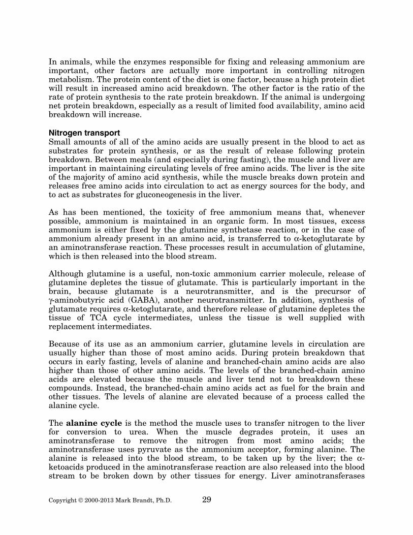
In animals, while the enzymes responsible for fixing and releasing ammonium are important, other factors are actually more important in controlling nitrogen metabolism. The protein content of the diet is one factor, because a high protein diet will result in increased amino acid breakdown. The other factor is the ratio of the rate of protein synthesis to the rate protein breakdown. If the animal is undergoing net protein breakdown, especially as a result of limited food availability, amino acid breakdown will increase. Nitrogen transport Small amounts of all of the amino acids are usually present in the blood to act as substrates for protein synthesis, or as the result of release following protein breakdown. Between meals (and especially during fasting), the muscle and liver are important in maintaining circulating levels of free amino acids. The liver is the site of the majority of amino acid synthesis, while the muscle breaks down protein and releases free amino acids into circulation to act as energy sources for the body, and to act as substrates for gluconeogenesis in the liver. As has been mentioned, the toxicity of free ammonium means that, whenever possible, ammonium is maintained in an organic form. In most tissues, excess ammonium is either fixed by the glutamine synthetase reaction, or in the case of ammonium already present in an amino acid, is transferred to α-ketoglutarate by an aminotransferase reaction. These processes result in accumulation of glutamine, which is then released into the blood stream. Although glutamine is a useful, non-toxic ammonium carrier molecule, release of glutamine depletes the tissue of glutamate. This is particularly important in the brain, because glutamate is a neurotransmitter, and is the precursor of γ-aminobutyric acid (GABA), another neurotransmitter. In addition, synthesis of glutamate requires α-ketoglutarate, and therefore release of glutamine depletes the tissue of TCA cycle intermediates, unless the tissue is well supplied with replacement intermediates. Because of its use as an ammonium carrier, glutamine levels in circulation are usually higher than those of most amino acids. During protein breakdown that occurs in early fasting, levels of alanine and branched-chain amino acids are also higher than those of other amino acids. The levels of the branched-chain amino acids are elevated because the muscle and liver tend not to breakdown these compounds. Instead, the branched-chain amino acids act as fuel for the brain and other tissues. The levels of alanine are elevated because of a process called the alanine cycle. The alanine cycle is the method the muscle uses to transfer nitrogen to the liver for conversion to urea. When the muscle degrades protein, it uses an aminotransferase to remove the nitrogen from most amino acids; the aminotransferase uses pyruvate as the ammonium acceptor, forming alanine. The alanine is released into the blood stream, to be taken up by the liver; the α-ketoacids produced in the aminotransferase reaction are also released into the blood stream to be broken down by other tissues for energy. Liver aminotransferases

Copyright © 2000-2013 Mark Brandt, Ph.D. 30
begin the process of diverting the alanine nitrogen to urea production, and the pyruvate produced from alanine is used as a gluconeogenic substrate. The glucose thus produced is then released into the bloodstream. The muscle can take up glucose for energy and in order to generate more pyruvate for additional nitrogen transport. Like the Cori cycle for lactate, the alanine cycle allows the muscle to “borrow” the liver mitochondria for energy production, and allows the muscle to transfer nitrogen to the liver while using amino acid breakdown for energy.
Amino acid breakdown pathways The amino acids are also frequently divided into families, based on the type of amino acid, or the final product formed from the amino acid. C3 Family: Amino acids convertible to pyruvate Alanine is converted directly to pyruvate by an aminotransferase reaction.
Serine can be converted to pyruvate by serine dehydratase; this reaction is common in some animals, but is a relatively minor pathway in humans. In humans the main pathway for metabolism of both serine and glycine is the conversion of
Pyruvate Glucose Glucose
Pyruvate
Alanine
Protein
Amino acidsα-ketoacids
AlanineNH4
Glutamine Branched-chainamino acids
Urea
MuscleLiver
UreaExcretionProducts
NH4Kidney
Alanine cycle
Brain
2 ATP4 ATP + 2 GTP
O O
O
O
O
+
O O
H3NAlanine
aminotransferase
α-Ketoglutarate Alanine
O O
H3N
O
O
+
O O
O
Glutamate Pyruvate

Copyright © 2000-2013 Mark Brandt, Ph.D. 31
serine to glycine by serine hydroxymethyltransferase, and glycine to carbon dioxide and ammonium by glycine synthase. These reactions both generate N5,N10-methylene tetrahydrofolate, which can then be used for other biosynthetic processes. This pathway therefore requires free tetrahydrofolate. Both reactions are reversible; if methylene tetrahydrofolate is available, serine levels tend to increase, and serine may be converted to pyruvate.
Cysteine can be metabolized by more than one pathway, generally producing pyruvate as the end product. These pathways begin with an aminotransferase reaction that removes the ammonium. The next steps vary depending on the organism and on conditions. The best-characterized pathway releases the toxic hydrogen sulfide (as shown below); other, less well-understood pathways appear to release sulfate. Threonine can be converted to glycine by serine hydroxymethyltransferase (the same enzyme that converts serine to glycine; in both reactions, this pyridoxal phosphate-dependent enzyme catalyzes the cleavage of the bond between the α- and β-carbons); in this case, the reaction involves the loss of acetaldehyde, which can be converted to acetyl CoA. The enzymes mentioned above then metabolize the glycine formed. This means that threonine can be used as a source of either glycine or
Serinehydroxymethyl
transferase
O O
H3N
Glycine
N5,N10-MethyleneTetrahydrofolateTHF
NH4+ O C O
Glycinecleavage enzyme
(Glycine synthase)
N5,N10-MethyleneTetrahydrofolate
NAD+
THF
NADH+
O O
H3N
OH
Serine
NH4
SerinedehydrataseO O
CH3O
Pyruvate
O O
H3N
OH
Threonine
CH3
Serinehydroxymethyl
transferaseH
O
CH3
Acetaldehyde
NADNADH
O O
H3N
O
2-Amino-3-ketobutyrate
CH3
Threoninedehydrogenase
CoA-SH
S
O
CH3CoA 2-Amino-3-ketobutyrateCoA lyase Aldehyde
dehydrogenase
FAD FADH2
O
O
CH3
Acetate
ATP+ CoA-SH
AMP+ PPi
Acetyl-CoASynthetase 2
S
O
CH3CoA
AlanineaminotransferaseO O
CH3H3N
Alanine
Glutamateα-Ketoglutarate
O O
H3N
SH
Cysteine
Alanineaminotransferase
Glutamate α-Ketoglutarate
O O
O
SH
Thiolpyruvate
H2S 3-Mercaptopyruvatesulfurtransferase
Acetyl-CoA
Acetyl-CoA
H2O+
NH4H2O +Threoninedehydratase
O O
CH2O
CH3
NAD+
CoA-SH
NADH+ CO2
Propionyl-CoA S
CH2O
CH3
CoA α-Ketobutyrateα-Ketoaciddehydrogenase

Copyright © 2000-2013 Mark Brandt, Ph.D. 32
serine. The reverse, however, is not true; humans cannot synthesize threonine. Threonine can also be converted to α-ketobutyrate by threonine dehydratase; the four-carbon α-ketobutyrate is converted to the three-carbon propionyl-CoA by α-ketoacid dehydrogenase, an enzyme complex similar to pyruvate dehydrogenase. The α-ketoacid dehydrogenase reaction is driven by the loss of carbon dioxide. Threonine can therefore be both glucogenic and ketogenic: breakdown of threonine results in either synthesis of the ketogenic acetyl CoA and the potentially glucogenic glycine, or in the production of the glucogenic propionyl-CoA. Side Note: Ethanol and Acetaldehyde Acetaldehyde is produced by the action of serine hydroxymethyltransferase on threonine. It is also produced from ethanol by alcohol dehydrogenase. Acetaldehyde is mildly toxic. In addition, the reduction of NAD to NADH by alcohol dehydrogenase can inhibit gluconeogenesis by inhibiting the conversion of lactate to pyruvate, resulting in lactate accumulation, and decreased plasma pH. These and other factors can result in liver damage following chronic ethanol ingestion. The liver has more than one pathway for metabolism of ethanol. One major pathway for the conversion of ethanol or acetaldehyde to acetyl-CoA is shown below.
C4 Family: amino acids convertible to oxaloacetate Asparagine can be converted to aspartate by release of free ammonium in a reaction catalyzed by asparaginase.
Aspartate is converted to oxaloacetate by a variety of aminotransferases. However, aspartate can also be converted to fumarate as part of the urea cycle, and in the adenylosuccinate lyase reaction of the adenosine biosynthesis pathway.
Ethanol
H2C
OH
CH3H
C
O
CH3
AcetaldehydeAldehyde
dehydrogenase
FAD FADH2
OC
O
CH3
Acetate
ATP+ CoA-SH
AMP+ PPi
Acetyl-CoASynthetase 2
SC
O
CH3CoA
Acetyl-CoAAlcoholdehydrogenase
NADNADH
H2O NH4
AsparaginaseO O
H3N
NH2
Asparagine
O
O O
H3N
O
Aspartate
O
Fumarate
O O
O
OO
Oxaloacetate
Aspartateaminotransferase
Glutamateα-Ketoglutarate
O O
H3N
OO
Aspartate
O O
OO
Copyright © 2000-2013 Mark Brandt, Ph.D. 33
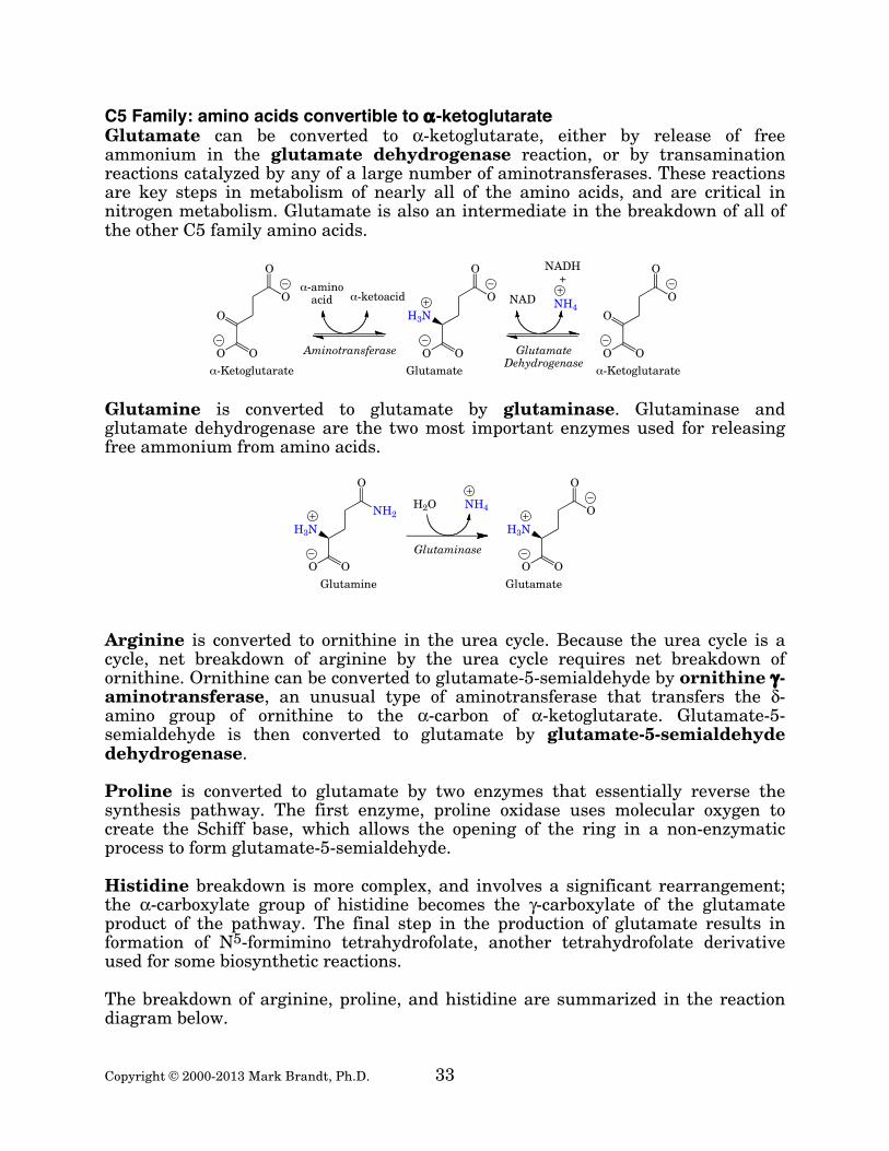
C5 Family: amino acids convertible to α-ketoglutarate Glutamate can be converted to α-ketoglutarate, either by release of free ammonium in the glutamate dehydrogenase reaction, or by transamination reactions catalyzed by any of a large number of aminotransferases. These reactions are key steps in metabolism of nearly all of the amino acids, and are critical in nitrogen metabolism. Glutamate is also an intermediate in the breakdown of all of the other C5 family amino acids.
Glutamine is converted to glutamate by glutaminase. Glutaminase and glutamate dehydrogenase are the two most important enzymes used for releasing free ammonium from amino acids.
Arginine is converted to ornithine in the urea cycle. Because the urea cycle is a cycle, net breakdown of arginine by the urea cycle requires net breakdown of ornithine. Ornithine can be converted to glutamate-5-semialdehyde by ornithine γ-aminotransferase, an unusual type of aminotransferase that transfers the δ-amino group of ornithine to the α-carbon of α-ketoglutarate. Glutamate-5-semialdehyde is then converted to glutamate by glutamate-5-semialdehyde dehydrogenase. Proline is converted to glutamate by two enzymes that essentially reverse the synthesis pathway. The first enzyme, proline oxidase uses molecular oxygen to create the Schiff base, which allows the opening of the ring in a non-enzymatic process to form glutamate-5-semialdehyde. Histidine breakdown is more complex, and involves a significant rearrangement; the α-carboxylate group of histidine becomes the γ-carboxylate of the glutamate product of the pathway. The final step in the production of glutamate results in formation of N5-formimino tetrahydrofolate, another tetrahydrofolate derivative used for some biosynthetic reactions. The breakdown of arginine, proline, and histidine are summarized in the reaction diagram below.
O O
O
O
O
α-Ketoglutarate
O O
O
O
O
α-Ketoglutarate
Aminotransferase O O
H3N
Glutamate
O
O
NADH+
NAD
GlutamateDehydrogenase
NH4
α-aminoacid α-ketoacid
H2O NH4
Glutaminase
O O
H3N
O
NH2
Glutamine
O O
H3N
Glutamate
O
O

Copyright © 2000-2013 Mark Brandt, Ph.D. 34
Branched-chain amino acids The branched-chain amino acids valine, isoleucine, leucine are metabolized by similar pathways. (Note: threonine also contains a β-branch, but it is broken down in other pathways.) The first step is a reversible transamination reaction catalyzed by branched-chain aminotransferase. The second step is catalyzed by branched-chain α-ketoacid dehydrogenase complex, an enzyme complex similar to the pyruvate dehydrogenase complex. This reaction results in branched-chain CoA derivatives, with the α-carboxylate lost as carbon dioxide. The third step is a dehydrogenase reaction similar to the first step in fatty acid β-oxidation. Because the same enzymes catalyze these initial steps, a defect in one enzyme results in elevated levels of partial breakdown products from all three amino acids. A deficiency in branched-chain α-ketoacid dehydrogenase complex results in severe
H2O
O O
H3N
NH3
Arginine
Arginase
Ornithine
O O
H3N
HNC
NH2
NH2
OC
NH2
NH2
Urea
Ornithineδ-aminotransferase
Glutamate
α-Keto-glutarate
O O
H3N
O
Glutamateγ-semialdehyde
H
NH2O
O
H2O
Proline oxidase
1/2 O2
NHO
O
Proline
H2O
Non-enzymatic
NAD(P)
NAD(P)H
Glutamate5-semialdehydedehydrogenase
O O
H3N
O
Glutamate
O
Histidine
O O
H3N
NH
N
NH4
Histidineammonia lyase
O O
NH
N
H2O
urocanate hydratase
O O
NH
N
O
H2O
O O
HN
O
N-Formiminoglutamate
OHNTHF
N5-Formimino-THF
imidazolonepropionase
glutamate-formimino-transferase
Pyrroline-5-carboxylate
Urocanate
4-Imidazolone-5-propionate

Copyright © 2000-2013 Mark Brandt, Ph.D. 35
neurological problems and death unless treated. The urine of affected individuals smells like maple syrup, hence the name Maple Syrup Urine Disease.
The later steps vary depending on the structure of the amino acid. Isoleucine is metabolized by reactions identical to those in a β-oxidation spiral, yielding an acetyl-CoA and a propionyl-CoA. The metabolism of valine is slightly more complex, but the pathway also results in propionyl-CoA. Propionyl-CoA is converted to the TCA cycle intermediate succinyl-CoA; these amino acids can therefore be used to synthesize glucose. Isoleucine is both glucogenic and ketogenic, because it also results in acetyl-CoA formation.
O O
H3N
Leucine
O O
H3N
Valine
O O
H3N
Isoleucine
Branched-chainamino acid
aminotransferase
Glutamate
α-Ketoglutarate
Branched-chainamino acid
aminotransferase
Glutamate
α-Ketoglutarate
Branched-chainamino acid
aminotransferase
Glutamate
α-Ketoglutarate
O O
O
O O
O
α-KetoisovalerateO O
O
α-Keto-β-methylvalerate α-Ketoisocaproic acid
NAD + CoA-SH
NADH + CO2
Branched-chainα-ketoaciddehydrogenase
NAD + CoA-SH
NADH + CO2
Branched-chainα-ketoaciddehydrogenase
NAD + CoA-SH
NADH + CO2
Branched-chainα-ketoaciddehydrogenase
S
O
S
O
S
O
α-Methylbutyryl-CoACoA CoA CoA
Isobutyryl-CoA Isovaleryl-CoA
4 steps 5 steps 4 steps
Acetoacetate+
Acetyl-CoA
Propionyl-CoAPropionyl-CoA+
Acetyl-CoA

Copyright © 2000-2013 Mark Brandt, Ph.D. 36
Leucine is converted to HMG-CoA (the ketone body precursor), and is then metabolized by cellular pathways that use HMG-CoA as an intermediate; leucine is thus a purely ketogenic amino acid. One of the enzymes in the leucine breakdown pathway, β-methylcrotonyl-CoA carboxylase, is the fourth biotin-dependent enzyme in humans. Biotin is thus critical for branched-chain amino acid metabolism; both conversion of propionyl-CoA to succinyl-CoA and conversion of leucine to HMG-CoA require biotin-dependent enzymes. Aromatic amino acids and Lysine Phenylalanine is converted to tyrosine by phenylalanine hydroxylase. This step is essential both in the production of tyrosine from phenylalanine, and in the breakdown of phenylalanine. A genetic deficiency in phenylalanine hydroxylase causes phenylketonuria, a moderately rare disorder (~1 in 10,000 live births) in which phenylketones such as phenylpyruvate accumulate to the point that they become detectable in the urine. Phenylketones interfere with brain development; untreated individuals with phenylketonuria experience irreversible abnormalities in brain functioning. Treatment involves a diet with strict limitations on phenylalanine levels. After the age of about ten, the brain development is essentially complete, and dietary restrictions are less critical. Because phenylketones are readily detectable, and because the effects of untreated phenylketonuria are so severe, in the United States screening for phenylketonuria is routinely performed at birth. The disorder phenylketonuria illustrates the fact that the ascorbate-dependent enzyme p-Hydroxyphenyl pyruvate hydroxylase requires the hydroxyl on the aromatic ring for activity.
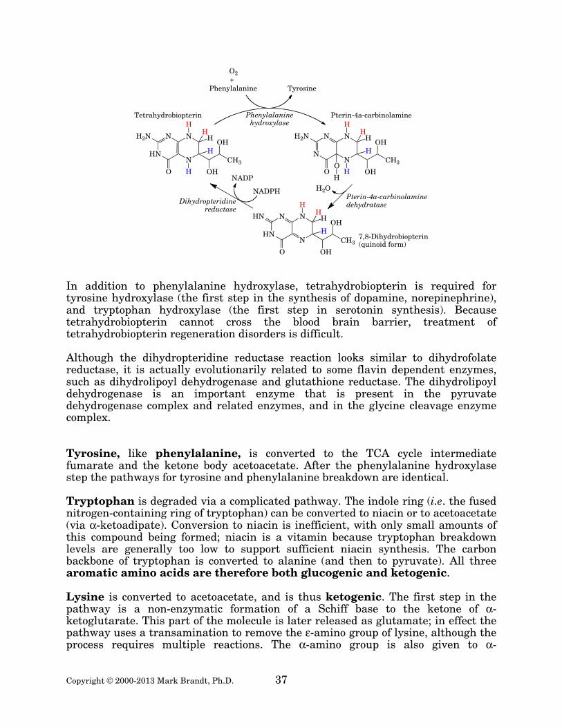
Phenylalanine hydroxylase uses tetrahydrobiopterin as a coenzyme. The tetrahydrobiopterin must then be regenerated using NADPH and a short pathway.
TyrosineaminotransferaseO O
CH2H3N
Tyrosine
Glutamateα-Ketoglutarate
O O
CH2H3N
Phenylalanine
Phenylalaninehydroxylase
O2
OH
O O
CH2O
p-Hydroxyphenylpyruvate
OH
CO2
p-Hydroxyphenylpyruvate
hydroxylase
OH
OH
Tetrahydro-biopterin
Pterin-4a-carbinolamine
Ascorbate
Dihydroascorbate
O2
H2O
O
O3 steps
O
OO
Acetoacetate
O
O
O
OFumarate
+
Homogentisate
H
H
+
+
+
Copyright © 2000-2013 Mark Brandt, Ph.D. 37
In addition to phenylalanine hydroxylase, tetrahydrobiopterin is required for tyrosine hydroxylase (the first step in the synthesis of dopamine, norepinephrine), and tryptophan hydroxylase (the first step in serotonin synthesis). Because tetrahydrobiopterin cannot cross the blood brain barrier, treatment of tetrahydrobiopterin regeneration disorders is difficult. Although the dihydropteridine reductase reaction looks similar to dihydrofolate reductase, it is actually evolutionarily related to some flavin dependent enzymes, such as dihydrolipoyl dehydrogenase and glutathione reductase. The dihydrolipoyl dehydrogenase is an important enzyme that is present in the pyruvate dehydrogenase complex and related enzymes, and in the glycine cleavage enzyme complex. Tyrosine, like phenylalanine, is converted to the TCA cycle intermediate fumarate and the ketone body acetoacetate. After the phenylalanine hydroxylase step the pathways for tyrosine and phenylalanine breakdown are identical. Tryptophan is degraded via a complicated pathway. The indole ring (i.e. the fused nitrogen-containing ring of tryptophan) can be converted to niacin or to acetoacetate (via α-ketoadipate). Conversion to niacin is inefficient, with only small amounts of this compound being formed; niacin is a vitamin because tryptophan breakdown levels are generally too low to support sufficient niacin synthesis. The carbon backbone of tryptophan is converted to alanine (and then to pyruvate). All three aromatic amino acids are therefore both glucogenic and ketogenic. Lysine is converted to acetoacetate, and is thus ketogenic. The first step in the pathway is a non-enzymatic formation of a Schiff base to the ketone of α-ketoglutarate. This part of the molecule is later released as glutamate; in effect the pathway uses a transamination to remove the ε-amino group of lysine, although the process requires multiple reactions. The α-amino group is also given to α-
HN
N
N
N
OH
H2N
O
H
HH
H
HCH3
OH
Tetrahydrobiopterin
N
N
N
N
OH
H2N
O
H
HH
H
HCH3
OH
Pterin-4a-carbinolamine
O
H
TyrosinePhenylalanine
Phenylalaninehydroxylase
O2+
HN
N
N
N
OH
HN
O
H
HH
HCH3
OH
H2OPterin-4a-carbinolaminedehydratase
7,8-Dihydrobiopterin(quinoid form)
NADP
NADPHDihydropteridine
reductase

Copyright © 2000-2013 Mark Brandt, Ph.D. 38
ketoglutarate in an aminotransferase reaction. The remainder of the pathway converges with the indole degradation pathway at the intermediate α-ketoadipate. The arrow from α-ketoadipate to acetoacetate represents a series of seven different reactions. (Note: some evidence suggests other pathways break down lysine to glucogenic compounds; however, it is likely that the major pathway for lysine breakdown results in synthesis of the ketone body acetoacetate.)
Methionine is metabolized by conversion to S-adenosylmethionine. The S-adenosylmethionine is then used for either methyl donor functions or as part of polyamine synthesis (a process which uses essentially the entire carbon skeleton of methionine). The homocysteine produced from S-adenosylmethionine is used to produce cysteine. If the goal is amino acid breakdown, the sulfur is metabolized using cysteine as an intermediate. The remainder of the methionine carbon skeleton is converted to propionyl-CoA.
O O
CH2H3N
Tryptophan
NH
O O
CH2H3N
N-Formylkynurenine
O
NH
O H
O O
CH2H3N
Kynurenine
O
NH2
O H
O O
CH2H3N
3-Hydroxykynurenine
O
NH2
OH
O
NH2
OH
O
O2
Tryptophan2,3-dioxygenase
H2O O
Formamidase
O2 + NADPH
H2O + NADP
H2O
Kynureninase[PLP]O O
CH3H3N
Alanine
+
6 steps
Kynurenine3-monooxygenase
OO
O
O
O
α-Ketoadipate
7 stepsO
OO
AcetoacetateO O
H3N
Lysine
NH3 4 steps
2Glutamate
2 α-Keto-glutarate
N
O
O Manysteps
Nicotinicacid
(Niacin)

Copyright © 2000-2013 Mark Brandt, Ph.D. 39
P
O
O
O O P
O
O
O
OHOHH
CH2O
N
N N
N
NH2
P
O
O
O
Adenosine Triphosphate(ATP)
+ P
O
O
O OH
Inorganicphosphate
+ P
O
O
O O P
O
O
OH
Pyrophosphate
O
OHOH
CH2
N
N N
N
NH2
SO
O
NH3
CH3
O O
H3N
Methionine
SCH3
Methionineadenosyltransferase
+
Methyltransferase
R CH3
R
O
OHOH
CH2
N
N N
N
NH2
SO
O
NH3
H2OAdenosine
hydrolase
SHO
O
NH3
Homocysteine
Methyl-cobalamin
Cobalamin
Methioninesynthase
S-Adenosylmethionine
S-Adenosylhomocysteine

Copyright © 2000-2013 Mark Brandt, Ph.D. 40
Summary Amino acids are a major energy source, especially during conditions in which glucose availability is limited. Most of the amino acids can be used as substrates for gluconeogenesis, and are termed glucogenic. In contrast leucine and lysine are converted to ketone bodies or to acetyl-CoA, and are therefore termed ketogenic. The three aromatic amino acids, threonine, and isoleucine are converted into both gluconeogenic compounds and ketogenic compounds, therefore are both glucogenic and ketogenic. The amino acids are grouped into families. The C3 family (alanine, serine, glycine, threonine, and cysteine) can all be converted to the 3-carbon α-ketoacid pyruvate, although some are converted to other molecules under appropriate conditions. The C4 family (aspartate and asparagine) are converted to the 4-carbon compounds oxaloacetate or fumarate. The branched-chain amino acids (leucine, isoleucine, and valine) are broken down by a series of common enzymes into CoA derivatives. These are then metabolized by separate pathways depending on the structure of the original compound. Leucine can be converted into HMG-CoA, the substrate for ketone body production, and is exclusively ketogenic. Valine is converted to propionyl-CoA and is exclusively glucogenic. Isoleucine is converted to acetyl-CoA and propionyl-CoA, and is therefore both ketogenic and glucogenic. The C5 family (glutamate, glutamine, histidine, proline, and arginine) are all converted to glutamate, and then to α-ketoglutarate. The aromatic amino acids phenylalanine and tyrosine result in formation of acetoacetate and fumarate, while tryptophan results in formation of acetoacetate and (via alanine) pyruvate. Each of these compounds is therefore both ketogenic and glucogenic. Lysine is broken down by a pathway that is related to that for the tryptophan indole ring, and forms acetoacetate. Methionine is used to produce a variety of biosynthetic intermediates; it can be converted into propionyl-CoA, and is therefore glucogenic.