3 D cell culture product selection guide for organoids...
12
3D cell culture product selection guide for organoids and spheroids
Transcript of 3 D cell culture product selection guide for organoids...

3D cell culture product selection guide for organoids and spheroids

2
Organoids and spheroids show great potential in many applications, including drug discovery, toxicology, and disease modeling. These 3D cell models offer opportunities to better understand complex biology in a physiologically relevant context.
As advances in culturing organoids and spheroids become more common, the need for cell culture guidance and product recommendations is becoming more prevalent. This selection guide is intended to give researchers a helpful starting point to facilitate the transition from 2D monolayer cultures to 3D cell models.
Introduction
Neural organoid cultured on a Thermo Scientific™ Nunclon™ Sphera™ 6-Well Plate, which allows cells to grow with virtually no attachment. The organoid was stained with antibodies conjugated to Invitrogen™ Alexa Fluor™ 488 and Alexa Fluor™ 594 dyes and imaged on an Invitrogen™ EVOS™ FL Auto 2 Imaging System at 10x magnification.
Introduction 2
Cell sources 4
Matrices and plasticware 5
Media systems to support growth 6
Tools for monitoring and detection 8
Tools for imaging and analysis 9
Key protocols and methods 10
Custom biology solutions 12
Contents

4
Researchers utilize cell lines to investigate disease models of interest. Gibco™ cell lines allow you to closely mimic the in vivo state and generate more physiologically relevant data from organoid and spheroid cultures.
The table below shows a selection of our Gibco™ primary and stem cells.
Cell sourcesPhysiologically relevant tissue models
Ordering informationProduct Cat. No.
Primary cells: liver
Human Plateable Hepatocytes, Induction Qualified HMCPIS
Cryopreserved Human Kupffer Cells HUKCCS
Stellate Cells Custom
HepaRG Cells, Cryopreserved HPRGC10
Liver Sinusoidal Endothelial Cells Custom
Primary cells: endothelial Human Umbilical Vein Endothelial Cells (HUVEC) C0035C
Primary cells: skin
Human Epidermal Keratinocytes, Neonatal (HEKn) C0015C
Human Epidermal Keratinocytes, Adult (HEKa) C0055C
Human Epidermal Melanocytes (HeMn), light, medium, or dark pigmentation
C0025C
C1025C
C2025C
Human Dermal Fibroblasts, Neonatal (HDFn) C0045C
Human Dermal Fibroblasts, Adult (HDFa) C0135C
Human Microvascular Endothelial Cells, Neonatal Dermis (HMVECnd) C0105C
Stem cellsHuman Episomal iPSC Line A18945
Cas9 iPSC Line Custom
Skin tissue model established from Gibco™ Human Epidermal Keratinocytes (HEKa) on Thermo Scientific™ Nunc™ Cell Culture Inserts and Carrier Plate System.

When growing 3D cell cultures, the surface you choose is essential for reproducible results.
Scaffold-based systems are used to provide physical structures to support the assembly of cells into 3D models and to expand to significant numbers. Scaffold-free systems are matrix-free alternatives and are generally more adaptable to forming 3D cell models that are naturally established by endogenous adhesion molecules and extracellular matrices. Porous membrane–based systems are advantageous when polarization and differentiation of epithelial cells are needed in constructing 3D tissue models. Selecting the right culture platform is an important first step in developing a successful culture system for organoids and spheroids.
Matrices and plasticwareScaffold-based and scaffold-free offerings
Ordering informationProduct Cat. No.
Scaffold-based systemsGeltrex LDEV-Free Reduced Growth Factor Basement Membrane Matrix A1413201
AlgiMatrix 3D Culture System, 96-Well Plate 12684015
Scaffold-free systems
Nunclon Sphera Microplates, 96U-Well Plate 174925
Nunclon Sphera Multidishes, 24-Well Plate 174930
Nunclon Sphera Dishes, 35 mm Dish 174943
Nalgene Single-Use PETG Erlenmeyer Flasks with Plain Bottom, 250 mL 41120250
Porous membrane–based and
multilayer systems
Nunc Polycarbonate Cell Culture Inserts in Multi-Well Plates, 0.4 µm pore size, 24-well 140620
Nunc 24-Well Carrier Plate with Cell Culture Inserts, 0.4 μm pore size 141002
HeLa spheroid cultured in a scaffold-free Thermo Scientific™ Nunclon™ Sphera™ 96U-well plate.

6
The Gibco™ brand is the one most cited for media and reagents in peer-reviewed publications on organoid and spheroid research.* Gibco™ products are widely used and trusted for consistency in the growth, differentiation, and maturation of 3D cell models. Using the right combination of media and growth factors is vital to supporting the formation of disease-relevant 3D organoids and spheroids from specialized cell types like stem cells or cancer cell lines.
Media systems to support growthGrowth, differentiation, and maturation of 3D cell models
Ordering informationProduct Cat. No.
Pluripotent stem cells
Media
StemFlex Medium A3349401
Essential 8 Medium A1517001
Essential 8 Flex Medium Kit A2858501
StemPro hESC SFM 1000701
Essential 6 Medium A1516401
ReagentsKnockOut Serum Replacement 10828010
RevitaCell Supplement A2644501
Skin cells
Media
Keratinocyte-SFM 17005042
Medium 154 M154500
EpiLife Medium MEPI500CA
Medium 254 M254500
Medium 106 M106500
MCDB 131 Medium 10372019
Reagents
Human Keratinocyte Growth Supplement (HKGS) S0015
EpiLife Defined Growth Supplement (EDGS) S0125
Supplement S7 S0175
Human Melanocyte Growth Supplement (HMGS) S0025
Low Serum Growth Supplement (LSGS) S00310
Microvascular Growth Supplement (MVGS) S00525
Cancer cells and tumoroids MediaDMEM/F-12, GlutaMAX Supplement 10565042
Essential 8 Medium 11875093
* Based on a third-party market report covering papers cited for disease modeling of organoids and spheroids with primary or stem cells as the starting cell type.
Growth factors High-quality Gibco™ growth factors are designed to give you high biological activity, high purity (95% pure), and <0.1 ng endotoxin per microgram. Our growth factors are verified with Gibco media for proven compatibility.
For a list of growth factors, go to thermofisher.com/growthfactors
Ordering information (cont.)Product Cat. No.
Hepatic cells
MediaWilliams’ E Medium A1217601
Advanced DMEM/F-12 12634010
ReagentsHepExtend Supplement A2737501
Primary Hepatocyte Maintenance Supplements CM4000
Brain cells
Media Neurobasal Medium 21103049
Reagents
B-27 Supplement, serum free 17504044
B-27 Supplement, minus insulin A1895601
B-27 Supplement, minus vitamin A 12587010
N-2 Supplement 17502001
Multiple cell types MediaDMEM/F-12, GlutaMAX Supplement 10565042
RPMI 1640 Medium 11875093
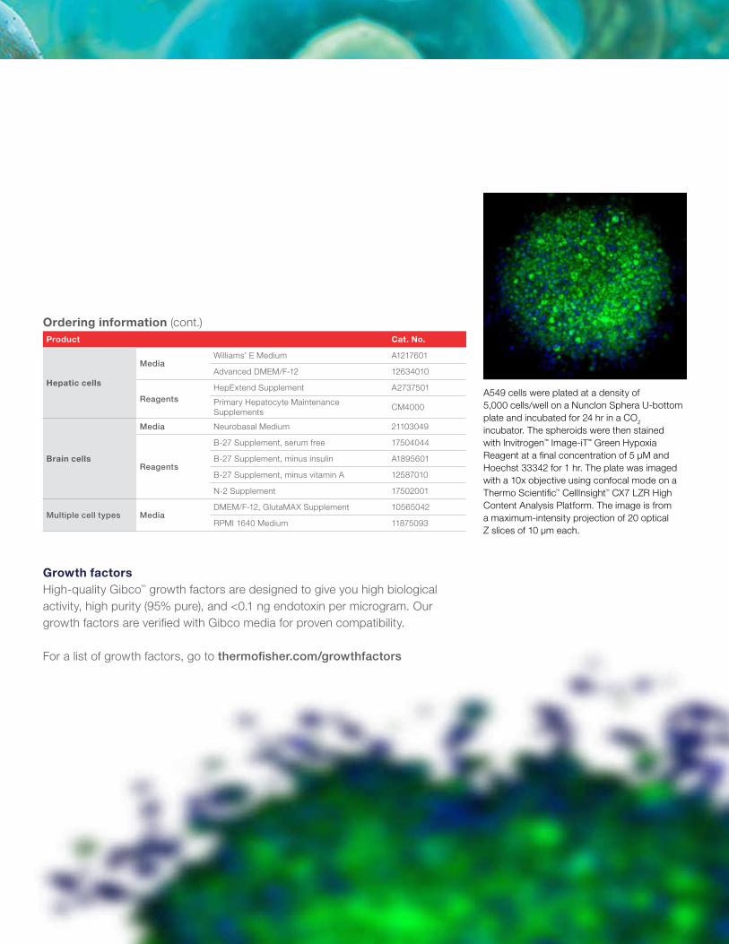
A549 cells were plated at a density of 5,000 cells/well on a Nunclon Sphera U-bottom plate and incubated for 24 hr in a CO2 incubator. The spheroids were then stained with Invitrogen™ Image-iT™ Green Hypoxia Reagent at a final concentration of 5 µM and Hoechst 33342 for 1 hr. The plate was imaged with a 10x objective using confocal mode on a Thermo Scientific™ CellInsight™ CX7 LZR High Content Analysis Platform. The image is from a maximum-intensity projection of 20 optical Z slices of 10 µm each.

8
qPCRWhen, where, and under what conditions are genes expressed? What triggers, or prevents, this expression? Scientists are discovering the surprisingly wide range of transcription and translation products, and how these different expression products determine the growth and health of an organism.
TaqMan AssaysOver 1.8 million predesigned Applied Biosystems™ TaqMan® Gene Expression Assays covering a growing list of model species have been predesigned using long-standing bioinformatics expertise in primer and probe design.
For more details, go to thermofisher.com/taqman
Tools for monitoring and detectionGene expression
Ordering informationProduct Cat. No.
QuantStudio real-
time PCR systems
QuantStudio 3 Real-Time PCR System, 96-well, 0.2 mL
A28137
QuantStudio 5 Real-Time PCR System, 96-well, 0.2 mL
A28139
QuantStudio 6 Flex Real-Time PCR System, 96-well, laptop
4485689
QuantStudio 7 Flex Real-Time PCR System, array card, laptop
4485700
QuantStudio 12K Flex Real-Time PCR System, Fast 96-well block, desktop
4471088
Spheroid staining using Invitrogen™ CellROX™ Deep Red Reagent. HeLa spheroids were pretreated with 100 μM menadione. Cells showing oxidative stress are stained red, and live-cell nuclei are stained blue.
Ordering informationProduct Cat. No.
Cell health reagents
Image-iT Fixation/Permeabilization Kit R37602
NucBlue Live ReadyProbes Reagent R37605
Click-iT Plus EdU Alexa Fluor 488 Imaging Kit
C10337
Image-iT Red Hypoxia Reagent H10498
Image-iT Green Hypoxia Reagent I14834
CellEvent Caspase-3/7 Green Detection Reagent
C10723
LIVE/DEAD Viability/Cytotoxicity Kit L3224
CellTracker Deep Red Dye C34565
CellROX Deep Red Reagent C10422
MitoTracker Orange M7510
ProLong Glass Antifade Mountant P36980
LysoTracker Deep Red L12492
Calcein, AM C3100MP
Cell healthReagents for 3D models Ensuring cells are maintaining the appropriate physiological morphology, markers, and activity is paramount to ensuring successful research outcomes. We have a full line of plate readers, imaging systems, and high-content analysis platforms to help you image and analyze your spheroids and organoids. These easy-to-use systems, combined with our broad portfolio of Invitrogen™ fluorescent reagents for cellular assays, allow researchers to effectively evaluate and understand 3D cell models.

Growing 3D models is a large investment in time and resources, and you need reassurance that your investment is going to give you the 3D models that you anticipate. Our visualization tools allow you to monitor the formation of your organoids and 3D models to give you confidence that you are heading in the right direction. These imaging and high-content analysis platforms have been recognized as trustworthy systems for analyzing organoid and spheroid cultures. In addition, Invitrogen™ antibodies are validated* to help ensure specificity and reproducibility in research results.
Tools for imaging and analysisVisualization of 3D cell models
Ordering informationProduct Cat. No.
Instruments
Varioskan LUX Multimode Microplate Reader VLB000D0
EVOS FL Auto 2 Imaging System AMAFD2000
CellInsight CX7 LZR High Content Analysis Platform CX7A1110LZR
EVOS XL Core Imaging System AMEX1000
EVOS XL Core Attachable Mechanical Stage AMEP4712
* The use or any variation of the word “validation” refers only to research use antibodies that were subject to functional testing to confirm that the antibody can be used with the research techniques indicated. It does not ensure that the product(s) was validated for clinical or diagnostic use.
HeLa cells were plated at a density of 5,000 cells/well on a Nunclon Sphera U-bottom plate and incubated for 24 hr in a CO2 incubator. The spheroids were then fixed with 4% formaldehyde and permeabilized with 0.25% Triton™ X-100. The spheroids were blocked with 3% BSA and then stained with Ki-67 antibody conjugated to Alexa Fluor 647 dye using the Invitrogen™ Zip Alexa Fluor™ 647 Rapid Antibody Labeling Kit. The plate was imaged with a 10x objective using confocal mode on a CellInsight CX7 LZR High Content Analysis Platform. The image is from a maximum-intensity projection of 20 optical Z slices of 10 µm each.

10
There is a wide range of protocols and methods for 3D cell model formation published to date. Table 1 is a selection of the seminal publications for different cell types, to help you get started on your journey to formation of 3D cell models.
Key protocols and methodsMost-cited publications
Table 1. Publications for 3D cell models.Organ Model type Cell type Differentiated cell type Relevant growth factor Relevant medium Key publication
Brain OrganoidPluripotent stem cells
• Ventricular zone radial glial cells• Cortical neurons
NADMEM/F-12, Neurobasal
Iefremova V et al. (2017) Cell Reports 19:50-59
• Cortical progenitor cells• Cortical neurons
BDNF, GDNF, NT-3, laminin, bFGF
DMEM/F-12, Essential 8, Essential 6, Neurobasal
Zhou T et al. (2017) Cell Stem Cell 21:274-283
• Cortical neurons DKK1 DMEM/F-12Phillips AW et al. (2017) J Vis Exp 125:e55799.
• Cortical neurons• Astrocytes
FGF2, EGF, BDNF, NT-3DMEM/F-12, Neurobasal
Pasca AM et al. (2015) Nature Methods 12:671-678
• Cortical neurons• Radial glial stem cells• Retinal cells
FGF2DMEM/F-12, Neurobasal
Lancaster MA et al. (2013) Nature 501:373-379
• Cortical neurons DKK1, BMPR1a-FcDMEM/F-12, Neurobasal
Mariani J et al. (2012) Proc Natl Acad Sci U S A 109:12770-12775
Liver
Spheroid Primary cells • Hepatocytes NA HepaRGProctor WR et al. (2017) Arch Toxicol 91:2849-2863
Organoid
Pluripotent stem cells
• Hepatocytes• Septum transversum mesenchyme• Endothelial cells
BMP4, VEGF, activin, PDGF-BB, FGF2
RPMI 1640, DMEM/F-12, StemPro-34 SFM
Takebe T et al. (2017) Cell Reports 21:2661-2670
• Hepatocytes• Cholangiocytes
Activin, BMP4, FGF2, FGF10, EGF, HGF
DMEM/F-12, RPMI 1640
Guan Y et al. (2017) JCI Insight 2:e94954
• HepatocytesActivin, TGFbeta, FGF2, BMP4, FGF4, DKK1, OSM, HGF
IMDM, F-12Pettinato G et al. (2016) Sci Rep 6:32888
Adult stem cells
• HepatocytesEGF, FGF10, HGF, noggin, BMP7, FGF19
Advanced DMEM/F-12
Huch M et al. (2015) Cell 160:299-312
Adult stem cells
• HepatocytesFGF10, HGF, EGF, noggin
Advanced DMEM/F-12
Huch M et al. (2013) Nature 494:247-250
Pluripotent stem cells
• Hepatocytes• HUVECs• Mesenchymal stem cells
FGF2, BMP4, activin, HGF, EGF
RPMI 1640Takebe T et al. (2013) Nature 499:481-484
Spheroid cell viability assay. Spheroid cell viability was evaluated using the Invitrogen™ LIVE/DEAD™ Cell Imaging Kit, where live cells are stained green and dead cells are stained red. Scale bar = 1,000 μm.
HCT 116 (1,000 cells/well)

11
Table 1. Publications for 3D cell models. (cont.)Organ Model type Cell type Differentiated cell type Relevant growth factor Relevant medium Key publication
Lung Organoid
Pluripotent stem cells, fetal cells
• Fibroblasts• Mesenchymal stem cells• HUVECs• Type I alveolar cells• Type II alveolar cells
FGF2 DMEM/F-12Wilkinson DC et al. (2017) Stem Cells Transl Med 6:622-633
Pluripotent stem cells
• Basal cells• Ciliated cells• Club cells• Mesenchymal stem cells• Alveolar progenitor cells
Activin, FGF4, FGF2, noggin, SHH
RPMI 1640, Advanced DMEM/F-12
Dye BR et al. (2015) eLife 4:e05098
Intestine Organoid
Pluripotent stem cells
• Goblet cells• Enteroendocrine cells• Mesenchymal stem cells
Activin, FGF4, BMP2, EGF, noggin, SHH
Advanced DMEM/F-12, RPMI 1640
Munera JO et al. (2017) Cell Stem Cell 21:51-64
• Enterocytes• Paneth cells• Goblet cells• Enteroendocrine cells• Innervated smooth muscle cells
IGF1, FGF2 KnockOut DMEMUchida H et al. (2017) JCI Insight 2:e86492
• Enterocytes• Paneth cells• Goblet cells• Enteroendocrine cells• Tuft cells• Smooth muscle cells
Activin, FGF4, EGF, noggin
RPMI 1640, Advanced DMEM/F-12
Watson CL et al. (2014) Nat Med 20:1310-1314
• Enterocytes• Paneth cells• Goblet cells• Enteroendocrine cells• Mesenchymal stem cells
Activin, FGF4, noggin, EGF
DMEM/F-12, RPMI 1640, Advanced DMEM/F-12
Spence JR et al. (2011) Nature 470:105-109
Adult stem cells
• Enterocytes• Paneth cells• Goblet cells• Enteroendocrine cells
Noggin, EGF, FGF10DMEM, Advanced DMEM/F-12
Sato T et al. (2011) Gastroenterology 141:1792-1772
• Enterocytes• Paneth cells• Goblet cells• Enteroendocrine cells
EGF, nogginAdvanced DMEM/F-12
Sato T et al. (2009) Nature 459:262-265
Kidney Organoid
Fetal tissue–derived cells
• Nephron progenitor cells• Glomerular cells• Proximal tubule epithelial cells• Loop of Henle epithelial cells• Distal tubule epithelial cells
BMP7, FGF2, LIF DMEM/F-12Li Z et al. (2016) Cell Stem Cell 19:516-529
Pluripotent stem cells
• Proximal tubule epithelial cells• Loop of Henle epithelial cells• Distal tubule epithelial cells• Podocytes
FGF2, noggin, activin, FGF9
Advanced RPMI 1640
Morizane R et al. (2015) Nat Biotechnol 33:1193-1200
Pluripotent stem cells
• Metanephric mesenchyme• Ureteric epithelial cells
FGF2, BMP4, activin, BMP7, FGF9
DMEM/F-12Takasato M et al. (2015) Nature 526:564-568

Find out more at thermofisher.com/3dmodel
For Research Use Only. Not for use in diagnostic procedures. © 2018 Thermo Fisher Scientific Inc. All rights reserved. All trademarks are the property of Thermo Fisher Scientific and its subsidiaries unless otherwise specified. TaqMan is a registered trademark of Roche Molecular Systems, Inc., used under permission and license. Triton is a trademark of Union Carbide Corporation. COL22613 0518
Scientists today are continually being asked to transition their research to more physiologically relevant disease models. Whether your team needs help developing the right cell model or is interested in outsourcing steps of the research project, our CellModel Services team can help provide a custom 3D cell model tailored to your research needs. Reach out to us at thermofisher.com/cellmodels.
Custom biology solutionsExtend your research capabilities and partner with our custom biology team
A549 cells were plated at a density of 5,000 cells/well on a Nunclon Sphera U-bottom plate and incubated for 24 hr in a CO2 incubator. The spheroids were then stained with Hoechst 33342 for 1 hr. The plate was imaged with a 10x objective using confocal mode on a CellInsight CX7 LZR High Content Analysis Platform. The image is from a maximum-intensity projection of 20 optical Z slices of 10 µm each.
A549 cells were plated at a density of 5,000 cells/well on a Nunclon Sphera U-bottom plate and incubated for 24 hr in a CO2 incubator. The spheroids were then fixed with 4% formaldehyde and permeabilized with 0.25% Triton X-100. The spheroids were blocked with 3% BSA and then stained with Ki-67 antibody conjugated to Alexa Fluor 647 dye using the Zip Alexa Fluor 647 Rapid Antibody Labeling Kit and Hoechst 33342. The plate was imaged with a 10x objective using confocal mode on a CellInsight CX7 LZR High Content Analysis Platform. The image is from a maximum-intensity projection of 20 optical Z slices of 10 µm each.