2nd Edition - JAYPEE BROTHERS: MEDICAL PUBLISHER · Includes 12-Lead ECG Interpretation ......
25
The Art and Science of Cardiac Physical Examination with Heart Sounds, Jugular and Precordial Pulsations on CD 2nd Edition Includes 12-Lead ECG Interpretation System requirements: • Operating System—Windows Vista or above • Recommended Web Browser—Google Chrome & Mozilla Firefox • Essential plugins—Java & Flash player ▪ Facing problems in viewing content—it may be your system does not have Java enabled. ▪ If Videos do not show up—it may be the system requires Flash player or need to manage Flash setting. To learn more about Flash setting click on the link in the help section. ▪ You can test Java and Flash by using the links from the help section of the CD/DVD. Accompanying CD/DVD Rom is playable only in Computer and not in DVD player. CD/DVD has Autorun function—it may take few seconds to load on your computer. If it does not works for you then follow the steps below to access the contents manually: • Click on My Computer • Select the CD/DVD drive and click open/explore—this will show list of files in the CD/DVD • Find and double click file—“launch.html” For more information about troubleshoot of Autorun click on: http://support.microsoft.com/kb/330135.
Transcript of 2nd Edition - JAYPEE BROTHERS: MEDICAL PUBLISHER · Includes 12-Lead ECG Interpretation ......
The Art and Science of Cardiac Physical Examination
with Heart Sounds, Jugular and Precordial Pulsations on CD
2nd EditionIncludes 12-Lead ECG Interpretation
System requirements:
• Operating System—Windows Vista or above • Recommended Web Browser—Google Chrome & Mozilla Firefox • Essential plugins—Java & Flash player
▪ Facing problems in viewing content—it may be your system does not have Java enabled.
▪ If Videos do not show up—it may be the system requires Flash player or need to manage Flash setting. To learn more about Flash setting click on the link in the help section.
▪ You can test Java and Flash by using the links from the help section of the CD/DVD.
Accompanying CD/DVD Rom is playable only in Computer and not in DVD player.
CD/DVD has Autorun function—it may take few seconds to load on your computer. If it does not works for you then follow the steps below to access the contents manually: • Click on My Computer • Select the CD/DVD drive and click open/explore—this will show list of files in the
CD/DVD • Find and double click file—“launch.html”
For more information about troubleshoot of Autorun click on:http://support.microsoft.com/kb/330135.
Prelims.indd 1 30-05-2015 13:57:13

Prelims.indd 2 30-05-2015 13:57:13

The Art and Science of Cardiac Physical Examination
with Heart Sounds, Jugular and Precordial Pulsations on CDIncludes 12-Lead ECG Interpretation
New Delhi | London | Philadelphia | Panama
The Health Sciences Publisher
Narasimhan Ranganathan MBBS FRCP(C) FACP FACC FAHA
Associate Professor in MedicineUniversity of Toronto, Ontario, Canada
Senior Cardiology Consultant St. Joseph’s Health CentreToronto, Ontario, Canada
Vahe Sivaciyan BSc MD FRCP(C)
Assistant Professor in Medicine University of Toronto, Ontario, Canada
Staff Cardiologist, St. Joseph’s Health Centre Toronto, Ontario, Canada
Franklin B Saksena MD CM FACP FRCP(C) FACC FAHA
Associate Professor in MedicineNorthwestern University School of Medicine
Chicago, Illinois, USA
ForewordSriram Rajagopal MD DM
Prelims.indd 3 30-05-2015 13:57:14
Jaypee Brothers Medical Publishers (P) LtdHeadquartersJaypee Brothers Medical Publishers (P) Ltd.4838/24, Ansari Road, DaryaganjNew Delhi 110 002, IndiaPhone: +91-11-43574357Fax: +91-11-43574314E-mail: [email protected]
Inquiries for bulk sales may be solicited at: [email protected]
The Art and Science of Cardiac Physical ExaminationSecond Edition: 2016ISBN: 978-93-5152-777-0Printed at:
Jaypee-Highlights Medical Publishers Inc.City of Knowledge, Bld. 237, ClaytonPanama City, PanamaPhone: +1 507-301-0496Fax: +1 507-301-0499E-mail: [email protected] Brothers Medical Publishers (P) Ltd.17/1-B, Babar Road, Block-B, ShaymaliMohammadpur, Dhaka-1207BangladeshMobile: +08801912003485E-mail: [email protected]
Overseas OfficesJ.P. Medical Ltd.83, Victoria Street, LondonSW1H 0HW (UK)Phone: +44-20 3170 8910Fax: +44(0)20 3008 6180E-mail: [email protected] Medical Inc.The Bourse111 South Independence Mall EastSuite 835Philadelphia, PA 19106, USAPhone: +1 267-519-9789E-mail: [email protected] Brothers Medical Publishers (P) Ltd.Bhotahity, Kathmandu, NepalPhone: +977-9741283608E-mail: [email protected]: www.jaypeebrothers.comWebsite: www.jaypeedigital.com
© 2016, Jaypee Brothers Medical Publishers
The views and opinions expressed in this book are solely those of the original contributor(s)/author(s) and do not necessarily represent those of editor(s) of the book.All rights reserved. No part of this publication and CD-ROM may be reproduced, stored or transmitted in any form or by any means, electronic, mechanical, photo copying, recording or otherwise, without the prior permission in writing of the publishers. All brand names and product names used in this book are trade names, service marks, trademarks or registered trademarks of their respective owners. The publisher is not associated with any product or vendor mentioned in this book.Medical knowledge and practice change constantly. This book is designed to provide accurate, authoritative information about the subject matter in question. However, readers are advised to check the most current information available on procedures included and check information from the manufacturer of each product to be administered, to verify the recommended dose, formula, method and duration of administration, adverse effects and contra indications. It is the responsibility of the practitioner to take all appropriate safety precautions. Neither the publisher nor the author(s)/editor(s) assume any liability for any injury and/or damage to persons or property arising from or related to use of material in this book.This book is sold on the understanding that the publisher is not engaged in providing professional medical services. If such advice or services are required, the services of a competent medical professional should be sought.Every effort has been made where necessary to contact holders of copyright to obtain permission to reproduce copyright material. If any have been inadvertently overlooked, the publisher will be pleased to make the necessary arrangements at the first opportunity.
Prelims.indd 4 30-05-2015 13:57:14
DedicationNarasimhan Ranganathan, Vahe Sivaciyan and Franklin B Saksena wish to dedicate this book
to their respective wives, Saroja, Ayda and Kathleen. For without their support, this book would not
have been possible.
Prelims.indd 5 30-05-2015 13:57:14

Prelims.indd 6 30-05-2015 13:57:14
Foreword
I feel privileged to be asked to write a foreword to this book The Art and Science of Cardiac Physical Examination. The authors are very experienced and senior clinicians, and have more than three decades of rigorous, scientific research into clinical signs and their mechanisms. They have made seminal contributions to the literature in this field, particularly in the area of the jugular venous pulse. Further, they are deeply committed to teaching and communicating the knowledge and insights that they have acquired over the years. The past few decades have seen considerable changes in the science and practice of cardiology. The plethora of new discoveries, new imaging modalities and newer modes of treatment has tended to overshadow the importance of sound clinical examination. Indeed, there is a widespread feeling that both the time and the importance accorded to the formal teaching of clinical skills in many contemporary cardiology training programs are inadequate. The authors' effort in bringing out this excellent book (and companion CD) serves as an effective and timely step to correct this trend. The treatment of the subject matter is comprehensive, with each of the main chapters starting with a detailed review of the normal physiology underlying a clinical phenomenon as well as the pathophysiology in different abnormal states, providing a clear understanding of the basis of the clinical sign. The chapters on the arterial pulse and jugular venous pulse are particularly illuminative in this respect. The correct technique of elicitation of the finding is then lucidly outlined, often with unique methods to demonstrate phenomena and insightful tips to improve bedside skills. Finally, the interpretation and integration of the information obtained is rightly emphasized, so that the finding can be placed in the context of the larger clinical picture in a cogent and meaningful manner. The summary at the end of each chapter provides a concise and rapid review to enhance learning. The chapters are extensively referenced providing rich material for further learning. The creative and original methods described in the chapter entitled “Elements of Auscultation” serve to beautifully unify the “Art” and “Science” aspects of auscultation. A separate chapter on “Pathophysiologic Basis of Symptoms and Signs in Cardiac Disease” serves to reiterate concepts described elsewhere in the book in the particular context of specific conditions. The novel use of audiovisual aids in the companion CD further remarkably enhances the value of this book as a learning resource. Examples from years of clinical observation have been carefully documented and painstakingly converted to video and audio clips that provide an unprecedented level of realism. The readers are provided with a “clinical experience” where
Prelims.indd 7 30-05-2015 13:57:14
The Art and Science of Cardiac Physical Examinationviii
they can literally see and hear the findings and can verify their skills of observation and interpretation in a “reallife” setting. This edition introduces two new chapters on electrocardiography, now widely regarded as part of the clinical evaluation. The first of these chapters provides extensive coverage of the principles of electrocardiography and interpretation, while the second chapter on “Integration of ECG into the Cardiac Diagnosis” provides a succinct account of the correlation of ECG findings in a wide range of cardiac disorders with the pathophysiology of these conditions. The section on selfassessment is also a valuable educational aid and serves to reinforce the message on the integration of information from different sources. This book is bound to be of immense value to any individual interested in clinical cardiology, from the fresh medical student (who will benefit from a sound and lucid introduction to the subject) to the senior and experienced clinician (who will gain new understanding and insight). The companion CD is wellsuited to serve as an important tool for both individual and group teaching. The authors are to be commended for their extraordinary effort in distilling decades of clinical experience into this extremely valuable contribution to the important field of clinical cardiology.
Sriram Rajagopal MD DM
Chief CardiologistSouthern Railway Headquarters Hospital
Chennai, Tamil Nadu, India
Prelims.indd 8 30-05-2015 13:57:14
Preface to the 2nd Edition
The first edition of our book was the result of our longlasting interest in promoting the usefulness and value of proper cardiac physical examination in the assessment of cardiac patients. It is a culmination of our longlasting experience in teaching and training physicians and students of cardiology. We have offered a course annually of the same title in Toronto over the last 35 years. Modern technological advances both invasive and noninvasive have contributed significantly to our knowledge and understanding of cardiac physical signs and their pathophysiologic correlates. Both students and the teachers alike become impressed by these technological tools to the extent of neglecting the ageold art as well as the substantial body of science behind the cardiac physical examination. These technological advances are here to stay. However, some have even gone to the extent of suggesting that a “physician should have an all purpose tool in his or her pocket that would be more in keeping with the 21st century than the stethoscope, a 200yearold technology whose time should be over”.1 One must never forget that any tool or instrument is only as good as the person using it. The information that can be derived from the proper assessment of the jugular contours, the precordial pulsations, the arterial pulses as well as cardiac auscultation can never be considered waste in terms of the assessment of a cardiac patient, in our opinion. It is not only cost effective and satisfying and can never be counterproductive to the patient’s needs. In addition, it could be lifesaving under certain circumstances (such as in remote locations, during power failure and times of disaster). Neglect of these basic skills, expected of physicians and cardiologists to be, will not augur well for the future generation of the physicians and patients alike. The positive features of our book include among other things innovative and proven effective teaching methods with the use of recordings of not only heart sounds and murmurs but also the actual videorecordings of both normal and abnormal jugular pulsations as well the precordial pulsations together with arterial flow signals and/or the heart sounds for timing of the events in relation to the cardiac cycle. We were pleased and not totally surprised however, when we discovered that our book was translated into Chinese, a few years ago.2 It suggests also that not all physicians share the opinion of some who would like to name the stethoscope as “archaic instrument” and lock it up in their office chest. In addition, it indicates a need to reach out to more medical schools and the institutions in many developed and developing nations. We are hoping that it would achieve that goal with our current publishers of this new and improved second edition.
Prelims.indd 9 30-05-2015 13:57:15
The Art and Science of Cardiac Physical Examinationx
In addition to the ‘The Art and Science of Cardiac Physical Examination’, we have also been interested in teaching 12lead ECG interpretation to physicians and trainees for many years offering annual courses. ECG is often considered an integral part of the office assessment of a cardiac patient and almost considered to be an extension of cardiac physical assessment. Most physicians either have or have access to an ECG machine in their offices. ECG is also indispensable in the assessment of patients presenting with acute symptoms of chest pain and or dyspnea. Therefore, when we were faced with the opportunity of providing a second edition, we wanted to make the book even more comprehensive. In addition to updating new and relevant information in several of the previous chapters of the first edition, we have included three new chapters. These consist of the following: a complete chapter consisting of six different sections which cover fully the 12lead Electrocardiogram Interpretation, a second chapter showing how to integrate the ECG into Cardiac Diagnosis and a third and final chapter for SelfAssessment at the end with several interesting clinical cases from our own practice. In addition, we have added a selfassessment section in the companion CD with several new clinical examples. We believe that these selfassessment sections would serve as a good review as well as being useful for reinforcement purposes both in selfteaching and/or group learning sessions. Before we end this preface, we would like to take the opportunity to reminisce and thank for the friendship and the association we have had both during the formative years of becoming a cardiologist as well as in the later years of career as a practicing cardiologist and as a teacher. During the years of training, I (the senior author) had the opportunity to work with some of the wellknown cardiologists including Dr George E Burch and Dr John Phillips of the reputed Tulane University medical school as well as Dr E Douglas Wigle and Dr Malcolm Silver (Cardiac Pathologist) of the University of Toronto. However, the longest association of teaching both cardiac physical examination and 12lead ECG interpretation was with Dr Jules Constant from the State University of Buffalo, New York, USA. We in fact used to invite him over to teach along with us in Toronto almost annually for many years in our annual cardiac physical examination course. I have also taken part in teaching along with him in ‘the 12lead ECG interpretation courses’ which he used to organize in the month of February in the warmer southern climate. He had a fine sense of empathy for the beginners, which was admirable. One’s own teaching technique also becomes more refined watching other masters perform. In fact, Dr. Constant was still alive at the time of the release of our first edition of the book. We wish to list the names of these individuals here in order not only to recognize their contribution in the field of cardiology but also to express our gratitude.
Prelims.indd 10 30-05-2015 13:57:15
Preface to the 2nd Edition xi
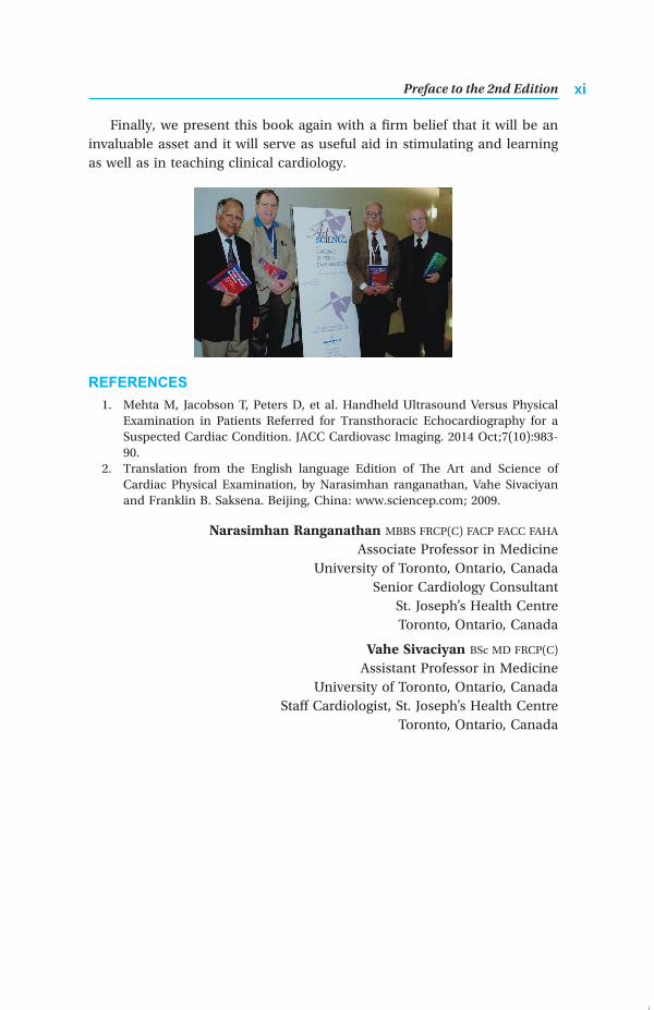
Finally, we present this book again with a firm belief that it will be an invaluable asset and it will serve as useful aid in stimulating and learning as well as in teaching clinical cardiology.
REFERENCES 1. Mehta M, Jacobson T, Peters D, et al. Handheld Ultrasound Versus Physical
Examination in Patients Referred for Transthoracic Echocardiography for a Suspected Cardiac Condition. JACC Cardiovasc Imaging. 2014 Oct;7(10):98390.
2. Translation from the English language Edition of The Art and Science of Cardiac Physical Examination, by Narasimhan ranganathan, Vahe Sivaciyan and Franklin B. Saksena. Beijing, China: www.sciencep.com; 2009.
Narasimhan Ranganathan MBBS FRCP(C) FACP FACC FAHA
Associate Professor in MedicineUniversity of Toronto, Ontario, Canada
Senior Cardiology Consultant St. Joseph’s Health CentreToronto, Ontario, Canada
Vahe Sivaciyan BSc MD FRCP(C)
Assistant Professor in Medicine University of Toronto, Ontario, Canada
Staff Cardiologist, St. Joseph’s Health Centre Toronto, Ontario, Canada
Prelims.indd 11 30-05-2015 13:57:15

Prelims.indd 12 30-05-2015 13:57:15
Preface to the 1st Edition
It has been our experience that teaching of the physical examination of the heart in medical schools has been deteriorating since the advent of the modern diagnostic tools such as the 2dimensional echocardiography and nuclear imaging. At best it has been sketchy and too superficial for the student to appreciate the pathophysiologic correlates. Both the invasive and the noninvasive modern technological advances have contributed substantially to our knowledge and understanding of the cardiac physical signs and their pathophysiological correlates. However, both the students and the teachers alike appear to be mesmerized by these technologic advances to the neglect of the ageold art as well as the substantial body of science of the cardiac physical examination. It is also sad to see that reputed journals also give low priority to articles related to the clinical examination. Our experience is substantiated by a nationwide survey of the teaching and practice of cardiac auscultation during internal medicine and cardiology training which concluded that there was in fact low emphasis on this and perhaps also on other bedside diagnostic skills.1 The state of the problem is wellreflected in the concerns expressed in previous publications.24 including the editorial in the American Journal of Medicine 2001;110:2335, entitled “Cardiac auscultation and teaching rounds: how can cardiac auscultation be resuscitated?” as well as in the rebuttal, “Selections from current literature. Horton hears a Who but no murmurs—does it matter?”.5 The latter goes to the extent of suggesting that auscultation be performed only when cardiac symptoms are encountered in patients. This appears to be based on an exaggerated concern for the waste of time and resources. Implicit in this statement if one chooses to agree with it, will be the acknowledgment of one’s failure as a physician caring for patients. On the contrary, we not only share the opinion of others that cardiac auscultation is a cost effective diagnostic skill6 and we would like to go one step further and suggest that all aspects of cardiac physical examination are very costeffective and rewarding in many ways. A properly obtained detailed and complete history of the patient’s problems and a thorough physical examination are never counterproductive to the interests of the patient. Modern technological advances are here to stay and they should be adjunct to the clinical examination of the patient but should not be allowed to replace them. Let us not forget that many of these tools do add to the rising costs of healthcare all over the world. A well carried out physical examination of the heart often provides the critical information necessary to choose the right investigative tool and to avoid the unnecessary ones. Even if one ignores the cost factor, a physician caring for a patient under
Prelims.indd 13 30-05-2015 13:57:15
The Art and Science of Cardiac Physical Examinationxiv
conditions where these techniques may not be accessible (at nights and on the weekends in some institutions, remote locations, during power failure and during times of natural or other disasters) should be able to assess and diagnose cardiac function and probable underlying pathology using the fives senses, a stethoscope and a sphygmomanometer. Mackenzie integrated the jugular venous pulse as part of the cardiovascular physical examination.7 Wood further went on to show that the precise analysis of the jugular venous pulse waveforms and the measurement of the venous pressure with reference to the sternal angle is possible at the bedside.8 Interpretation of the jugular venous pulse contour and the assessment of the pressure yet remains an occult art practiced only by experienced clinicians. Poor, illdefined and vague terms such as jugular venous distension are commonly used and written about even in reputed journals when cardiac physical findings are mentioned. One of the satisfying features of medicine aside from contributing to the clinical improvement of an ailing patient, is the intellectual excitement and satisfaction of arriving at the right conclusion through proper reasoning based on clues derived from the clinical examination of the patient. In addition, not surprisingly some of the physical signs have also been shown in this day and age of ‘Evidence based Medicine’, to be of prognostic importance. For instance elevated jugular venous pressure and the third heart sound in patients with symptomatic heart failure had been shown to have independent prognostic information.9 We believe that a proper understanding of the pathophysiologic correlates of the various signs and symptoms would help in developing skills of logical thinking required of a good clinician at work. It is all the more important if one were to plan to study the validity or the worthiness of the detection of an abnormal sound or sign in relationship to other cardiac measurements.10 Improper understanding would only result in testing of wrong hypotheses and misleading conclusion. The purpose of this book is to arm the student of cardiology with the proper techniques and understanding of the art and science of the cardiac physical examination, to dispel myths and confusion and to help develop skills required of any astute clinician. This book is a culmination of our efforts resulting from our longstanding experience of teaching and training physicians and trainees and students of cardiology. In fact an annual course entitled by the same name as our book has been organized and offered by us at our institution in Toronto over 25 years. They have been always well received and extremely appreciated for the teaching methods and the content by the attendees. Audio recordings of heart sounds and murmurs, as well as video recordings of jugular and precordial pulsations with simultaneously recorded sounds and flow signals for timing from actual patients collected over many years of clinical
Prelims.indd 14 30-05-2015 13:57:15

Preface to the 1st Edition xv
practice have been utilized in this course. Video display of the actual sounds and murmurs provides a realtime playback effect and enhances the group teaching and learning experience using multiple listening devices with infrared transmission of sounds. The teaching material and methods have been developed and refined over many years, stimulated by enthusiastic and inquisitive students and trainees and aided by our own research and studies particularly with reference to the jugular venous flows and pulsations as well as with regard to the precordial pulsations. The organization of the material presented in this book warrants some elaboration. We believe that the information is presented in a fashion integrating the science with concepts useful for logical integration into clinical applications. The teaching method adopted is somewhat unique and we believe totally original in some sections. This would be evident in chapters on Jugular Venous Pulse, the Precordial pulsations as well as the Arterial Pulse. The approach to the interpretation of the jugular venous pulsations presented here brings to the forefront the proper method of integration of the art with the science at the bedside. We believe that it is different in many ways from other books dealing with cardiac examination. Every important topic has a summary of salient and practical points from the point of view of clinical assessment. This would serve for quick review as well as act as pointers needing reinforcement. Many illustrations of sounds and murmurs used in the text are derived from digital display of actual audio recordings from patients The pathophysiology of some of the important clinical cardiac conditions are shown in flow diagrams as well as in tabular format permitting logical review and reinforcement. The references given at the end of the chapters are specially chosen to provide a variety of pertinent as well as the classic papers. A special chapter deals with local and systemic manifestations of cardiovascular disease authored by our colleague and friend Dr Franklin B Saksena (Senior Attending Physician, Division of Adult Cardiology, John Stroger, Jr. Hospital of Cook County and Assistant Professor of Medicine, Northwestern University, Chicago, Attending Physician, Swedish Covenant Hospital, Attending Physician, Saint Mary of Nazareth Hospital). It provides several useful illustrations as well as a major list of references. The audio and the video recordings of sounds and murmurs, the jugular and the precordial pulsations from actual patients with a variety of clinical cardiac problems are also available in a companion CD which will have all the videofiles playable through the Windows Media player on any new modern computer. These video recordings made from actual patients are meant to further enhance individual learning as well as group teaching of students and trainees in cardiology. They will provide a real time play back effect of heart sounds and murmurs displayed on an oscilloscopic screen. Another unique feature in the videofiles includes the presentation
Prelims.indd 15 30-05-2015 13:57:15
The Art and Science of Cardiac Physical Examinationxvi
of simultaneous recordings of the 2dimensional echocardiographic images together with the audiorecordings of the heart murmurs from a few patients with specific cardiac lesions. We present this book with a firm belief that it will be an invaluable asset and hope it will serve as a very useful tool in learning and teaching clinical cardiology.
REFERENCES 1. Mangione S, Nieman LZ, Gracely E, et al. The teaching and practice of cardiac
auscultation during internal medicine and cardiology training. A nationwide survey. Ann Intern Med 1993;119:4754.
2. Schneiderman H. Cardiac auscultation and teaching rounds: how can cardiac auscultation be resuscitated? Am J Med 2001;110:2335.
3. Lok CE, Morgan CD, Ranganathan N. The accuracy and interobserver agreement in detecting the ‘gallop sounds’ by cardiac auscultation. Chest 1998; 114:12838.
4. Tavel ME. Cardiac auscultation. A glorious past—but does it have a future? Circulation 1996;93:12503.
5. KopesKerr CP. Selections from current literature. Horton hears a Who but no murmurs—does it matter? Fam Pract 2002;19:4225.
6. Shaver. J. A. Cardiac auscultation: a costeffective diagnostic skill. Curr Probl Cardiol 1995;7:441530.
7. Mackenzie J. The study of the Pulse. London: Pentland, 1902. 8. Wood P. Diseases of the Heart and Circulation. Philadelphia,: JB Lippincott
Vo, 1956. 9. Drazner MH, Rame JE, Stevenson LW, et al. Prognostic importance of elevated
jugular venous pressure and a third heart sound in patients with heart failure. N Engl J Med 2001;345:57481.
10. Marcus GM, Gerber IL, McKeown BH, et al. Association between phonocardiographic third and fourth heart sounds and objective measures of left ventricular function. Jama 2005;293:223844.
Narasimhan Ranganathan MBBS FRCP(C) FACP FACC FAHA
Associate Professor in MedicineUniversity of Toronto, Ontario, Canada
Senior Cardiology ConsultantSt. Joseph’s Health CentreToronto, Ontario, Canada
Vahe Sivaciyan BSc MD FRCP(C)
Assistant Professor in Medicine University of Toronto, Ontario, Canada
Staff Cardiologist, St. Joseph’s Health Centre Toronto, Ontario, Canada
Prelims.indd 16 30-05-2015 13:57:15
Acknowledgments to the 2nd Edition
First of all, I would like to express my sincere thanks to all my colleagues at both the St. Michael’s Hospital as well as the St. Joseph’s Health Centre who have been supportive of my teaching endeavors during the years of my hospital practice. In addition we wish to express our sincere gratitude to all of our patients who had volunteered their time in this regards for the purposes of cardiac teaching. This second edition however, could not be successfully completed without the help of Mr. Roger Harris. He was also quite helpful in the preparation of the first edition of the book as mentioned in the previous acknowledgments. We wish to express and record here our profound thanks and acknowledgments for the invaluable assistance of Mr. Roger Harris in the production of not only the newer illustrations but also in the preparation of the companion CD and its content files for the second edition. Mr. Roger Harris is currently the retired chief of the audiovisual department of the St. Joseph’s Health Centre in Toronto. We had numerous audio recordings from actual clinical patients to choose from. But our desire to provide them in a similar format as the previous CD with real time playback effect had some tremendous obstacles to overcome. Video recordings of the oscilloscopic tracings of the phono signals and adjusting them for asynchrony between the video and the audio signals due to playback and recording involving different devices was the most difficult and time consuming of these. Luckily for us, the efforts of Mr. Roger Harris were not only very fruitful and victorious but also timely. Thus we are really indebted to him and therefore sincerely express our thanks to Roger for his invaluable assistance. We wish to express our sincere gratitude and thanks to Dr. Sriram Rajagopal, Chief of Cardiology, at the Southern Railway Headquarters Hospital, Chennai, a premier tertiary cardiac care centre in India with recognized post graduate training program in Cardiology, for writing the Foreword to this second edition. Finally, we would also like to thank Mr. Jitendar P Vij (Group Chairman), Mr. Ankit Vij (Group President), Ms Chetna Malhotra Vohra (Associate Director), Ms Angima Shree (Development Editor) and Production team of Jaypee Brothers Medical Publishers (P) Ltd., New Delhi, India.
Narasimhan Ranganathan
Prelims.indd 17 30-05-2015 13:57:15

Prelims.indd 18 30-05-2015 13:57:15
Acknowledgments to the 1st Edition
I wish to express on behalf of all the authors our sincere thanks and gratitude to many individuals who had helped either directly or indirectly our efforts in teaching of bedside clinical cardiology over the years and thereby had made the publication of this work possible. First our thanks go to all the patients who had kindly volunteered their time for the purpose of medical education and teaching. I wish to express also my sincere thanks to all my colleagues in the Cardiology division of the St. Michael’s Hospital, University of Toronto with whom I had worked between the years of 1970 through 1988, my colleagues at the St. Joseph’s Health Centre from 1989 up to the present as well as the administration of the St. Joseph’s Health Centre for their support of our educational programs and endeavours. A special thanks is also due to Mr John Cooper and his family whose kind donation towards the Cardiology service at St. Joseph’s Health Centre allowed the acquisition of a computer with a fast processor and modern video editing capabilities, which eventually helped in the conversion of old technology to modern technology. We would like to express our thanks also to Professor Emeritus Rashmi Desai from the Department of Physics at the University of Toronto and his colleague Dr Katrin Rohlf from the Department of Chemistry, University of Toronto for their input and comments. Our profound gratitude and sincerest thanks however, we owe to Mr. Roger Harris, who is the head of the AudioVisual department at St. Joseph’s Health Centre, without whose ingenuity and dedicated and continued assistance, the publication of this book and the companion video CD would not be possible. Most of the audio recordings were originally made on a four channel Cambridge magnetic disc recorder of the 1960s. In fact we originally used to play these discs even during our annual continuing medical education courses, using a storage oscilloscope with the help of a television camera connected to large monitors for instant display of the waveforms. In 1989, I had the good fortune of associating with Mr Roger Harris after I joined St. Joseph’s Health Centre. With his assistance and advice, the audio recordings were initially converted to video recordings. When reliable video editing programs with good and acceptable synchrony between the audio and the video tracks became available, Roger helped to digitise and archive these video recordings. In addition it is through his efforts we have made the successful transition to current technology with display capability through the Windows Media player on any modern computer. Furthermore, his assistance has been invaluable for the production of all of the illustrations in the text as well as the production of the companion Video CD. Therefore his dedication and contributions are gratefully acknowledged and very much appreciated.
Prelims.indd 19 30-05-2015 13:57:15
The Art and Science of Cardiac Physical Examinationxx
Finally, we also wish to express our sincere gratitude and appreciation to Mr Balu Srinivasan for his timely and dedicated professional assistance in the preparation of the final design and format of the companion Video CD.
Narasimhan Ranganathan
Prelims.indd 20 30-05-2015 13:57:15

1. Approach to the Physical Examination of the Cardiac Patient 1• Reasons for which Cardiac Assessment is Sought 2• Cardiac Symptoms and their Appraisal 4• Generation of Working List of Possible Diagnoses 6• The Approach to a Focused Physical Examination 7• Practical Points to a Focused Cardiac Physical Examination 18
2. Arterial Pulse 20• Physiology of the Arterial Pulse 20• Assessment of the Arterial Pulse 43• Practical Points in the Clinical
Assessment of the Arterial Pulse 57
3. Blood Pressure and its Measurement 62• Physiology of Blood Flow and Blood Pressure 62• Physiology of BP Measurement 63• Points to Remember when Making the BP Measurement 66• Factors which Affect Blood Pressure Readings 68• Interpretation of Blood Pressure Measurements 69• Use of BP Measurement in Special Clinical Situations 70
4. Jugular Venous Pulse 87• Normal RA Pressure Pulse Contours 88• Jugular Venous Inflow Velocity Patterns and the
Relationship to the Right Atrial Pressure Pulse 90• Jugular Venous Flow Events and their Relationship to Jugular
Venous Pulse Contours 92• Normal Jugular Venous Pulse Contour and its
Recognition at the Bedside 98• Individual Components of the Right Atrial Pressure Pulse,
their Determinants and their Recognition in the Jugulars 104• Abnormal Jugular Venous Pulse Contours as
Related to Abnormal JVF Velocity Patterns 114• Abnormal Jugular Contours 121• Assessment of Jugular Venous Pressure 131• Clinical Assessment of the Jugular Venous Pulse 134• Points to Remember 134
5. Precordial Pulsations 141• The Mechanics and Physiology of the
Normal Apical Impulse 141• Physical Principles Governing the Formation of
the Apical Impulse 143
Contents
Prelims.indd 21 30-05-2015 13:57:15
The Art and Science of Cardiac Physical Examinationxxii
• Normal Apical Impulse and its Determinants 147• Assessment of the Apical Impulse 148• Left Parasternal and Sternal Movements 164• Right Parasternal Movement 165• Pulsations Over the Clavicular Heads 166• Pulsations Over the Second and/or
Third Left Intercostal Spaces 166• Subxiphoid Impulse 166• Practical Points in the Clinical Assessment of
the Precordial Pulsations 166
6. Heart Sounds 173• Principles of Sound Formation in the Heart 173• First Heart Sound (S1) 174• Clinical Assessment of S1 and its Components 192• Second Heart Sound 195• Normal S2 195• Abnormal S2 198• Clinical Assessment of S2 214• Opening Snap 220• Clinical Assessment of the OS 226• Third Heart Sound (S3) 227• Clinical Features of S3 243• Clinical Assessment of S3 244• Fourth Heart Sound (S4) 246• Clinical Assessment of S4 251
7. Heart Murmurs (Part I) 259• Principles Governing Murmur Formation 259• Hemodynamic Factors and Cardiac Murmurs 263• Frequencies of Murmurs 263• The Grading of the Murmurs 265• Systolic Murmurs 265• Ejection Murmurs 266• Regurgitant Systolic Murmurs 287• Mitral Regurgitation 288• Tricuspid Regurgitation 312• Ventricular Septal Defect (VSD) 319• Clinical Assessment of Systolic Murmurs 327
8. Heart Murmurs (Part II) 339• Diastolic Murmurs 339• Diastolic Murmurs of Mitral Origin 339• Diastolic Murmurs of Tricuspid Origin 347• Semilunar Valve Regurgitation 349• Aortic Regurgitation 349• Pulmonary Regurgitation 358
Prelims.indd 22 30-05-2015 13:57:16
Contents xxiii
• Clinical Assessment of Diastolic Murmurs 361• Continuous Murmurs 363• Persistent Ductus Arteriosus 366• Aortopulmonary Window 368• Clinical Assessment of Continuous Murmurs 371• Pericardial Friction RUB 373• Innocent Murmurs 374
9. Elements of Auscultation 380• The Stethoscope 380• The Methodical Way of Auscultation 381
10. Pathophysiologic Basis of Symptoms and Signs in Cardiac Disease 394• Pathophysiology of Mitral Regurgitation 394• Pathophysiology of Aortic Regurgitation 399• Pathophysiology of Mitral Stenosis 403• Pathophysiology of Aortic Stenosis 406• Pathophysiology of Myocardial Ischemia/Infarction 408• Pathophysiology of Hypertensive Heart Disease 412• Pathophysiology of Dilated Cardiomyopathy 415• Pathophysiology of Hypertrophic
Obstructive Cardiomyopathy 416• Pathophysiology of Atrial Septal Defect 420• Pathophysiology of Diastolic Dysfunction 422• Pathophysiology of Constrictive Pericarditis 423• Pathophysiology of Cardiac Tamponade 426• Appendix 428
11. Local and Systemic Manifestation of Cardiovascular Disease 440• General Observations 440• Congenital Syndromes/Diseases 444• Vascular Diseases 449• Valvular Heart Disease 453• Endocrine and Metabolic Diseases 454• Inflammatory Diseases 458• Diseases of Connective Tissue and Joints 459• Pharmacological Drugs 462• Musculoskeletal Diseases 465• Tumors 467• Synopsis 468• Acknowledgment 472
12. 12-Lead Electrocardiogram Interpretation 479• Section I Basic Principles and the Electrocardiogram (ECG)
of the Normal Patients 479
Prelims.indd 23 30-05-2015 13:57:16
The Art and Science of Cardiac Physical Examinationxxiv
• Section II: Axis Deviations and Intra-Ventricular Conduction Defects 516
• Section III: Chamber Enlargement, Hypertrophy, Overloads 548• Section IV: Myocardial Infarction 566• Section V: Ventricular Pre-Excitation/Pericarditis: 590• Section VI: Abnormalities of ST-T Waves/QT
Intervals/ST Segment Deviations/T Waves 600• Appendix 625
13. Integration of ECG into Cardiac Diagnosis 642• Diagnostic ECG Features and Associated Conditions 643• Acute Clinical States 644• Sudden Death, Cardiac Arrest/
Syncope/Ventricular Tachyarrhythmias 644• Valvular Disease 646• Myocardial Diseases 647• Congenital Heart Defects 648• Cardiac Involvement in Systemic Disorders 655• Other Miscellaneous Conditions 658
14. Self-Assessment 663• Patient 1 663• Patient 2 666• Patient 3 669• Patient 4 672• Patient 5 674• Patient 6 676• Patient 7 678• Patient 8 680• Patient 9 682• Patient 10 685• Patient 11 686• Patient 12 688• Patient 13 690• Patient 14 692• Patient 15 694• Patient 16 696• Patient 17 698• Patient 18 702• Patient 19 704• Patient 20 707
Index 713
Prelims.indd 24 30-05-2015 13:57:16
CD Content List
Chapter 4: Jugular Venous PulseChapter 5: Precordial PulsationsChapter 6: Heart SoundsChapter 7: Heart Murmurs Part IChapter 8: Heart Murmus Part IIChapter 14: Self-Assessment
Heart Sounds, Jugular and Precordial Pulsations
Prelims.indd 25 30-05-2015 13:57:16