2nd Bi-Annual UK Endocrine Pathology Society/RCPath ......Thursday 6th - Friday 7th February 2020...
87
EFM 06022020 – 07022020 @UEndopath @rcpathendo 2nd Bi-Annual UK Endocrine Pathology Society/RCPath Endocrine Pathology Update Thursday 6 th - Friday 7 th February 2020 Held at Royal College of Pathologists 6 Alie Street, London E1 8QT
Transcript of 2nd Bi-Annual UK Endocrine Pathology Society/RCPath ......Thursday 6th - Friday 7th February 2020...

EFM 06022020 – 07022020 @UEndopath
@rcpathendo
2nd Bi-Annual UK Endocrine Pathology Society/RCPath Endocrine Pathology Update
Thursday 6th - Friday 7th February 2020
Held at
Royal College of Pathologists 6 Alie Street, London E1 8QT

EFM 05022022 - 0602022
@UEndopath
General Information
Certificates of attendance Certificates of attendance will be emailed to all attendees, within a fortnight of the conference. This conference is eligible for 9 CPD credits Speaker presentations Where permission has been given, speaker presentations are included in this pack or will be uploaded to the RCPath website Feedback A link to an online feedback form will be emailed to you after the conference, please do complete. All comments are confidential, and will be taken into consideration in an effort to improve future conferences Mobile phones For the benefit of other delegates and speakers, please ensure your phone is on silent mode Cloakroom The cloakroom is situated on the basement floor and is a self-service facility. Any items that you leave in this area will be left at your own risk Toilets There are toilets available in the basement and on the second floor of the building, situated by the lifts Wi-Fi There is free Wi-Fi available throughout the building. The network name is ‘RCP_Guest’ and the password is 2chtrcpath Fire There are no fire alarms scheduled, so should the fire alarm go off, all guests will need to vacate via the stairs. The operations manager on duty will assist Twitter
Feel free to use the below hashtag to tweet your experiences from todays event. Please be careful if taking photos and make sure to ask and get consent before uploading to social media #rcpathdigipath @UEndopath

EFM 06022022 - 0702022
@UEndopath
@rcpathendo
Programme
Thursday 6th February 2020
09.30 Registration 10.00 Introduction and welcome 10.05 The Thyroid Cancer ‘Epidemic’ – Professor Manuel Sobrinho-Simoes, Porto 10.30 WHO 2017- update on encapsulated follicular patterned lesions, NIFTP, FTUMP, WDTUMP, WDCa-NOS with emphasis on diagnostic pitfalls and how to avoid them – Professor Giovanni Tallini, Bologna 11.30 Refreshments 12.00 Reporting thyroid FNA using the UK Thy system with emphasis on practical application - Dr Sarah Johnson, Newcastle 12.40 Diagnosing papillary thyroid carcinoma with emphasis on non-encapsulated variants and illustrative cases - Dr Mufaddal Moonim, London 13.20 Lunch 14.00 Risk stratification of thyroid cancer with emphasis on high grade cases and illustrative examples - Dr Ronald Ghossein, New York 15.00 Surgical approaches to thyroid cancer and what the surgeon needs from the pathologist before and after surgery - Mr Sabapathy Balasubramanian, Sheffield 15.50 Refreshments 16.10 Unexpected pathology in the thyroid: case based discussions - Professor Giovanni Tallini and Professor Manuel Sobrinho-Simoes 17.10 Closing remarks

EFM 06022022 - 0702022
@UEndopath
@rcpathendo
Friday 7th February 2020
09.00 Registration 09.30 Welcome 09.35 Pitfalls and medicolegal issues in thyroid diagnosis and how to avoid them, with illustrative cases - Dr Ronald Ghossein, New York 10.35 Refreshments 11.00 The thyroid cancer patient: what the oncologist needs from pathology and an update on new therapies and clinical trials - Professor Jonathan Wadsley, Sheffield 11.55 Why the Thyroid Multidisciplinary Team is so important with emphasis on common practical issues - Dr David Poller, Portsmouth 12.55 Closing remarks 13.00 Meeting close

EFM 06022022 - 0702022
@UEndopath
@rcpathendo
Presenters
Dr Mufaddal Moonim, London
Dr Mufaddal Moonim is consultant Histopathologist at Guy’s & St Thomas’ Hospitals NHS Foundation Trust, London and prior to this worked for many years at the Tata Memorial Cancer center in Mumbai India. He is lead for Haematopathology and Endocrine Pathology at GSTT. He is also meetings secretary for the UK Endocrine Pathology Society (UKEPS) and co-author of the RCPath Adrenal tumour dataset. His interests include adrenal tumours, SDHB expression in Phaeochromocytoma, thyroid tumours, thyroid FNA and minimally invasive diagnostics.
Prof. Manuel Sobrinho-Simoes, Porto
Manuel Sobrinho Simões, MD, PhD is Professor of Pathology of the Medical Faculty/University Hospital S. João and Director of the Institute of Molecular Pathology and Immunology of the University of Porto (IPATIMUP). MSS was President of the European Society of Pathology from 1999 to 2001, after having been Secretary from 1989 to 1997. MSS has co-authored many chapters of the 2nd, 3rd and 4th editions (1988, 2004 & 2017) of the WHO Book on Endocrine Tumours. 5th editions (1988, 2004 & 2017) of the WHO Book on Endocrine Tumours.
Dr Ronald Ghossein, New York
Dr Ronald Ghossein is Attending Pathologist and Director of Head and Neck Pathology at Memorial Sloan-Kettering Cancer Center in New York City. His research focuses on the histologic reclassification of thyroid carcinomas into clinically relevant entities. He was the senior author of the publication that renamed the non-invasive encapsulated follicular variant of papillary carcinoma as non-invasive follicular thyroid neoplasm with papillary like nuclei (NIFTP). He also published extensively on the predictive value of various histologic and molecular parameters such as extent of vascular invasion, extra-thyroid extension, nodal burden and BRAFV600E. He also analyses mouse models used in thyroid cancer pathogenesis.

EFM 06022022 - 0702022
@UEndopath
@rcpathendo
Professor Giovanni Tallini
Giovanni Tallini, MD trained in Anatomic Pathology with Dr. J. Rosai in the US, was Faculty at the Department of Pathology of Yale University School of Medicine, and after 15 years in the US moved back to his native country to become Professor of Pathology at the University of Bologna School of Medicine. In addition to routine anatomic pathology commitments, he has set up and currently directs the Molecular Pathology Unit of the regional health care agency (Azienda USL di Bologna) for the Italian NHS. Over the years Dr. Tallini has developed an interest in thyroid gland pathology and pathobiology, a field to which he has made significant contributions. He has been President of the Endocrine Pathology Society, co-editor of the 2014 AFIP Tumor Pathology Atlas
for the thyroid gland, working group member for the WHO books Tumors of Endocrine Organs (3d and 4th editions).
Dr D Poller, Portsmouth
Dr David Poller is a Consultant Pathologist & Reader in Pathology at The Queen Alexandra Hospital in Portsmouth. His interests are the pathology & cytology of thyroid & endocrine cancer with a number of publications in this area. Dr Poller is Treasurer and Membership Secretary of the UK Endocrine Pathology Society, and is a member of UK RCPath guideline and NCRI research groups that relate to diagnosis of thyroid cancer. He was member of The Endocrine Pathology Society’s NIFTP working group in 2015/6 and frequently lectures on thyroid disease & FNA thyroid cytology.
Dr Sarah Johnson, Newcastle
Consultant Cyto/histopathologist at the Royal Victoria Infirmary in Newcastle upon Tyne. Lead for Endocrine Pathology and Lead for Cytology (cervical screening and nongynae cytology) for Newcastle upon Tyne Hospitals NHS Foundation Trust. Co-author of RCPath Endocrine Cancer Datasets, Thyroid Cytology Guidance document, Endocrine Tissue Pathways and Thyroid Cytology Audit Template. Author of publications on thyroid, parathyroid and NET pathology including ongoing thyroid cytology-histology correlation. Member of NCRI Thyroid Cancer Subgroup. President of UKEPS, previously Secretary.
Professor Saba Balasubramanian
Saba Balasubramanian is an endocrine surgeon and honorary professor at the University of Sheffield. He is lead clinician for thyroid cancer in Sheffield and educational lead for the British Association of Endocrine and Thyroid surgeons. In addition to numerous book chapters, he has published in peer reviewed journals on thyroid, parathyroid, adrenal surgery and breast cancer and is a chief investigator on several translational and clinical research projects. His current research interests include post-surgical hypoparathyroidism (PoSH), use of novel technology for parathyroid identification and preservation and thyroid cancer epidemiology. He is currently the chief investigator for the NIHR EME funded multicentre trial on fluorescent imaging of parathyroids (the NIFTy - Near Infra Red Fluorescent Imaging in Thyroid surgery – trial) and co-investigator in the NIHR HTA funded multicentre trial on hemi versus total thyroidectomy in low risk differentiated thyroid cancer (the HoT
trial).

EFM 06022022 - 0702022
@UEndopath
@rcpathendo
Professor Jon Wadsley, Sheffield
Professor Wadsley was appointed as Consultant Clinical Oncologist at Weston Park Hospital, Sheffield in 2004. His clinical interests are in thyroid cancer, neuroendocrine tumours and pancreatic/biliary tract cancers, with a particular interest in molecular radiotherapy. His research mirrors these clinical interests, with a particular focus on thyroid cancer and neuroendocrine tumours. He currently chairs the NCRI Thyroid Cancer Subgroup and is Chief Investigator of the SELIMETRY trial. He is Clinical Director of the Sheffield Cancer Clinical Trials Centre, Cancer Specialty Lead for the Yorkshire and Humber Clinical Research Network and NIHR National Specialty Lead for Radiotherapy and Imaging.

EFM 06022022 - 0702022
@UEndopath
@rcpathendo
Abstracts and References
The Thyroid Cancer “Epidemic”
Professor Manuel Sobrinho-Simões
Learning points
1.The existence of thyroid cancer “epidemic”: Relationship between obesity, diabetes, human development index and the risk of thyroid cancer 2. Discuss whether or not thyroid cancer “epidemic” also occurs in low- and middle-income countries and, if yes, what about the cancer histotypes? 3. Discuss the increasing incidence in USA, paying attention to differential trends of thyroid cancer, namely regarding classic PTC and follicular variant PTC 4. How to deal with subcentimeter non-invasive, encapsulated, follicular variant PTC in terms of cancer incidence? 5. How to deal, in general, with microcarcinomas (mPTC and mMTC) in terms of thyroid cancer incidence and mortality. The problem of so-called cancer overdiagnosis. Abstract
There is an on-going discussion about the reasons underlying the increased thyroid cancer incidence (throughout the world?). I will not specifically address the (putative) role played by environmental and technological factors in the so-called thyroid cancer “epidemic”. Instead, I will concentrate on the following points: 1. Role played by the utilization of FNA with and/or without molecular data. 2. Is it true that increasing smaller sized thyroid carcinomas are seen in most institutions? And both regarding classic papillary carcinoma – PTC – and follicular variant of PTC? 3. Diagnosis of papillary microcarcinomas (mPTC).3a. Issues concerning histopathology, immunohistochemistry and genetics; 3b. mPTC inside benign lesions; 3c. Differential diagnosis of mPTC and microNIFTP; 3d. Evaluation of risk of mPTC displaying microscopical invasion of perithyroid adipose tissue. 4. Diagnosis of medullary microcarcinoma (mMTC): Histopathological, immunohistochemical and molecular features. References 1. Davies L, Welch HG. Current thyroid cancer trends in the United States. JAMA Otolaryngol Head
Neck Surg 140:317-322, 2014
2. Goodarzi E. Epidemiology, incidence and mortality of thyroid cancer and their relationship with the
human development index in the world: An ecology study in 2018. Adv Hum Biol 9:162-167, 2019
3. Lortet-Tieulent J et al. Thyroid cancer "epidemic" also occurs in low- and middle-income countries.
Int J Cancer 144:2082-2087, 2019
4. Mao Y, Xing M. Recent incidences and differential trends of thyroid cancer in the USA. Endocr Relat
Cancer 23:313-322, 2016
5. Oberman B et al. Relationship between obesity, diabetes and the risk of thyroid cancer. Am J
Otolaryngol. 36:535-41, 2015
6. Xu B et al. Should subcentimeter non-invasive encapsulated, follicular variant of papillary thyroid
carcinoma be included in the noninvasive follicular thyroid neoplasm with papillary-like nuclear
features category? Endocrine 59:143-150, 2018

EFM 06022022 - 0702022
@UEndopath
@rcpathendo
Unexpected Pathology in the Thyroid: Case based discussions Professor Manuel Sobrinho-Simões
Learning points
1. Whenever facing a difficult or questionable thyroid tumour, immunohistochemistry for thyroglobulin, TTF1 and calcitonin is mandatory. 2. The most important differential diagnosis, outside classic papillary carcinoma (cPTC) and metastatic carcinomas, is the demonstration of wide invasion and/or vascular (venous) invasion 3. The evaluation of encapsulation of any thyroid carcinoma is more important than the histopathological features per se (e.g. columnar cell PTC, hobnail variant of PTC, cribriform morular carcinoma). 4. The morphological appearance of the neoplastic cells fitting with the so-called “intermediate nuclei” usually lack any typical molecular alterations and lead to the diagnosis of well differentiated tumour or carcinoma. 5. Searching for molecular alterations in cytology specimens in useless for two main reasons: RAS mutations do not indicate malignancy (they are frequently detected in Follicular adenoma and NIFTP) and BRAF mutations are almost exclusively detected in classic PTC that can be readily diagnosed by cytology. Abstracts
1. What to do in adenomas and NIFTPs in which the cytological study and/or the surgical specimen find a RAS mutation? 2. Is there room to use the diagnosis of “Well differentiated carcinoma” instead of pushing towards papillary or follicular thyroid carcinoma? The same applies to differentiated tumours of uncertain malignant potential? 3. How to diagnose tumours that look like fairly differentiated thyroid carcinomas that are negative for thyroglobulin and TTF1? 4. Is there room for diagnosing poorly differentiated carcinomas based upon high grade features? 5. How to deal with (very) aggressive histological features (e.g. hobnail variant of PTC) occurring in encapsulated tumours? References 1. Baloch ZW, LiVolsi VA. Special types of thyroid carcinoma. Histopathology 72:40-52, 2018 2. Cameselle-Teijeiro JM et al. Hobnail variant of papillary thyroid carcinoma: Clinicopathologic and molecular evidence of progression to undifferentiated carcinoma in 2 Cases. Am J Surg Pathol. 41:854-860, 2017 3. Lloyd RV et al: WHO classification of tumours of endocrine organs. 4th Edition, Lyon: IARC Press, 2017 4. McLeod DAS et al. Contemporary debates in adult papillary thyroid cancer management. Endocr Rev 40:1481-1499, 2019 5. Melo M et al. TERT, BRAF, and NRAS in primary thyroid cancer and metastatic disease. J Clin Endocrinol Metab 102:1898-1907, 2017 6. Rooper LM et al. Recurrent DICER1 hotspot mutations in malignant thyroid gland teratomas: Molecular characterization and proposal for a separate classification. Am J Surg Pathol Jan 7. 2020

EFM 06022022 - 0702022
@UEndopath
@rcpathendo
WHO 2017- update on encapsulated follicular patterned lesions, NIFTP, FTUMP, WDTUMP, WDCa-NOS with emphasis on diagnostic pitfalls and how to avoid them Professor Giovanni Tallini Learning points
1. Encapsulated follicular patterned lesions (NIFTP, FTUMP, WDTUMP, WDCa-NOS): the context for the WHO 2017 update 2. Classification of well differentiated follicular patterned thyroid nodules 3. Criteria for the diagnosis of NIFTP (Non-invasive follicular thyroid neoplasm with papillary-like nuclear features) 4. Criteria for the diagnosis of Tumours of uncertain malignant potential 5. Understand the importance of accurate sampling and careful examination of thyroid nodules 6. Avoid pitfalls in the diagnosis of well differentiated follicular patterned thyroid nodules Abstracts
In the routine practice of pathology a combination of four basic morphologic features is utilized to diagnose tumors of follicular cell derivation: i) papillary growth pattern; ii) follicular growth pattern iii); presence of a tumor capsule and of its invasion (capsular or vascular invasion); iv) presence of alterations of nuclear morphology typical of papillary carcinoma. The relative weight given to these four features has shifted considerably over the years: it has now become apparent that all of them need to be taken into account in a balanced manner. Some encapsulated thyroid nodules pose considerable diagnostic problems because of uncertainties whether the nuclear changes are sufficient to justify a diagnosis of papillary carcinoma, or whether there is bona fide capsular or vascular invasion. The 2017 WHO classification promotes a pragmatic approach for these difficult cases. It has acknowledged the term of “Non-invasive follicular thyroid neoplasm with papillary-like nuclear features (NIFTP)” for non-invasive neoplasms of thyroid follicular cells with a follicular growth pattern and nuclear features of papillary carcinoma that are well-developed (non-invasive follicular variant papillary carcinoma) or expressed incompletely. This new terminology includes many tumors that in the past have been diagnosed as encapsulated follicular variant papillary carcinoma. When non-invasive, the encapsulated follicular variant papillary carcinoma has an extremely low malignant potential, with a recurrence rate <1%. It is important to recognize that NIFTP is not a “new” tumor, but simply a new diagnostic term: the absence of the “carcinoma” label makes it easy to avoid aggressive forms of treatment for the patients. The new WHO classification also endorses the concept of “tumors of uncertain malignant potential” to diagnose cases with equivocal signs of invasion (of the tumor capsule and/or of vessels):"Well differentiated tumor of uncertain malignant potential (WDT-UMP)" for tumors with well to incompletely expressed nuclear alterations of papillary carcinoma, and "Follicular tumor of uncertain malignant potential (FT-UMP)" if the nuclear features of papillary carcinoma are absent. The consistent utilization of the NIFTP and UMP diagnostic terms is essential. Data are accumulating to support their use in the practice of thyroid pathology.

EFM 06022022 - 0702022
@UEndopath
@rcpathendo
References Williams ED. Guest Editorial: Two Proposals Regarding the Terminology of Thyroid Tumors. IJSP.2000;8(3):181-183. PubMed PMID: 11493987. Rosai J. The encapsulated follicular variant of papillary thyroid carcinoma: back to the drawing board. Endocr Pathol. 2010 Mar;21(1):7-11. Rosai, J, DeLellis, RA, Carcangiu, ML, Frable, WJ, Tallini, G. Tumors of the Thyroid and Parathyroid Glands. AFIP Atlas of Tumor Pathology. American Registry of Pathology, 2014. Cancer Genome Atlas Research Network. Integrated genomic characterization of papillary thyroid carcinoma. Cell. 2014;159(3):676-90. Nikiforov YE et al. Nomenclature Revision for Encapsulated Follicular Variant of Papillary Thyroid Carcinoma: A Paradigm Shift to Reduce Overtreatment of Indolent Tumors. JAMA Oncol. 2016;2(8):1023-9. Chan JKC, Nikiforov YE, Tallini G. Introduction - Other encapsulated follicular-patterned thyroid tumours. World Health Organization Classification of Tumours of Endocrine Organs. Lloyd RV, Osamura RY, Klöppel G, Rosai J editors. WHO/IARC Classification of Tumours, 4th Edition, Volume 10. IARC: Lyon (France), 2017, pp.75-76. Chan JKC, Tallini G. Tumours of uncertain malignant potential - Other encapsulated follicular-patterned thyroid tumours. World Health Organization Classification of Tumours of Endocrine Organs. Lloyd RV, Osamura RY, Klöppel G, Rosai J editors. WHO/IARC Classification of Tumours, 4th Edition, Volume 10. IARC: Lyon (France), 2017, pp.76-77. Nikiforov YE, Ghossein RA, Kakudo K, LiVolsi V, Papotti M, Randolph GW, Tallini G, Thompson LDR, Tuttle RM. Non-invasive follicular thyroid neoplasm with papillary-like nuclear features - Other encapsulated follicular-patterned thyroid tumours. World Health Organization Classification of Tumours of Endocrine Organs. Lloyd RV, Osamura RY, Klöppel G, Rosai J editors. WHO/IARC Classification of Tumours, 4th Edition, Volume 10. IARC: Lyon (France), 2017, pp.78-80. Xu B, Tallini G, Scognamiglio T, Roman BR, Tuttle RM, Ghossein RA. Outcome of Large Noninvasive Follicular Thyroid Neoplasm with Papillary-Like Nuclear Features. Thyroid. 2017 Apr;27(4):512-517. doi: 10.1089/thy.2016.0649. Xu B, Farhat N, Barletta JA, Hung YP, Biase D, Casadei GP, Onenerk AM, Tuttle RM, Roman BR, Katabi N, Nosé V, Sadow P, Tallini G, Faquin WC, Ghossein R. Should subcentimeter non-invasive encapsulated, follicular variant of papillary thyroid carcinoma be included in the noninvasive follicular thyroid neoplasm with papillary-like nuclear features category? Endocrine. 2018 Jan;59(1):143-150. doi: 10.1007/s12020-017-1484-1. Nikiforov YE, Baloch ZW, Hodak SP, Giordano TJ, Lloyd RV, Seethala RR, Wenig BM. Change in Diagnostic Criteria for Noninvasive Follicular Thyroid Neoplasm With Papillary like Nuclear Features. JAMA Oncol. 2018 Aug 1;4(8):1125-1126. doi: 10.1001/jamaoncol.2018.1446. Xu B, Reznik E, Tuttle RM, Knauf J, Fagin JA, Katabi N, Dogan S, Aleynick N, Seshan V, Middha S, Enepekides D, Casadei GP, Solaroli E, Tallini G, Ghossein R, Ganly I. Outcome and molecular characteristics of non-invasive encapsulated follicular variant of papillary thyroid carcinoma with oncocytic features. Endocrine. 2019 Apr;64(1):97-108. doi: 10.1007/s12020-019-01848-6.

EFM 06022022 - 0702022
@UEndopath
@rcpathendo
Reporting thyroid FNA using the UK Thy system with emphasis on practical application Dr Sarah J Johnson, Newcastle Learning Points
1. To understand the role that FNA has in the investigation of thyroid nodules.
2. To understand the strengths and limitations of thyroid FNA.
3. To know how to improve adequacy, quality and accuracy of thyroid FNA cytology locally.
4. To know how to use the RCPath Thy categories for reporting thyroid FNAs.
Abstract A thyroid nodule should initially be investigated by ultrasound scan (USS) by an experienced thyroid radiologist, with allocation of a score (eg BTA U score). Nodules with indeterminate or worse features (U3 and above) should also undergo USS-guided fine needle aspiration (FNA) for cytological examination by an experienced cytopathologist. Such FNA is a crucial part of the investigation of a thyroid nodule and can stratify the patients for reassurance and discharge, follow up with repeat USS +/- FNA, or for diagnostic or therapeutic surgery. The strengths of thyroid FNA are its ability to identify non-neoplastic lesions (eg colloid nodule) and to make definite diagnosis of certain malignancies (eg PTC, MTC, ATC, some metastases). There are, however, important limitations of thyroid FNA such as the often high unsatisfactory rates, varied usage nationally, interobserver variation in allocation of some Thy categories, and the inherent difficulty of follicular-patterned lesions. These will be discussed, with some practical pointers to assist in improving specimen quality and cytological diagnosis. There has been a recent meta-analysis published of the Risk of Malignancy after Thy categorisation of FNA and these data will be given, as well as a discussion of the Thy categories compared to other international systems. Brief mention will be made of molecular testing, still not routinely available in the UK, and of core biopsies of thyroid. References
1. Guidance on the reporting of thyroid cytology specimens, RCPath, 2nd ed, 2016.
2. BTA Guidelines for the management of thyroid cancer, 3rd ed, Clin Endocrinol 2014;81(S1):1-
122.
3. Parkinson D et al. Thyroid cytology-histology correlation using the RCPath terminology for thyroid cytology reporting. J Clin Pathol 2017;70:648-655.
4. Poller DN, Bongiovanni M, Trimboli P. Risk of malignancy in the various categories of the UK
Royal College of Pathologists Thy terminology for thyroid FNA cytology: a systematic review and meta-analysis. Cancer Cyopathol 2020;128:36-42.
5. Poller DN, Johnson SJ, Bongiovanni M. Measures to reduce diagnostic error and improve clinical decision making in thyroid fine needle aspiration cytology: a proposed framework. In press in Cancer Cytopathol.

EFM 06022022 - 0702022
@UEndopath
@rcpathendo
Diagnosing papillary thyroid carcinoma with emphasis on non-encapsulated variants and illustrative cases. Dr Mufaddal Moonim, London Papillary thyroid carcinoma (PTC) is the commonest malignant tumour of the thyroid. The talk discusses the histologic criteria required for diagnosis, including architectural patterns, nuclear criteria and pitfalls associated with the use of these criteria. Papillary carcinoma has myriad morphologies and there are various variants, some of which are morphologic entities and others are associated with prognostic significance. The latter include Tall cell variant, columnar cell variant, cribriform-morular variant, diffuse sclerosing variant and hobnail cell variant. The diagnostic criteria of these are covered including relevant immunohistochemistry and molecular findings. Risk stratification of thyroid cancer with emphasis on high grade cases and illustrative example. Dr Ronald Ghossein, New York The treatment of thyroid carcinoma of follicular cell origin has undergone important changes in the last two decades. The “one size fits all approach” to management has been abandoned for a therapy based on risk stratification. The pathologist has a key role in reporting on the numerous prognostically relevant histopathological features that will determine initial risk grouping. This in turn will guide the need for completion thyroidectomy and/or postoperative radioactive iodine therapy. Among these crucial parameters are the presence and extent of vascular invasion, extrathyroidal extension, surgical margin status, tumor size, grade and the characteristics of nodal metastasis. While molecular alterations are not currently required for prognostication, some are promising markers of outcome such as TERT promoter and BRAFV600E mutations. The latter is currently used to select patients for targeted therapies in radioactive iodine refractory thyroid carcinoma and most recently in anaplastic thyroid carcinoma with impressive responses. This lecture aims to summarize the diagnostic criteria, the controversies, the prognostic impact and the challenges of these pathological characteristics, focusing specifically on the parameters that are incorporated into the American Joint Committee on Cancer (AJCC) staging system, the College of American Pathologists (CAP) reporting template, the International Collaboration on Cancer Reporting (ICCR), the American Thyroid Association (ATA) and the National Comprehensive Cancer Network (NCCN) guidelines. Pitfalls and medicolegal issues in thyroid diagnosis and how to avoid them, with illustrative cases. Dr Ronald Ghossein, New York There are numerous diagnostic pitfalls in the surgical pathology examination of thyroid tumors. Some lead to an erroneous diagnosis of carcinoma in a benign lesion such as pseudo-capsular invasion due to FNA related changes, reactive endothelial proliferation simulating vascular invasion, nuclear clearing in benign conditions such as lymphocytic thyroiditis mimicking the nuclear features of papillary carcinoma, parasitic nodules involved by chronic inflammation simulating metastatic thyroid carcinoma to lymph node and excessive reliance on immunohistochemical stains to diagnose papillary thyroid carcinoma. Other situations result in underdiagnosis or misclassification of thyroid malignancies. Poor sampling of an encapsulated lesion can lead to undertreatment of a carcinoma with extensive angioinvasion. Because of false positive immunostains in oncocytic lesions, Hurthle cell carcinomas can be misdiagnosed as medullary carcinoma. Passive diffusion of thyrogobulin can lead to a wrong diagnosis of carcinoma in a thyroid lymphoma. Very rare but clinically important variants of papillary carcinoma can be mistyped such as a cribriform morular variant being misdiagnosed as columnar cell variant depriving the patients of an early and life saving diagnosis of Familial Adenomatosis Polyposis (FAP). Overdiagnosis of anaplastic carcinoma is a particularly dramatic occurrence. The pathologist may mistake post-FNA reactive spindle cell proliferation, endocrine atypia in a Hurthle adenonma, spindle cell metaplasia and especially squamous metaplasia in papillary carcinoma as signs of anaplastic transformation. These errors often lead to wrong or

EFM 06022022 - 0702022
@UEndopath
@rcpathendo
unnecessary treatments with their attached morbidity and in some instances a reduced chance of cure. The thyroid cancer patient: what the oncologist needs from pathology and an update on new therapies and clinical trials Professor Jon Wadsley Learning Points 1.To understand evolving trends in the management of low risk differentiated thyroid cancer and requirements of pathological reporting to allow optimal clinical decision making. 2.To understand recent developments in treatment of iodine refractory differentiated thyroid cancer and how pathology might help better select patients for treatment. 3.To understand recent developments in treatment of advanced medullary thyroid cancer and how pathology can help select patients for treatment, with a particular focus on the development of RET specific inhibitors. 4.To understand recent developments in research and treatment in anaplastic thyroid cancer. Abstract Developments in thyroid cancer are occurring at both ends of the spectrum of disease aggressiveness. In low risk differentiated thyroid cancer the trend is to treatment de-escalation, to avoid the potential long term morbidities of unnecessary treatment. In advanced, aggressive diseases such as iodine refractory differentiated thyroid cancer, advanced medullary thyroid cancer and anaplastic thyroid cancer, new targeted treatments are being developed which are capable of significantly slowing the progression of the disease and improving patients’ survival and quality of life. All of these developments are dependent on detailed and accurate pathogical information to allow appropriate stratification and selection of patients for the correct treatment. This session will review these developments and the information that an oncologist requires from pathology to allow optimal management of patients with thyroid cancer. References 1. Dehbi, H. et al (2019). Recurrence after low-dose radioiodine ablation and recombinant human thyroid-stimulating hormone for differentiated thyroid cancer (HiLo): long-term results of an open-label, non-inferiority randomised controlled trial.. Lancet Diabetes Endocrinol, 7(1), 44-51. 2. Brose, M.et al. (2014) Sorafenib in radioactive iodine-refractory, locally advanced or metastatic differentiated thyroid cancer:a randomised, double-blind, phase 3 trial. Lancet 384; 319-28. 3.Schlumberger, M. et al (2015) Lenvatinib versus Placebo in Radioiodine- Refractory Thyroid Cancer. New England Journal of Medicine 372; 621-30 4.Wells, S. et al (2011) Vandetanib in Patients With Locally Advanced or Metastatic Medullary Thyroid Cancer: A Randomized, Double-Blind Phase III Trial. Journal of Clinical Oncology 5.Elisei, R. et al. (2013). Cabozantinib in Progressive Medullary Thyroid Cancer. Journal of Clinical Oncology 31(29); 3639-46 6. Subbiah, V et al (2017) Dabrafenib and Trametinib Treatment in Patients With Locally Advanced or Metastatic BRAF V600–Mutant

EFM 06022022 - 0702022
@UEndopath
@rcpathendo
Anaplastic Thyroid Cancer. Journal of Clinical Oncology 36(1); 7-13 Declaration of Interest
Received research funding from AstraZeneca and Sanofi-Genzyme.
Received honoraria from AstraZeneca, Sanofi-Genzyme, Eisai, Ipsen, Lilly, AAA, Novartis.
Importance of the Thyroid Multidisciplinary Team (MDT) Common Practical Problems Dr David N. Poller Portsmouth Multidisciplinary (MDT) working is mandatory in modern management of thyroid cancer. The majority of cancers & borderline tumours of the thyroid present little or no risk to life. The other major difference between thyroid cancer & most other tumours is that the intra/interobserver reproducibility of many of the diagnostic criteria is relatively poor & hence there is considerable subjectivity in diagnoses for both FNA cytology and histopathology at MDT’s. Published studies show k statistics of ~0 .2 for capsular and vascular invasion in thyroid cancer & k statistics for NIFTP vs. adenoma of ~0.45. A brief review of the existing UK & international guidelines for histopathology & cytopathology relevant to thyroid cancer is presented, the diagnostic pitfalls of histopathology and cytopathology, problems of interobserver reproducibility of papillary carcinoma-type nuclei, capsular invasion and vascular invasion, NIFTP,WTUMP, & FTUMP. Risk stratification of thyroid carcinoma is discussed according to American Thyroid Association (ATA) 2015 guidance, describing the low-risk profile of majority of the cases discussed at typical thyroid MDT & also seen in requests for external thyroid histopathology opinions. In Portsmouth in 2019 88% of 25 external cases received were either final diagnosis benign, tumour of uncertain malignant potential/NIFTP, or ATA low risk thyroid cancer without vascular invasion. The results of a 2018 national peer review of thyroid cancer is presented as an example of some of the practical issues pathologists face working within the thyroid multidisciplinary teams with a series of recommendations for improving cytological & histopathology decision-making in MDT’s.

EFM 06022022 - 0702022
@UEndopath
@rcpathendo
PRESENTATIONS

Reporting thyroid FNA using the UK Thy system with emphasis on practical application
Dr Sarah J Johnson, Newcastle upon TyneConsultant Cyto/Histopathologist and Clinical Lead for Endocrine PathologyPresident of UKEPS

• Place of FNA in investigation of thyroid nodules• RCPath Thy categories and PPVs / ROMs• Strengths and limitations of thyroid FNA• Role of FNA report• How can quality and accuracy be improved?• Problem areas• Additional or alternative approaches• Summary
Contents of presentation

• All patients being investigated for thyroid cancer should have USS of neck in secondary care by appropriate competent practitioner
• Radiology should be scoredeg U system or TIRADS(subjective)
• Then USS-guided FNA if U3, U4, U5
• And U1, U2 if statistically high risk of cancer
Place of FNA in investigation of a thyroid nodule
BTA Guidelines for the management of thyroid cancerClin Endocrinol 2014 Jan;81(S1):1-122.

RCPath Thy categories and PPVs / ROMsRCPath categories RCPath indicative
PPV (2016) - all cases in category
Meta-analysis (2019) – only cases with histology
Thy1 Non-diagnostic for cytological diagnosis
1% 12%(95% CI 5%-22%)
Thy1c Cystic, non-diagnostic
Thy2 Non-neoplastic 1.4% 5% (95% CI 3%-9%)
Thy2c Cystic, non-neoplastic
Thy3a Neoplasm possible – atypia 17% 25% (95% CI 20%-31%)
Thy3f Neoplasm possible –suggesting follicular neoplasm
Up to 40% 31% (95% CI, 24%-39%); Thy3 22% (18-26%)
Thy4 Suspicious of malignancy Up to 68% 79% (95% CI, 70%-87%)
Thy5 Malignant Up to 100% 98% (95% CI, 97%-99%).
Guidance on the reporting of thyroid cytology specimens, 2nd ed 2016, RCPathPoller et al Cancer Cytopathol 2020 Jan;128(1):36-42. doi: 10.1002/cncy.22201. Epub 2019 Nov 13.

…which map to international categories

• Is very useful for• Colloid nodules• Diagnosis of some malignancies
• PTC, MTC, ATC, (lymphoma)
• But has some major problems:1. varied use nationally2. can have high unsatisfactory rate3. interobserver variation4. limited value for follicular lesions
Strengths and limitations of thyroid FNA

1. Huge variation in use of categories nationally2. Plus some high rates of inadequacy
UKEPS audit7941 cases from 9 centresYears• 2013 – 2015 from most• 2008 – 2014 from twoCases per year per centre • mostly 150-400 (12–571)
Limitations of thyroid FNA

3. Interobserver variation, especially Thy3a and Thy4
Kocjan et al Am J Clin Pathol 2011;135:852-859.

4. limited use in follicular lesions
Differential diagnosis of follicular lesions includes:
• non-neoplasms, eg hyperplastic nodules
• encapsulated follicular-patterned neoplasms• follicular adenomas• follicular carcinoma• encapsulated FVPTC • WDCa-NOS• NIFTP• WDTUMP• FTUMP• oncocytic adenoma• oncocytic carcinoma
Which all need a HISTOLOGICAL diagnosis


Stratifying nodule for further clinical action:• Reassurance as to high likelihood of benignity, no further action
needed (if asymp, benign USS, no clinical concern)
• Thy2• More investigations needed - usually repeat USS +/- FNA
• Thy1 • Thy3a • Thy4 - if repeat likely to help• (Thy5 if lymphoma and core biopsy needed to subtype)
• Needs diagnostic surgery (hemithyroid or isthmus)• Thy3f
• Thy4 – if repeat unlikely to help• Diagnosis of malignancy made – needs therapeutic surgery or
other treatment• Thy5 – PTC, MTC, ATC, (lymphoma)
In practice, role of thyroid FNA report

• Experienced staff – aspirators, cytopathologists, clinicians
• Maximise quality of specimens
• Communicate with aspirators
• Adequacy and ROSE?
• Consider relevant clinical and radiological findings
• MDT discussion pre-surgery with central review of cytology
• Ongoing audit of cytology / histology correlation
• Understand limitations of cytology and manage expectations
How can quality and accuracy be improved?
Poller et al in press in Cancer Cytopathol.

Not standardised across UK
Specimens• Thyroid - FNA and/or cyst fluid• Lymph node - FNA
May be mix of• Direct spreads
• Airdried then Giemsa• Fixed then Pap
• LBC• Surepath™• Thinprep™
• Cell blocks or clots – FFPE, H&E
In Newcastle upon Tyne• direct spreads all airdried, stained with MGG• needle washout for Surepath™ LBC • very rarely a spontaneous clot present
– processed for H&E
Maximise quality of specimens

• Provision of information on request form
• Liaise about individual cases
• Essential to give feedback on quality so can improve
Communicate with aspirators

• Solid lesions – need at least 6 groups of thyroid follicular epithelial cells, each with at least 10 well visualised cells
• Report should state reason for inadequacy
• Related to technique – Thy1• acellular• just blood or blood obscures cells• too few epithelial cells or badly preserved cells
• Related to lesion• cyst macrophages without
abundant colloid - Thy1c
• Aspirators should audit their adequacy rates
Adequacy

Adequacy (cont)
Thy1 Thy1c
UKEPS audit 4 - 48% 7 - 22%
NuTH
2008-10 16.1% 10.6%
2011-14 9.8% 10.7%
2015 11.9% 9.6%
2016 9.6% 4.5%
2017 7.6% 8.6%
2018 4.9% 9.9%
Parkinson et al J Clin Pathol 2017;70:648-655.Unpublished NuTH audit by SJ Johnson.

• ROSE can help if non-diagnostic rate is high
• Done by BMS or cytopathologist
• Current review of 25 published articles:• Overall 13.6% Thy1 (3.0-43.3)• Only 3 separated out Thy1c which reduced overall Thy1 rate
• ROSE, 6 articles – Thy1 6.0% (3.0-10.9)• No ROSE, 17 articles – Thy1 18.5% (7.9 – 43.3)
To ROSE or not to ROSE?
Poller et al in press in Cytopathology.

MDTM discussion• All Thy4 and Thy5, with review of cytology• Others by local arrangement and clinical need
Central review of cytology – NuTH audits 2014-15:• Often changes Thy category – up to 65%• Changes usually add clinical value
o Reduces nondiagnostic (Thy1)o Increases non-neoplastic (Thy2)o Reduces nonspecified Thy3 o Reduces Thy3ao Increases suspicious and malignant (Thy4 and Thy5)
• Improves accuracy compared to histology• Better pre-op diagnosis of malignancy• Increases clinician confidence
Central review of cytology for MDTM
Guidance on the reporting of thyroid cytology specimens, 2nd ed 2016, RCPath.Unpublished audits by SJ Johnson.

• Need to know frequency of use and ROM for each category locally• Local clinicians need to know ROM – send them audits
or include in reports• ROM = probability lesion is malignant on histology
• Need to know calculation• Only cases with surgery (= PPV) – more relevant for
Thy3a, Thy3f, Thy4, Thy5• All cases in that cytology category – more relevant for
Thy1, Thy2 • Remember subjectivity in histology diagnoses
• Audit can inform reporting
Ongoing audit of cytology / histology correlation
Parkinson et al J Clin Pathol 2017;70:648-655.Poller et al in press in Cancer Cytopathol.Audit template, RCPath, 2019.

• Colloid – present or not?
• What can be accepted in “non-neoplastic”
• When to use “atypia” or Thy3a
• When to suggest follicular-patterned lesion / neoplasm
• When to be suspicious of malignancy
• Diagnosing malignancy
Problem areas – personal view

• MGG - thick colloid
Colloid – present or not?

• MGG - thin colloid
Colloid? (cont)

Often mimicked
eg LN FNA
Breast FNA
• Pap• Surepath LBC –
thick colloid
Can crack and mimic psammoma bodies
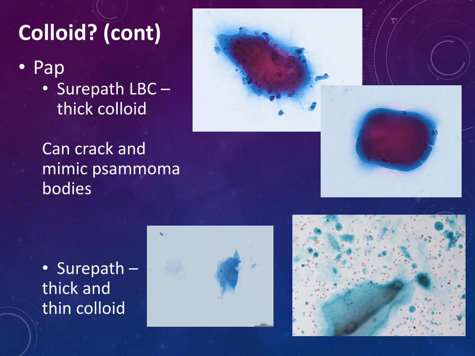
• Surepath –thick and thin colloid
Colloid? (cont)

• Pap
• Surepath LBC –thin colloid
• Direct spreadsand• Thinprep LBC - both very difficult!
Colloid? (cont)

• Can be cellular with a few microfollicles but not prominent microfollicles
• Can have plump cyst lining cells if nuclei bland
• Or slight nuclear changes
• Can have some oncocytic cells, in fact a mixture is reassuring
• May have cytoplasmic lipofuscin granules (I find reassuring)
What can be accepted in “non-neoplastic”, ie Thy2

• Definition: cytological features raising possibility of neoplasia but not enough for another category
• Text should indicate problem• Should be a minority
• TBS1 <7%, TBS2 <10%• RCPath suggests 5-10%
Easy to over-use• Instead of Thy2• Instead of Thy4 – increases ROM of Thy3a & Thy4
Report should be very clear on interpretation, possibilities and limitations
When to use “atypia” or Thy3a?

Thy3a (cont)Highly variable frequency of use nationally:UKEPS audit 2.2 – 25%
NuTH Thy3a Thy3a/Thy5 ratio (1.0-3.0)
2008-10 11%
2011-14 14.4% 2.73
2015 11.4% 2.0
2016 9.6% 1.13
2017provisional
18.4% 4.25
2018provisional
19.3% 3.9

Thy3a
Thy3a (cont)
Conceptually:
Not a progression
More anoverlap…with subjectivity at all the intersections…
Thy2
Thy3fThy2
Thy4 Thy5Thy1
Thy3a Thy3f Thy4 Thy5

• Should be subcategorised, examples• architectural atypia - too many microfollicles and/or
too little colloid - cannot exclude FN

• Scanty focal nuclear/cytological atypia – higher ROM
• other features when cannot exclude PTC, egpsammoma body-like

• atypical cyst lining cells
• predominance of lymphoid cells - cannot exclude lymphoma

When to suggest follicular lesion / neoplasm, ie Thy3f = surgery
• Cytological features• Reduced / no colloid• High cellularity with frequent microfollicles and/or cell clustersor • Predominance of oncocytes(beware MTC and metastatic RCC)
(Rememberintrathyroidal parathyroids)

Thy2 vs Thy3 in follicular setting? – my approach
Amount of colloid
Cellularity and microfollicles
Colloid nodule
Cellular colloid nodule
Favour cellular colloid
nodule but cannot exclude
follicular neoplasm
Differential diagnosis is
between follicular
neoplasm and cellular colloid
nodule
Favour follicular neoplasm but cannot
exclude cellular colloid nodule
Suggests follicular neoplasm
Thy2 Thy2 Thy3a Thy3f Thy3f Thy3f


• Subjective• Comes with experience and review of cases once have histology• Usually for PTC, so features falling short of diagnostic (Thy5) but
more marked than atypical (Thy3a)
When to be suspicious of malignancy, ie Thy4 vs Thy3a

• Papillary thyroid carcinoma• No cytological features are pathognomic of PTC
• Need qualitative and quantitative judgement
• Complicated by recent entity of NIFTP• Use Thy4 if
• Significant follicular architecture with nuclear abnormalities but not too many intranuclear inclusions
• Especially if U3 on radiology• Flag in report that it may be a lower risk lesion such as NIFTP or eFVPTC
• Go to Thy5 if• Papillary structures• Psammoma bodies• Multiple intranuclear inclusions
Diagnosing malignancy, ie Thy5


PTC

PTC

Problem of PTC vs oncocytes
Both can have Differences
Large cells with dense well demarcated cytoplasm
PTC – smoothOncocytes - granular
n/c ratios PTC – high (usually, beware TCV)Oncocytes – low
Large cell sheets with vessels PTC – papillary structuresOncocytic neoplasm – traversing vessels
Thick or inspissated colloid can mimic psammoma bodies
Both can have giant cells with dense cytoplasm especially in Hashimoto’s
Nuclei – large nucleoli of oncocytes can mimic intranuclear inclusions of PTC

Thy3f oncocyticneoplasm

Thy5 PTC(Histol = PTC, 30% TCV)

• Medullary thyroid carcinoma
• Less common so can be overlooked unless considered
• Remember possibility of MTC with oncocytic lesions
• Immunostaining is useful• Positive for TTF1, CEA, calcitonin• Negative for thyroglobulin
• If cellular, and many features present, but some uncertainty (eg no confirmation on immunostaining), could report as Thy4 but state could be regarded as diagnostic in clinical context of a significantly raised serum calcitonin

MTC

MTC in LN

• Anaplastic thyroid carcinoma

• High grade lymphoma

• Also counts as Thy5• Check patient history and imaging• Beware metastatic RCC – mimics oncocytic neoplasm
• Metastatic SCC
• Metastatic carcinoma

1. Immunocytochemistry• PTC – careful – CK19, HBME1, CD56, (BRAF)• MTC – CEA, calcitonin, TTF1, thyroglobulin• ATC vs lymphoma• Intrathyroidal parathyroid gland – PTH, TTF1, thyroglobulin• Metastases
2. Molecular testing• Huge increase in understanding of genetics of TC• Could refine diagnosis of indeterminate cytology• Point mutation testing for BRAF V600E mutation – 99% PPV PTC, but
not sensitive• “rule in” – to therapeutic surgery for malignancy
• 7 mutation panel – also RET/PTC, PAX8/PPARG, RAS• NGS, eg Thyroseq3 and other panels in development
• “rule out” – to avoid diagnostic surgery• Affirma by Veracyte• NGS, eg Thyroseq3 and other panels in development
Additional or alternative approaches

3. Core biopsies• No UK guidelines • Useful guidance from Korea, suggests categories for reporting:
I. Non-diagnosticII. Benign lesionIII. Indeterminate lesion – IIIA nuclear atypia, IIIB architectural
atypiaIV. Follicular neoplasm – IVA without nuclear atypia, IVB with
nuclear atypiaV. Suspicious for malignancyVI. Malignant
• And indications:• Previous unsatisfactory FNA, or cystic change• Previous AUS/FLUS• FN vs non-neoplastic but not for FA vs FC• Calcified nodules• Uncommon eg lymphoma vs ATC
• Low rate of inadequate samples• Facilitates ancillary tests
Jung et al J Pathol Trans Med 2015;49:288-299.

• Thyroid FNA is a valuable pre-operative investigation but has limitations, many can be reduced, some are inherent
• Essential to• Maximise quality of specimens• Use prose and categories for reporting• Use additional tests and second opinions
appropriately• Be aware of limitations• Audit so know ROMs locally• Have experienced staff throughout
• aspirators• cytopathologists• clinicians
Summary

Thankyou for listening
And enjoy reporting thyroid cytology!

04/02/2020
1
The thyroid cancer patient: what the oncologist needs from pathology and an update on new therapies and clinical trials
Prof Jon WadsleyConsultant Clinical Oncologist, Weston Park HospitalChair of NCRI Thyroid Cancer Subgroup2nd Bi-Annual UKEPS/RCPath Endocrine Pathology Update7th February 2020
Overview
• Low risk differentiated thyroid cancer– Trend to de-escalate treatment– On going trials
• Iodine refractory differentiated thyroid cancer– Treatment options– Biomarkers and further research
• Medullary thyroid cancer– Treatment options– RET mutations and further research
• Anaplastic thyroid cancer– iNATT– BRAF mutations
Low risk differentiated thyroid cancer
• What is low risk?
• Questions about management
• Trend to de-escalation
– HiLo
– IoN
– HoT
• What does the oncologist need from pathology?
What is low risk?TNM staging
Tumour• T0: There is no evidence of a tumor.• T1: The tumor is 2 centimeters (cm) or smaller and limited to the thyroid.• T1a: The tumor is 1 cm or smaller.• T1b: The tumor is larger than 1 cm but less than 2 cm.• T2: The tumor is larger than 2 cm but smaller than 4 cm and is limited to the thyroid.• T3: The tumor is larger than 4 cm, but the tumor does not extend beyond the thyroid gland.• T4: The tumor is any size and has extended beyond the thyroid.• T4a: The tumor has spread beyond the thyroid to nearby soft tissues, the larynx, trachea, esophagus, or recurrent laryngeal
nerve.• T4b: The tumor has spread beyond the regions in T4a (above).
Node• NX: The regional lymph nodes cannot be evaluated.• N0: There is no evidence of cancer in the regional lymph nodes.• N1: Cancer has spread to the lymph nodes.• N1a: Cancer has spread to the lymph nodes around the thyroid (called the central compartment; the pretracheal,
paratracheal, and prelaryngeal lymph nodes).• N1b: Cancer has spread beyond the central compartment, including unilateral cervical (lymph nodes on 1 side of the neck),
bilateral cervical (lymph nodes on both sides of the neck), contralateral cervical (the opposite side of the tumor), or mediastinal (the chest) lymph nodes.
Metastasis• MX: Distant metastasis cannot be evaluated.• M0: Cancer has not spread to other parts of the body.• M1: Cancer has spread to other parts of the body.

04/02/2020
2
What is low risk?Stage
Papillary or follicular* under 55 years
Stage I Any T Any N M0
Stage II Any T Any N M1
Papillary or follicular 55 years or over
Stage I T1a, T1b, T2 N0 M0
Stage II T3 N0 M0
T1, T2, T3 N1 M0
Stage III T4a Any N M0
Stage IVA T4b Any N M0
Stage IVB Any T Any N M1
What is low risk? ATA Postoperative risk stratification
Low risk Intermediate risk High risk
No local or distant metastases
All macroscopic tumour resected
No tumour invasion of loco-regional tissues or structures
No aggressive histology (eg tall cell, insular) or vascular invasion
No I131 uptake outside the thyroid bed on post RAI scan
Clinical N0 or </=5 N1 pathological micrometastases- <0.2cm
Intrathyroidal encapsulated FVPTC
Intrathyroidal well differentiated follicular cancer with capsular but no (or minimal) vascular invasion
Microscopic invasion of tumour into perithyroidal soft tissue
I131 uptake in the neck but outside the thyroid bed on post RAI imaging
Tumour with aggressive histology or vascular invasion
Clinical N1 or >5 pathologic nodes, all nodes <3cm
Intrathyroidal papillary cancer 1-4cm with V600E BRAF mutation
Macroscopic tumour invasion
Incomplete tumour resection
Distant metastases
Raised thyroglobulin out of proportion to what is seen on post RAI scan
Pathologic N1 with a node >3cm
Follicular cancer with extensive vascular invasion (>4 foci)
What is low risk?Dynamic risk stratification
Excellent response Indeterminate response Incomplete response
Suppressed and stimulated Thyroglobulin<1mcg/l
No evidence of disease on neck ultrasound scan
Cross-sectional and/or nuclear imaging clear (if done)
Suppressed thyroglobulin <1 and stimulated thyroglobulin 1-10mcg/l
Neck ultrasound with non-specific changes or stable sub-cm lymph nodes
Cross-sectional and/or nuclear imaging not completely normal but non-specific findings
Suppressedthyroglobulin>1 or Stimulated thyroglobulin>10mcg/l
Rising thyroglobulin
Persistent or new disease on imaging
What about molecular pathology?Comments from ATA 2015 Guidelines
‘While not routinely recommended for initial post-operative risk stratification in DTC, the mutational status of BRAF, and potentially other mutations such as TERT, have the potential to refine risk estimates when interpreted in the context of other clinico-pathologic risk factors.’
‘The role of molecular testing in guiding post-operative RAI use has yet to be established, therefore no molecular testing to guide post-operative RAI use can be recommended at this time.’
Not currently universally available in the UK.
Likely to be addressed in NICE Thyroid Cancer Guidelines

04/02/2020
3
Questions about management
• What extent of surgery is necessary-lobectomy vs total thyroidectomy?
• Is radioiodine ablation required?
• If so what activity of I131 is required?
• Is subsequent TSH suppression necessary?
• How long should patients be followed up?
Trend to de-escalate treatment
Drivers-
1. Generally excellent outcomes with very high overall survival
2. Concerns about morbidity of treatment
-vocal cord palsy
-hypothyroidism
-hypoparathyroidism
-second malignancy
HiLo trial
• HiLo recruited patients with well-differentiated thyroid cancer requiring radioiodine (RAI) ablation therapy after total thyroidectomy
• Randomised to receive (factorial trial)– 1.1GBq vs 3.7GBq I131
– Thyroid hormone withdrawal (THW) vs recombinant TSH (rhTSH)
• Primary endpoint was ablation success rate at 6-9 months defined by both:– Stimulated thyroglobulin
– RAI uptake scan
• Longer term follow up recently reported- median follow up 78 months (maximum 127 months)
HiLo Trial Results-Ablation Success Rate
• 438 patients from 29 UK centres randomised 2007-2010
• 421 had both Tg and RAI scan at 6-9 months post-ablation
• Tumour stage T1-T3; N1a or N1b; no metastatic disease
• Trial objective of non-inferiority achieved for both main comparisons
1.1 GBq(n=214)
3.7 GBq(n=207)
% ablation success
85% 89%
Thyrogen
(n=210)Hormone withdrawal(n=211)
% ablation success
87% 87%

04/02/2020
4
HiLo Trial Results- RecurrenceHigh dose RAI and THW versus
Low dose RAI and recombinant TSH
HiLo Trial Results-Summary
• Overall thyroid cancer recurrence rate 5% (21/434)• Risk of thyroid cancer recurrence not higher in patients having
(i) 1.1GBq RAI compared with 3.7GBq RAI(ii) recombinant TSH compared with THW
• These findings were consistent across T- and N- stages• Several recurrences were seen after more than 5 years follow
up• Confirmed second malignancy found in 3 patients in 1.1GBq
RAI group and 4 patients in 3.7GBq RAI group, diagnosed 6 months-7 years post ablation
• This has led to change in BTA Guidelines and UK practice
IoN Trial• Patients are randomly
assigned to receive radioiodine ablation or not following total thyroidectomy
• Close follow up with annual USS neck
• Primary endpoint- progression free survival at 5 years
• Target of 450 patients has been reached but on going recruitment agreed due to lower than anticipated event rate
IoN Trial- Eligibility Criteria
• INCLUSION CRITERIA
Papillary thyroid cancer (PTC):• Non aggressive histological features (small foci of aggressive
histology allowed• at the discretion of the MDT)• VI lymph nodes (pN1a)• pT1a(m): all individual foci 1cm• pT1b and pT1b(m): >1-2cm• pT2 and pT2(m): >2-4cm• pT3 and pT3(m): >4cm confined to the thyroid• pT3 R0 +/- (m): any size with minimal ETE if recommended by
the MDT• pN0• pN1a• pNX
Follicular thyroid cancer (FTC) (including oncocytic or Hürthle cell cancer):• minimally invasive FTC which are considered low risk and are• recommended by the specialist MDT based on overall clinico-
pathological• assessment• pT1b and pT2: >1-4cm intrathyroidal• pT3 R0:any size up to 4 cm with minimal ETE if recommended
by the MDT
• EXLUSION CRITERIA
• pT1a -without any positive nodes or unfavourable clinical features, treated by lobectomy.
• Up to 4cm non-invasive Encapsulated Follicular Variant of Papillary Thyroid
• Cancer (eFVPTC) with no capsular or vascular invasion (>4 cm can be included at
• the discretion of the MDT)• non-invasive follicular tumour with papillary-like nuclei (NIFTP)• Anaplastic, poorly differentiated or medullary carcinoma• R1 or R2 thyroidectomy• Patients with:• pN1b• M1• Aggressive Papillary thyroid cancer with any of the following
features:• Widely invasive• Poorly differentiated• Anaplastic• Tall cell• Columnar cell• Diffuse sclerosing variants• Follicular thyroid cancer/Hürthle cell cancer with any of the
following features:• Tumours greater than 4cm• Widely invasive• Poorly differentiated• Anaplastic• Incomplete resection or lobectomy• pT4a and pT4b or macroscopic and microscopic tumour invasion
of loco-regional tissues or structures

04/02/2020
5
HoT Trial (Dae Kim)
• Lobectomy vs thyroidectomy for patients with low-risk thyroid cancer
• Extensive engagement with surgical community and patient groups
• NIHR HTA grant application successful
• Final protocol currently in development
• Hoping to be open to recruitment mid 2020
• Will have strict eligibility criteria
Summary- what does the oncologist need from pathology?
• Increased importance of detailed pathological staging because this does now affect what further treatment is given– Extent of surgery– Whether to give I131 and what activity– Duration of TSH suppression and follow up– Eligibility for clinical trials
• Possible emerging role for molecular pathology-BRAF and TERT- but currently to guide prognosis rather than treatment
Iodine refractory differentiated thyroid cancer
• Overview
• Currently available treatments
• Recent Trials
• Future role of biomarkers
Iodine Refractory Differentiated Thyroid Cancer
• Relatively uncommon- develops in 5-10% of differentiated thyroid cancer patients
• Some variation in criteria– One or more measurable lesions that do not demonstrate iodine
uptake on a previous radioiodine scan (diagnostic uptake or post therapy)
– One or more measurable lesions that have progressed within 12 months of I-131 therapy, despite demonstrable radioiodine avidity at the time of that treatment
• Some include FDG-PET positive lesions• Poor prognosis- 10 years survival <10%• Until recently few treatment options
– Single agent Doxorubicin
• More recent interest in multi-targeted kinase inhibitors

04/02/2020
6
Potential molecular targetsAgents tested in iodine refractory
differentiated thyroid cancer
• Sorafenib
• Sunitinib
• Pazopanib
• Vandetanib
• Axitinib
• Motesanib
• Lenvatinib
Sorefenib in radioactive iodine-refractory, locally advanced or metastatic differentiated thyroid cancer: a randomised, double-blind,
phase 3 trialBrose M et al Lancet 2014; 384:319-28
• Phase 3 sorafenib vs placebo
• 417 patients randomised
• Papillary, follicular (hurtle cell), poorly differentiated thyroid cancer
• Evidence of radiological progression (RECIST) within last 14 months
• At least one measurable lesion
• Crossover allowed at progression
Sorafenib- Outcomes
• Primary endpoint: progression free survival
• Median Progression Free Survival 10.8 vs 5.8 months in favour of Sorafenib
• 6 month disease control rate 54.1% vs 33.8%
• Objective response rate 12.2% vs 0.5%
• No significant difference in overall survival (median not reached at time of reporting)
• BRAF and RAS mutations not predictive of PFS benefit from Sorafenib

04/02/2020
7
Sorafenib (contd)Toxicity
• Hand foot syndrome (76.3%)
• Diarrhoea (68.6%)
• Alopecia (67.1%)
• Rash or desquamation (50.2%)
• Fatigue (49.8%)
• Weight loss (46.9%)
• Hypertension (40.6%)
• Squamous cell carcinoma of the skin (7 patients)
• 18.8% patients had to discontinue Sorafenib due to toxicity
Sorafenib (contd)
Lenvatinib versus Placebo in Radioiodine-Refractory Thyroid CancerSchlumberger et al NEJM Feb 2015
• Phase 3 lenvatinib vs placebo• 392 patients randomised (2:1)• Papillary, poorly differentiated, follicular, Hurtle cell
included• Independently reviewed evidence of radiological
progression within the previous 13 months• At least 1 measurable lesion• Crossover allowed at progression• Primary endpoint: PFS
Lenvatinib- Outcomes
• Median Progression Free Survival 18.3 vs 3.6 months in favour of lenvatinib
• 6 month PFS 77.5% vs 25.4%• PFS benefit maintained in all
subgroups– previous tyrosine kinase
inhibitor– Histology– BRAF or RAS mutation status
• Response rate 64.8% vs 1.5%• 4 complete responses to
Lenvatinib• Overall survival HR 0.73,
p=0.10

04/02/2020
8
PFS by prior TKI exposure Lenvatininb- Toxicity
• Toxicity– Hypertension (67.8%)
– Diarrhoea (59.4%)
– Fatigue (59.0%)
– Weight loss (46.4%)
– Hand foot syndrome (31.8%)
– Proteinuria (31.0%)
• Treatment related deaths– 6 deaths considered to be drug-related
– 1 pulmonary embolism, 1 haemorrhagic stroke
Re-differentiation therapy
• Since I-131 is such an effective treatment for iodine-avid advanced thyroid cancer, several attempts have been made to re-instate iodine uptake in iodine-refractory cells
– Lithium
– Retinoids
– Recent targeted approaches
A more targeted approach to increasing iodine uptake
• Expression of NaI symporter regulated by MAPKinase signalling pathway
• Mutations leading to activation of the pathway switches off NaI expression in thyroid cells
• Inhibiting the pathway might therefore allow re-expression of NaI

04/02/2020
9
Selumetinib
• MEK inhibitor
• Blocks MAPK pathway signalling
• Early phase trials show little evidence of direct cytotoxic activity on thyroid cancer cells
• Preclinical work in mice demonstrated that iodine refractory thyroid cancer cells with BRAF mutations treated with a MEK inhibitor regain the ability to accumulate radioiodine
Selumetinib-Enhanced Radioiodine Uptake in Advanced Thyroid Cancer
Ho et al NEJM 2013
• Pilot study
• 20 patients with iodine refractory DTC
• Given selumetinib 75mg bd for 4 weeks
• 12/20 had increased iodine uptake as assessed by I124 PET
• 8 received further I131 therapy
• 5 had partial radiological response to treatment
• All 8 patients had a fall in serum thyroglobulin
SELIMETRY
• UK multicentre single arm phase 2 trial
• Aiming to replicate findings of the pilot study
• Aiming to recruit 60 patients
• Currently open at– Royal Marsden
– Guildford
– Oxford
– Bristol
– Southampton

04/02/2020
10
Summary- what does the oncologist need from pathology?
• Multi-kinase inhibitors demonstrate improvements in progression free survival
• Significant costs in terms of toxicity
• Novel approaches being explored to allow further I131 therapy
• Better biomarkers are needed to predict benefit from treatment
• ?role for circulating tumour DNA
Medullary thyroid cancer
• Overview
• Currently available treatments
• The development of RET specific inhibitors
Advanced Medullary Carcinoma of the thyroid
• Arises from parafollicular C cells
• 5% of thyroid cancers
• 25% hereditary- RET mutations
• In locally advanced/metastatic disease
– 10 year overall survival 40%
• Neither radiotherapy nor conventional chemotherapy demonstrate durable responses
Treatment Approaches in Advanced Medullary Thyroid Cancer
• Historical
– Cytotoxic chemotherapy
– IFN
– Somatostatin analogues
• Current
– Tyrosine kinase inhibitors
– Radioisotope therapies- MIBG, Dotatate

04/02/2020
11
Vandetanib in patients with locally advanced or metastatic medullary thyroid cancer: a randomised, double blind, phase III trial
Wells et al, JCO 2011
• Phase 3 Vandetanib 300mg/day vs placebo
• 2:1 randomisation
• 331 patients with advanced MTC recruited
• No progression criteria prior to trial entry
• Crossover allowed at progression
• Primary endpoint- progression free survival
Vandetanib- outcomes
• Significant improvement in Progression Free Survival demonstrated– HR 0.46 (0.31-0.69)
• Benefits consistent across all pre-specified subgroups– Performance status 0 vs 1– Metastatic vs locally advanced
disease– Hereditary vs sporadic– Prior therapy
• Higher response rate noted in tumours RET M918T mutation positive (but too many unknown mutation status cases to comment on significance of RET mutation status)
Vandetanib- toxicity
• Median duration of treatment 90.1 months• 12% stopped Vandetanib due to toxicity• Most common toxicities
– Diarrhoea– Rash– Nausea– Hypertension– Asthenia– QTc prolongation– Increased thyroxine requirement
Vandetanib- toxicity
• QTc interval prolongation
• Single measurement >550ms or >100ms above baseline
• Repeated measurements 500-550ms or 60-100ms above baseline
• Dose reduction required in 35% patients
04/02/2020
12
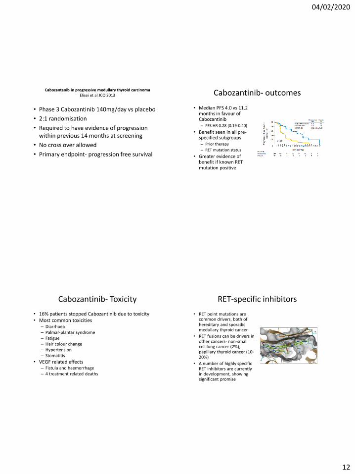
Cabozantanib in progressive medullary thyroid carcinomaElisei et al JCO 2013
• Phase 3 Cabozantinib 140mg/day vs placebo
• 2:1 randomisation
• Required to have evidence of progression within previous 14 months at screening
• No cross over allowed
• Primary endpoint- progression free survival
Cabozantinib- outcomes
• Median PFS 4.0 vs 11.2 months in favour of Cabozantinib– PFS HR 0.28 (0.19-0.40)
• Benefit seen in all pre-specified subgroups– Prior therapy
– RET mutation status
• Greater evidence of benefit if known RET mutation positive
Cabozantinib- Toxicity
• 16% patients stopped Cabozantinib due to toxicity• Most common toxicities
– Diarrhoea– Palmar-plantar syndrome– Fatigue– Hair colour change– Hypertension– Stomatitis
• VEGF related effects– Fistula and haemorrhage– 4 treatment related deaths
RET-specific inhibitors
• RET point mutations are common drivers, both of hereditary and sporadic medullary thyroid cancer
• RET fusions can be drivers in other cancers- non-small cell lung cancer (2%), papillary thyroid cancer (10-20%)
• A number of highly specific RET inhibitors are currently in development, showing significant promise

04/02/2020
13
BLU-667 BLU-667
• 69 patients with RET alterations treated to date
• Some impressive responses seen
• Generally very well tolerated with only mild (G1/2) toxicity
BLU-667 LOXO-292 (Selpercatinib)

04/02/2020
14
LOXO-292 (Selpercatinib) RET-specific inhibitors
• Phase 3 trials of both BLU-667 and LOXO-292 are in development and likely to open in the UK within the next 12 months
• Will require testing for somatic RET mutations in MTC patients with advanced progressive disease
Summary- what does the oncologist need from pathology?
• Germline RET mutation testing already standard for new diagnoses of MTC
• Will be increasingly important to be able to access somatic RET mutation status for non-inherited cases
– Prognostic information
– To select patients for treatment
• Genomics England test directory
Anaplastic thyroid cancer
• Rare (1-2% of thyroid cancers-estimated approx 150 cases in UK pa)
• Highly aggressive, undifferentiated
• Important differential- thyroid lymphoma
• Rarely operable, current treatments generally ineffective
• Median survival only 5-12 months
• 20-50% have BRAF V600E mutations

04/02/2020
15
iNATT
• Anaplastic thyroid cancer tissue bank
• 114 samples collected so far
• A number of projects using the tissue underway
• Contact: Dr Laura Moss, Velindre Hospital, Cardiff
Dabrafenib and Trametinib Treatment in Patients With Locally Advanced or Metastatic BRAF V600–Mutant Anaplastic Thyroid Cancer.
Subbiah et al JCO 2017
• 28 patients with ATC screened
• 16 found to be eligible
• 15/16 had BRAF V600E mutations
• Durable responses seen- estimated 1 year survival rate 80%
• Significant toxicity-fatigue, pyrexia, nausea
BRAF- Dabrafenib/Tametinib
• UK Compassionate access scheme has been negotiated to allow patients of good performance status with BRAF mutation to access these drugs
• Immunohistochemical testing is sufficient in the first instance, but ideally should be confirmed by NGS
Summary- what does the oncologist need from pathology?
• Important to distinguish from other more treatable conditions (eg lymphoma)
– Core biopsy may be preferable to FNA
• Rapid turnaround is essential
• BRAF testing may lead to additional treatment options in fitter patients

04/02/2020
16
Overall Summary
• Low risk thyroid cancer
– Trend to treatment de-escalation
– Requirement for detailed pathological reporting to ensure appropriate stratification
• Advanced disease
– Increasing treatment options available
– Need for molecular testing to determine suitability for treatment likely to increase

EFM 06022022 - 0702022
@UEndopath
@rcpathendo
FORTHCOMING EVENTS

More info:https://www.iaoplondon2020.com