
2635
2
Author Disclosure: F. Xia, None; E. Nam, None; D. Hallahan, None. 2635 Early Experience With Highly Focal Radiation Delivery to the Mouse Brain Using Our Small Animal Radiation Research Platform (SARRP) E. Tryggestad 1 , J. Wong 1 , T. A. Chan 1 , L. E. Dillehay 1 , C. W. Kennedy 1 , J. Zhang 2 , T. L. DeWeese 1 1 Johns Hopkins University, Department of Radiation Oncology and Molecular Radiation Sciences, Baltimore, MD, 2 Johns Hopkins University, Department of Radiology, Baltimore, MD Purpose/Objective(s): Validate that we can effectively deliver radiation to a confined region of the mouse brain as evidenced by comparison of pre and post-irradiation, in-vivo MR images. Materials/Methods: Two NCR athymic nude mice were focally irradiated with 15 beams spaced equally over 210° in the coronal plane to single-fraction maximum doses of 22 and 33 Gy (10%). The x-ray source position remained fixed while the prone mice were rotated on a movable stage. A constant-voltage source provided 225 kVp x rays, while a 0.75 mm beam aperture defined a 1.5 mm (FWHM) circular radiation field at the treatment isocenter. T2-weighted micro MR scans were acquired both pre and 20 hours post-irradiation with an 11.7 Tesla NMR spectrometer (Bruker Biospin, Billerica, MA, USA) using a fast spin-echo sequence. The total imaging time required was 45 minutes per mouse, per session. Figure 1 shows three orthogonal cuts through the volume for one of the mice prior to treatment. CT treatment simulation and dose planning was used prior to treatment to accurately localize the right frontal lobe region of each mouse brain and determine the effective depth to be used for the dose calculations. Figure 2 shows the CT-based dose distribution and brain DVH for the mouse that received 33 Gy using our preliminary Pinnacle (ADAC/Philips) Monte Carlo model. Results: These data are satisfying in that they closely mimic clinically-based radiation planning and treatment delivery to the human brain. The time between irradiation and the post-treatment MR proved to be insufficient, as no changes were observed qualitatively comparing the sets of images for each mouse. We anticipate that later MR follow-up imaging will show localized contrast differences; we endeavor to extend this investigation. Conclusions: We gratefully acknowledge Dr. Susumu Mori from the Department of Radiology for providing the MR capabilities demonstrated here. This work has been supported in part by grant NCI R01–108449. S563 Proceedings of the 48th Annual ASTRO Meeting
Transcript of 2635
Author Disclosure: F. Xia, None; E. Nam, None; D. Hallahan, None.
2635 Early Experience With Highly Focal Radiation Delivery to the Mouse Brain Using Our Small AnimalRadiation Research Platform (SARRP)
E. Tryggestad1, J. Wong1, T. A. Chan1, L. E. Dillehay1, C. W. Kennedy1, J. Zhang2, T. L. DeWeese1
1Johns Hopkins University, Department of Radiation Oncology and Molecular Radiation Sciences, Baltimore, MD, 2JohnsHopkins University, Department of Radiology, Baltimore, MD
Purpose/Objective(s): Validate that we can effectively deliver radiation to a confined region of the mouse brain as evidencedby comparison of pre and post-irradiation, in-vivo MR images.
Materials/Methods: Two NCR athymic nude mice were focally irradiated with 15 beams spaced equally over 210° in thecoronal plane to single-fraction maximum doses of 22 and 33 Gy (�10%). The x-ray source position remained fixed while theprone mice were rotated on a movable stage. A constant-voltage source provided 225 kVp x rays, while a 0.75 mm beamaperture defined a 1.5 mm (FWHM) circular radiation field at the treatment isocenter.
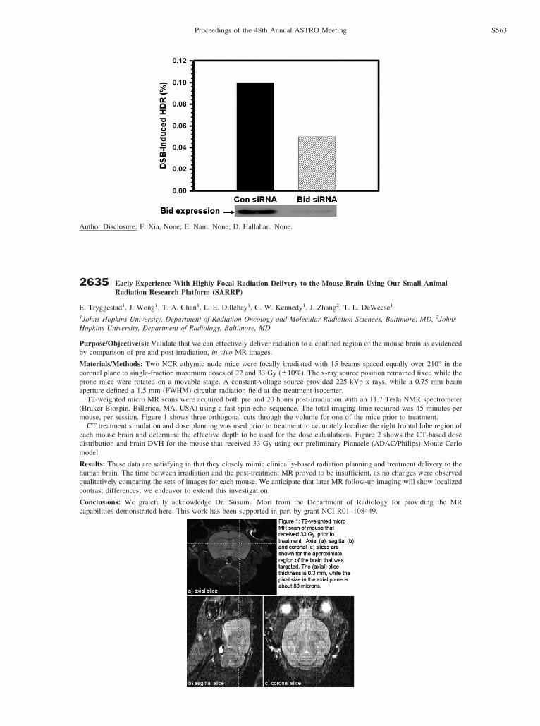
T2-weighted micro MR scans were acquired both pre and 20 hours post-irradiation with an 11.7 Tesla NMR spectrometer(Bruker Biospin, Billerica, MA, USA) using a fast spin-echo sequence. The total imaging time required was 45 minutes permouse, per session. Figure 1 shows three orthogonal cuts through the volume for one of the mice prior to treatment.
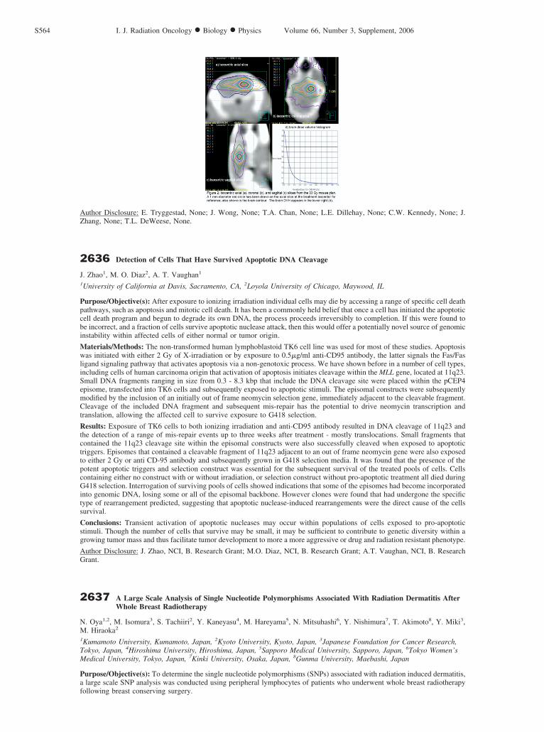
CT treatment simulation and dose planning was used prior to treatment to accurately localize the right frontal lobe region ofeach mouse brain and determine the effective depth to be used for the dose calculations. Figure 2 shows the CT-based dosedistribution and brain DVH for the mouse that received 33 Gy using our preliminary Pinnacle (ADAC/Philips) Monte Carlomodel.
Results: These data are satisfying in that they closely mimic clinically-based radiation planning and treatment delivery to thehuman brain. The time between irradiation and the post-treatment MR proved to be insufficient, as no changes were observedqualitatively comparing the sets of images for each mouse. We anticipate that later MR follow-up imaging will show localizedcontrast differences; we endeavor to extend this investigation.
Conclusions: We gratefully acknowledge Dr. Susumu Mori from the Department of Radiology for providing the MRcapabilities demonstrated here. This work has been supported in part by grant NCI R01–108449.
S563Proceedings of the 48th Annual ASTRO Meeting
Author Disclosure: E. Tryggestad, None; J. Wong, None; T.A. Chan, None; L.E. Dillehay, None; C.W. Kennedy, None; J.Zhang, None; T.L. DeWeese, None.
2636 Detection of Cells That Have Survived Apoptotic DNA Cleavage
J. Zhao1, M. O. Diaz2, A. T. Vaughan1
1University of California at Davis, Sacramento, CA, 2Loyola University of Chicago, Maywood, IL
Purpose/Objective(s): After exposure to ionizing irradiation individual cells may die by accessing a range of specific cell deathpathways, such as apoptosis and mitotic cell death. It has been a commonly held belief that once a cell has initiated the apoptoticcell death program and begun to degrade its own DNA, the process proceeds irreversibly to completion. If this were found tobe incorrect, and a fraction of cells survive apoptotic nuclease attack, then this would offer a potentially novel source of genomicinstability within affected cells of either normal or tumor origin.
Materials/Methods: The non-transformed human lymphoblastoid TK6 cell line was used for most of these studies. Apoptosiswas initiated with either 2 Gy of X-irradiation or by exposure to 0.5�g/ml anti-CD95 antibody, the latter signals the Fas/Fasligand signaling pathway that activates apoptosis via a non-genotoxic process. We have shown before in a number of cell types,including cells of human carcinoma origin that activation of apoptosis initiates cleavage within the MLL gene, located at 11q23.Small DNA fragments ranging in size from 0.3 - 8.3 kbp that include the DNA cleavage site were placed within the pCEP4episome, transfected into TK6 cells and subsequently exposed to apoptotic stimuli. The episomal constructs were subsequentlymodified by the inclusion of an initially out of frame neomycin selection gene, immediately adjacent to the cleavable fragment.Cleavage of the included DNA fragment and subsequent mis-repair has the potential to drive neomycin transcription andtranslation, allowing the affected cell to survive exposure to G418 selection.
Results: Exposure of TK6 cells to both ionizing irradiation and anti-CD95 antibody resulted in DNA cleavage of 11q23 andthe detection of a range of mis-repair events up to three weeks after treatment - mostly translocations. Small fragments thatcontained the 11q23 cleavage site within the episomal constructs were also successfully cleaved when exposed to apoptotictriggers. Episomes that contained a cleavable fragment of 11q23 adjacent to an out of frame neomycin gene were also exposedto either 2 Gy or anti CD-95 antibody and subsequently grown in G418 selection media. It was found that the presence of thepotent apoptotic triggers and selection construct was essential for the subsequent survival of the treated pools of cells. Cellscontaining either no construct with or without irradiation, or selection construct without pro-apoptotic treatment all died duringG418 selection. Interrogation of surviving pools of cells showed indications that some of the episomes had become incorporatedinto genomic DNA, losing some or all of the episomal backbone. However clones were found that had undergone the specifictype of rearrangement predicted, suggesting that apoptotic nuclease-induced rearrangements were the direct cause of the cellssurvival.
Conclusions: Transient activation of apoptotic nucleases may occur within populations of cells exposed to pro-apoptoticstimuli. Though the number of cells that survive may be small, it may be sufficient to contribute to genetic diversity within agrowing tumor mass and thus facilitate tumor development to more a more aggressive or drug and radiation resistant phenotype.
Author Disclosure: J. Zhao, NCI, B. Research Grant; M.O. Diaz, NCI, B. Research Grant; A.T. Vaughan, NCI, B. ResearchGrant.
2637 A Large Scale Analysis of Single Nucleotide Polymorphisms Associated With Radiation Dermatitis AfterWhole Breast Radiotherapy
N. Oya1,2, M. Isomura3, S. Tachiiri2, Y. Kaneyasu4, M. Hareyama5, N. Mitsuhashi6, Y. Nishimura7, T. Akimoto8, Y. Miki3,M. Hiraoka2
1Kumamoto University, Kumamoto, Japan, 2Kyoto University, Kyoto, Japan, 3Japanese Foundation for Cancer Research,Tokyo, Japan, 4Hiroshima University, Hiroshima, Japan, 5Sapporo Medical University, Sapporo, Japan, 6Tokyo Women’sMedical University, Tokyo, Japan, 7Kinki University, Osaka, Japan, 8Gunma University, Maebashi, Japan
Purpose/Objective(s): To determine the single nucleotide polymorphisms (SNPs) associated with radiation induced dermatitis,a large scale SNP analysis was conducted using peripheral lymphocytes of patients who underwent whole breast radiotherapyfollowing breast conserving surgery.
S564 I. J. Radiation Oncology ● Biology ● Physics Volume 66, Number 3, Supplement, 2006


