25-1 Human Anatomy, First Edition McKinley & O'Loughlin Chapter 25 Lecture Outline: Respiratory...
56
25-1 Human Anatomy, First Edition McKinley & O'Loughlin Chapter 25 Lecture Outline: Respiratory System
-
Upload
juniper-daniel -
Category
Documents
-
view
240 -
download
7
Transcript of 25-1 Human Anatomy, First Edition McKinley & O'Loughlin Chapter 25 Lecture Outline: Respiratory...

25-1
Human Anatomy, First Edition
McKinley & O'Loughlin
Chapter 25 Lecture Outline:Respiratory System

25-2
Organization and Functions of the Respiratory System Consists of an upper respiratory tract and a
lower respiratory tract. Conducting portion transports air.
includes the nose, nasal cavity, pharynx, larynx, trachea, and progressively smaller airways, from the primary bronchi to the bronchioles
Respiratory portion carries out gas exchange. composed of small airways called respiratory
bronchioles and alveolar ducts as well as air sacs called alveoli

3
25-4
Respiratory System Functions Breathing (pulmonary ventilation).
consists of two cyclic phases: inhalation, also called inspiration exhalation, also called expiration
Inhalation draws gases into the lungs. Exhalation forces gases out of the
lungs. Gas exchange, gas conditioning, sound
production, olfaction, and defense.

25-5
Upper Respiratory Tract Composed of the nose and nasal cavity,
paranasal sinuses, pharynx (throat), and associated structures.
All part of the conducting portion of the respiratory system.

6

7

25-8
Paranasal Sinuses Four bones of the skull contain paired air
spaces called the paranasal sinuses. decrease skull bone weight
Named for the bones in which they are housed. frontal ethmoidal sphenoidal maxillary
Communicate with the nasal cavity by ducts. Covered with the same pseudostratified ciliated
columnar epithelium as the nasal cavity.

9

25-10
Pharynx Common space used by both the respiratory
and digestive systems. Commonly called the throat. Funnel-shaped, meaning that it is slightly
wider superiorly and narrower inferiorly. Originates posterior to the nasal and oral
cavities and extends inferiorly near the level of the bifurcation of the larynx and esophagus.
Common pathway for both air and food.

25-11
Pharynx Walls are lined by a mucosa and contain
skeletal muscles that are primarily used for swallowing.
Flexible lateral walls are distensible in order to force swallowed food into the esophagus.
Partitioned into three adjoining regions: nasopharynx oropharynx laryngopharynx

25-12
Nasopharynx Superiormost region of the pharynx. Located directly posterior to the nasal cavity and
superior to the soft palate, which separates it from the posterior part of the oral cavity.
Normally, only air passes through. Material from the oral cavity and oropharynx is typically
blocked from entering the nasopharynx by the soft palate, which elevates when we swallow.
In the lateral walls of the nasopharynx, paired auditory tubes connect the nasopharynx to the middle ear.
Posterior nasopharynx wall also houses a single pharyngeal tonsil (commonly called the adenoids).

25-13
Oropharynx The middle pharyngeal region. Immediately posterior to the oral cavity. Bounded by the edge of the soft palate superiorly and
the hyoid bone inferiorly. Common respiratory and digestive pathway through
which both air and swallowed food and drink pass. 2 pairs of muscular arches, the anterior palatoglossal
arches and the posterior palatopharyngeal arches, form the entrance from the oral cavity.
Lymphatic organs here provide the “first line of defense” against ingested or inhaled foreign materials.
Palatine tonsils are on the lateral wall between the arches, and the lingual tonsils are at the base of the tongue.

25-14
Laryngopharynx Inferior, narrowed region of the pharynx. Extends inferiorly from the hyoid bone and is
continuous with the larynx and esophagus. Terminates at the superior border of the
esophagus and is equivalent to the inferior border of the cricoid cartilage in the larynx.
The larynx (voice box) forms the anterior wall Lined with a nonkeratinized stratified
squamous epithelium Permits passage of both food and air.
25-15
Lower Respiratory Tract Conducting airways (larynx, trachea, bronchi,
bronchioles and their associated structures). Respiratory portion of the respiratory system
(respiratory bronchioles, alveolar ducts, and alveoli).

25-16
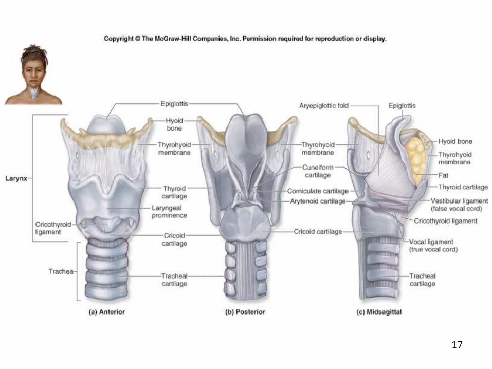
Larynx Voice box is a short, somewhat cylindrical airway
bounded posteriorly by the laryngopharynx and inferiorly by the trachea.
Prevents swallowed materials from entering the lower respiratory tract.
Conducts air into the lower respiratory tract. Produces sounds. Supported by a framework of nine pieces of
cartilage (three individual pieces and three cartilage pairs) that are held in place by ligaments and muscles.
17

25-18
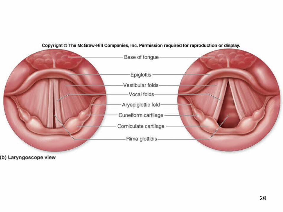
Sound Production Inferior ligaments, called vocal ligaments, covered by a
mucous membrane are called the vocal folds. are “true vocal cords” because they produce sound
when air passes between them Superior ligaments are called vestibular ligaments,
along with the mucosa covering them are called the vestibular folds.
Are “false vocal cords” because they have no function in sound production, but protect the vocal folds.
The vestibular folds attach to the corniculate cartilages. The tension, length, and position of the vocal folds
determine the quality of the sound.

19
20

21
25-22
Trachea A flexible, slightly rigid tubular organ often referred to
as the “windpipe.” Extends through the mediastinum and lies immediately
anterior to the esophagus, inferior to the larynx, and superior to the primary bronchi of the lungs.
Anterior and lateral walls of the trachea are supported by 15 to 20 C-shaped tracheal cartilages.
cartilage rings reinforce and provide some rigidity to the tracheal wall to ensure that the trachea remains open (patent) at all times
cartilage rings are connected by elastic sheets called anular ligaments

23

25-24
Trachea At the level of the sternal angle, the trachea
bifurcates into two smaller tubes, called the right and left primary bronchi.
Each primary bronchus projects laterally toward each lung.
The most inferior tracheal cartilage separates the primary bronchi at their origin and forms an internal ridge called the carina.
25-25
Bronchial Tree A highly branched system of air-conducting passages
that originate from the left and right primary bronchi. Progressively branch into narrower tubes as they
diverge throughout the lungs before terminating in terminal bronchioles.
Incomplete rings of hyaline cartilage support the walls of the. primary bronchi to ensure that they remain open.
Right primary bronchus is shorter, wider, and more vertically. oriented than the left primary bronchus.
Foreign particles are more likely to lodge in the right primary bronchus.

25-26
Bronchial Tree The primary bronchi enter the hilum of each lung together
with the pulmonary vessels, lymphatic vessels, and nerves. Each primary bronchus then branches into several
secondary bronchi (or lobar bronchi). The left lung has two secondary bronchi since it has two
lobes. The right lung has three lobes and three secondary bronchi. They further divide into tertiary bronchi. The right lung is supplied by 10 tertiary bronchi, and the
left lung is supplied by 8 to 10 tertiary bronchi. Each tertiary bronchus is called a segmental bronchus
because it supplies a part of the lung called a bronchopulmonary segment.

27

28

29

25-30
Respiratory Bronchioles, Alveolar Ducts, and Alveoli Contain small saccular outpocketings called alveoli. An alveolus is about 0.25 to 0.5 millimeter in
diameter. Its thin wall is specialized to promote diffusion of
gases between the alveolus and the blood in the pulmonary capillaries.
Gas exchange can take place in the respiratory bronchioles and alveolar ducts as well as in the lungs, which contain approximately 300–400 million alveoli.
The spongy nature of the lung is due to the packing of millions of alveoli together.

31

32

25-33
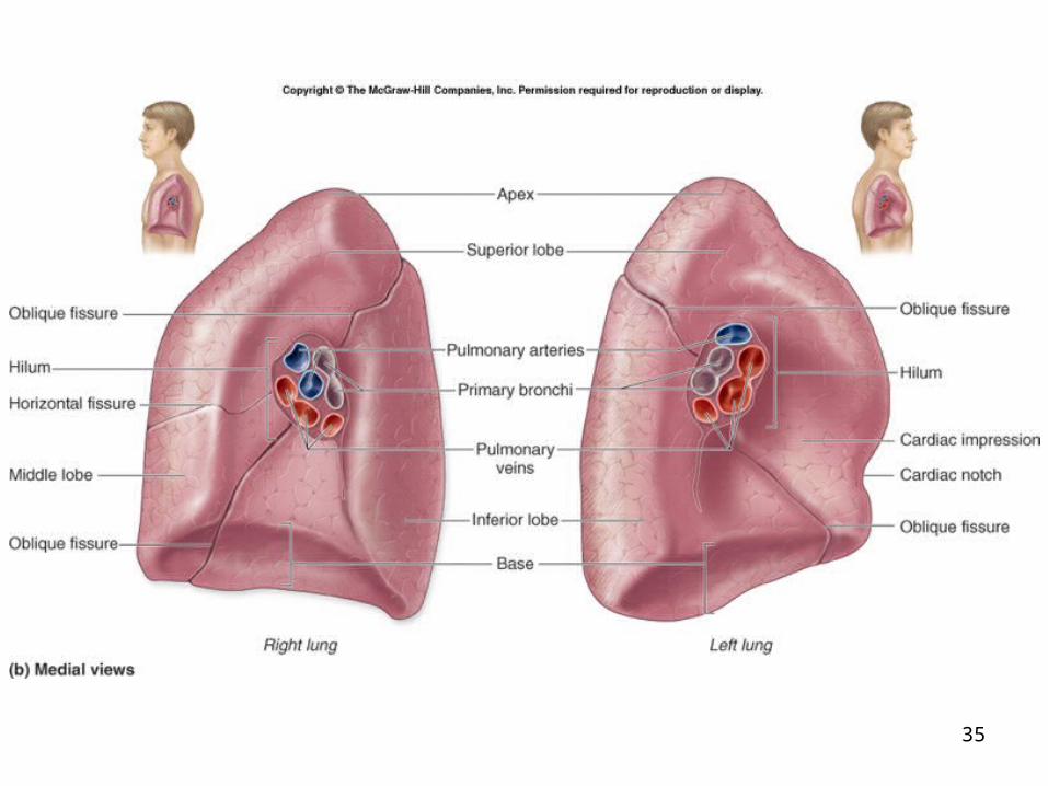
Gross Anatomy of the Lungs Each lung has a conical shape. Its wide, concave base rests upon the muscular
diaphragm. Its relatively blunt superior region, called the apex or
(cupola), projects superiorly to a point that is slightly superior and posterior to the clavicle.
Both lungs are bordered by the thoracic wall anteriorly, laterally, and posteriorly, and supported by the rib cage.
Toward the midline, the lungs are separated from each other by the mediastinum.
The relatively broad, rounded surface in contact with the thoracic wall is called the costal surface of the lung.

34
35

36

37

38

25-39
Pleura and Pleural Cavities The outer surface of each lung and the
adjacent internal thoracic wall are lined by a serous membrane called pleura, which is formed from simple squamous epithelium.
The outer surface of each lung is tightly covered by the visceral pleura, while the internal thoracic walls, the lateral surfaces of the mediastinum, and the superior surface of the diaphragm are lined by the parietal pleura.
The parietal and visceral pleural layers are continuous at the hilum of each lung.

25-40
Pleura and Pleural Cavities The outer surface of each lung is tightly covered by the
visceral pleura, while the internal thoracic walls, the lateral surfaces of the mediastinum, and the superior surface of the diaphragm are lined by the parietal pleura.
The potential space between these serous membrane layers is a pleural cavity.
The pleural membranes produce a thin, serous fluid that circulates in the pleural cavity and acts as a lubricant, ensuring minimal friction during breathing.

41

25-42
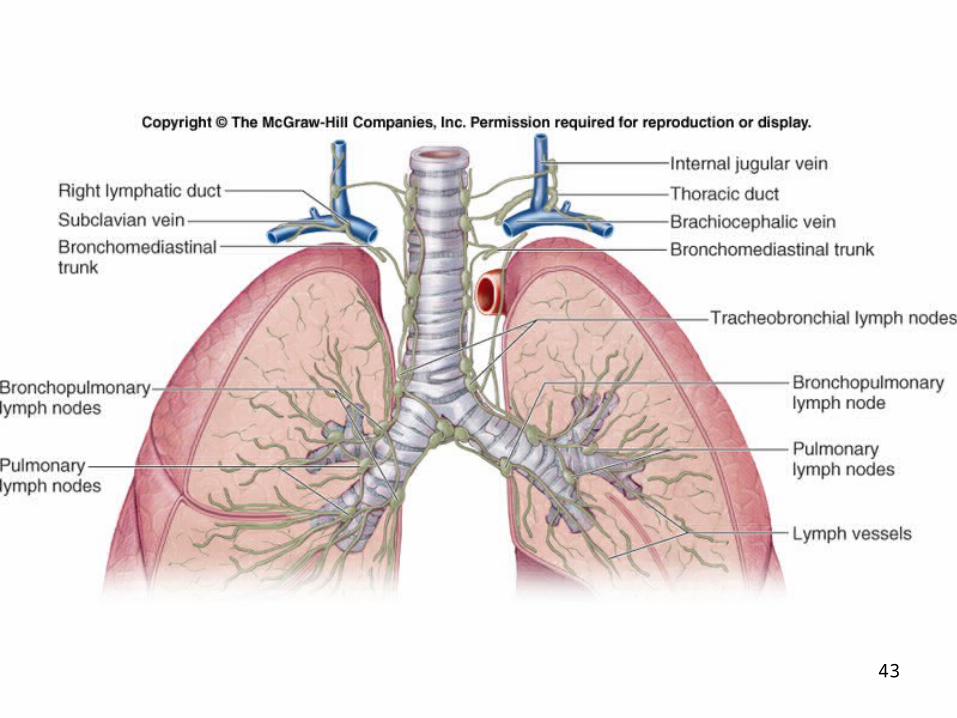
Lymphatic Drainage Lymph nodes and vessels are located within
the connective tissue of the lung as well as around the bronchi and pleura.
The lymph nodes collect carbon, dust particles, and pollutants that were not filtered out by the pseudostratified ciliated columnar epithelium.
43

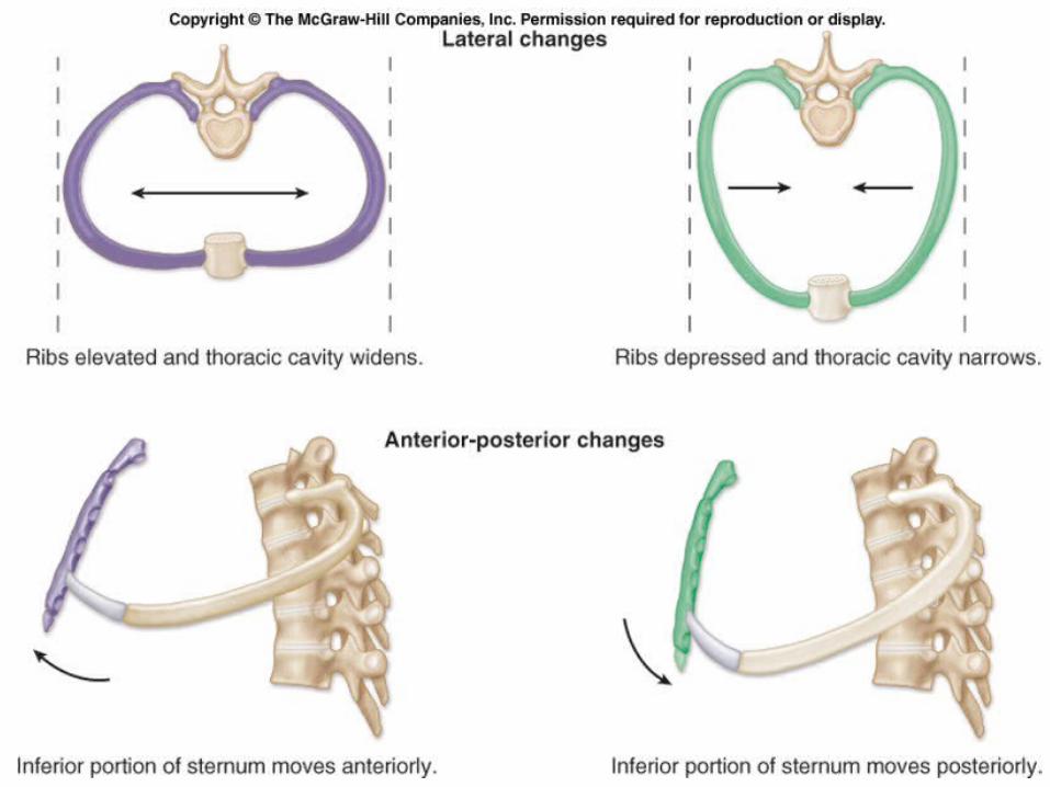
25-44
Thoracic Wall Dimensional Changes During Respiration Lateral dimensional changes occur with rib
movements. Elevation of the ribs increases the lateral
dimensions of the thoracic cavity, while depression of the ribs decreases the lateral dimensions of the thoracic cavity.

25-45
Muscles that Move the Ribs The scalenes help increase thoracic cavity dimensions
by elevating the first and second ribs during forced inhalation.
The ribs elevate upon contraction of the external intercostals, thereby increasing the transverse dimensions of the thoracic cavity during inhalation.
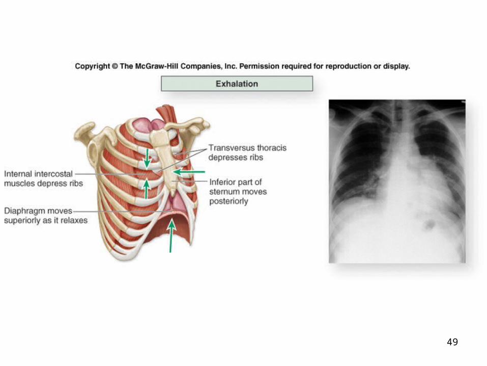
Contraction of the internal intercostals depresses the ribs, but this only occurs during forced exhalation.
Normal exhalation requires no active muscular effort. A small transversus thoracis extends across the inner
surface of the thoracic cage and attaches to ribs 2–6. It helps depress the ribs.

25-46
Muscles that Move the Ribs Two posterior thorax muscles also assist with respiration.
These muscles are located deep to the trapezius and latissimus dorsi, but superficial to the erector spinae muscles.
The serratus posterior superior elevates ribs 2–5 during inhalation, and the serratus posterior inferior depresses ribs 8–12 during exhalation.
In addition, some accessory muscles assist with respiratory activities.
The pectoralis minor, serratus anterior, and sternocleidomastoid help with forced inhalation, while the abdominal muscles (external and internal obliques, transversus abdominis, and rectus abdominis) assist in active exhalation.

47

48
49
50
51

25-52
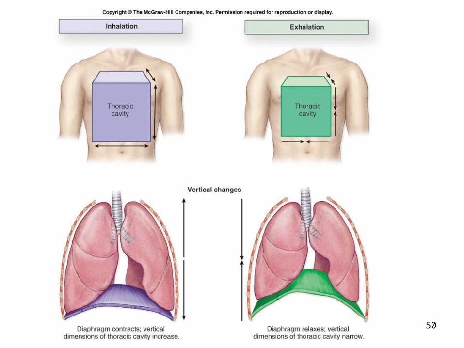
Boyle’s Law “The pressure of a gas decreases if the volume of the
container increases, and vice versa.” When the volume of the thoracic cavity increases even
slightly during inhalation, the intrapulmonary pressure decreases slightly, and air flows into the lungs through the conducting airways.
Air flows into the lungs from a region of higher pressure (the atmosphere) into a region of lower pressure (the intrapulmonary region).
When the volume of the thoracic cavity decreases during exhalation, the intrapulmonary pressure increases and forces air out of the lungs into the atmosphere.

25-53
Ventilation Control by Respiratory Centers of the Brain The trachea, bronchial tree, and lungs are innervated by
the autonomic nervous system. The autonomic nerve fibers that innervate the heart
also send branches to the respiratory structures. The involuntary, rhythmic activities that deliver and
remove respiratory gases are regulated in the brainstem.
Regulatory respiratory centers are located within the reticular formation through both the medulla oblongata and pons.

54

25-55
Aging and the Respiratory System Becomes less efficient with age due to several structural
changes. Decrease in elastic connective tissue in the lungs and the
thoracic cavity wall. Loss of elasticity reduces the amount of gas that can be
exchanged with each breath and results in a decrease in the ventilation rate.
Emphysema may cause a loss of alveoli or their functionality
Reduced capacity for gas exchange can cause an older person to become “short of breath” upon exertion.
Carbon, dust, and pollution material gradually accumulate in our lymph nodes and lungs.

56