2,3,5-Trimethoxy-4-cresol, an anti-metastatic constituent ...for.nchu.edu.tw/up_book/Leader...
9
ORIGINAL PAPER 2,3,5-Trimethoxy-4-cresol, an anti-metastatic constituent from the solid-state cultured mycelium of Antrodia cinnamomea and its mechanism Chin-Chung Lin 1,2 • Ching-Chun Chen 1 • Yueh-Hsiung Kuo 3,4 • Jong-Tar Kuo 2 • K. J. Senthil Kumar 5 • Sheng-Yang Wang 5,6,7 Received: 28 January 2015 / Accepted: 20 April 2015 / Published online: 8 May 2015 Ó The Japanese Society of Pharmacognosy and Springer Japan 2015 Abstract Antrodia cinnamomea is a valuable and unique edible fungus originating from the forests of Taiwan. In this study, an anti-metastatic compound, 2,3,5-trimethoxy- 4-cresol (TMC), was isolated from the solid-state cultured mycelium of A. cinnamomea. According to the results obtained from cell wound healing, cell migration and in- vasion assays, TMC effectively suppressed movement, migration and invasion of lung cancer cells at the dosage of 5–40 lM, which was non-toxic to A549 cells. In addition, TMC reduced protein expression of Akt, MMP-2 and MMP-9 and enhanced E-cadherin and TIMP-1 protein expression, which are known to regulate cell adhesion, migration and invasion. Taken together, TMC effectively suppresses movement, migration and invasion of lung cancer cells, and achieves an anti-cancer metastasis effect. Keywords Antrodia cinnamomea Á 2,3,5-Trimethoxy-4- cresol Á Anti-metastasis Á EMT Abbreviations DMSO Dimethyl sulfoxide DPBS Dulbecco’s phosphate-buffered saline ECM Extracellular matrix EMT Epithelial–mesenchymal transition FAK Focal adhesion kinase FBS Fetal bovine serum GA Gallic acid MMP Matrix metalloproteinase MTT 3-(4,5-Dimethylthiazol-2-yl)-2,5,- diphenyltetrazolium bromide NSCLC Non-small-cell lung cancer SDS Sodium dodecyl sulfate TEMED N,N,N’,N’-Tetra-methylethylenediamine TIMP-2 Tissue inhibitor of metalloproteinase-2 TNF-a Tumor necrosis factor alpha Introduction Non-small-cell lung cancer (NSCLC) is the leading cause of cancer death worldwide, and one of the most metastatic tumors. Although the 5-year survival rate for patients with localized NSCLC is more than 50 %, 57 % of patients have advanced/metastatic disease at the time of diagnosis, which carries a survival rate of less than 5 %. The primary cause of death in patients with lung cancer is tumor metastasis [1, 2]. Currently, surgery is the treatment of choice for early stage NSCLC to improve patient survival [3], but surgery also increases the risk to the patient, and therefore & Jong-Tar Kuo [email protected] & Sheng-Yang Wang taiwanfi[email protected] 1 Taiwan Leader Biotech Company, Taipei, Taiwan 2 Department of Biological Science and Technology, China University of Science and Technology, Taipei, Taiwan 3 Graduate Institute of Chinese Pharmaceutical Science, China Medical University, Taichung, Taiwan 4 Department of Biotechnology, Asia University, Taichung, Taiwan 5 Department of Forestry, National Chung Hsing University, 250-Kuo-Kung Road, Taichung 402, Taiwan 6 Agricultural Biotechnology Center, National Chung-Hsing University, Taichung, Taiwan 7 Agricultural Biotechnology Research Institute, Academia Sinica, Taipei, Taiwan 123 J Nat Med (2015) 69:513–521 DOI 10.1007/s11418-015-0916-6
Transcript of 2,3,5-Trimethoxy-4-cresol, an anti-metastatic constituent ...for.nchu.edu.tw/up_book/Leader...

ORIGINAL PAPER
2,3,5-Trimethoxy-4-cresol, an anti-metastatic constituentfrom the solid-state cultured mycelium of Antrodia cinnamomeaand its mechanism
Chin-Chung Lin1,2 • Ching-Chun Chen1 • Yueh-Hsiung Kuo3,4 • Jong-Tar Kuo2 •
K. J. Senthil Kumar5 • Sheng-Yang Wang5,6,7
Received: 28 January 2015 / Accepted: 20 April 2015 / Published online: 8 May 2015
� The Japanese Society of Pharmacognosy and Springer Japan 2015
Abstract Antrodia cinnamomea is a valuable and unique
edible fungus originating from the forests of Taiwan. In
this study, an anti-metastatic compound, 2,3,5-trimethoxy-
4-cresol (TMC), was isolated from the solid-state cultured
mycelium of A. cinnamomea. According to the results
obtained from cell wound healing, cell migration and in-
vasion assays, TMC effectively suppressed movement,
migration and invasion of lung cancer cells at the dosage of
5–40 lM, which was non-toxic to A549 cells. In addition,
TMC reduced protein expression of Akt, MMP-2 and
MMP-9 and enhanced E-cadherin and TIMP-1 protein
expression, which are known to regulate cell adhesion,
migration and invasion. Taken together, TMC effectively
suppresses movement, migration and invasion of lung
cancer cells, and achieves an anti-cancer metastasis effect.
Keywords Antrodia cinnamomea � 2,3,5-Trimethoxy-4-
cresol � Anti-metastasis � EMT
Abbreviations
DMSO Dimethyl sulfoxide
DPBS Dulbecco’s phosphate-buffered saline
ECM Extracellular matrix
EMT Epithelial–mesenchymal transition
FAK Focal adhesion kinase
FBS Fetal bovine serum
GA Gallic acid
MMP Matrix metalloproteinase
MTT 3-(4,5-Dimethylthiazol-2-yl)-2,5,-
diphenyltetrazolium bromide
NSCLC Non-small-cell lung cancer
SDS Sodium dodecyl sulfate
TEMED N,N,N’,N’-Tetra-methylethylenediamine
TIMP-2 Tissue inhibitor of metalloproteinase-2
TNF-a Tumor necrosis factor alpha
Introduction
Non-small-cell lung cancer (NSCLC) is the leading cause
of cancer death worldwide, and one of the most metastatic
tumors. Although the 5-year survival rate for patients with
localized NSCLC is more than 50 %, 57 % of patients have
advanced/metastatic disease at the time of diagnosis, which
carries a survival rate of less than 5 %. The primary cause
of death in patients with lung cancer is tumor metastasis [1,
2]. Currently, surgery is the treatment of choice for early
stage NSCLC to improve patient survival [3], but surgery
also increases the risk to the patient, and therefore
& Jong-Tar Kuo
& Sheng-Yang Wang
1 Taiwan Leader Biotech Company, Taipei, Taiwan
2 Department of Biological Science and Technology, China
University of Science and Technology, Taipei, Taiwan
3 Graduate Institute of Chinese Pharmaceutical Science, China
Medical University, Taichung, Taiwan
4 Department of Biotechnology, Asia University, Taichung,
Taiwan
5 Department of Forestry, National Chung Hsing University,
250-Kuo-Kung Road, Taichung 402, Taiwan
6 Agricultural Biotechnology Center, National Chung-Hsing
University, Taichung, Taiwan
7 Agricultural Biotechnology Research Institute, Academia
Sinica, Taipei, Taiwan
123
J Nat Med (2015) 69:513–521
DOI 10.1007/s11418-015-0916-6

chemotherapy, i.e., anti-metastatic and/or anti-invasion
drugs, may be recommended to prevent local recurrence of
the primary tumor and spread of the tumor cells.
Antrodia cinnamomea (syn. Antrodia camphorata and
Taiwanofungus camphorata) is a valuable and unique
edible fungus that originates from the forests of Taiwan. It
has long been used as a folk remedy for treating various
diseases including liver diseases, hypertension, abdominal
pain and cancer [4]. Due to its high commercial value, A.
cinnamomea has attracted much research and development
interest in Taiwan over the past decade, and has been
awarded a ‘‘National Health Food’’ certificate by Taiwan’s
Department of Health. Numerous studies have been con-
ducted on the physiology and biochemical and pharmaco-
logical properties of A. cinnamomea [4–11], and many
compounds identified from A. cinnamomea have been re-
ported to possess health-promoting activities [12, 13].
Among the various activities reported, the anti-cancer ac-
tivities of A. cinnamomea are of particular interest. Re-
cently, increasing importance has been attached to the anti-
metastatic activity of A. cinnamomea and its bioactive
compounds [14–16]. Our previous study demonstrate that
antrodin C, a melamide derivative isolated from the
mycelia of A. cinnamomea, inhibits the metastatic potential
of human breast carcinoma (MCF-7 cells) through the in-
hibition of epithelial–mesenchymal transition via suppres-
sion of the Smad2/3 signaling pathway [17]. Antrodon, a
glycoprotein isolated from the mycelia of A. cinnamomea,
inhibits the metastatic potential of Lewis lung carcinoma
(LLC) in vitro via down-regulation of matrix metallopro-
teinase (MMP)-2, MMP-9, tumor necrosis factor alpha
(TNF-a), interleukin (IL)-6 and IL-8, and up-regulation of
tissue inhibitor of metalloproteinase (TIMP)-1 and TIMP-2
[18]. However, studies on anti-metastatic compounds of the
fruiting body or mycelium of A. cinnamomea are limited.
In the present study, a potent anti-metastatic constituent,
2,3,5-trimethoxy-4-cresol (TMC) (Fig. 1), was isolated
from the solid-state cultural mycelium product of Antrodia
cinnamomea. The biochemical steps including the role of
TMC in the metastatic process in lung cancer cells (A549)
have been elucidated. Curcumin has been reported to be a
potent anti-metastatic phyto-compound [19], so in the
present study a non-cytotoxic concentration of curcumin
was used as a positive drug control.
Results and Discussion
Most cancer cells can acquire metastatic capability leading
to secondary tumors. Here we first used a low dose of
purified extract, pure compound TMC, to explore the cy-
totoxicity of the treatment. A549 cells treated with differ-
ent concentrations of TMC underwent MTT assay and were
analyzed after 24 h. Cell viability was not significantly
different in cells treated with TMC at concentrations of
5–40 lM compared to the control group (DMSO-treated
A549 cells) (Fig. 2). This data is consistent with a previous
report stating that below the concentration of 20 lM, TMC
(previously known as 2,3,6-trimethoxy-4-methylphenol)
was not cytotoxic to murine colorectal CT26 and human
leukemia cancer cell lines, with an IC50 value of[20 lM
[20]. TMC was not cytotoxic to A549 cells at these dosa-
ges. Therefore, in subsequent experiments, TMC was ap-
plied in this concentration range. In addition, the positive
drug control curcumin (10 lM) does not show cytotoxicity
to A549 cells (data not shown).
Wound healing is generally characterized by cell pro-
liferation, remodeling of the extracellular matrix, cell in-
vasion and migration [21]. In this study, a wound-healing
assay was conducted to investigate cell migration ability. A
wound line was created by scraping seeded monolayer
A549 cells, which were then cultured in low serum-free
medium plus TMC. The wound-healing activity of cells
was observed between 0 and 12 h. The results indicated
that TMC significantly decreased the wound-healing ability
of A549 cells in a dose-dependent manner (Fig. 3). Next, in
order to investigate the invasive and migratory capability
of tumor cells, a trans-well migration assay was performed.
The results showed that inhibition occurred in a concen-
tration-dependent manner with higher doses of TMC
Fig. 1 2,3,5-Trimethoxy-4-
cresol (TMC)Fig. 2 Effect of TMC on the viability of A549 cells. Cells were
treated with or without TMC at various concentrations (0, 5, 10, 20,
40 lM) for 24 h. Cell viability was analyzed by MTT assay. The
results represent the average of three independent experiments ± SD.
*P\ 0.05, **P\ 0.005 represent significant difference relative to
the control group
514 J Nat Med (2015) 69:513–521
123

resulting in fewer cells migrating to the lower chamber
(Fig. 4a). When TNF-a was added to induce A549 cell
growth, the same dose of TMC was used to explore the
inhibition of A549 cell migration capability (Fig. 4b). We
also used a trans-well chamber coated with Matrigel to
analyze the invasion capability of cells. As shown in Fig. 5,
significantly fewer cells passed into the lower chamber
with higher doses of TMC, in a dose-dependent manner.
Previous studies have suggested that enhanced tumor cell
adhesiveness correlates with metastatic ability [22]. When
A549 cells were treated with different dosages of TMC
(0–40 lM), after 8 h the adhesion ability of cells was in-
hibited in a dose-dependent manner (Fig. 6). The inhibition
ratio of adhesion activity of cells on the Matrigel mem-
brane was 70 % of that of the control group. This result
indicates that TMC significantly reduced the cell adhesion
ability of A549 cells. With reference to previous reports
[23, 24], the positive drug control of this study, a non-
cytotoxic concentration of curcumin (10 lM), showed a
strong inhibition of migration, invasion and adhesion of
A549 cells; the inhibitory effect of TMC was highly
comparable with curcumin.
Matrix metalloproteinases (MMPs) belong to a zinc-de-
pendent family (ECM) of endopeptidases and are the prin-
cipal mediators of the alterations observed in the
microenvironment during cancer progression [25]. MMPs
are implicated in a variety of physiological processes, in-
cluding wound healing, uterine involution and organo-
genesis, as well as in pathological conditions such as
inflammation, vascular and auto-immune disorders, and
carcinogenesis [26, 27]. MMPs have been considered to be
important therapeutic targets for cancer because the
metastatic potential of various cancers is correlated with the
ability of cancer cells to degrade the basement membrane
[27]. The other key protease involved in degrading the
components of the basement membrane is urokinase plas-
minogen activator (u-PA). u-PA is a serine protease that
cleaves the ECM and converts the inactive plasminogen to
active plasmin, which can directly mediate the invasion by
degrading matrix components [27]. In order to investigate
whether MMPs and u-PA are involved in the TMC inhibition
of cancer cell invasion and motility, a gelatin and casein
zymography assay was performed to check MMP and u-PA
activity. As shown in Fig. 7, TMC significantly reduced the
MMP-2 (Fig. 7a) and MMP-9 activity (Fig. 7b). The inhi-
bition of MMP-2 and MMP-9 expression in A549 cells was
52 and 35 %, respectively, at the dosage of 40 lM. The u-PA
activity in A549 cells was also inhibited by TMC (Fig. 8).
Epithelial–mesenchymal transition (EMT) has an im-
portant role in the progression of tumor metastasis [28]. It has
been reported that protein kinase Akt induces EMT through
down-regulation of the epithelial marker E-cadherin and
upregulation of the mesenchymal marker vimentin in cancer
progression [29]. E-cadherin is responsible for epithelial
cell–cell adhesion and maintenance of cytoskeleton organi-
zation, and it is the major transmembrane component of
adherens junctions. Cancer cell proliferation, invasion and
metastasis increases with loss of function of E-cadherin [30].
During the EMT process, most of the tumor suppressor
proteins (E-cadherin) are down-regulated, and the
Fig. 3 Effects of TMC on the morphology and migration of A549
cells in wound healing assay. A549 cell monolayers treated with or
without different dosages of TMC (0, 5, 10, 20, 40 lM) were scraped
and the number of cells in the denuded zone (i.e., the wound) was
quantitated after 0, 6 and 12 h through light microscopy observation.
The results represent the average of three independent ex-
periments ± SD. *P\ 0.05, **P\ 0.005 represent significant dif-
ference relative to the control group
J Nat Med (2015) 69:513–521 515
123

oncoproteins (vimentin, N-cadherin) are up-regulated [31].
E-cadherin promotes homotypic tumor cell adhesion func-
tion to maintain intercellular contacts that confine cells to the
primary tumor site and prevent metastatic potential [32]. To
further confirm the inhibition mechanism of TMC in
metastasis, the effects of TMC on E-cadherin, p-AKT and
TIMP-1 were examined. As shown in Fig. 9a, TMC sig-
nificantly enhanced levels of the epithelial marker E-cad-
herin, and decreased expression of p-Akt. Moreover, TIMP-
1 expression was also increased (Fig. 9b). In conclusion, our
data suggest that treatment of lung cancer cells with TMC
induces molecular changes, which are consistent with a less
motile, migratory and invasive phenotype of lung cancer
cell. In conclusion, 2,3,5-trimethoxy-4-cresol (TMC) ef-
fectively suppresses movement, migration and invasion of
lung cancer cells, and achieves an anti-cancer metastasis
effect.
Fig. 4 TMC inhibited the migration of A549 cells in trans-well
assay. a A549 cells were seeded in the upper chamber of the trans-
well filters (pore size, 8 lm) after treatment with various concentra-
tions of TMC (0, 5, 10, 20, 40 lM) and curcumin (10 lM).
Chemoattractant was placed in the bottom chamber. A549 cells
migrate through the small pores in the transmembrane in response to
the chemoattractant. FBS (20 %) was used as the inducer. b A549
cells treated with TMC (0, 5, 10, 20, 40 lM) for 24 h, then seeded in
the upper chamber of the trans-well and incubated for 12 h; 20 %
FBS and TNF-a were used as the inducer. *P\ 0.05, **P\ 0.005
represent significant difference relative to the control group. Cur
curcumin
Fig. 5 TMC inhibited the invasive ability of A549 cells. A549 cells
were treated with TMC (0, 5, 10, 20, 40 lM) or curcumin (10 lM)
and seeded in the upper chamber of the trans-well which was coated
Matrigel, and incubated for 12 h; 20 % FBS was used as the inducer.
*P\ 0.05, **P\ 0.005 show significant difference relative to the
control group. Cur curcumin
Fig. 6 TMC inhibited the adhesion of A549 cells through Matrigel.
Matrigel was coated in the 24-well incubator for 24 h, and A549 cells
were treated with TMC (0, 5, 10, 20, 40 lM) or curcumin (10 lM) for
24 h, then cells were subtracted and seeded in the 24-well incubator
for 8 h. Results are presented as mean ± SD of three assays.
*P\ 0.05, **P\ 0.005 show significant difference relative to the
control group. Cur curcumin
516 J Nat Med (2015) 69:513–521
123

Experimental section
Chemical and reagents
2,3,5-Trimethoxy-4-cresol was previously isolated and
identified from the solid-state cultured mycelium of A.
cinnamomea and was known as 2,3,6-trimethoxy-4-
methylphenol [20]. In this study, we also isolated it from
the solid-state cultured mycelium of A. cinnamomea (strain
no. LEACS-002) obtained from the R&D Center of Taiwan
Leader Biotechnology Corp. (Taichung, Taiwan). The pu-
rity of TMC was above 99 %, as confirmed by HPLC and1H-NMR analysis. Ham’s F12 medium was purchased
from Amimed/BioConcept (Allschwil, Switzerland); fetal
bovine serum (FBS) was obtained from Gibco-BRL (Chi-
na). Antibodies against MMP-2, MMP-9, and b-actin were
purchased from Santa Cruz Biotechnology (Heidelberg,
Germany); Akt, cadherin, NF-jB, and TIMP1 were pur-
chased from Cell Signaling (Danvers, MA, USA). The
3-(4,5-dimethylthiazol-2-yl)-2,5-diphenyltetrazolium bro-
mide (MTT) and curcumin were obtained from Sigma–
Aldrich (St. Louis, MO, USA). Millicell hanging cell cul-
ture insert was obtained from Millipore (Billerica, MA,
USA); Matrigel was purchased from BD Biosciences (San
Jose, CA, USA).
Cell culture
The human non-small-cell lung cancer cell line A549 was
obtained from the American Type Culture Collection
(ATCC, Manassas, VA, USA). The cell line was cultured
in Ham’s F12 medium supplemented with 10 % heat-
inactivated FB and, 2 mM glutamine, and cell cultures
were maintained at 37 �C in an incubator with 5 % CO2.
MTT assay
Cell viability was determined by MTT colorimetric assay
as described previously [33]. A549 cells (2 9 105 cells/24-
Fig. 7 Effect of TMC-dependent MMP activity inhibition on A549
cells. A549 cells were treated with TMC (0, 5, 10, 20, 40 lM) in
culture medium. After 24 h, the cells were cultured in serum-free
medium and then subjected to gelatin zymography to analyze the
activities of a MMP-2 and b MMP-9. The activities of these proteins
were subsequently quantified by densitometric analysis. Data from 3
independent experiments are shown. *P\ 0.05, **P\ 0.005 repre-
sent significant difference relative to the control
Fig. 8 TMC inhibited uPA activity on A549 cells. Cells were treated
with up to 40 lM TMC for 24 h and were then subjected to casein
zymography to analyze uPA activity, which was subsequently
quantified by densitometry analysis. Data from 3 independent
experiments are shown. *P\ 0.05, **P\ 0.005 represent significant
difference relative to the control group
J Nat Med (2015) 69:513–521 517
123

well plates) were seeded and incubated overnight. Cells
were then treated with various concentrations of TMC (5,
10, 20 and 40 lM) for 24 h. After incubation, the medium-
deprived cells were incubated with MTT (10 lg/mL) in
100 lL of fresh DMEM for 1 h at 37 �C. The MTT-gen-
erated violet formazan crystals were dissolved in dimethyl
sulfoxide (DMSO) and the absorbance was measured at
570 nm (A570) using an ELISA microplate reader (lQuant,
Bio-Tek Instruments, Winooski, VT, USA). Cell viability
(%) was calculated as: (A570 of treated cells/A570 of un-
treated cells) 9100.
Cell wound-healing repair assay
Cell proliferation and migration was observed in A549
cells using the wound-healing assay [34]. A549 cells
Fig. 9 Inhibition effects of
TMC on expression of proteins
involved in regulation of cell
motility and invasion of A549
cells. A549 cells were treated
with TMC (0, 5, 10, 20, 40 lM)
for 24 h. Immunoblots are
shown for a p-Akt, E-cadherin,
b MMP-2, MMP-9, TIMP-1 and
b-actin (control) in A549 cell
lines. The cell lysates were
prepared for Western blotting as
described in Experimental
Section. Data from 3
independent experiments are
shown. *P\ 0.05, **P\ 0.005
represent significant difference
relative to the control
518 J Nat Med (2015) 69:513–521
123
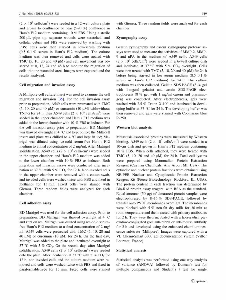
(2 9 107 cells/cm2) were seeded in a 12-well culture plate
and grown to confluence or near ([90 %) confluence in
Ham’s F12 medium containing 10 % FBS. Using a sterile
200 lL pipet tip, separate wounds were scratched, and
cellular debris and FBS were removed by washing with
PBS; cells were then starved in low-serum medium
(0.5–0.1 % serum in Ham’s F12 medium). The culture
medium was then removed and cells were treated with
TMC (5, 10, 20 and 40 lM) and cell movement was ob-
served at 0, 12, 24 and 48 h to monitor the migration of
cells into the wounded area. Images were captured and the
results analyzed.
Cell migration and invasion assay
A Millipore cell culture insert was used to examine the cell
migration and invasion [35]. For the cell invasion assay
prior to preparation, A549 cells were pretreated with TMC
(5, 10, 20 and 40 lM) or curcumin (10 lM) with/without
TNF-a for 24 h, then A549 cells (2 9 105 cells/cm2) were
seeded in the upper chamber, and Ham’s F12 medium was
added to the lower chamber with 10 % FBS as inducer. For
the cell invasion assay prior to preparation, BD Matrigel
was thawed overnight at 4 �C and kept on ice; the Millicell
insert and plate was chilled to 4 �C and kept in ice; Ma-
trigel was diluted using ice-cold serum-free Ham’s F12
medium to a final concentration of 2 mg/ml, After Matrigel
solidification, A549 cells (2 9 105 cells/cm2) were seeded
in the upper chamber, and Ham’s F12 medium was added
to the lower chamber with 10 % FBS as inducer. Both
migration and invasion assays were conducted after incu-
bation at 37 �C with 5 % CO2 for 12 h. Non-invaded cells
in the upper chamber were removed with a cotton swab,
and invaded cells were washed twice with PBS and fixed in
methanol for 15 min. Fixed cells were stained with
Giemsa. Three random fields were analyzed for each
chamber.
Cell adhesion assay
BD Matrigel was used for the cell adhesion assay. Prior to
preparation, BD Matrigel was thawed overnight at 4 �Cand kept on ice. Matrigel was diluted using ice-cold serum-
free Ham’s F12 medium to a final concentration of 2 mg/
ml. A549 cells were pretreated with TMC (5, 10, 20 and
40 lM) or curcumin (10 lM) for 24 h. On the first day,
Matrigel was added to the plate and incubated overnight at
37 �C with 5 % CO2. On the second day, after Matrigel
solidification, A549 cells (2 9 105 cells/cm2) were seeded
onto the plate. After incubation at 37 �C with 5 % CO2 for
12 h, non-invaded cells and the culture medium were re-
moved and cells were washed twice with PBS and fixed in
paraformaldehyde for 15 min. Fixed cells were stained
with Giemsa. Three random fields were analyzed for each
chamber.
Zymography assay
Gelatin zymography and casein zymography protease as-
says were used to measure the activities of MMP-2, MMP-
9 and uPA in the medium of A549 cells. A549 cells
(2 9 105 cells/cm2) were seeded in a 6-well culture dish
and incubated at 37 �C with 5 % CO2 overnight. Cells
were then treated with TMC (5, 10, 20 and 40 lM) for 24 h
before being starved in low-serum medium (0.5–0.1 %
serum in Ham’s F12 medium) for 24 h. The culture
medium was then collected. Gelatin SDS-PAGE (8 % gel
with 1 mg/ml gelatin) and casein SDS-PAGE elec-
trophoresis (8 % gel with 1 mg/ml casein and plasmino-
gen) was conducted. After electrophoresis, gels were
washed with 2.5 % Triton X-100 and incubated in devel-
oping buffer at 37 �C for 24 h. The developing buffer was
then removed and gels were stained with Coomassie blue
R-250.
Western blot analysis
Metastasis-associated proteins were measured by Western
blotting. A549 cells (2 9 105 cells/cm2) were seeded in a
10-cm dish and grown in Ham’s F12 medium containing
10 % FBS. When cells attached, they were treated with
TMC (5, 10, 20 and 40 lM) for 24 h. Total cell lysates
were prepared using Mammalian Protein Extraction
Reagent (Cayman Chemicals, Ann Arbor, MI, USA). The
cytosolic and nuclear protein fractions were obtained using
NE-PER Nuclear and Cytoplasmic Protein Extraction
Reagent Kit (Pierce Biotechnology, Rockford, IL, USA).
The protein content in each fraction was determined by
Bio-Rad protein assay reagent, with BSA as the standard.
Equal amounts (50 lg) of denatured protein samples were
electrophoresed by 8–15 % SDS-PAGE, followed by
transfer onto PVDF membranes overnight. The membranes
were blocked with 5 % non-fat dry milk for 30 min at
room temperature and then reacted with primary antibodies
for 2 h. They were then incubated with a horseradish per-
oxidase-conjugated goat anti-rabbit or anti-mouse antibody
for 2 h and developed using the enhanced chemilumines-
cence substrate (Millipore). Images were captured with a
VL Chemi-Smart 3000 gel documentation system (Vilber
Lourmat, France).
Statistical analysis
Statistical analysis was performed using one-way analysis
of variance (ANOVA) followed by Duncan’s test for
multiple comparisons and Student’s t test for single
J Nat Med (2015) 69:513–521 519
123

comparison. The data are reported as mean ± SD. The
numbers of independent experiments assessed are given in
the figure legends.
Acknowledgments Parts of this study were supported by the Min-
istry of Science and Technology, Taiwan, Republic of China (NSC-
103-2911-I-005-301, NSC-102-2911-I-005-301) and the Ministry of
Education, Taiwan, R.O.C. under the ATU plan.
Conflict of interest No competing financial interests exist.
References
1. Simon GR (2014) nab-Paclitaxel for the treatment of advanced
squamous none small-cell lung cancer: a comprehensive update.
Clin Lung Cancer 15:391–397
2. Riihimaki M, Thomsen H, Hemminki A, Sundquist K, Hemminki
K (2013) Comparison of survival of patients with metastases
from known versus unknown primaries: survival in metastatic
cancer. BMC Cancer 13:36
3. Nichols L, Saunders R, Knollmann FD (2012) Causes of death of
patients with lung cancer. Arch Pathol Lab Med 136:1552–1557
4. Wang HC, Chu FH, Chian SC, Liao JW, Hsieh HW, Li WH, Lin
CC, Shaw JF, Kuo YH, Wang SY (2012) Establishment of the
metabolite profile for an Antrodia cinnamomea health food pro-
duct and investigation of its chemoprevention activity. J Agric
Food Chem 61:8556–8564
5. Hsieh YH, Chu FH, Wang YS, Chien SC, Chang ST, Shaw JF,
Chen CY, Hsiao WW, Kuo YH, Wang SY (2010) Antrocamphin
A, an anti-inflammatory principal from the fruiting body of
Taiwanofungus camphoratus, and its mechanisms. J Agric Food
Chem 58:3153–3158
6. Gokila Vani M, Senthil Kumar KJ, Liao JW, Chien SC, Mau JL,
Chiang SS, Lin CC, Kuo YH, Wang SY (2013) Antcin C from
Antrodia cinnamomea protects liver cells against free radical-
induced oxidative stress and apoptosis in vitro and in vivo
through Nrf2-dependent mechanism. Evid Based Complement
Alternat Med 2013:296082
7. Lin TY, Chen CY, Chien SC, Hsiao WW, Chu FH, Li WH, Lin
CC, Shaw JF, Wang SY (2011) Metabolite profiles for Antrodia
cinnamomea fruiting bodies harvested at different culture ages
and substrates from different wood. J Agric Food Chem
59:7626–7635
8. Senthil Kumar KJ, Chu FH, Hsieh HW, Liao JW, Li WH, Lin
CC, Shaw JF, Wang SY (2011) Antroquinonol from ethanolic
extract of mycelium of Antrodia cinnamomea protects hepatic
cells from ethanol-induced oxidative stress through Nrf-2 acti-
vation. J Ethnopharmacol 136:168–177
9. Lin TY, Chien SC, Kuo YH, Wang SY (2012) Distinguishing
between R- and S-antcin C and their cytotoxicity. Nat Prod
Commun 7:835–836
10. Lu MC, El-Shazly M, Wu TY, Du YC, Chang TT, Chen CF, Hsu
YM, Lai KH, Chiu CP, Chang FR, Wu YC (2013) Recent re-
search and development of Antrodia cinnamomea. Pharmacol
Ther 139:124–156
11. Chen YC, Liu YL, Li FY, Chang CI, Wang SY, Lee KY, Li SL,
Chen YP, Jinn TR, Tzen JT (2011) Antcin A, a steroid-like
compound from Antrodia camphorata, exerts anti-inflammatory
effect via mimicking glucocorticoids. Acta Pharmacol Sin
32:904–911
12. Geethangili M, Tzeng YM (2011) Review of pharmacology ef-
fects of Antrodia camphorata and its bioactivities compounds.
Evid Based Complement Alternat Med 2011:212641
13. Ao ZH, Xu ZH, Lu ZM, Xu HY, Zhang XM, Dou WF (2009)
Niuchangchih (Antrodia camphorata) and its potential in treating
liver diseases. J Ethnophrarmcol 121:194–212
14. Liu FC, Lai MT, Chen YY, Lin WH, Chang SJ, Sheu MJ, Wu CH
(2013) Elucidating the inhibitory mechanisms of the ethanolic
extract of the fruiting body of the mushroom Antrodia cin-
namomea on the proliferation and migration of murine leukemia
WEHI-3 cells and their tumorigenicity in a BALB/c allograft
tumor model. Phytomedicine 20:874–882
15. Chen YY, Chou PY, Chien YC, Wu CH, Wu TS, Sheu MJ (2012)
Ethanol extracts of fruiting bodies of Antrodia cinnamomea ex-
hibit anti-migration action in human adenocarcinoma CL1-0 cells
through the MAPK and PI3 K/AKT signaling pathways. Phy-
tomedicine 19:768–778
16. Yang HL, Kuo YH, Tsai CT, Huang YT, Chen SC, Chang HW,
Lin E, Lin WH, Hseu YC (2011) Anti-metastatic activities of
Antrodia camphorata against human breast cancer cells mediated
through suppression of the MAPK signaling pathway. Food
Chem Toxicol 49:290–298
17. Senthil Kumar KJ, Vani MG, Chueh PJ, Mau JL, Wang SY
(2015) Antrodin C inhibits epithelial-to-mesenchymal transition
and metastasis of breast cancer cells via suppression of Smad2/3
and b-Catenin signaling pathways. PLoS One 10:e0117111
18. Fa KN, Yang CM, Chen PC, Lee YY, Chyau CC, Hu ML (2015)
Anti-metastatic effect of antrodan, the Antrodia cinnamomea
mycelia glycoprotein in lung carcinoma cells. Int J Biol Macro-
mol 74:476–482
19. Shishodia S, Chaturvedi MM, Aggarwal BB (2007) Role of
curcumin in cancer therapy. Curr Probl Cancer 31:243–305
20. Lee TH, Chen CC, Chen JJ, Liao HF, Chang HS, Sung PJ, Tseng
MH, Wang SY, Ko HH, Kuo YH (2014) New cytotoxic com-
ponents from Antrodia camphorate. Molecules 19:21378–21385
21. Dauer DJ, Ferraro B, Song L, Yu B, Mora L, Buettner R, Enke-
mann S, Jove R, Haura EB (2005) Stat3 regulates genes commonto both wound healing and cancer. Oncogene 24:3397–3408
22. Albelda S (1993) Role of integrins and other cell adhesion
molecules in tumor progression and metastasis. Lab Invest J Tec
Meth Pathology 68:4–17
23. Chen HW, Lee JY, Huang JY, Wang CC, Chen WJ, Su SF,
Huang CW, Ho CC, Chen JJW, Tsai MF, Yu SL, Yang PC (2008)
Curcumin inhibits lung cancer cell invasion and metastasis
through the tumor suppression HLJ1. Cancer Res 68:7428–7438
24. Chen QY, Zheng Y, Jiao DM, Chen FY, Hu HZ, Wu YQ, Song J,
Yan J, Wu LJ, Lv GY (2014) Curcumin inhibits lung cancer cell
migration and invasion through Rac1-dependent signaling path-
way. J Nutr Biochem 25:177–185
25. Kessenbrock K, Plaks V, Werb Z (2010) Matrix metallopro-
teinases: regulators of the tumor microenviroment. Cell 141:52–67
26. Gialeli C, Theocharis AD, Karamanos NK (2010) Roles of matrix
metalloproteinases in cancer progression and their pharmaco-
logical targeting. FEBS J 278:16–27
27. Roy DM, Walsh LA (2014) Candidate prognostic markers in
breast cancer: focus on extracellular proteases and their in-
hibitors. Breast Cancer 6:81–91
28. Thiery JP (2002) Epithelial–mesenchymal transitions in tumour
progression. Nat Rev Cancer 2:442–454
29. Grille SJ, Bellacosa A, Upson J, Klein-Szanto AJ, van Roy F,
Lee-Kwon W, Donowitz M, Tsichlis PN, Larue L (2003) The
protein kinase Akt induces epithelial mesenchymal transition and
promotes enhanced motility and invasiveness of squamous cell
carcinoma lines. Cancer Res 63:2172–2178
30. Xiao D, He J (2010) Epithelial mesenchymal transition and lung
cancer. J Thoracic Disease 2:154–159
31. Tania M, Khan MA, Fu J (2014) Epithelial to mesenchymal
transition inducing transcription factors and metastatic cancer.
Tumour Biol 35:7335–7342
520 J Nat Med (2015) 69:513–521
123

32. Zetter BR (1993) Adhesion molecules in tumor metastasis. Semin
Cancer Biol 4:219–229
33. Lin CT, Senthil Kumar KJ, Tseng YH, Wang ZJ, Pan MY, Xiao
JH, Chien SC, Wang SY (2009) Anti-inflammatory activity of
Flavokawain B from Alpinia pricei Hayata. J Agric Food Chem
57:6060–6065
34. Wu YY, Peck K, Chang YL, Pan SH, Cheng YF, Lin JC, Yang
RB, Hong TM, Yang PC (2011) SCUBE3 is an endogenous TGF-
b receptor ligand and regulates the epithelial-mesenchymal
transition in lung cancer. Oncogene 30:3682–3693
35. Meng XN, Jin Y, Yu Y, Bai J, Liu GY, Zhu J, Zhao YZ, Wang Z,
Chen F, Lee KY, Fu SB (2009) Characterisation of fibronectin-
mediated FAK signalling pathways in lung cancer cell migration
and invasion. Br J Cancer 101:327–334
J Nat Med (2015) 69:513–521 521
123


![Molecular Formula H C CH - jubl.com 2,3,5-Collidine [695-98-7] 2,3,5-Trimethylpyridine C8H11N Omeprazole; Esomeprazole Commercial - Regular Production ... Molecular Formula](https://static.fdocuments.net/doc/165x107/5aa0e20d7f8b9a8e178ea119/molecular-formula-h-c-ch-jubl-235-collidine-695-98-7-235-trimethylpyridine.jpg)
