21-1 Blood, Blood Vessels & Circulation. 21-2 Cardiovascular System Blood vessels: Types A. Arteries...
50
21-1 Blood, Blood Vessels & Circulation
-
Upload
gervase-wilson -
Category
Documents
-
view
221 -
download
0
Transcript of 21-1 Blood, Blood Vessels & Circulation. 21-2 Cardiovascular System Blood vessels: Types A. Arteries...

21-1
Blood, Blood Vessels & Circulation

21-2
Cardiovascular System
Blood vessels: Types
A. Arteries -carry oxygenated blood (most of the time)away from the heart-thicker than veins-three layers: inner endothelium
middle smooth muscle outer connective tissue
-arteriole = small artery B. Veins -carry deoxygenated blood (most ofthe time) – toward the heart-same three layers as arteries(less SM and connective tissue)-thinner and more expansive thanarteries-contain valves - to help the flowof blood back to heart
-small vein = venule
C. Capillaries -site of gas exchange with tissues-connect arterioles and venules-network of microscopic vessels(one cell thick) = capillary bed-site of exchange: gases, nutrients,wastes-can be closed off when not needed

21-3
21-4
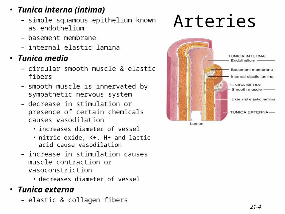
Arteries• Tunica interna (intima)
– simple squamous epithelium known as endothelium
– basement membrane– internal elastic lamina
• Tunica media– circular smooth muscle & elastic fibers– smooth muscle is innervated by
sympathetic nervous system– decrease in stimulation or presence of
certain chemicals causes vasodilation• increases diameter of vessel• nitric oxide, K+, H+ and lactic acid
cause vasodilation
– increase in stimulation causes muscle contraction or vasoconstriction
• decreases diameter of vessel
• Tunica externa– elastic & collagen fibers

21-5
Blood Distribution – Arterial circuit
21-6
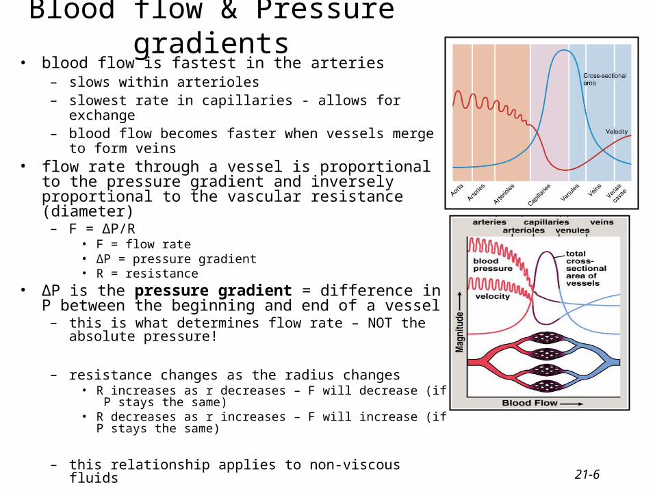
Blood flow & Pressure gradients• blood flow is fastest in the arteries
– slows within arterioles– slowest rate in capillaries - allows for
exchange– blood flow becomes faster when vessels merge to form veins
• flow rate through a vessel is proportional to the pressure gradient and inversely proportional to the vascular resistance (diameter)
– F = ΔP/R• F = flow rate• ΔP = pressure gradient• R = resistance
• ΔP is the pressure gradient = difference in P between the beginning and end of a vessel
– this is what determines flow rate – NOT the absolute pressure!
– resistance changes as the radius changes• R increases as r decreases – F will decrease (if P stays the same)• R decreases as r increases – F will increase (if P stays the same)
– this relationship applies to non-viscous fluids

21-7
Blood flow & Pressure gradients
• frictional losses (resistance) causes a drop in P as the blood travels through a section of vessel
– caused by friction between the moving fluid and the stationary wall
– if pressure gradient is unchanged – then increasing R will inhibit blood flow and decrease F
– R depends on three factors• 1. blood viscosity (η) - # of circulating
RBCs• 2. vessel length (L)• 3. vessel radius (r) – major determinant of
R– R = 1/r4
21-8
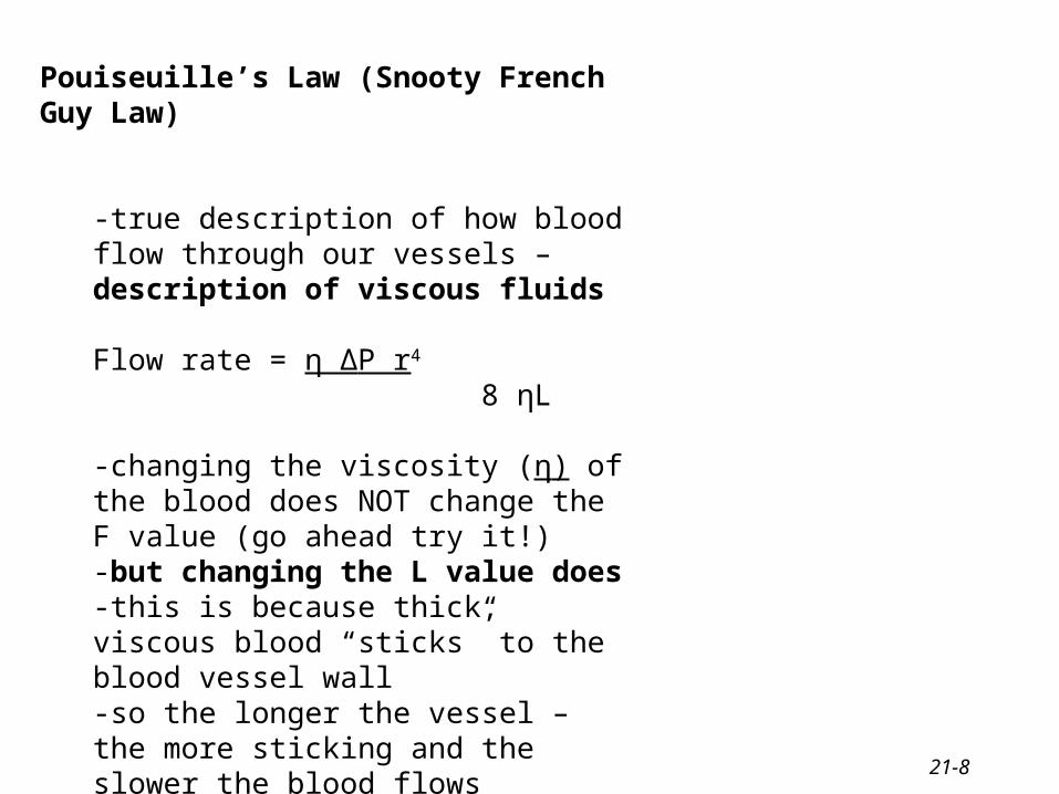
Pouiseuille’s Law (Snooty French Guy Law)
-true description of how blood flow through our vessels – description of viscous fluids
Flow rate = η ΔP r4
8 ηL
-changing the viscosity (η) of the blood does NOT change the F value (go ahead try it!)-but changing the L value does-this is because thick, viscous blood “sticks” to the blood vessel wall-so the longer the vessel – the more sticking and the slower the blood flows

21-9
Blood Pressure• Pressure exerted by blood on walls of a
vessel – depends on the volume of blood within the vessel and the
distensibility of the vessel– caused by contraction of the ventricles– highest in aorta
• difference between systole and diastole – pulse pressure• the volume of blood entering an artery is not the same as the
volume leaving it– during ventricular systole – the SV leaving the artery is 1/3 of
that entering the artery– the artery will distend with increasing volumes– during diastole – the recoil of the vessel drives the exit of the blood
• BUT no blood enters the vessel
• mean arterial pressure – average pressure driving blood forward into the tissues throughout the cardiac cycle
– at resting heart rate – about 2/3 of the cardiac cycle is spent in diastole
– MAP = diastolic pressure + 1/3 (systolic pressure – diastolic pressure)
• MAP falls steadily insystemic circulation with distance from left ventricle
– 35 mm Hg entering the capillaries– 0 mm Hg entering the right atrium
21-10
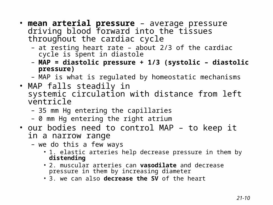
• mean arterial pressure – average pressure driving blood forward into the tissues throughout the cardiac cycle– at resting heart rate – about 2/3 of the cardiac cycle is spent in
diastole– MAP = diastolic pressure + 1/3 (systolic – diastolic pressure) – MAP is what is regulated by homeostatic mechanisms
• MAP falls steadily insystemic circulation with distance from left ventricle– 35 mm Hg entering the capillaries– 0 mm Hg entering the right atrium
• our bodies need to control MAP – to keep it in a narrow range– we do this a few ways
• 1. elastic arteries help decrease pressure in them by distending• 2. muscular arteries can vasodilate and decrease pressure in them by
increasing diameter• 3. we can also decrease the SV of the heart
21-11
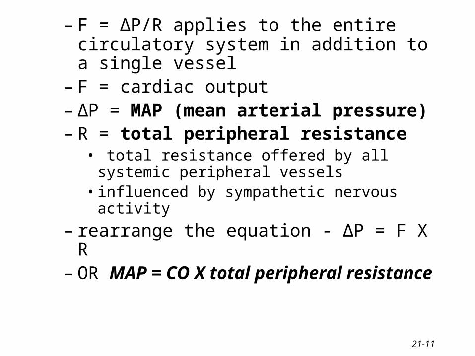
– F = ΔP/R applies to the entire circulatory system in addition to a single vessel
– F = cardiac output– ΔP = MAP (mean arterial pressure)– R = total peripheral resistance
• total resistance offered by all systemic peripheral vessels
• influenced by sympathetic nervous activity
– rearrange the equation - ΔP = F X R – OR MAP = CO X total peripheral resistance
21-12
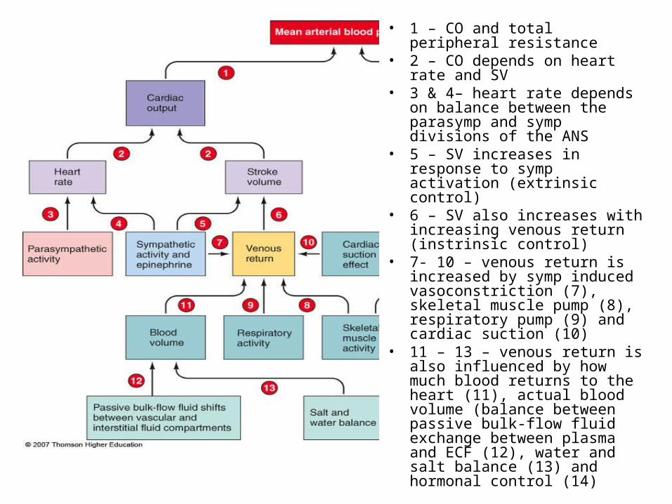
• 1 – CO and total peripheral resistance
• 2 – CO depends on heart rate and SV
• 3 & 4– heart rate depends on balance between the parasymp and symp divisions of the ANS
• 5 – SV increases in response to symp activation (extrinsic control)
• 6 – SV also increases with increasing venous return (instrinsic control)
• 7- 10 – venous return is increased by symp induced vasoconstriction (7), skeletal muscle pump (8), respiratory pump (9) and cardiac suction (10)
• 11 – 13 – venous return is also influenced by how much blood returns to the heart (11), actual blood volume (balance between passive bulk-flow fluid exchange between plasma and ECF (12), water and salt balance (13) and hormonal control (14)
21-13
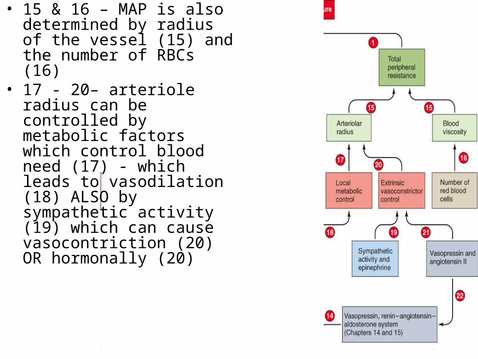
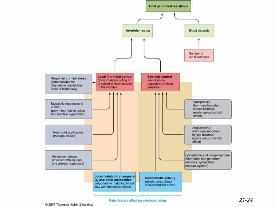
• 15 & 16 – MAP is also determined by radius of the vessel (15) and the number of RBCs (16)
• 17 - 20– arteriole radius can be controlled by metabolic factors which control blood need (17) - which leads to vasodilation (18) ALSO by sympathetic activity (19) which can cause vasocontriction (20) OR hormonally (20)

21-14
Arterioles• major resistance vessels in the vascular tree• radius is small enough to offer resistance to flow• high arteriolar resistance causes a marked
drop in the MAP as blood flows through these vessels– MAP arteriole entrance = 93 mm Hg– MAP arteriole exit = 37 mm Hg
• resistance also converts the pulsatile nature of systolic-diastolic pressure to nonpulsatile pressure within the capillaries
• radius of the arteriole can be adjusted to– 1. variably distribute cardiac output among the organs– 2. help regulate arterial blood pressure
21-15
Arterioles and Arteriolar resistance
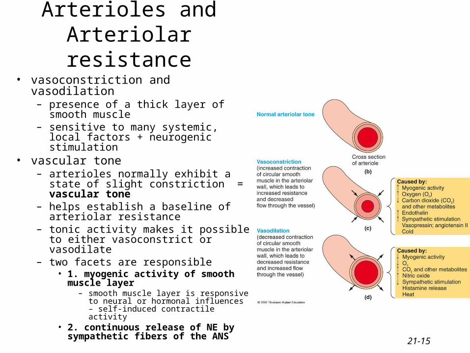
• vasoconstriction and vasodilation– presence of a thick layer of smooth
muscle– sensitive to many systemic, local factors
+ neurogenic stimulation• vascular tone
– arterioles normally exhibit a state of slight constriction = vascular tone
– helps establish a baseline of arteriolar resistance
– tonic activity makes it possible to either vasoconstrict or vasodilate
– two facets are responsible• 1. myogenic activity of smooth muscle
layer– smooth muscle layer is responsive to
neural or hormonal influences – self-induced contractile activity
• 2. continuous release of NE by sympathetic fibers of the ANS

21-16
Arteriolar diameter
• Intrinsic Factors/Local changes – at the tissue, organ specific– local chemical factors
• metabolic factors• vasoactive mediators
– histamine– local physical factors
• hot/cold• passive stretch of arteriole• shear stress within arteriole
• Extrinsic factors– sympathetic nervous system– hormones

21-17
Chemical influences and arteriole radius = local metabolic changes
• most important local chemical influences on arteriolar smooth muscle are local changes in metabolism within that organ
• local metabolic changes can affect the diameter of an arteriole without neural influence• active hyperemia = local arteriolar vasodilation that increases blood flow into an organ
– arterioles are found within an organ and can be directly affected by that organ’s metabolic needs
• local metabolic factors– decreased/increased oxygen = vasodilation/vasoconstriction– increase carbon dioxide = vasodilation– increased carbonic acid= vasodilation– increased K+ - repeated APs that outpace the Na/K pump’s ability to correct ionic changes =
vasodilation– increased osmolarity – concentration of solutes accumulates in actively metabolic cells =
vasodilation– prostaglandin release = vasodilation
• relative concentration of these factors can determine the state of arteriolar muscle tone
21-18
Vasoactive Mediators
• these local chemical changes do not act directly on smooth muscle but act on the endothelial cells
• ECs – simple squamous epithelia cells– found lining the inside of the arteriole and
capillary
• ECs then release chemical factors called vasoactive mediators– e.g. endothelin = vasoconstriction
– e.g. nitric oxide = vasodilation by relaxing arteriolar smooth muscle
• inhibits entrance of calcium into the smooth muscle which inhibits the opening of the foot proteins on the sarcoplasmic reticulum
• ECs have multiple roles
21-19
Histamine• NOT released by metabolic changes• NOT produce by endothelial cells• released upon pathology• released by connective tissue cells within the organ or by
circulating white blood cells (mast cells, basophils)• usually released in response to organ damage• causes vasodilation to increase blood flow and speed healing
21-20
Local Physical factors• application of heat or cold
– heat causes localized arteriolar vasodilation• increases blood flow
– cold – counteracts histamine-induced swelling by inducing vasoconstriction • shear stress
– blood flowing over the endothelial lining creates friction = shear stress– increase in shear stress can cause increased release of NO – promotes
vasodilation– increased blood flow now reduces shear stress
• myogenic response to stretch– increased MAP drives more blood into the arteriole which pushes out against the
vessel wall = passive stretch• leads to vasoconstriction to reduce this blood volume
– arteriolar smooth muscle responds to passive stretch by increasing its tone through vasoconstriction
– this increased tone acts to resist the passive stretch– done by the release of vasoactive mediators from the EC (e.g. endothelin)
– arterial occlusion can block blood flow and reduce this stretch• arterioles will dilate in response=reactive hyperemia
21-21
Extrinsic control of arteriolar diameter• includes both neural and hormonal control• sympathetic division innervates arteriolar smooth muscle everywhere
except the brain• NO parasympathetic innervation of arteriolar smooth muscle!
(exception – ciltoris and penis)• sympathetic activity contributes to arteriolar vascular tone• increased sympathetic activity induces vasodilation of arterioles in
heart and skeletal muscle– drops arteriolar resistance and changes MAP
• main area of the brain to adjust sympathetic output = cardiovascular control center
• other brain regions involved – hypothalamus• Hormones: ADH, angiotensin II, epinephrine, norepinephrine

21-22
HORMONAL CONTROL OF ARTERIOLAR DIAMETER:
•angiotensin II – converted from angiotensin I by the enzyme ACE (produced in the lungs)
– regulates body’s salt balance– causes the release of aldosterone from the adrenal cortex
– increased salt reabsorption– powerful vasoconstrictor
•vasopressin (ADH) – released from the posterior pituitary in response to changes in water volume
– drop in water content, release of vasopressin, decreased urine volume, increased water retention
– also construction of peripheral vessels = vasoconstrictor
– plays a role in reducing hemorrhage
21-23
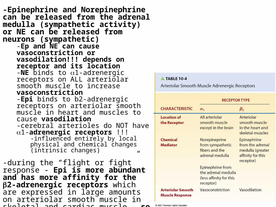
-Epinephrine and Norepinephrine can be released from the adrenal medulla (sympathetic activity) or NE can be released from neurons (sympathetic)
-Ep and NE can cause vasoconstriction or vasodilation!!! depends on receptor and its location-NE binds to 1-adrenergic receptors on ALL arteriolar smooth muscle to increase vasoconstriction-Epi binds to b2-adrenergic receptors on arteriolar smooth muscle in heart and muscles to cause vasodilation-cerebral arterioles do NOT have 1-adrenergic receptors !!!
-influenced entirely by local physical and chemical changes (intrinsic changes)
-during the “flight or fight” response - Epi is more abundant and has more affinity for the 2-adrenergic receptors which are expressed in large amounts on arteriolar smooth muscle in skeletal and cardiac muscle – so overall effect is vasodilation to heart and skeletal muscles
21-24
21-25
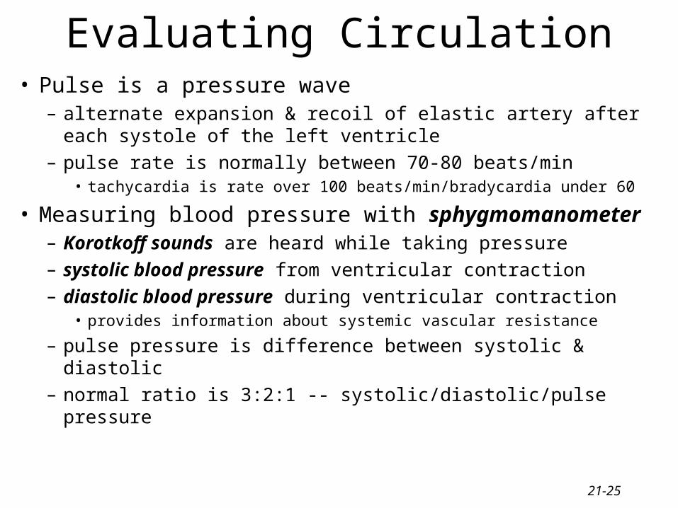
Evaluating Circulation• Pulse is a pressure wave
– alternate expansion & recoil of elastic artery after each systole of the left ventricle
– pulse rate is normally between 70-80 beats/min• tachycardia is rate over 100 beats/min/bradycardia under 60
• Measuring blood pressure with sphygmomanometer– Korotkoff sounds are heard while taking pressure
– systolic blood pressure from ventricular contraction
– diastolic blood pressure during ventricular contraction• provides information about systemic vascular resistance
– pulse pressure is difference between systolic & diastolic
– normal ratio is 3:2:1 -- systolic/diastolic/pulse pressure
21-26
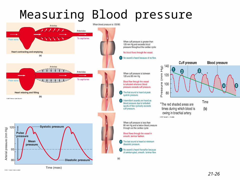
Measuring Blood pressure
21-27
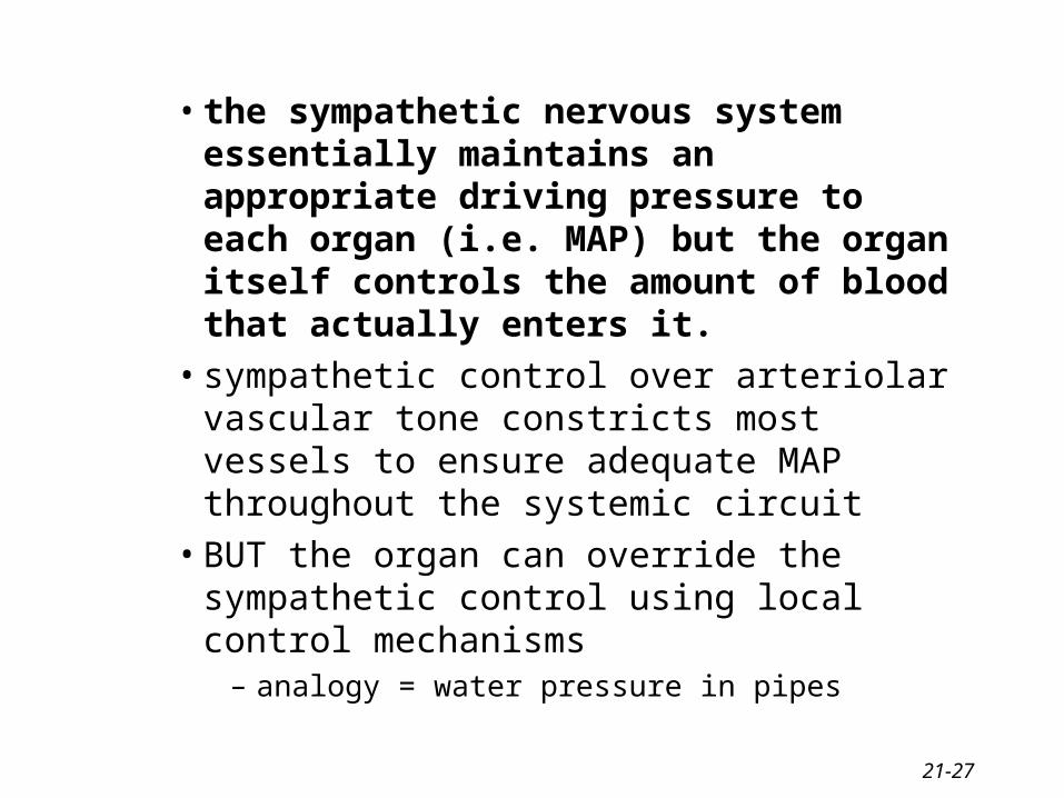
• the sympathetic nervous system essentially maintains an appropriate driving pressure to each organ (i.e. MAP) but the organ itself controls the amount of blood that actually enters it.
• sympathetic control over arteriolar vascular tone constricts most vessels to ensure adequate MAP throughout the systemic circuit
• BUT the organ can override the sympathetic control using local control mechanisms
– analogy = water pressure in pipes
21-28
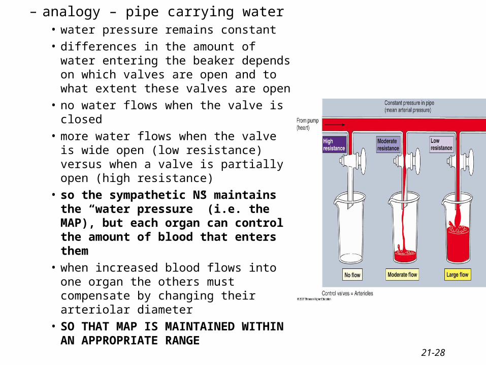
– analogy – pipe carrying water• water pressure remains constant
• differences in the amount of water entering the beaker depends on which valves are open and to what extent these valves are open
• no water flows when the valve is closed
• more water flows when the valve is wide open (low resistance) versus when a valve is partially open (high resistance)
• so the sympathetic NS maintains the “water pressure” (i.e. the MAP), but each organ can control the amount of blood that enters them
• when increased blood flows into one organ the others must compensate by changing their arteriolar diameter
• SO THAT MAP IS MAINTAINED WITHIN AN APPROPRIATE RANGE

21-29
Regulation of BP• regulation of mean arterial pressure by the body done mainly through arteriolar
diameter• Blood pressure = MAP within a small length of blood vessel• our body uses BP to immediately determine heart rate and contraction strength• BP is measured constantly by baroreceptors
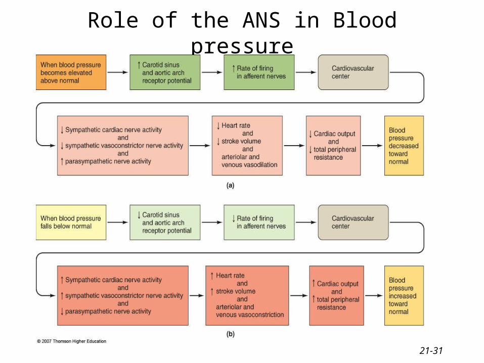
– changes in pressure within blood vessels– initiates either short-term or long-term reflexes– two major BRs: carotid sinus & aortic arch baroreceptors (mechanoreceptors)
• as the BP within the vessels increase –increases the rate of firing of the afferent neurons within these BRs
• decreasing pressure decreases the rate of firing• role of cardiovascular center in the medulla
– help regulate heart rate & stroke volume– integrating center for information sent by the carotid and aortic BRs– divided into two centers: vasomotor & cardiac centers (made up of
cardioacceleratory, cardioinhibitory divisions)– alters the ratio between sympathetic and parasympathetic activity to the heart and
BVs– Vasomotor center = specific neurons regulate blood vessel diameter - sympathetic
vasomotor nerves– Cardiac center = signals sent out through vagus & cardiac accelerator nerves -
changes heart rate
21-30
• Higher brain centers such as cerebral cortex, limbic system (emotions) & hypothalamus
– anticipation of competition– increase in body temperature
• Hypothalamus– osmoreceptors control salt and water balance and therefore long-term
regulation of BP– cerebral cortex-hypothalamus preprograming – fight or flight responses,
sexual orgasm, blushing– control over cutaneous arterioles for temperature regulation takes control
over the cardiovascular center’s control over these vessels for regulation BP• therefore BP can fall dramatically as arteries dilate to allow cooling
• Proprioceptors– input during physical activity
• Chemoreceptors– monitor concentration of chemicals in the blood (O2, CO2 and H+ions)– also located in the carotid and aortic arteries– increase respiratory activity to bring in more O2 or blow off more CO2– also increase BP by sneding excitatory impulses to the cardiovascular center
21-31
Role of the ANS in Blood pressure
21-32
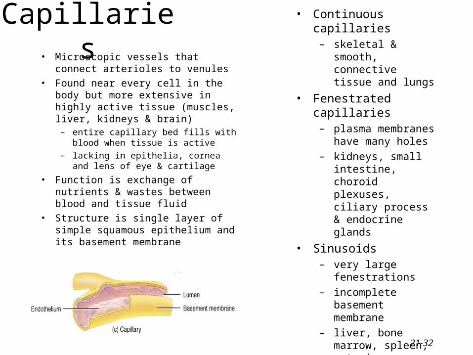
Capillaries• Microscopic vessels that connect
arterioles to venules
• Found near every cell in the body but more extensive in highly active tissue (muscles, liver, kidneys & brain)
– entire capillary bed fills with blood when tissue is active
– lacking in epithelia, cornea and lens of eye & cartilage
• Function is exchange of nutrients & wastes between blood and tissue fluid
• Structure is single layer of simple squamous epithelium and its basement membrane
• Continuous capillaries– skeletal & smooth,
connective tissue and lungs
• Fenestrated capillaries– plasma membranes have
many holes
– kidneys, small intestine, choroid plexuses, ciliary process & endocrine glands
• Sinusoids– very large fenestrations
– incomplete basement membrane
– liver, bone marrow, spleen, anterior pituitary, & parathyroid gland
21-33
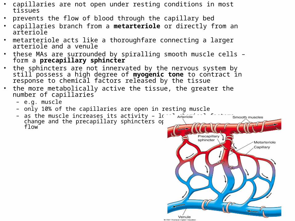
• capillaries are not open under resting conditions in most tissues• prevents the flow of blood through the capillary bed• capillaries branch from a metarteriole or directly from an arteriole• metarteriole acts like a thoroughfare connecting a larger arteriole and a venule• these MAs are surrounded by spiralling smooth muscle cells – form a
precapillary sphincter• the sphincters are not innervated by the nervous system by still possess a high
degree of myogenic tone to contract in response to chemical factors released by the tissue
• the more metabolically active the tissue, the greater the number of capillaries– e.g. muscle– only 10% of the capillaries are open in resting muscle – as the muscle increases its activity – local chemical factors change and the
precapillary sphincters open to allow more blood flow

21-34
• ECs fit together like a jigsaw puzzle with considerable gaps in between the cells = pores
• sizes vary from capillary to capillary• brain capillaries have EC cells held together by tight junctions – no pores• most tissue capillaries allow the passage of small water-soluble substances
like but glucose, small amino acids and peptides and ions– prevents the passage of larger proteins like plasma proteins
• this transport may actually be regulated by the capillary itself– ECs may secrete substances that “tighten” up their junctions– histamine increases the gaps by inducing a contractile response in the EC and
widening the gaps (actin-myosin interaction in the cytoskeleton)• in the liver, the capillary walls have larger pores to allow the passage of
proteins– liver synthesizes the plasma proteins which must be allowed to pass into the
circulatory system
21-35
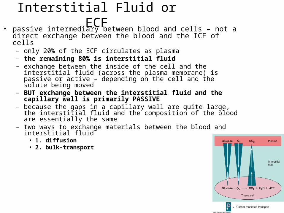
Interstitial Fluid or ECF• passive intermediary between blood and cells – not a direct exchange
between the blood and the ICF of cells– only 20% of the ECF circulates as plasma– the remaining 80% is interstitial fluid– exchange between the inside of the cell and the interstitial fluid (across
the plasma membrane) is passive or active – depending on the cell and the solute being moved
– BUT exchange between the interstitial fluid and the capillary wall is primarily PASSIVE
– because the gaps in a capillary wall are quite large, the interstitial fluid and the composition of the blood are essentially the same
– two ways to exchange materials between the blood and interstitial fluid• 1. diffusion• 2. bulk-transport
21-36
Capillary Exchange• Movement of materials in & out of a capillary
– diffusion (most important method)• substances move down concentration gradient
• all plasma solutes except large proteins pass freely across– through lipid bilayer, fenestrations or intercellular clefts
– blood brain barrier does not allow diffusion of water-soluble materials (nonfenestrated epithelium with tight junctions)
– transcytosis (vesicular transport)• passage of material across endothelium in tiny vesicles by endocytosis and
exocytosis – large, lipid-insoluble molecules such as insulin or maternal antibodies passing
through placental circulation to fetus
– bulk flow• movement of large amount of dissolved or suspended material in same direction
• move in response to pressure - from area of high pressure to area of low
• faster rate of movement than diffusion or osmosis
• regulates relative volumes of blood & interstitial fluid
• comprised of: filtration (movement of material into interstitial fluid) + reabsorption (movement from interstitial fluid into capillaries)
21-37
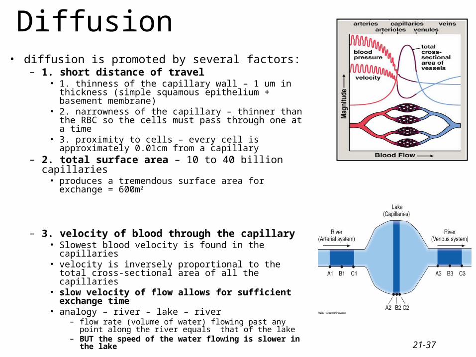
Diffusion• diffusion is promoted by several factors:
– 1. short distance of travel• 1. thinness of the capillary wall – 1 um in thickness
(simple squamous epithelium + basement membrane)• 2. narrowness of the capillary – thinner than the RBC so
the cells must pass through one at a time• 3. proximity to cells – every cell is approximately
0.01cm from a capillary– 2. total surface area – 10 to 40 billion capillaries
• produces a tremendous surface area for exchange = 600m2
– 3. velocity of blood through the capillary• Slowest blood velocity is found in the capillaries• velocity is inversely proportional to the total cross-
sectional area of all the capillaries • slow velocity of flow allows for sufficient exchange
time• analogy – river – lake – river
– flow rate (volume of water) flowing past any point along the river equals that of the lake
– BUT the speed of the water flowing is slower in the lake
21-38
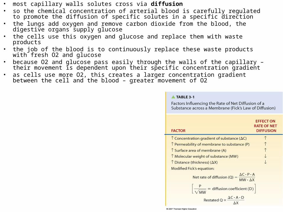
• most capillary walls solutes cross via diffusion• so the chemical concentration of arterial blood is carefully regulated to promote the diffusion of
specific solutes in a specific direction• the lungs add oxygen and remove carbon dioxide from the blood, the digestive organs supply
glucose• the cells use this oxygen and glucose and replace them with waste products• the job of the blood is to continuously replace these waste products with fresh O2 and glucose• because O2 and glucose pass easily through the walls of the capillary – their movement is dependent
upon their specific concentration gradient• as cells use more O2, this creates a larger concentration gradient between the cell and the blood –
greater movement of O2

21-39
21-40
Bulk-flow• a volume of protein-free plasma filters out of the capillary and mixes
with the surrounding interstitial fluid and then is reabsorbed = bulk flow
• the various components are moved in bulk in contrast to the movement of individual components as seen in diffusion
• when blood pressure inside the capillary exceeds that of the osmotic pressure of the blood – fluid is pushed out through the pores in the capillary wall = ultrafiltration
• with the fluid comes the movement of multiple other components• when inward driving osmotic pressure exceeds the outward blood
pressure– net inward movement of fluid and fluid components = reabsorption
• ultrafiltration and reabsorption are collectively known as bulk flow• does NOT play a role in the exchange of individual solutes• does play a role in regulating the distribution of ECF between the
circulating plasma and interstitial fluid• this is the way we establish the composition of our interstitial fluid
21-41
Bulk Flow
• (Pc + πIF) – (πP + PIF) = outward pressure – inward pressure
• major factors are Pc and πP

21-42
• factors affecting bulk flow– 1. Pc = capillary blood pressure
• fluid or hydrostatic pressure exerted on the inside of the capillary wall by blood• tends to force fluid OUT of the capillaries into the interstitial fluid• in general the blood pressure at the arteriolar end of a capillary = 37 mm Hg due to frictional
losses in the upstream arterioles - declines to 17 mm Hg at the venous end of a capillary– 2. πP = plasma osmotic pressure (oncotic pressure)
• osmotic pressure encourages inward movement of fluid into the capillary• 25 mm Hg – doesn’t change
– 3. PIF = interstitial fluid hydrostatic pressure• fluid pressure exerted on the outside of the capillary wall by interstitial fluid• tends to force fluid into the capillaries and into the blood plasma
– 4. πIF = interstitial fluid-colloid osmotic pressure (πIF is close to zero)• does not normally contribute significantly to bulk flow• Caused by the osmotic pressure of proteins in the IF - promotes the osmotic movement of water
out of the blood and into the fluid • but during pathologic conditions – i.e. release of histamine which widens the capillary pores –
increased leakage of plasma proteins into the interstitial fluid and πIF can increase
21-43
Net exchange of fluid across capillaries• Net exchange pressure = (Pc + πIF) – (πP + PIF) = outward pressure – inward pressure• two pressures forcing fluid out of the plasma are capillary blood pressure and
interstitial fluid-osmotic pressure• two pressures forcing fluid into the plasma are plasma-osmotic pressure and interstitial
fluid hydrostatic pressure• the imbalance between the forces produces a net exchange of fluid at a given point
along the capillary wall• as the blood enters the capillary – outward forces exceed that of inward and fluid is
forced out into the interstitial fluid (no protein movement!) – ultrafiltration forces result
• at the venular end the inward pressure now exceeds the outward pressure and reabsorption will take place
• as the blood moves through the capillary – pressure drops• so the amount of fluid moving out of the blood drops as the blood travels through the
capillary region and progressively increasing amounts of fluid re-enter the blood as the blood reaches the venular end of the capillary
• BUT these pressure values are controversial

21-44
Lymph• even under normal circumstances –
more fluid is filtered from the blood plasma into the interstitial fluid than is reabsorbed back into the blood plasma
• excess fluid = lymph• this excess fluid is directed into a
series of vessels that are similar in structure and composition to veins – lymphatic vessels
• one-way system of vessels that leads back to the circulatory system via the right lymphatic duct and the thoracic duct
21-45
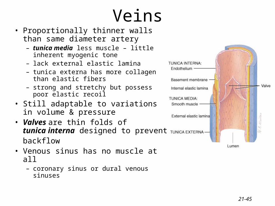
Veins• Proportionally thinner walls than same
diameter artery– tunica media less muscle – little inherent
myogenic tone– lack external elastic lamina– tunica externa has more collagen than
elastic fibers – strong and stretchy but possess poor
elastic recoil
• Still adaptable to variationsin volume & pressure
• Valves are thin folds of tunica interna designed to prevent backflow
• Venous sinus has no muscle at all– coronary sinus or dural venous sinuses
21-46
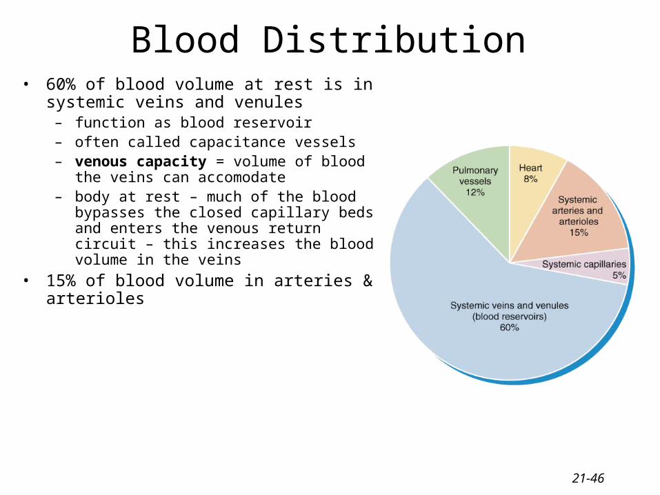
Blood Distribution• 60% of blood volume at rest is in systemic veins
and venules– function as blood reservoir– often called capacitance vessels– venous capacity = volume of blood the veins can
accomodate– body at rest – much of the blood bypasses the
closed capillary beds and enters the venous return circuit – this increases the blood volume in the veins
• 15% of blood volume in arteries & arterioles

21-47
Veins & Venules• veins & venules have little tone and resistance• the arteriole communicates chemically with the
venule to ensure that inflow and outflow surrounding the capillary region matches
• venous return is enhanced by many extrinsic factors– venous return = volume of blood entering the atria
from the veins (pulmonary and systemic)– much of the driving force on the blood (blood
pressure) has been lost as it enters the veins– BP averages on 17 mm Hg in the veins– but because the pressure of the blood entering the
right atrium in o mm Hg – there is still a small driving force that helps to return the blood through the veins
– clinical note – if atrial pressure becomes pathologically elevated (e.g. leaky AV valves) – this pressure differential can be lost, reducing the venous return
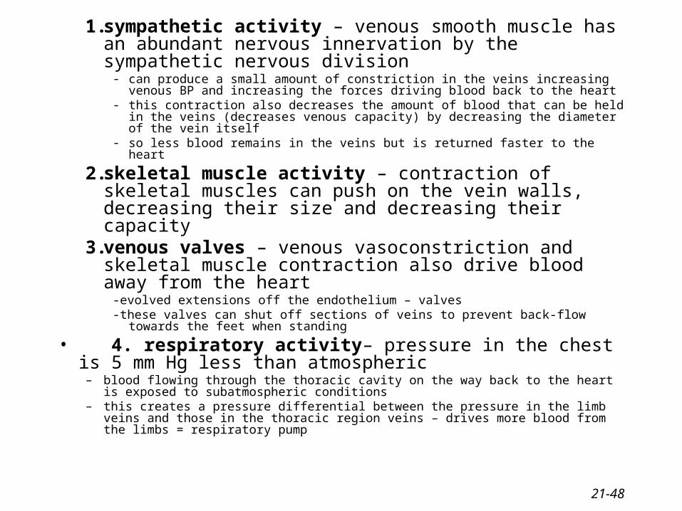
1. sympathetic activity – venous smooth muscle has an abundant nervous innervation by the sympathetic nervous division
- can produce a small amount of constriction in the veins increasing venous BP and increasing the forces driving blood back to the heart
- this contraction also decreases the amount of blood that can be held in the veins (decreases venous capacity) by decreasing the diameter of the vein itself
- so less blood remains in the veins but is returned faster to the heart
2. skeletal muscle activity – contraction of skeletal muscles can push on the vein walls, decreasing their size and decreasing their capacity
3. venous valves – venous vasoconstriction and skeletal muscle contraction also drive blood away from the heart
-evolved extensions off the endothelium – valves-these valves can shut off sections of veins to prevent back-flow towards the feet
when standing
• 4. respiratory activity– pressure in the chest is 5 mm Hg less than atmospheric
– blood flowing through the thoracic cavity on the way back to the heart is exposed to subatmospheric conditions
– this creates a pressure differential between the pressure in the limb veins and those in the thoracic region veins – drives more blood from the limbs = respiratory pump
21-48

21-49
Veins and gravity- gravity – when lying down, forces of gravity are equally
applied to all veins-when standing, vessels below the heart become more subject to
gravitational forces-so the veins increase their volume to counteract the increased
volume-this reduces the amount of blood returning to the heart –
decreases CO-also increases the pressure in the capillaries – forces more fluid
out into the tissues from the blood-compensatory mechanisms – fall in MAP triggers the
sympathetic NS to induce venous constriction to drive some of the pooled blood up toward the heart + skeletal muscle tone will increase to help propel blood to the heart

21-50