2014_BKCS Lee and Baek
5
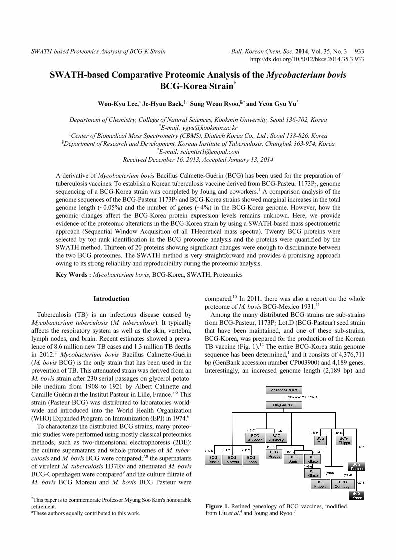
SWATH-based Proteomics Analysis of BCG-K Strain Bull. Korean Chem. Soc. 2014, Vol. 35, No. 3 933 http://dx.doi.org/10.5012/bkcs.2014.35.3.933 SWATH-based Comparative Proteomic Analysis of the Mycobacterium bovis BCG-Korea Strain † Won-Kyu Lee, a Je-Hyun Baek, ‡,a Sung Weon Ryoo, §,* and Yeon Gyu Yu * Department of Chemistry, College of Natural Sciences, Kookmin University, Seoul 136-702, Korea * E-mail: [email protected] ‡ Center of Biomedical Mass Spectrometry (CBMS), Diatech Korea Co., Ltd., Seoul 138-826, Korea § Department of Research and Development, Korean Institute of Tuberculosis, Chungbuk 363-954, Korea * E-mail: [email protected] Received December 16, 2013, Accepted January 13, 2014 A derivative of Mycobacterium bovis Bacillus Calmette-Guérin (BCG) has been used for the preparation of tuberculosis vaccines. To establish a Korean tuberculosis vaccine derived from BCG-Pasteur 1173P 2 , genome sequencing of a BCG-Korea strain was completed by Joung and coworkers. 1 A comparison analysis of the genome sequences of the BCG-Pasteur 1173P 2 and BCG-Korea strains showed marginal increases in the total genome length (~0.05%) and the number of genes (~4%) in the BCG-Korea genome. However, how the genomic changes affect the BCG-Korea protein expression levels remains unknown. Here, we provide evidence of the proteomic alterations in the BCG-Korea strain by using a SWATH-based mass spectrometric approach (Sequential Window Acquisition of all THeoretical mass spectra). Twenty BCG proteins were selected by top-rank identification in the BCG proteome analysis and the proteins were quantified by the SWATH method. Thirteen of 20 proteins showing significant changes were enough to discriminate between the two BCG proteomes. The SWATH method is very straightforward and provides a promising approach owing to its strong reliability and reproducibility during the proteomic analysis. Key Words : Mycobacterium bovis, BCG-Korea, SWATH, Proteomics Introduction Tuberculosis (TB) is an infectious disease caused by Mycobacterium tuberculosis (M. tuberculosis). It typically affects the respiratory system as well as the skin, vertebra, lymph nodes, and brain. Recent estimates showed a preva- lence of 8.6 million new TB cases and 1.3 million TB deaths in 2012. 2 Mycobacterium bovis Bacillus Calmette-Guérin (M. bovis BCG) is the only strain that has been used in the prevention of TB. This attenuated strain was derived from an M. bovis strain after 230 serial passages on glycerol-potato- bile medium from 1908 to 1921 by Albert Calmette and Camille Guérin at the Institut Pasteur in Lille, France. 3-5 This strain (Pasteur-BCG) was distributed to laboratories world- wide and introduced into the World Health Organization (WHO) Expanded Program on Immunization (EPI) in 1974. 6 To characterize the distributed BCG strains, many proteo- mic studies were performed using mostly classical proteomics methods, such as two-dimensional electrophoresis (2DE): the culture supernatants and whole proteomes of M. tuber- culosis and M. bovis BCG were compared; 7,8 the supernatants of virulent M. tuberculosis H37Rv and attenuated M. bovis BCG-Copenhagen were compared 9 and the culture filtrate of M. bovis BCG Moreau and M. bovis BCG Pasteur were compared. 10 In 2011, there was also a report on the whole proteome of M. bovis BCG-Mexico 1931. 11 Among the many distributed BCG strains are sub-strains from BCG-Pasteur, 1173P 2 Lot.D (BCG-Pasteur) seed strain that have been maintained, and one of these sub-strains, BCG-Korea, was prepared for the production of the Korean TB vaccine (Fig. 1). 12 The entire BCG-Korea stain genome sequence has been determined, 1 and it consists of 4,376,711 bp (GenBank accession number CP003900) and 4,189 genes. Interestingly, an increased genome length (2,189 bp) and † This paper is to commemorate Professor Myung Soo Kim's honourable retirement. a These authors equally contributed to this work. Figure 1. Refined genealogy of BCG vaccines, modified from Liu et al. 4 and Joung and Ryoo. 7
-
Upload
je-hyun-baek -
Category
Documents
-
view
22 -
download
1
Transcript of 2014_BKCS Lee and Baek
SWATH-based Proteomics Analysis of BCG-K Strain Bull. Korean Chem. Soc. 2014, Vol. 35, No. 3 933
http://dx.doi.org/10.5012/bkcs.2014.35.3.933
SWATH-based Comparative Proteomic Analysis of the Mycobacterium bovis
BCG-Korea Strain†
Won-Kyu Lee,a Je-Hyun Baek,‡,a Sung Weon Ryoo,§,* and Yeon Gyu Yu*
Department of Chemistry, College of Natural Sciences, Kookmin University, Seoul 136-702, Korea*E-mail: [email protected]
‡Center of Biomedical Mass Spectrometry (CBMS), Diatech Korea Co., Ltd., Seoul 138-826, Korea§Department of Research and Development, Korean Institute of Tuberculosis, Chungbuk 363-954, Korea
*E-mail: [email protected]
Received December 16, 2013, Accepted January 13, 2014
A derivative of Mycobacterium bovis Bacillus Calmette-Guérin (BCG) has been used for the preparation of
tuberculosis vaccines. To establish a Korean tuberculosis vaccine derived from BCG-Pasteur 1173P2, genome
sequencing of a BCG-Korea strain was completed by Joung and coworkers.1 A comparison analysis of the
genome sequences of the BCG-Pasteur 1173P2 and BCG-Korea strains showed marginal increases in the total
genome length (~0.05%) and the number of genes (~4%) in the BCG-Korea genome. However, how the
genomic changes affect the BCG-Korea protein expression levels remains unknown. Here, we provide
evidence of the proteomic alterations in the BCG-Korea strain by using a SWATH-based mass spectrometric
approach (Sequential Window Acquisition of all THeoretical mass spectra). Twenty BCG proteins were
selected by top-rank identification in the BCG proteome analysis and the proteins were quantified by the
SWATH method. Thirteen of 20 proteins showing significant changes were enough to discriminate between
the two BCG proteomes. The SWATH method is very straightforward and provides a promising approach
owing to its strong reliability and reproducibility during the proteomic analysis.
Key Words : Mycobacterium bovis, BCG-Korea, SWATH, Proteomics
Introduction
Tuberculosis (TB) is an infectious disease caused by
Mycobacterium tuberculosis (M. tuberculosis). It typically
affects the respiratory system as well as the skin, vertebra,
lymph nodes, and brain. Recent estimates showed a preva-
lence of 8.6 million new TB cases and 1.3 million TB deaths
in 2012.2 Mycobacterium bovis Bacillus Calmette-Guérin
(M. bovis BCG) is the only strain that has been used in the
prevention of TB. This attenuated strain was derived from an
M. bovis strain after 230 serial passages on glycerol-potato-
bile medium from 1908 to 1921 by Albert Calmette and
Camille Guérin at the Institut Pasteur in Lille, France.3-5 This
strain (Pasteur-BCG) was distributed to laboratories world-
wide and introduced into the World Health Organization
(WHO) Expanded Program on Immunization (EPI) in 1974.6
To characterize the distributed BCG strains, many proteo-
mic studies were performed using mostly classical proteomics
methods, such as two-dimensional electrophoresis (2DE):
the culture supernatants and whole proteomes of M. tuber-
culosis and M. bovis BCG were compared;7,8 the supernatants
of virulent M. tuberculosis H37Rv and attenuated M. bovis
BCG-Copenhagen were compared9 and the culture filtrate of
M. bovis BCG Moreau and M. bovis BCG Pasteur were
compared.10 In 2011, there was also a report on the whole
proteome of M. bovis BCG-Mexico 1931.11
Among the many distributed BCG strains are sub-strains
from BCG-Pasteur, 1173P2 Lot.D (BCG-Pasteur) seed strain
that have been maintained, and one of these sub-strains,
BCG-Korea, was prepared for the production of the Korean
TB vaccine (Fig. 1).12 The entire BCG-Korea stain genome
sequence has been determined,1 and it consists of 4,376,711
bp (GenBank accession number CP003900) and 4,189 genes.
Interestingly, an increased genome length (2,189 bp) and
†This paper is to commemorate Professor Myung Soo Kim's honourable
retirement.aThese authors equally contributed to this work.
Figure 1. Refined genealogy of BCG vaccines, modifiedfrom Liu et al.4 and Joung and Ryoo.7
934 Bull. Korean Chem. Soc. 2014, Vol. 35, No. 3 Won-Kyu Lee et al.
number of genes (156 genes) were observed in the BCG-
Korea strain compared to the genome of the BCG-Pasteur
strain (http://www.ncbi.nlm.nih.gov/genome/161?project_id
=58781), the mother strain of BCG-Korea. However, no
study has been reported showing the effects of genomic
changes in BCG-Korea on the expression profile of its pro-
teins.
In this study, we identified from 923 proteins in BCG-
Pasteur and BCG-Korea strains and extracted quantitative
information on 20 kind of most abundantly expressed BCG
proteins using a SWATH-based approach (Sequential Window
Acquisition of all THeoretical mass spectra), which is
straightforward, highly reproducible, reliable and applicable
to a large number of samples and characteristic proteomic
analyses.13 Ten proteins out of 20 showed different expre-
ssion levels in the two BCG strains within a 15-35% range.
The SWATH method provided evidence of genomic effects
on protein levels using the selected 20 BCG proteins.
Materials and Methods
Chemicals. Iodoacetamide was purchased from Sigma-
Aldrich (USA). Tris-HCl, urea and dithiothreitol (DTT) were
obtained from Merck (Germany). All other chemicals were
acquired from standard sources and were of molecular bio-
logy grade or higher.
BCG Culture. BCG-Korea (M. bovis BCG-Korea 1168P2)
was grown in 5 round beakers with 250 mL of Sauton's
liquid medium for 3 weeks at 37 °C under stationary condi-
tions. The freeze-dried BCG-Pasteur 1173 P2 secondary seed
LotD was reconstituted in Sauton’s medium (1 mL) and
cultured in Sauton’s potato medium in Roux tubes.
Preparation of Proteome Samples. For the preparation
of the protein extracts from the BCG strains, cells were
collected by centrifugation at 12,000 × g for 10 min and
washed 3 times with PBS buffer (pH 7.4). The collected
cells were lysed by ultra-sonication in ice-cold lysis buffer
(50 mM Tris-HCl, 10 mM MgCl2, 0.02% sodium azide, pH
7.4) that included protease and phosphatase inhibitors (Roche,
USA). The lysates were incubated for 3 h in denaturing
buffer (8 M urea, 50 mM Tris-HCl, pH 8.0), treated with 10
mM DTT at 37 °C for 1 h, and alkylated with 55 mM
iodoacetamide at room temperature for 30 min. The proteins
in the sample were further digested with modified trypsin
(Promega, USA) at 37 oC overnight. The peptides in the
mixture were recovered using a desalting C18 cartridge
(Sep-Pak, Waters, USA) and completely dried in a speed-
vacuum before mass analysis.
LC-MS/MS Analysis. Triple-TOF-TM 5600+ (AB Sciex,
Canada) was used for the BCG strain proteomes. The
instrument was coupled with an Eksigent NanoLC-2D+ with
Nanoflex cHiPLC system for reproducible peptide sample
separation. Solvent A contained 0.1% formic acid (v/v) in
water and solvent B contained of 0.1% formic acid (v/v) in
100% acetonitrile. The samples were loaded onto a trap
column (0.5 mm × 200 µm) at a flow rate of 1 µL/min, and
separated on an analytical column (15 cm × 75 µm) with a
linear gradient of 2% to 35% solvent B over 30 min at a flow
rate of 400 nL/min on the Nanoflex cHiPLC system. The
Chip nanoLC column was regenerated by washing woth
60% solvent B for 50 min and equilibrating with 2% solvent
B for 10 min. For data-dependent acquisition experiment, a
Triple-TOF-TM 5600+ mass spectrometer was used with a
50-ms survey scan (TOF-MS) and 50-ms automated MS/MS
scan for the 15 ions with the highest intensity. The MS/MS
triggering criteria for parent ions were as follows: precursor
intensity (> 150 counts) and charge state (> 1) with dynamic
exclusion option (exclusion time: 6 s). Ions were isolated
using a quadruple UNIT resolution (0.7 Da) and fragmented
in the collision cell using collision energy ramped from 15 to
45 eV within the 50-ms accumulation time. For SWATH
MS-based experiments, the mass spectrometer was operated
as previously described by Gillet et al.13 with minor changes.
In detail, Triple-TOF 5600+ was used in the looped product
ion mode with 20 Da/mass windows (each SWATH window
has a 1 Da overlap) in the range of 400 to 1000 Da (Experi-
ment 1: MS1 scan (400-1,600 Da); Experiment 2: 400-420
Da; Experiment 3: 419-444 Da ~ Experiment 24: 979-1,000
Da). The MS2 scan range was set to 100-1,600 m/z. The
collision energy for each window was determined based on
the appropriate collision energy for a two-charged ion
centered upon the window with a spread of 15 eV. An
accumulation time of 80 ms was used for each fragment ion
scan and for the survey scans (total duty cycle, 2.5 s; number
of total cycles, 1,406) in high sensitivity mode.
Data Analysis. All spectra generated from data-dependent
acquisitions were searched against the BCG database using
ProteinPilotTM (mycobacterium_bovis_bcg_1_proteins.fasta:
total 3,953 protein entries) with the following search para-
meters: fully tryptic digestion, precursor ion and fragment
ion mass tolerance for high-resolution Triple-TOF 5600,
fixed modifications for cysteine (+57 Da: carbamidomethyl-
ation) and biological modifications/artifact such as methi-
onine oxidation (+16 Da). For SWATH MS data, all SWATH
MS runs were processed using the PeakViewTM software
(Version 1.2). The peak extraction mass window was 50
ppm, and the top 20 proteins were selected for SWATH data
analysis. Five peptides per protein were selected and 5
transitions per peptide were also considered. All quantitative
analyses (e.g. normalization, analysis of variance (ANOVA)
and clustering) were processed using the Perseus software
v1.2.0.16 (Max Planck Institute of Biochemistry).
Results and Discussion
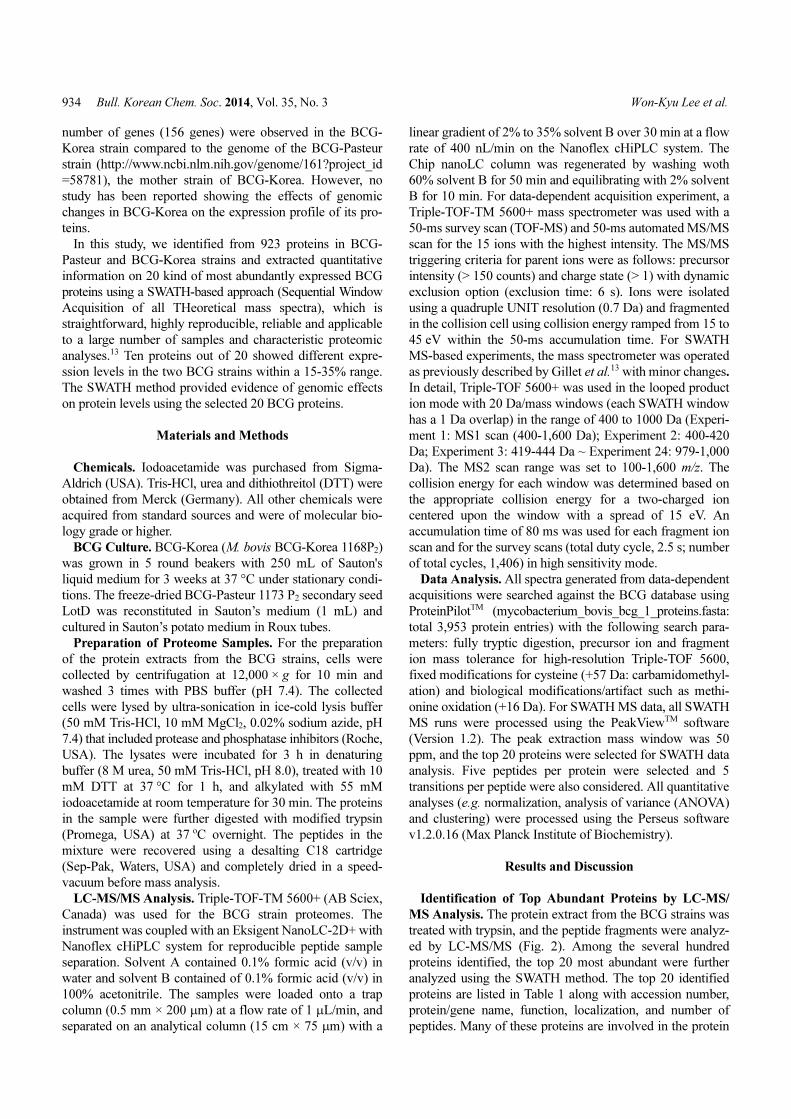
Identification of Top Abundant Proteins by LC-MS/
MS Analysis. The protein extract from the BCG strains was
treated with trypsin, and the peptide fragments were analyz-
ed by LC-MS/MS (Fig. 2). Among the several hundred
proteins identified, the top 20 most abundant were further
analyzed using the SWATH method. The top 20 identified
proteins are listed in Table 1 along with accession number,
protein/gene name, function, localization, and number of
peptides. Many of these proteins are involved in the protein
SWATH-based Proteomics Analysis of BCG-K Strain Bull. Korean Chem. Soc. 2014, Vol. 35, No. 3 935
folding and lipid biosynthesis pathway, such as GroEL1,
GroLE2, DnaK, ClpC, HtpG, Fas, Mas, and Pks13. The
abundance of these proteins indicates that they are critical
for the survival of the Mycobacterium species. These pro-
teins were known to be located in the cell wall, cytoplasm,
periplasmic space, and plasma membrane. Among the identi-
fied proteins, preprotein translocase subunit 1 (SecA1) is a
transmembrane protein involved in Mycobacterium virulen-
ence.14
Figure 2. A workflow of SWATH-based quantification for multipleBCG-Pasteur and BCG-Korea strains.
Table 1. Summary of 20 BCG proteins quantified by a SWATH-based approach
# Accession #Gene
nameDescription
Amino
Acid #Function Localizationa
#
Peptides
1 BCG_0479 GroEL2 60 kDa chaperonin 2 541 Chaperone Cp 99
2 BCG_2545c Fas Putative fatty acid synthase 3,070 Lipid synthesis Cw, Cs, Pm, Fsc 33
3 BCG_0389 DnaK Putative chaperone protein 626 Chaperone NA 31
4 BCG_2962c Mas Putative multifunctional mycocerosic acid syn-
thase
2,112 Lipid synthesis Cw, Pm 23
5 BCG_0717 RpoC DNA-directed RNA polymerase 1,317 Transcription DRPc, Cw, Pm 22
6 BCG_1194c MetE Putative 5-methyltetrahydropteroyltriglutamate-
homocysteine methyltransferase
760 Amino acid synthesis Cw, Cs, Pm 23
7 BCG_3269c SecA1 Putative preprotein translocase 950 Transport Cp, Pm 15
8 BCG_2578c AlaS Putative alanyl-tRNA synthetase 905 Protein synthesis Cp 12
9 BCG_3487c GroEL1 60 kDa chaperonin 1 540 Chaperone Bn, Cw, Cs, Pm 18
10 BCG_3862c Pks13 Polyketide synthase 1,734 Lipid synthesis Cw, Cs, Pm 15
11 BCG_1537c Can Putative aconitate hydratase 944 Metabolic reaction, cellular
respiration
NA 12
12 BCG_3661c ClpC Putative ATP-dependent clp proteasE 849 Chaperone Cw, Cs, Pm 13
13 BCG_1954 AceA Putative isocitrate lyase 767 Carboxylic acid metabolism Cp 12
14 BCG_3704c TopA DNA topoisomerase I 935 Intron homing Cw, Cs, Pm 11
15 BCG_1308c Kgd Multifunctional 2-oxoglutarate metabolism
enzyme
1,215 Metabolic reaction, cellular
respiration
Cw, Cs, Pm, PDc 11
16 BCG_2315c HtpG Putative chaperone protein 648 Chaperone Cp 15
17 BCG_0097c Idh Putative isocitrate dehydrogenase 746 Metabolic reaction, cellular
respiration
Pm 13
18 BCG_1509c Tal Putative transaldolase 374 Pentose shunt Cp 10
19 BCG_2496c Gdh Putative NAD-dependent glutamate dehydroge-
nase
1,624 Oxidoreductase Cw, Cs, Pm 11
20 BCG_0035 GyrB Dna gyrase 715 DNA topological change Ch, Cp 9
aSubcellular location was determined using UniProtKB-SubCell (http://www.uniprot.org/locations), Abbreviation key: Bn, bacterial nucleoid; Ch,chromosome; Cp, cytoplasm; Cs, cytosol; Cw, cell wall; DRPc, DNA-directed RNA polymerase complex; Fsc, fatty acid synthase complex; PDc,pyruvate dehydrogenase complex; Pm, plasma membrane; NA, information is not available.
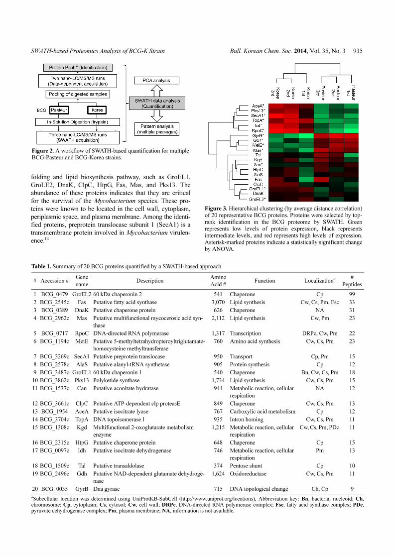
Figure 3. Hierarchical clustering (by average distance correlation)of 20 representative BCG proteins. Proteins were selected by top-rank identification in the BCG proteome by SWATH. Greenrepresents low levels of protein expression, black representsintermediate levels, and red represents high levels of expression.Asterisk-marked proteins indicate a statistically significant changeby ANOVA.

936 Bull. Korean Chem. Soc. 2014, Vol. 35, No. 3 Won-Kyu Lee et al.
Quantitative Analysis of the BCG top 20 Proteins. The
expression levels of the selected 20 proteins in the BCG-
Pasteur and BCG-Korea strains were compared using the
SWATH method and are color coded (Fig. 3). They were
divided into three groups according to their expression level
changes. The expression levels of the first group (AceA,
Pks13, SecA1, TopA, Icd, and RpoC) were slightly increased
in BCG-Korea. The expression levels of these proteins in
BCG Korea were 15-40% higher than those in BCG Pasteur
(Fig. 4(a)). It is notable that the proteins in the first group are
involved in nucleotide metabolism (RpoC and TopA), lipid
metabolism (Pks13), carboxylic acid metabolism (AceA),
cellular respiration (Icd) and protein transport (SecA1). The
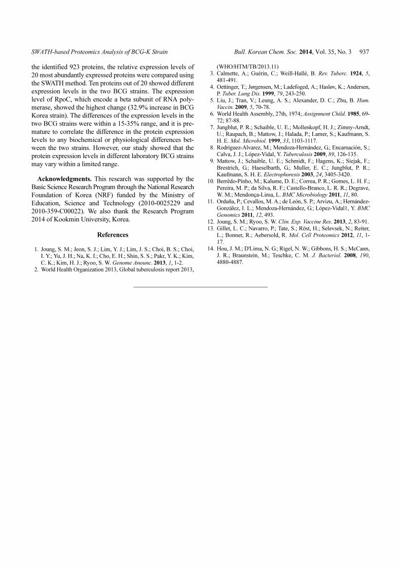
expression levels of the second group (GyrB, Gdh, MelE
and Mas) in BCG Korea were 10-20% lower than those in
BCG Pasteur (Fig. 4(b)). Proteins in the second group are
involved in lipid metabolism (Mas), amino acid metabolism
(MelE), DNA modification, or are members of the oxido-
reductase family (Gdh). The remainder (GroEL2, GroEL1,
Can, Tal, Kgd, Fas, DnaK, AlaS, ClpC, and HtpG) showed
similar expression levels. Interestingly, the expression levels
of the chaperone proteins (GloEL1, GloEL2, DnaK, ClpC,
and HtpG) were similar in the BCG Pasteur and BCG Korea
strains (Fig. 3). The constant expression levels of these
chaperones suggest that these proteins could serve as internal
standards when comparing the proteomes of different BCG
strains.
The sequences of the 20 genes investigated in this study
were identical in the BCG Pasteur and BCG Korea strains
(data not shown). In addition, all proteins, except GyrB, had
the same copy number in the BCG Pasteur and BCG Korea
strains. There are two copies of the gyrB gene in BCG-Pasteur
and a single copy in the BCG Korea strain. Interestingly, the
expression level of the GyrB protein was slightly reduced in
BCG Korea (Fig. 4(b)), suggesting that the higher copy
number of gyrB was not correlated to the expression level.
During the establishment of various BCG strains, several
regions of the genome sequence were deleted or duplicated.5
In this study, we comprehensively analyzed the proteomes
from two BCG strains with different passage numbers. From
Figure 4. Groups based on responsive expression pattern. All expression levels were normalized to total protein abundances. Errors werecalculated from three SWATH run replicates. The expression levels of proteins in BCG-Korea (grey bar) were compared to those of BCG-Pasteur (white bar).
SWATH-based Proteomics Analysis of BCG-K Strain Bull. Korean Chem. Soc. 2014, Vol. 35, No. 3 937
the identified 923 proteins, the relative expression levels of
20 most abundantly expressed proteins were compared using
the SWATH method. Ten proteins out of 20 showed different
expression levels in the two BCG strains. The expression
level of RpoC, which encode a beta subunit of RNA poly-
merase, showed the highest change (32.9% increase in BCG
Korea strain). The differences of the expression levels in the
two BCG strains were within a 15-35% range, and it is pre-
mature to correlate the difference in the protein expression
levels to any biochemical or physiological differences bet-
ween the two strains. However, our study showed that the
protein expression levels in different laboratory BCG strains
may vary within a limited range.
Acknowledgments. This research was supported by the
Basic Science Research Program through the National Research
Foundation of Korea (NRF) funded by the Ministry of
Education, Science and Technology (2010-0025229 and
2010-359-C00022). We also thank the Research Program
2014 of Kookmin University, Korea.
References
1. Joung, S. M.; Jeon, S. J.; Lim, Y. J.; Lim, J. S.; Choi, B. S.; Choi,
I. Y.; Yu, J. H.; Na, K. I.; Cho, E. H.; Shin, S. S.; Pakr, Y. K.; Kim,
C. K.; Kim, H. J.; Ryoo, S. W. Genome Anounc. 2013, 1, 1-2. 2. World Health Organization 2013, Global tuberculosis report 2013,
(WHO/HTM/TB/2013.11) 3. Calmette, A.; Guérin, C.; Weill-Hallé, B. Rev. Tuberc. 1924, 5,
481-491.
4. Oettinger, T.; Jørgensen, M.; Ladefoged, A.; Hasløv, K.; Andersen,P. Tuber. Lung Dis. 1999, 79, 243-250.
5. Liu, J.; Tran, V.; Leung, A. S.; Alexander, D. C.; Zhu, B. Hum.
Vaccin. 2009, 5, 70-78. 6. World Health Assembly, 27th, 1974; Assignment Child. 1985, 69-
72; 87-88.
7. Jungblut, P. R.; Schaible, U. E.; Mollenkopf, H. J.; Zimny-Arndt,U.; Raupach, B.; Mattow, J.; Halada, P.; Lamer, S.; Kaufmann, S.
H. E. Mol. Microbiol. 1999, 33, 1103-1117.
8. Rodríguez-Alvarez, M.; Mendoza-Hernández, G.; Encarnación, S.;Calva, J. J.; López-Vidal, Y. Tuberculosis 2009, 89, 126-135.
9. Mattow, J.; Schaible, U. E.; Schmidt, F.; Hagens, K.; Siejak, F.;
Brestrich, G.; Haeselbarth, G.; Muller, E. C.; Jungblut, P. R.;Kaufmann, S. H. E. Electrophoresis 2003, 24, 3405-3420.
10. Berrêdo-Pinho, M.; Kalume, D. E.; Correa, P. R.; Gomes, L. H. F.;
Pereira, M. P.; da Silva, R. F.; Castello-Branco, L. R. R.; Degrave,W. M.; Mendonça-Lima, L. BMC Microbiology 2011, 11, 80.
11. Orduña, P.; Cevallos, M. A.; de León, S. P.; Arvizu, A.; Hernández-
González, I. L.; Mendoza-Hernández, G.; López-Vidal1, Y. BMCGenomics 2011, 12, 493.
12. Joung, S. M.; Ryoo, S. W. Clin. Exp. Vaccine Res. 2013, 2, 83-91.
13. Gillet, L. C.; Navarro, P.; Tate, S.; Röst, H.; Selevsek, N.; Reiter,L.; Bonner, R.; Aebersold, R. Mol. Cell Proteomics 2012, 11, 1-
17.
14. Hou, J. M.; D'Lima, N. G.; Rigel, N. W.; Gibbons, H. S.; McCann,J. R.; Braunstein, M.; Teschke, C. M. J. Bacteriol. 2008, 190,
4880-4887.