2014-03 Val edward Jones Kytocel report - AllWeCare · MelBec Microbiology Ltd 1 ... or...
14
Professor Val Edwards-Jones MelBec Microbiology Ltd 1 Background Kytocel TM is a new dressing primarily focused on its absorbent and haemostatic properties. Initial studies and previous publications have demonstrated that the material within Kytocel TM , Chitosan, has antimicrobial properties. There are a number of methods that can be used to demonstrate antimicrobial activity of dressings but the most accepted methods are zone of inhibition (Thomas and McCubbin 2003), Log reduction assay (Gallant-Behm et al 2005) and challenge testing (Thomas and McCubbin 2003). These following studies were undertaken to assess the dressing for antimicrobial activity. 1.1 Zone of inhibition assay The basis of the zone of inhibition test is to place a dressing of known size on the surface of a Petri dish containing a suspension of a microorganism (at approx 10 6 cfu/ml) and incubate overnight at 37 o C. The zone of inhibition surrounding the dressing is measured. The effect underneath the dressing should also be assessed visually for growth and also sub cultured onto a sterile culture medium. 1.2.1 Method 1.2.1 Dressings for testing The dressings tested were:- Dressing 1 Tricotex Dressing 2 Aquacel Dressing 3 Kytocel Organisms used Staphylococcus aureus ATCC 6538P (methicillin resistant Staphylococcus aureus (MRSA) Pseudomonas aeruginosa ATCC 9027 Candida albicans NCTC 1363 Esherichia coli ATCC 8739
Transcript of 2014-03 Val edward Jones Kytocel report - AllWeCare · MelBec Microbiology Ltd 1 ... or...

Professor Val Edwards-Jones
MelBec Microbiology Ltd
1
Background
KytocelTM is a new dressing primarily focused on its absorbent and
haemostatic properties. Initial studies and previous publications have
demonstrated that the material within KytocelTM, Chitosan, has
antimicrobial properties. There are a number of methods that can be
used to demonstrate antimicrobial activity of dressings but the most
accepted methods are zone of inhibition (Thomas and McCubbin 2003),
Log reduction assay (Gallant-Behm et al 2005) and challenge testing
(Thomas and McCubbin 2003). These following studies were undertaken
to assess the dressing for antimicrobial activity.
1.1 Zone of inhibition assay
The basis of the zone of inhibition test is to place a dressing of known
size on the surface of a Petri dish containing a suspension of a
microorganism (at approx 106 cfu/ml) and incubate overnight at 37oC.
The zone of inhibition surrounding the dressing is measured. The effect
underneath the dressing should also be assessed visually for growth and
also sub cultured onto a sterile culture medium.
1.2.1 Method
1.2.1 Dressings for testing
The dressings tested were:-
Dressing 1 Tricotex
Dressing 2 Aquacel
Dressing 3 Kytocel
Organisms used
Staphylococcus aureus ATCC 6538P (methicillin resistant Staphylococcus
aureus (MRSA)
Pseudomonas aeruginosa ATCC 9027
Candida albicans NCTC 1363
Esherichia coli ATCC 8739

Professor Val Edwards-Jones
MelBec Microbiology Ltd
2
1.2.2. Method
Petri dishes containing a 5mm layer of Tryptone Soy Agar (TSA, LabM,
LAB011) for bacteria and Sabour aud Dextrose Agar (SDA, LabM,
LAB009) for the yeast were inoculated with 0.2ml of a log phase broth
culture of each test organism. The suspension was distributed uniformly
on the surface of the plate and dried for 15minutes. Portions of each
dressing measuring 40mm X 40mm were then placed on the agar, with
the wound contact surface downwards.
Each test was performed in triplicate and a similar piece of non-woven
gauze fabric (Tricotex) was used as a control. The plates were incubated
for 24hrs at 370C and the plates for fungi at 370C for 48hrs.
1.2.3. Bacteriocidal / Bacteriostatic activity
Following removal of the dressing, the original plate was re-incubated
for a further 24hrs if the area under the dressing after removal, showed
no microbial growth. After incubation, the area under the dressing was
sub-cultured onto a sterile TSA or SDA plate and examined for residual
growth to test for bacteriocidal/bacteriostatic or yeasticidal activity.
1.3.1 Results
There was no zone of inhibition observed with any of the dressings
tested. This demonstrated that there was no antimicrobial substance
diffusing laterally from the product.
1.3.2 Bacteriocidal/bacteriostatic and Yeasticidal activity
Following removal of Tricotex and Aquacel dressings from the 24hr
incubated plates, there was some evidence of reduced growth where
the dressing had been placed on the plate. When examined closely,
following a further 24hr incubation, this effect had disappeared and
there was visual growth under the Tricotex and Aquacel dressings.
However, under the Kytocel dressing there was no visible growth (see
figure 1) and there was no growth on sub culture. This demonstrated
that the Kytocel dressing was killing the organisms underneath the
dressing (bacteriocidal and Yeasticidal). However, there was evidence of

Professor Val Edwards-Jones
MelBec Microbiology Ltd
3
residual dressing on the plate where it had been co-cultured with
Candida albicans) (see figure 2)
Kytocel demonstrated antimicrobial activity against all four
microorganisms tested and killed the organisms on the surface of the
culture plate at a concentration of 106 cfu/ml.
1.4 Discussion
This experiment demonstrated that the antimicrobial activity observed
with the Kytocel dressing was not due to a diffusible antimicrobial
substance but an interaction with the organism and the dressing
material. The retention of the dressing on the surface of the agar plate
in the presence of Candida albicans is not easily understood. This may
have been due to an interaction of the culture medium and the dressing
or alternatively this could have been due to an unknown interaction with
the organism, dressing and culture medium.
Previous studies have shown that there is an interaction between
positively charged chitosan molecules and negatively charged microbial
cell membranes, altering cell permeability which leads to leakage of
intracellular constituents (Cheung et al 2004). There have been other
interactions described with intracellular components including DNA and
mRNA and inhibition of protein synthesis (Cheung et al 2004).
1.5 Key points Zone of Inhibition Assay
The antimicrobial effect of the dressing is not dependent on the release
of a soluble/diffusible compound but the effect is based upon the
interaction with the organism. This is probably due to an interaction of
the cationic charged dressing and negatively charged membrane.
The relevance of the dressing retention on the surface of the culture
plate in the presence of Candida albicans is interesting and further work
would have to be undertaken to fully understand this phenomenon.

Professor Val Edwards-Jones
MelBec Microbiology Ltd
4
Figure 1 The area underneath the dressing following removal of Kytocel dressing showing killing of the organisms.
Key: from left to right MRSA, Candida albicans, E.coli and Pseudomonas aeruginosa

Professor Val Edwards-Jones
MelBec Microbiology Ltd
5
Figure 2 Residual Kytocel dressing following testing with Candida albicans

Professor Val Edwards-Jones
MelBec Microbiology Ltd
6
2.0 Challenge testing
2.1 Principle of the test
A known number of organisms are added to a fluid that will maintain
numbers without allowing growth, and added to a piece of dressing at
time 0hrs. This is then incubated for 24hrs at 37 oC and at 2, 4 and 24hrs
the numbers of viable organisms are accurately counted following
vigorous mixing (to loosen organisms form the dressing that may have
adhered to it). This test helps to determine the speed of kill and the
time taken to reduce numbers or kill all organisms added to a dressing
within a 24hr period.
Objective:
To assess the time taken to kill four microorganisms using a modified
challenge test as described by Thomas and McCubbin 2003.
2.2 Materials and Methods
2.2.1 Dressings for testing
The dressings tested were :-
Dressing 1 Tricotex
Dressing 2 Aquacel
Dressing 3 Kytocel
Organisms used
Staphylococcus aureus ATCC 6538P (MRSA)
Pseudomonas aeruginosa ATCC 9027
Candida albicans NCTC 1363
Esherichia coli ATCC 8739
2.2.2 Method
1ml of a dilution of a log phase culture of each organism (approx.
106colony forming units (cfu)/ml)) was added to portions of the dressing
measuring 2.5 x 2.5cm (see figure 1). (Note; 1ml fluid in total was added

Professor Val Edwards-Jones
MelBec Microbiology Ltd
7
because the dressings absorbed approximately this amount and
remained moist up to 24hrs incubation).
The inoculated dressings were incubated at 370C in a sealed container
for 2,4,and 24hrs then transferred to 9ml of Letheen broth (Lab 184, Lab
M. UK) to neutralize any antimicrobial inhibitors) and vigorously vortex
mixed to remove any viable organisms remaining in the dressings. 0.1ml
of this was added to 9.9ml of PBS (Phosphate Buffered Saline) and the
number of viable organism present in each was determined using a
standard surface counting technique.
Each dressing was tested in triplicate and serial dilutions were
performed in triplicate on each extract (n=9) for each dressing.
The time kill kinetics was calculated up to a twenty four hour period.
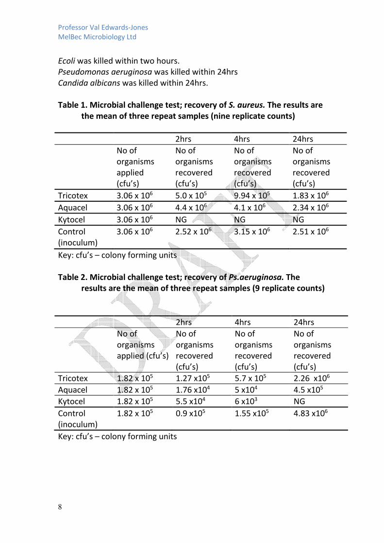
2.3.1. Results
The results of the challenge tests are shown in tables 1-4. These are the
mean values for nine estimations of viable counts.
All the microorganisms grew in the test system and there was no marked
decrease in organism numbers compared to the original numbers in the
original inoculum.
Dressing 1 – Tricotex (Control Dressing)
This dressing showed no antimicrobial activity against any organism
tested.
Dressing 2 – Aquacel
This dressing showed no antimicrobial activity against any organism
tested.
Dressing 3 – Kytocel
All microorganisms were killed within a 24hr period.
MRSA was killed within two hours.
Professor Val Edwards-Jones
MelBec Microbiology Ltd
8
Ecoli was killed within two hours.
Pseudomonas aeruginosa was killed within 24hrs
Candida albicans was killed within 24hrs.
Table 1. Microbial challenge test; recovery of S. aureus. The results are
the mean of three repeat samples (nine replicate counts)
2hrs 4hrs 24hrs
No of
organisms
applied
(cfu’s)
No of
organisms
recovered
(cfu’s)
No of
organisms
recovered
(cfu’s)
No of
organisms
recovered
(cfu’s)
Tricotex 3.06 x 10⁶ 5.0 x 10⁵ 9.94 x 10⁵ 1.83 x 10⁶
Aquacel 3.06 x 10⁶ 4.4 x 10⁶ 4.1 x 10⁶ 2.34 x 10⁶
Kytocel 3.06 x 10⁶ NG NG NG
Control
(inoculum)
3.06 x 10⁶ 2.52 x 10⁶ 3.15 x 10⁶ 2.51 x 10⁶
Key: cfu’s – colony forming units
Table 2. Microbial challenge test; recovery of Ps.aeruginosa. The
results are the mean of three repeat samples (9 replicate counts)
2hrs 4hrs 24hrs
No of
organisms
applied (cfu’s)
No of
organisms
recovered
(cfu’s)
No of
organisms
recovered
(cfu’s)
No of
organisms
recovered
(cfu’s)
Tricotex 1.82 x 10⁵ 1.27 x10⁵ 5.7 x 10⁵ 2.26 x10⁶
Aquacel 1.82 x 10⁵ 1.76 x10⁴ 5 x10⁴
4.5 x10⁵
Kytocel 1.82 x 10⁵ 5.5 x10⁴ 6 x10³ NG
Control
(inoculum)
1.82 x 10⁵ 0.9 x10⁵ 1.55 x10⁵
4.83 x10⁶
Key: cfu’s – colony forming units
Professor Val Edwards-Jones
MelBec Microbiology Ltd
9
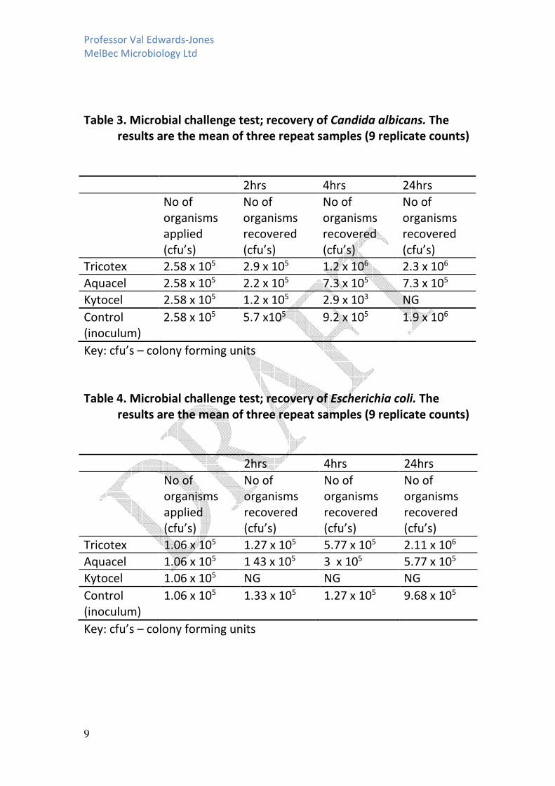
Table 3. Microbial challenge test; recovery of Candida albicans. The
results are the mean of three repeat samples (9 replicate counts)
2hrs 4hrs 24hrs
No of
organisms
applied
(cfu’s)
No of
organisms
recovered
(cfu’s)
No of
organisms
recovered
(cfu’s)
No of
organisms
recovered
(cfu’s)
Tricotex 2.58 x 10⁵ 2.9 x 10⁵ 1.2 x 10⁶ 2.3 x 10⁶
Aquacel 2.58 x 10⁵ 2.2 x 10⁵ 7.3 x 10⁵ 7.3 x 10⁵
Kytocel 2.58 x 10⁵ 1.2 x 10⁵ 2.9 x 10³ NG
Control
(inoculum)
2.58 x 10⁵ 5.7 x10⁵ 9.2 x 10⁵ 1.9 x 10⁶
Key: cfu’s – colony forming units
Table 4. Microbial challenge test; recovery of Escherichia coli. The
results are the mean of three repeat samples (9 replicate counts)
2hrs 4hrs 24hrs
No of
organisms
applied
(cfu’s)
No of
organisms
recovered
(cfu’s)
No of
organisms
recovered
(cfu’s)
No of
organisms
recovered
(cfu’s)
Tricotex 1.06 x 10⁵ 1.27 x 10⁵ 5.77 x 10⁵ 2.11 x 10⁶
Aquacel 1.06 x 10⁵ 1 43 x 105 3 x 10⁵ 5.77 x 10⁵
Kytocel 1.06 x 10⁵ NG NG NG
Control
(inoculum)
1.06 x 10⁵ 1.33 x 10⁵ 1.27 x 10⁵ 9.68 x 10⁵
Key: cfu’s – colony forming units

Professor Val Edwards-Jones
MelBec Microbiology Ltd
10
2.4 Discussion
Tricotex and Aquacel did not show any antimicrobial activity. This was
expected as no previous studies have demonstrated any antimicrobial
effect of the dressings. The model test system was appropriate as there
was no marked increase or decrease in cell numbers in the inoculum
control over the twenty four hour period, even when incubated at 37oC,
the ideal temperature for the organisms tested. The PBS contained no
nutrients but a buffered salt solution to maintain osmolality and
maintain pH. The dressings over the twenty four hour period showed a
slight increase in cell numbers but this was not markedly different from
the inoculum control, implying that the dressing itself had not been used
as a source of nutrients for any of the organisms tested.
Kytocel showed total kill within 24hrs for the three bacteria tested and
the fungal strain. Some strains had a quicker speed of kill with MRSA and
E.coli being killed within a two hour period. The yeast (Candida albicans)
and P. aeruginosa showed a marked reduction in numbers at four hours
and total kill at 24hrs. In order to calculate the exact time of kill, further
time points would have had to be included.
2.5 Key Points Challenge test
The importance of the speed of kill of an antimicrobial agent depends
upon the application. The dressings tested are applied to a wound to
absorb excess exudate and the number of times they are changed daily
would depend upon strikethrough. If a dressing is applied to a wound to
help reduce bacterial numbers and help resolve infection or colonization
then it would be important to have the quickest speed of kill as possible.
Most wounds are cleaned and may also be debrided. It is at this point
that the numbers of microorganisms in a wound will be reduced and
there is a known therapeutic window when an antimicrobial agent has
its maximum effect. Kytocel killed over one million cells of two
organisms within a two hour period (MRSA and E.coli) and the
remaining two organisms within a 24 hr period with a marked reduction
in cell numbers in 4hrs, demonstrating a good speed of kill for the
application as a dressing.
Professor Val Edwards-Jones
MelBec Microbiology Ltd
11
3. Log reduction assay
3.1 Principle of the test
A known number of organisms are added to a fluid that will maintain
numbers without allowing growth, and added to a piece of dressing at
time 0hrs. This is then incubated for 30minutes and 2hrs and the
numbers of viable organisms are accurately counted following vigorous
mixing (to loosen organisms from the dressing that may have adhered to
it). The reduction in organism numbers (expressed as log10) between
30minutes and 2hrs are recorded. This test helps determine how quickly
the antimicrobial substance acts against microorganisms and the level of
activity.
As defined by Gallant–Behm et al ., (2005) low antimicrobial activity was
considered to be less than 1 log reduction, moderate activity between 1
and 3 log reduction and high antimicrobial activity as greater than 3 log
reduction.
3.2 Materials and method
3.2.1 The dressings tested were:-
Dressing 1 Tricotex
Dressing 2 Aquacel
Dressing 3 Kytocel
Organisms used
Staphylococcus aureus ATCC 6538P
Pseudomonas aeruginosa ATCC 9027
Candida albicans NCTC 1363
Esherichia coli ATCC 8739
3.2.2 Method
1ml of a dilution of a log phase culture of each organism (approx
106colony forming units (cfu)/ml)) was added to portions of the dressing
measuring 2.5 x 2.5cm (see figure 1). (Note; 1ml fluid in total was added
because the dressings absorbed approx this amount and remained moist
throughout the incubation time).

Professor Val Edwards-Jones
MelBec Microbiology Ltd
12
The inoculated dressings were incubated at 370C in a sealed container
for 30minutes and 2hrs then transferred to 9ml of Letheen agar (Lab
184, Lab M. UK) to neutralize many antimicrobial inhibitors) and vortex
mixed to remove any viable organisms remaining in the dressings. 0.1ml
of this was added to 9.9ml of PBS and the number of viable organism
present in each was determined using a standard surface counting
technique.
Each dressing was tested in triplicate and serial dilutions were
performed in triplicate on each extract (n=9) for each dressing.
3.3 Results
Aquacel did not show any antimicrobial activity against any organism
compared to the control dressing (Tricotex) or the inoculum control.
Kytocel showed a high level of antimicrobial activity against MRSA with a
3log reduction at 30minutes and 6 log reduction after 2hrs.
Kytocel showed a high level of antimicrobial activity against E.coli with a
2 log reduction at 30minutes and a 5 log reduction after 2hrs.
Kytocel showed a low level of antimicrobial activity against
Pseudomonas aeruginosa and Candida albicans at 30minutes and 2hrs
with less than one log reduction for both organisms at both time
periods.
Table 2 showing log reduction (30minutes/120 minutes) induced by the
dressings under test compared to the Tricotex control.
Microorganism Aquacel Kytocel Tricotex
Control
MRSA 0.35/-0.14 3.61/6.47 0/0
E. coli 0.20/-0.06 1.95/5.02 0/0
P. aeruginosa 0.03/-0.39 0.66/0.13 0/0
C. albicans 0.07/0.11 -0.33/0.46 0/0

Professor Val Edwards-Jones
MelBec Microbiology Ltd
13
Discussion
Kytocel showed a high level of antimicrobial activity against two
common organisms found in wounds, MRSA and E.coli within a two hour
period showing a total kill within two hours equivalent to a 5-6 log
reduction. This would be extremely advantageous in wound care,
especially if there had been a biofilm on a chronic wound. Previous work
has shown that there is a small therapeutic window following
debridement when an antimicrobial dressing would have a beneficial
effect to help disrupt a biofilm and reduce numbers of common
colonisers in a wound.
Further Recommendations
The zone of inhibition assay showed Kytocel did not release any
diffusible antimicrobial properties but there was an interaction with the
dressing and the organisms on the surface of the culture plate as there
was total kill of the organisms underneath the dressing. This was not due
to absorbency, as Aquacel, which had similar absorbency properties to
Kytocel, think sentence needs finishing?
Kytocel has a rapid speed of kill and a 5-6 log reduction of common
pathogens, with a total kill observed within 24hrs on planktonic cells
(cells in suspension).
Therefore because Kytocel demonstrates kill of organisms on the surface
of a culture plate and also with planktonic cells in suspension, it would
be beneficial to test this dressing in a single organism and mixed
organism biofilm model to determine whether the mixed absorbent,
haemostatic and antimicrobial properties of the dressing would prevent
and inhibit a biofilm.
I recommend two further experiments:
1. Prevention of a biofilm formation in an explant pork model.
2. Eradication of a biofilm in an explant pork model.

Professor Val Edwards-Jones
MelBec Microbiology Ltd
14
Gallant-Behm C.L. et al., 2005 Comparison of in vitro disc diffusion and
time kill assays for the evaluation of antimicrobial wound dressing
efficiency Wound Repair and Regeneration 13; 4. 412-417.
Thomas and McCubbin 2003 A comparison of the antimicrobial effects of
four silver-containing dressings on three organisms J of Wound Care
2003; 12; 3; 101-107)
Ying-chien CHUNG,Ya-ping SU, Chiing-chang CHEN, Guang JIA ,
Huey-lan WANG, J C Gaston WU, Jaung-geng LIN 2004 Relationship
between antibacterial activity of chitosan and surface characteristics of
cell wall Acta Pharmacol ;25): 932-936.