2012-michael joiner-hypofractionation
45
Does the tumour radiobiology help in the move toward hypofractionation? Michael Joiner Wayne State University Radiation Oncology Detroit, Michigan [email protected]
-
Upload
fondas-vakalis -
Category
Health & Medicine
-
view
675 -
download
1
Transcript of 2012-michael joiner-hypofractionation
Does the tumour radiobiology help in the move toward
hypofractionation?
Michael Joiner
Wayne State University Radiation Oncology
Detroit, Michigan
Does the tumour radiobiology hinder in the move toward
hypofractionation?
Michael Joiner
Wayne State University Radiation Oncology
Detroit, Michigan
MCJ
What radiobiology?
Aug 12 3
• Research has focused on doses per fraction of 1–3 Gy: clinically relevant
• Radiobiology is quite mature
• Little incentive/funding for high-dose research
Historically…
MCJ
Why reconsider high dose fractions?
Aug 12 4
• Because we can: Physics • Patient convenience and demand • Lower cost of whole treatment • Evidence that it is very effective • Evidence of low !/" in some sites
The radiobiology has not yet caught up
MCJ
Why not LQ at high doses?
Aug 12 5
• Reponse is really linear at higher doses? • Vascular damage? • Increased apotosis? • Immunological effects? • Mixed tumor cell populations with different
response characteristics?
Answers likely depend on tissue type and tumor type / stage
MCJ
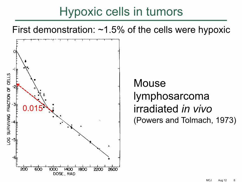
Hypoxic cells in tumors
Aug 12 6
First demonstration: ~1.5% of the cells were hypoxic
Mouse lymphosarcoma irradiated in vivo (Powers and Tolmach, 1973)
0.015
MCJ Aug 12 7
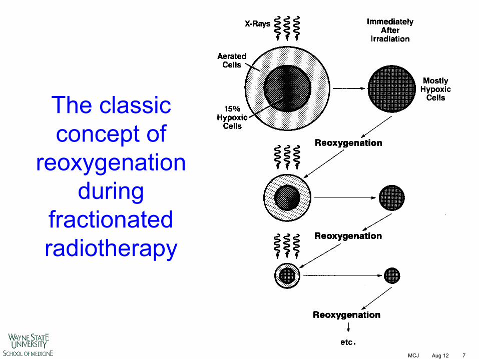
The classic concept of
reoxygenation during
fractionated radiotherapy
MCJ
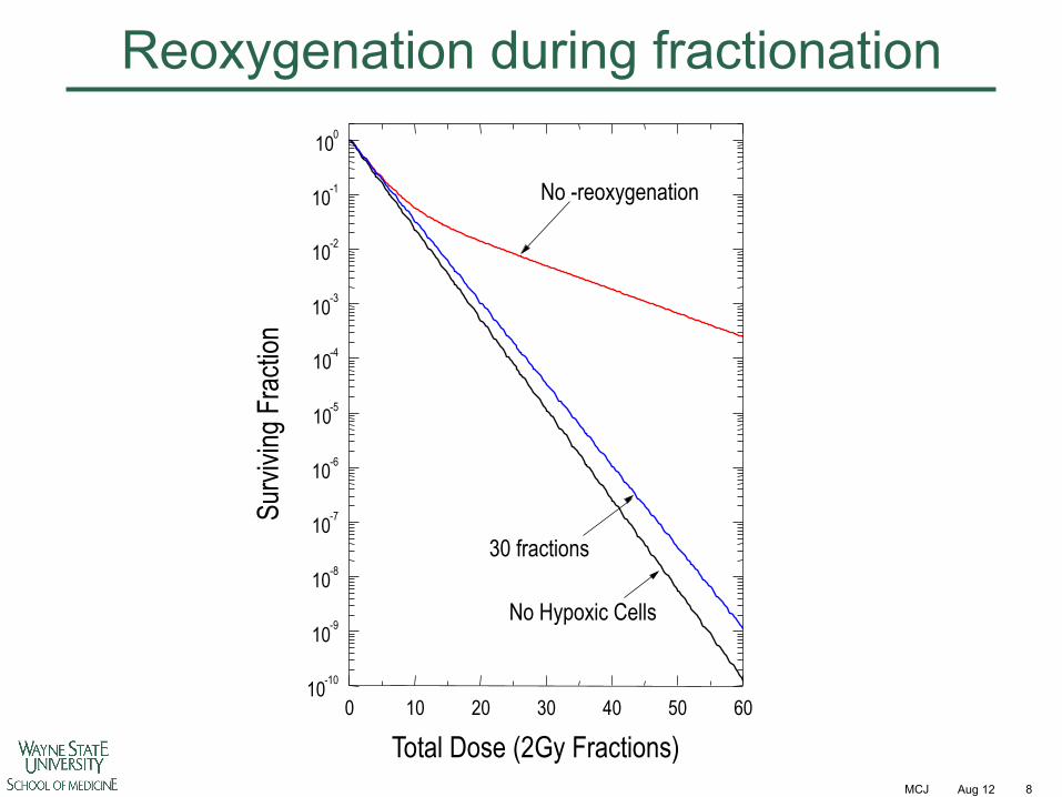
Reoxygenation during fractionation
Aug 12 8
No -reoxygenation
30 fractions
No Hypoxic Cells
0 10 20 30 40 50 60 10 -10 10 -9 10 -8 10 -7 10 -6
10 -5 10 -4
10 -3 10 -2 10 -1 10 0
Surv
iving
Fra
ction
Total Dose (2Gy Fractions)
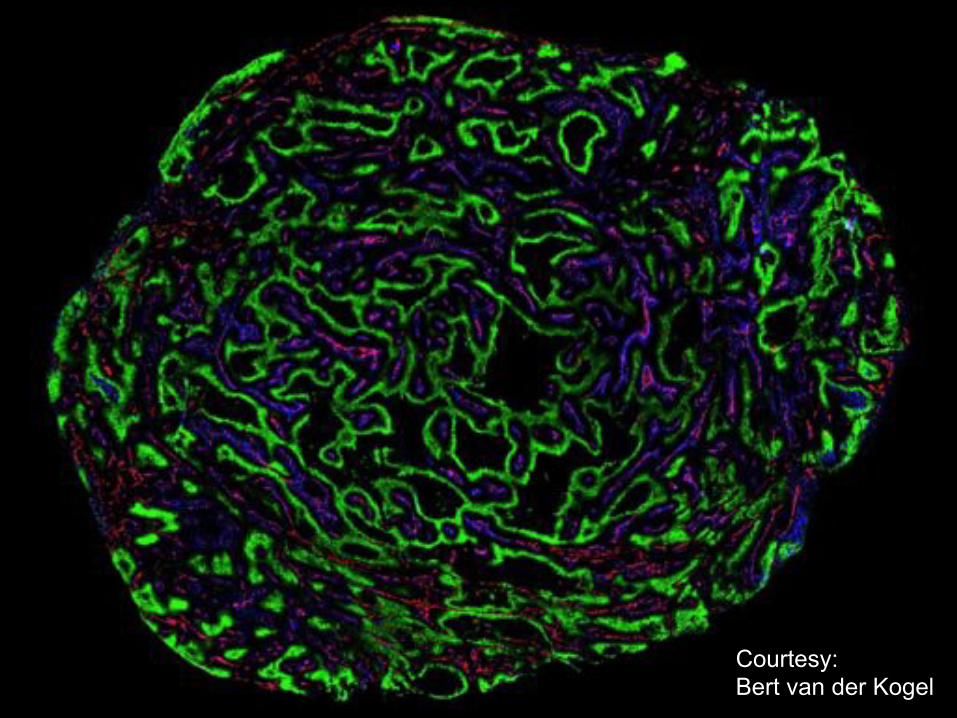
Courtesy: Bert van der Kogel
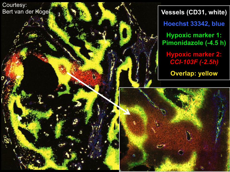
Vessels (CD31, white)
Hoechst 33342, blue
Hypoxic marker 1: Pimonidazole (-4.5 h)
Hypoxic marker 2: CCI-103F (-2.5h)
Overlap: yellow
Courtesy: Bert van der Kogel
MCJ
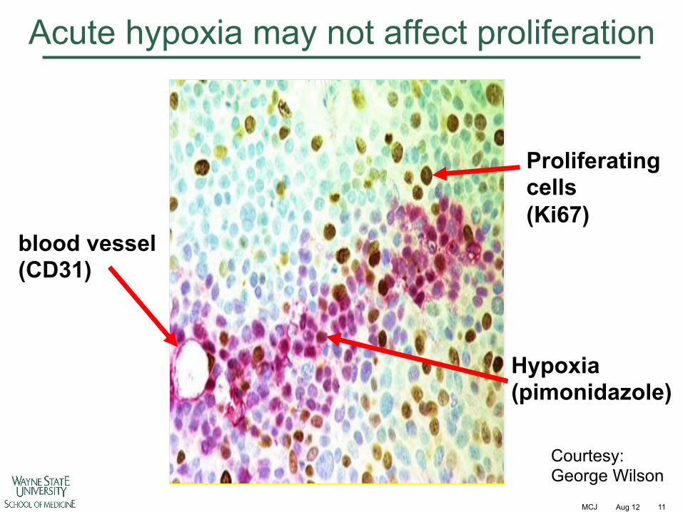
Acute hypoxia may not affect proliferation
Aug 12 11
blood vessel (CD31)
Proliferating cells (Ki67)
Hypoxia (pimonidazole)
Courtesy: George Wilson
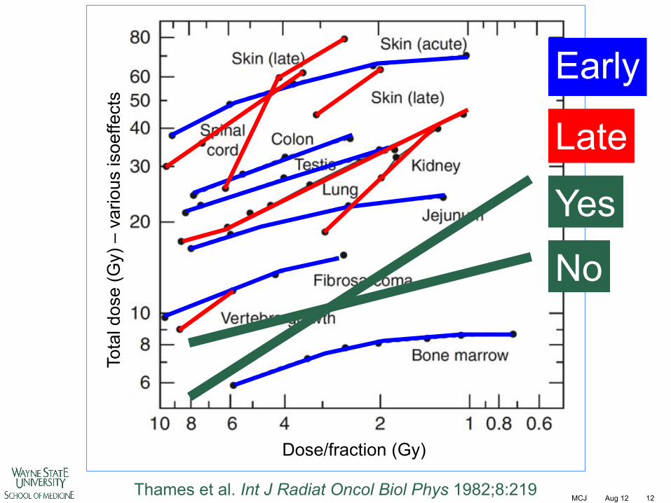
MCJ Aug 12 12 Thames et al. Int J Radiat Oncol Biol Phys 1982;8:219
Late
Early To
tal d
ose
(Gy)
– v
ario
us is
oeffe
cts
Dose/fraction (Gy)
No
Yes
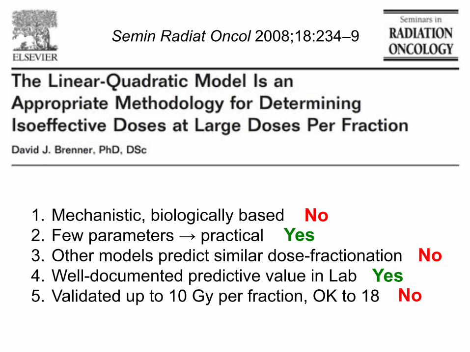
1. Mechanistic, biologically based 2. Few parameters ! practical 3. Other models predict similar dose-fractionation 4. Well-documented predictive value in Lab 5. Validated up to 10 Gy per fraction, OK to 18
Semin Radiat Oncol 2008;18:234–9
No Yes
No
No Yes
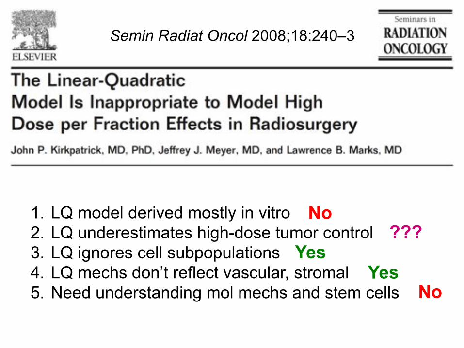
1. LQ model derived mostly in vitro 2. LQ underestimates high-dose tumor control 3. LQ ignores cell subpopulations 4. LQ mechs don’t reflect vascular, stromal 5. Need understanding mol mechs and stem cells
No ???
Yes
No Yes
Semin Radiat Oncol 2008;18:240–3
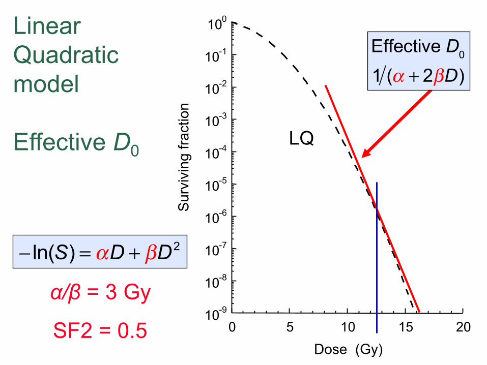
Linear Quadratic model Effective D0
10 -9
10 -8
10 -7
10 -6
10 -5
10 -4
10 -3
10 -2
10 -1
10 0
0 5 10 15 20
Sur
vivi
ng fr
actio
n
Dose (Gy)
LQ
!/" = 3 Gy
SF2 = 0.5
Effective D0
1 (! + 2"D)
! ln(S) = "D + #D2
MCJ
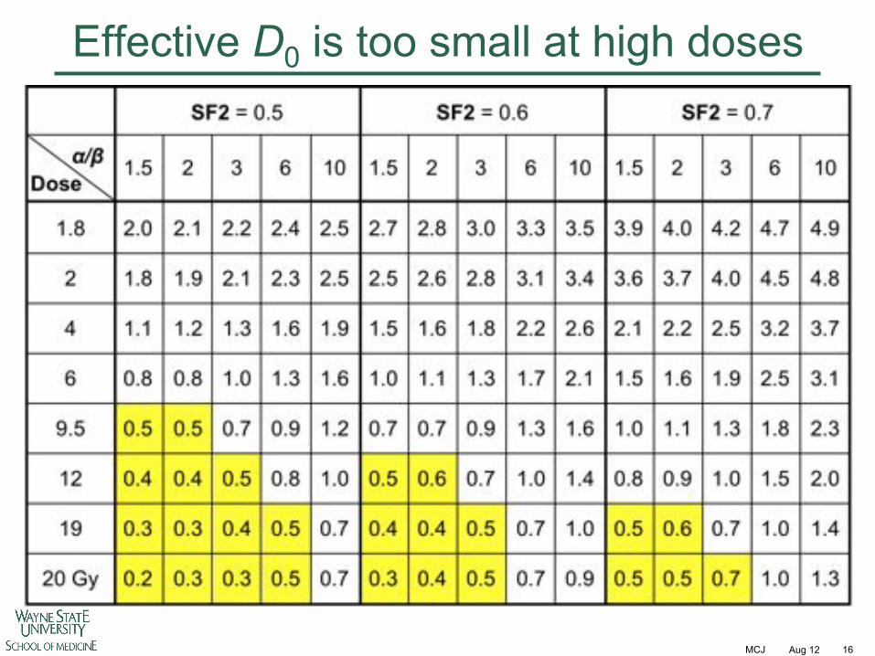
Effective D0 is too small at high doses
Aug 12 16
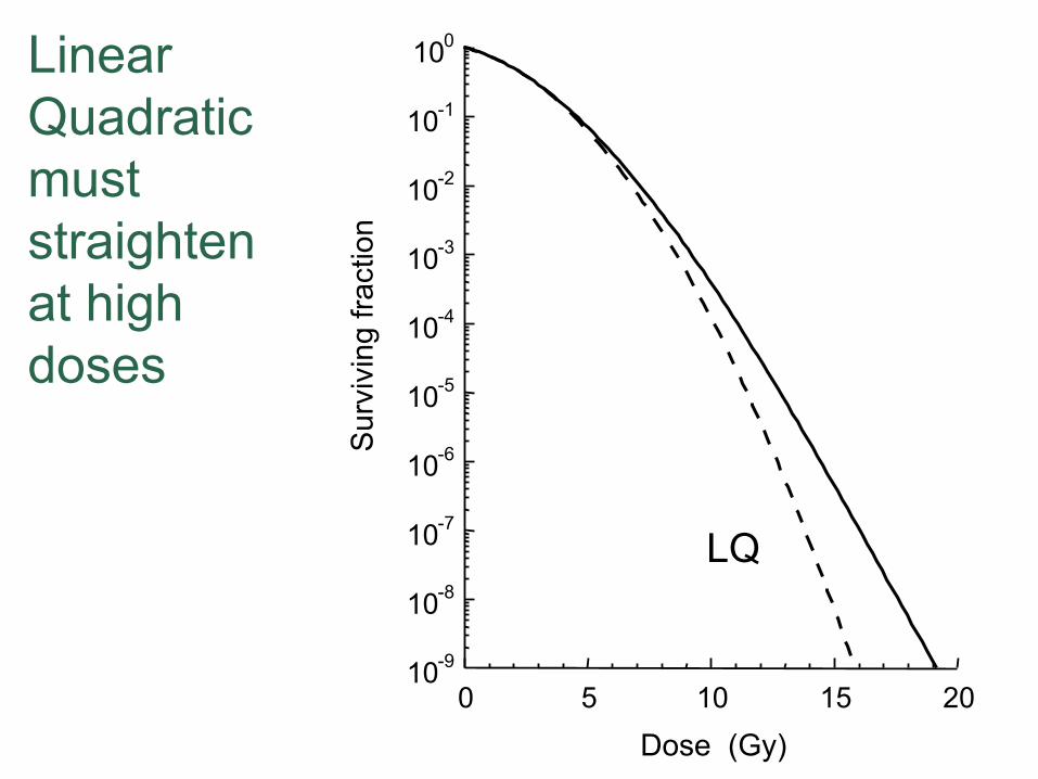
Linear Quadratic must straighten at high doses
10 -9
10 -8
10 -7
10 -6
10 -5
10 -4
10 -3
10 -2
10 -1
10 0
0 5 10 15 20
Sur
vivi
ng fr
actio
n
Dose (Gy)
LQ

LQC
! ln(S) ="D + #D2 !$D3
The Linear Quadratic Cubic model
10 -9
10 -8
10 -7
10 -6
10 -5
10 -4
10 -3
10 -2
10 -1
10 0
0 5 10 15 20
Sur
vivi
ng fr
actio
n
Dose (Gy)
LQ
! ln(S) ="D + #D2
!/" = 3 Gy
SF2 = 0.5
! = " 3DL( )
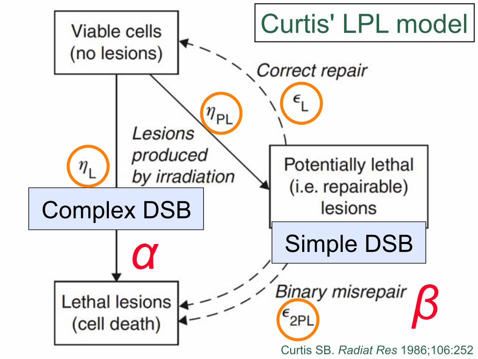
Simple DSB Complex DSB
! "
Curtis' LPL model
Curtis SB. Radiat Res 1986;106:252
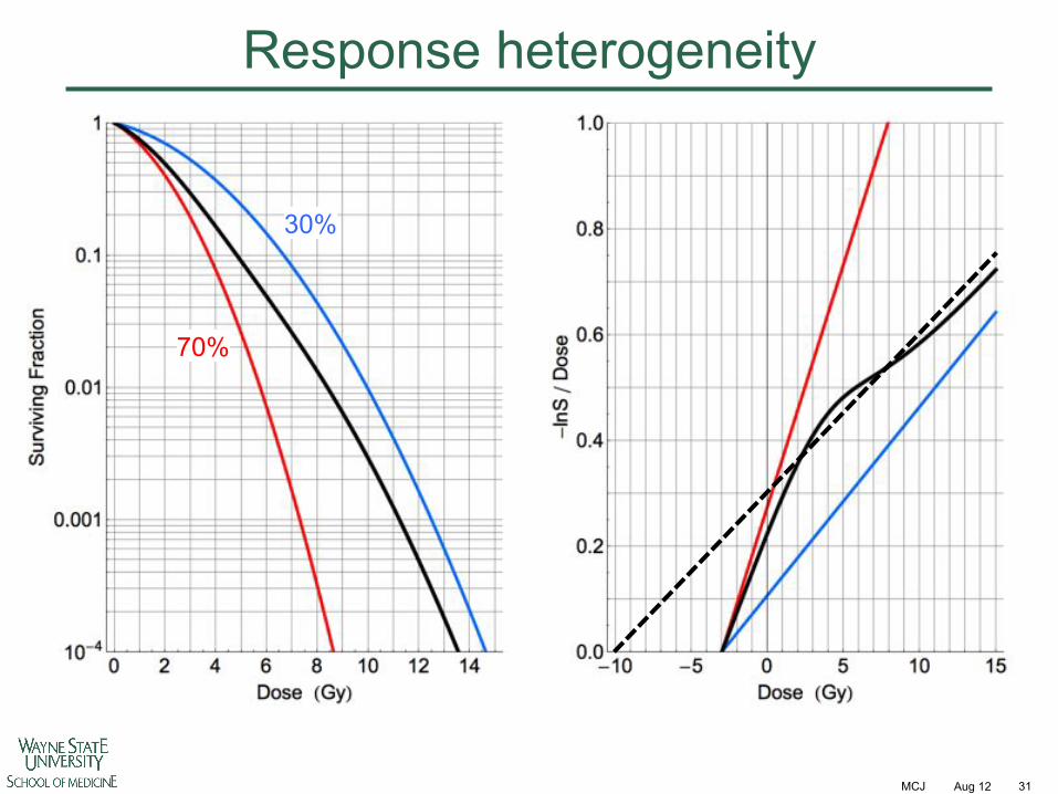
Response heterogeneity
Alternative damage response pathways and/or cell types which are dose dependent
MCJ
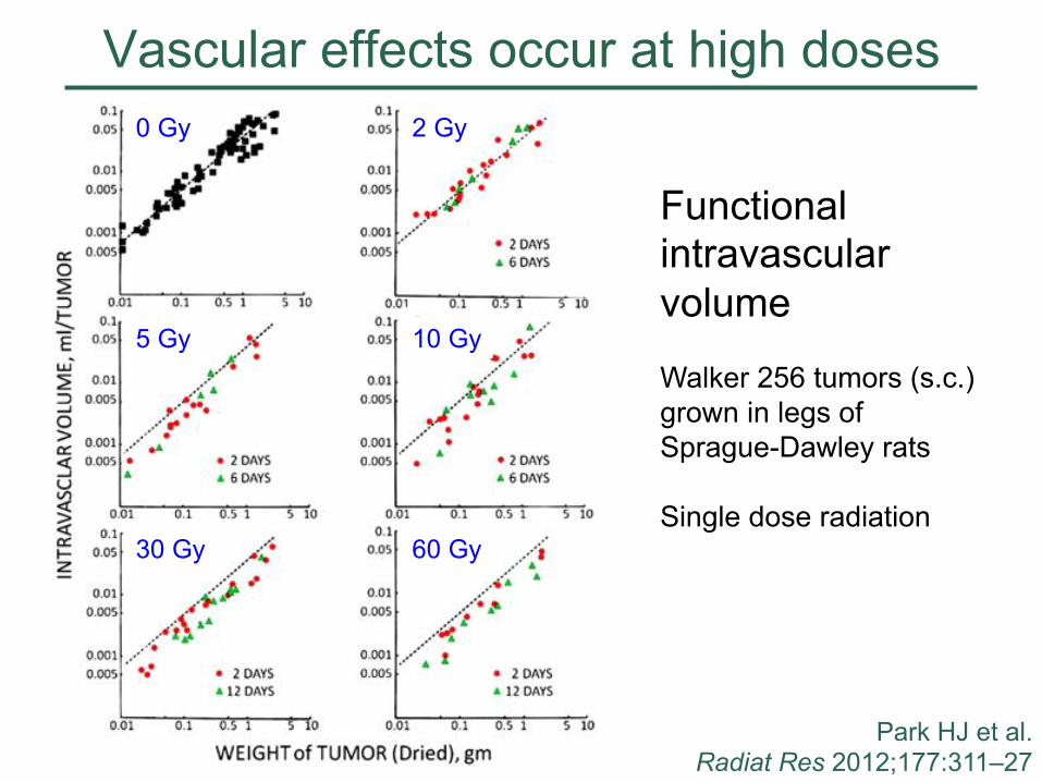
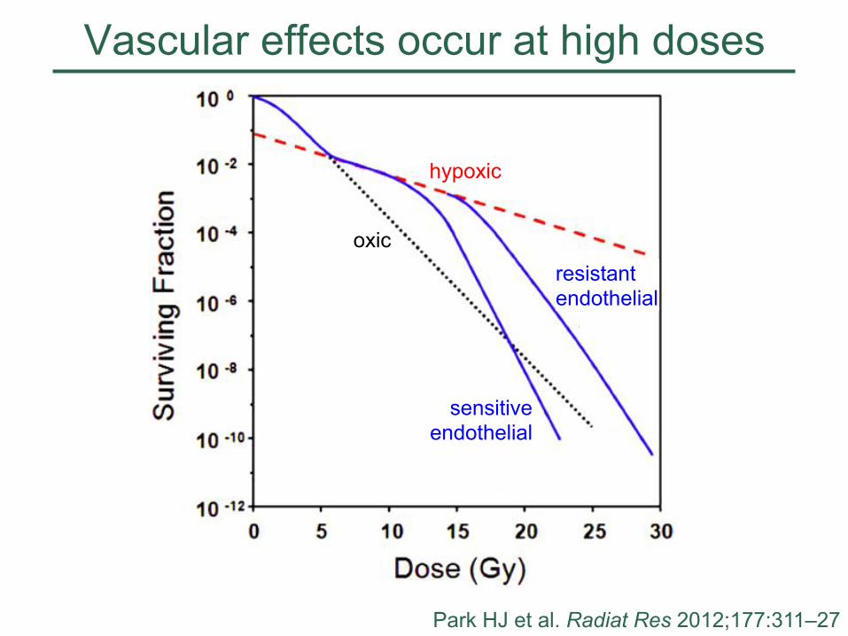
Vascular effects occur at high doses
Aug 12 21
Park HJ et al. Radiat Res 2012;177:311–27
Functional intravascular volume Walker 256 tumors (s.c.) grown in legs of Sprague-Dawley rats Single dose radiation
0 Gy 2 Gy
5 Gy 10 Gy
30 Gy 60 Gy
MCJ
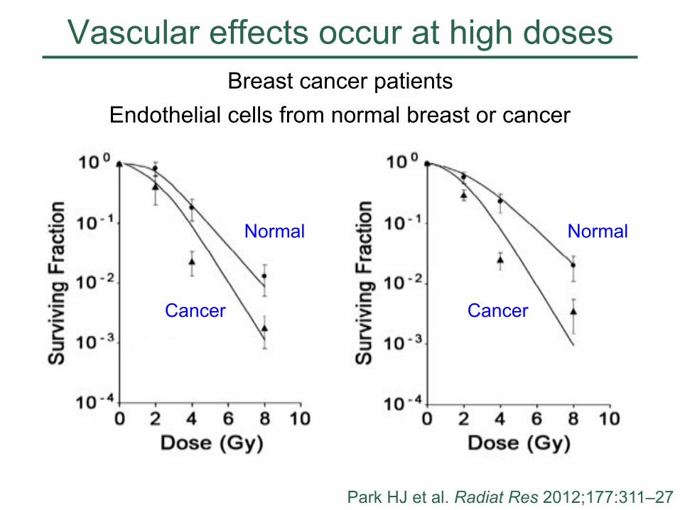
Vascular effects occur at high doses
Aug 12 22
Breast cancer patients Endothelial cells from normal breast or cancer
Park HJ et al. Radiat Res 2012;177:311–27
Normal
Cancer
Normal
Cancer
MCJ
Vascular effects occur at high doses
Aug 12 23 Park HJ et al. Radiat Res 2012;177:311–27
oxic resistant endothelial
hypoxic
sensitive endothelial
MCJ
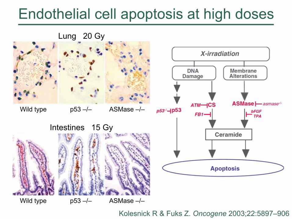
Endothelial cell apoptosis at high doses
Aug 12 24 Kolesnick R & Fuks Z. Oncogene 2003;22:5897–906
Intestines 15 Gy
Wild type p53 –/– ASMase –/–
Lung 20 Gy
Wild type p53 –/– ASMase –/–
MCJ
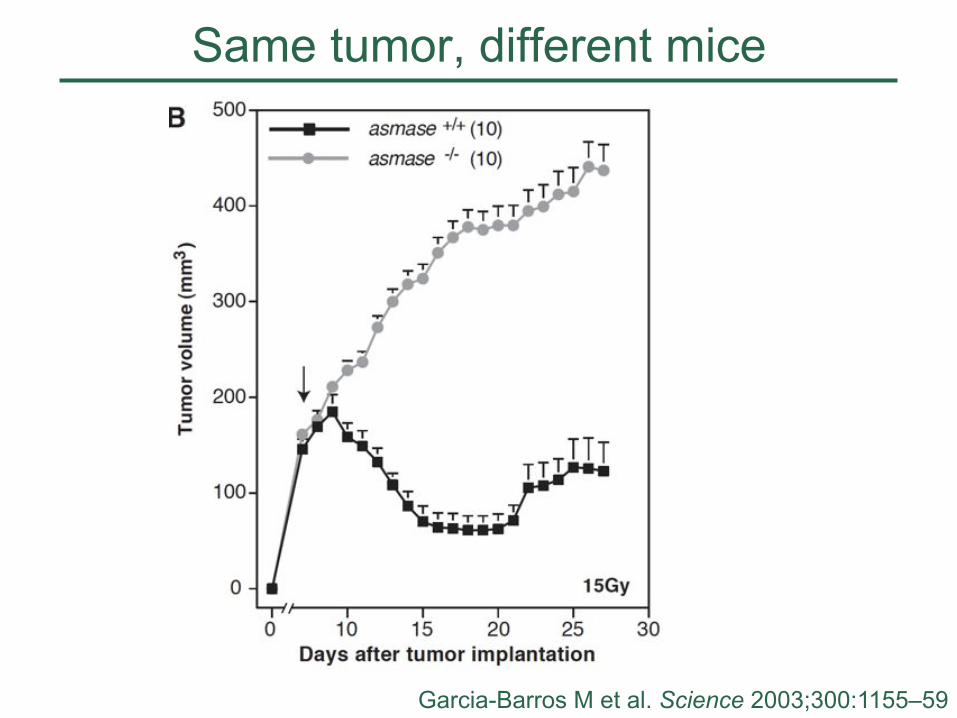
Same tumor, different mice
Aug 12 25 Garcia-Barros M et al. Science 2003;300:1155–59
MCJ
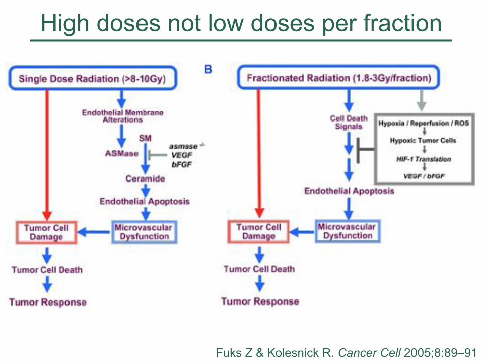
High doses not low doses per fraction
Aug 12 26 Fuks Z & Kolesnick R. Cancer Cell 2005;8:89–91
MCJ
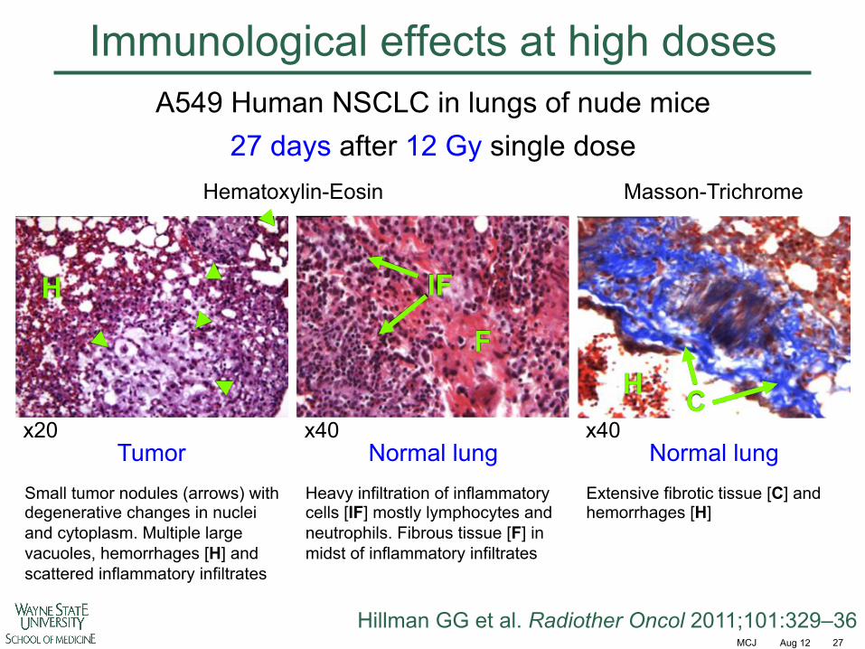
Immunological effects at high doses
Aug 12 27
Tumor Normal lung Normal lung x40 x40 x20
A549 Human NSCLC in lungs of nude mice 27 days after 12 Gy single dose
Hematoxylin-Eosin Masson-Trichrome
Hillman GG et al. Radiother Oncol 2011;101:329–36
H C
IF
F
H
Small tumor nodules (arrows) with degenerative changes in nuclei and cytoplasm. Multiple large vacuoles, hemorrhages [H] and scattered inflammatory infiltrates
Heavy infiltration of inflammatory cells [IF] mostly lymphocytes and neutrophils. Fibrous tissue [F] in midst of inflammatory infiltrates
Extensive fibrotic tissue [C] and hemorrhages [H]
Response heterogeneity
Mixed target cell populations with different sensitivites
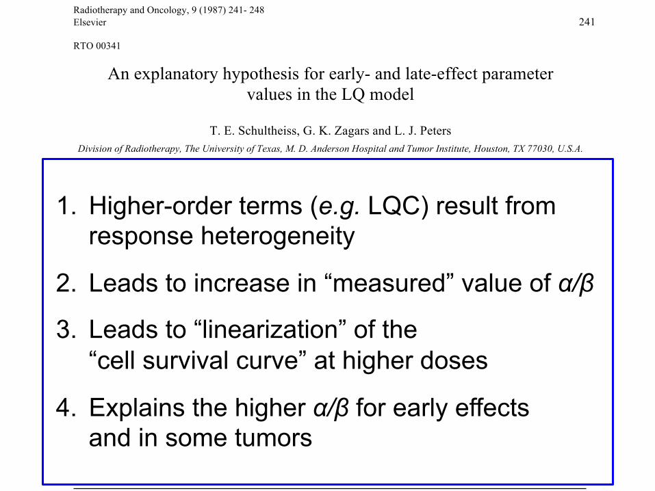
Radiotherapy and Oncology, 9 (1987) 241- 248 Elsevier 241 RTO 00341
An explanatory hypothesis for early- and late-effect parameter values in the LQ model
T. E. Schultheiss, G. K. Zagars and L. J. Peters Division of Radiotherapy, The University of Texas, M. D. Anderson Hospital and Tumor Institute, Houston, TX 77030, U.S.A.
(Received 3 November 1986, revision received 17 February 1987, accepted 17 February 1987) Key words: Cell survival; lsoeffect: LQ model; Late effect
Summary The isoeffect equation derived from the linear-quadratic (LQ) model of cell survival contains linear and quadratic terms in dose. Experimental studies have shown that higher-order terms may also be present. These terms have been previously attributed to the fact that the LQ model may be the first two terms in a power series approximation to a more complex model. This study shows that higher-order terms are introduced as a result of heterogeneity in the response of the cell population being irradiated. This heterogeneity is modeled by assuming that the parameters ! and " in the LQ model are distributed according to a bivariate normal distribution. Using this distribution, the expected value of cell survival contains third- and fourth-order terms in dose. These terms result in the previously observed downward curvature of Fe plots. Furthermore, these higher-order terms introduce bias in the estimated values of ! and ", if only the linear and quadratic terms of the LQ model are used, and higher-order terms are ignored. The bias is such that the estimated value of ! /" is substantially increased. Thus the higher values of ! /" observed for early effects as compared to late effects may be due to greater heterogeneity of response in early-responding tissues than in later-responding tissues. This differential effect is maintained even if the two cell populations have the same average values of ! and ".
1. Higher-order terms (e.g. LQC) result from response heterogeneity
2. Leads to increase in “measured” value of !/"
3. Leads to “linearization” of the “cell survival curve” at higher doses
4. Explains the higher !/" for early effects and in some tumors
MCJ
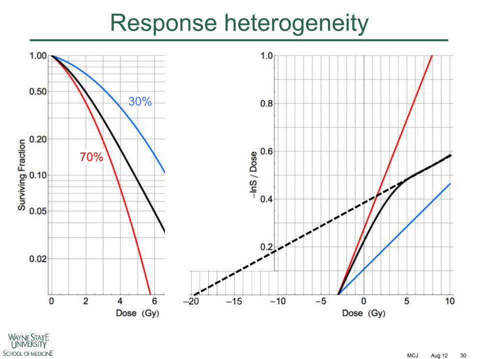
Response heterogeneity
Aug 12 30
–15 –20
30%
70%
MCJ
Response heterogeneity
Aug 12 31
30%
70%
Put more of this heterogeneity modeling in?
A model is a lie that helps you see the truth
- Howard Skipper
Thanks Patrick McDermott
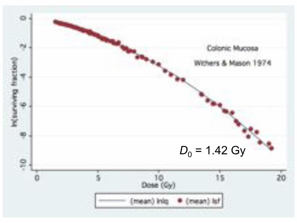
D0 = 1.42 Gy
MCJ Aug 12 35
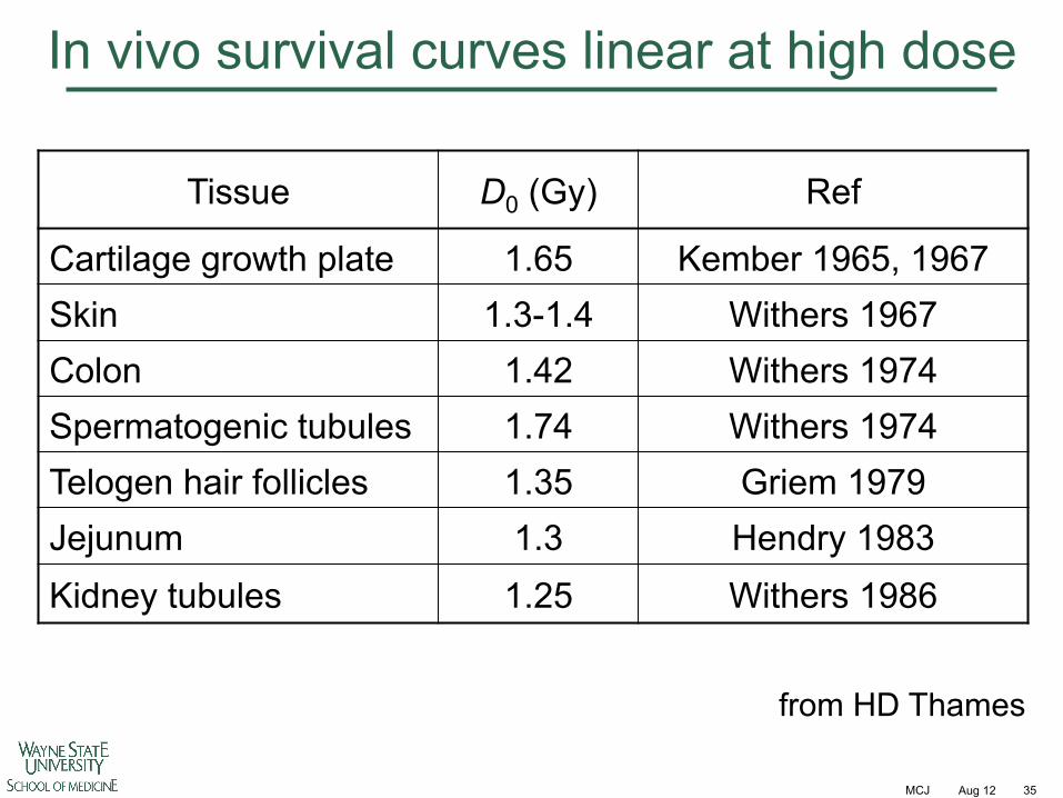
In vivo survival curves linear at high dose
Tissue D0 (Gy) Ref
Cartilage growth plate 1.65 Kember 1965, 1967 Skin 1.3-1.4 Withers 1967 Colon 1.42 Withers 1974 Spermatogenic tubules 1.74 Withers 1974 Telogen hair follicles 1.35 Griem 1979 Jejunum 1.3 Hendry 1983 Kidney tubules 1.25 Withers 1986
from HD Thames

MCJ Aug 12 36
Why does this make a difference? High-dose log-linear (HDLL) model predicts higher isoeffect dose than LQ as the curve goes linear
At what dose does this divergence become significant?
HDLL LQ
MCJ Aug 12 37
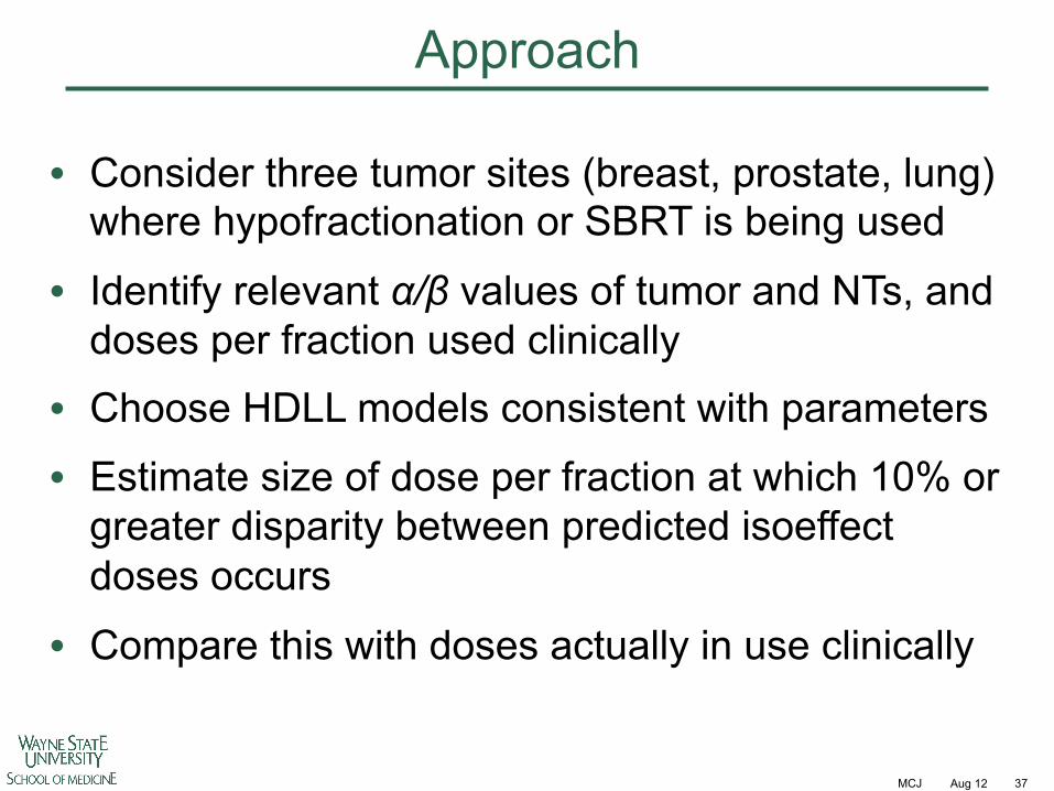
Approach
• Consider three tumor sites (breast, prostate, lung) where hypofractionation or SBRT is being used
• Identify relevant !/" values of tumor and NTs, and doses per fraction used clinically
• Choose HDLL models consistent with parameters
• Estimate size of dose per fraction at which 10% or greater disparity between predicted isoeffect doses occurs
• Compare this with doses actually in use clinically
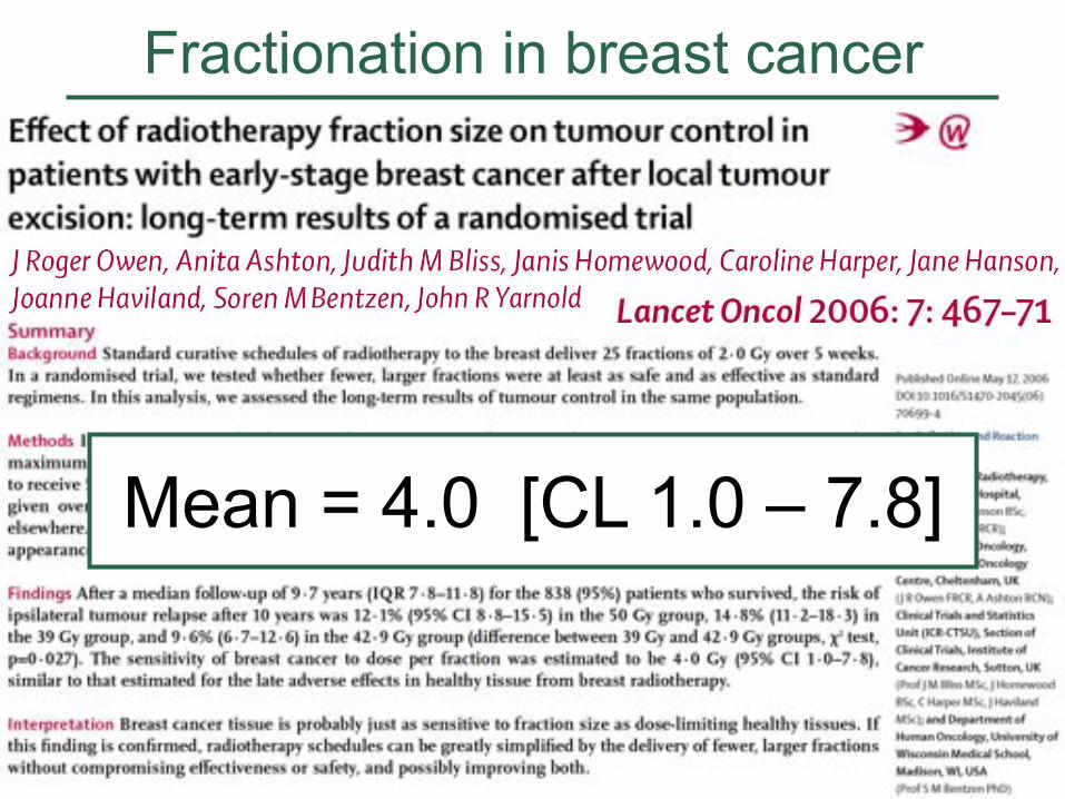
Fractionation in breast cancer
Mean = 4.0 [CL 1.0 – 7.8]
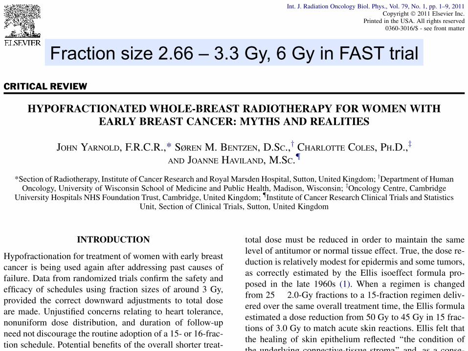
CRITICAL REVIEW
HYPOFRACTIONATED WHOLE-BREAST RADIOTHERAPY FOR WOMEN WITHEARLY BREAST CANCER: MYTHS AND REALITIES
JOHN YARNOLD, F.R.C.R.,* SØREN M. BENTZEN, D.SC.,y CHARLOTTE COLES, PH.D.,z
AND JOANNE HAVILAND, M.SC.{
*Section of Radiotherapy, Institute of Cancer Research and Royal Marsden Hospital, Sutton, United Kingdom; yDepartment of HumanOncology, University of Wisconsin School of Medicine and Public Health, Madison, Wisconsin; zOncology Centre, Cambridge
University Hospitals NHS Foundation Trust, Cambridge, United Kingdom; {Institute of Cancer Research Clinical Trials and StatisticsUnit, Section of Clinical Trials, Sutton, United Kingdom
INTRODUCTION
Hypofractionation for treatment of women with early breastcancer is being used again after addressing past causes offailure. Data from randomized trials confirm the safety andefficacy of schedules using fraction sizes of around 3 Gy,provided the correct downward adjustments to total doseare made. Unjustified concerns relating to heart tolerance,nonuniform dose distribution, and duration of follow-upneed not discourage the routine adoption of a 15- or 16-frac-tion schedule. Potential benefits of the overall shorter treat-ment time include greater convenience and improved localtumor control, although the latter benefit remains to betested. Adjusting fraction size across the breast is a goodway of matching dose to tumor relapse risk. Amodest reduc-tion in fraction size to breast tissue remote from the tumorbed and at low risk of local tumor relapse is expected to re-duce late adverse effects without significant loss of tumorcontrol. The corollary is that dose escalation to the indexquadrant, whether by hypofractionation or by a sequentialboost dose, will result in a greater relative increase in late ad-verse effects than tumor control, a therapeutic disadvantagethat can be overcome only by exploiting a marked dose-volume effect.
BRIEF HISTORY OF HYPOFRACTIONATION
Hypofractionation: What went wrong and whyIt has been understood for more than 100 years that the re-
lationship between total radiation dose and biological effectdepends on the dose per fraction. As fraction size increases,
total dose must be reduced in order to maintain the samelevel of antitumor or normal tissue effect. True, the dose re-duction is relatively modest for epidermis and some tumors,as correctly estimated by the Ellis isoeffect formula pro-posed in the late 1960s (1). When a regimen is changedfrom 25 ! 2.0-Gy fractions to a 15-fraction regimen deliv-ered over the same overall treatment time, the Ellis formulaestimated a dose reduction from 50 Gy to 45 Gy in 15 frac-tions of 3.0 Gy to match acute skin reactions. Ellis felt thatthe healing of skin epithelium reflected ‘‘the condition ofthe underlying connective-tissue stroma’’ and, as a conse-quence, he hypothesized that ‘‘apart from bone andbrain.the normal tissue tolerance dose, could be based onskin tolerance.’’ It was realized in the late 1970s and early1980s that dose reductions estimated using the Ellis formulawere insufficient for matching late side-effects (2–5). Lateeffects such as subcutaneous fibrosis and skintelangiectasia are more sensitive than acute reactions toaltered fraction size (6–7). In fairness, Ellis proposed hisformula as a hypothesis to be tested in the clinic, butradiation oncologists applied the formula in what, withhindsight, was an uncritical way.
The distinct fractionation sensitivities of early and late re-sponding normal tissues are well described using a linear-quadratic model in which an endpoint-specific quantity,the a/b ratio, offers a reliable way of describing these differ-ences (8–9). Assuming a typical a/b value of 3.0 Gy for latenormal tissue responses, a 15-fraction regimen reproducingthe effects of 25 fractions of 2.0 Gy requires a reduction intotal dose from 50 Gy to 42.8 Gy in fractions of 2.85 Gy
Reprint requests to: John Yarnold, Section of Radiotherapy,Institute of Cancer Research and Royal Marsden Hospital, SuttonSM2 5PT, UK. Tel: (+44) 208 661 3388. Fax: (+44) 208 6613107; E-mail: [email protected]. John Yarnold, F.R.C.R., and Dr. Charlotte Coles, Ph.D., are
supported by the National Institute of Health Research BiomedicalResearch Centre. Joanne Haviland, M.Sc., is supported by a core
grant to Institute of Cancer Research Clinical Trials and StatisticsUnit from Cancer Research UK. Prof. Søren Bentzen, D.Sc., ac-knowledges support from U.S. National Cancer Institute grant no.2P30 CA 014520-34.Conflict of interest: none.Received May 18, 2010, and in revised form Aug 9, 2010.
Accepted for publication Aug 17, 2010.
1
Int. J. Radiation Oncology Biol. Phys., Vol. 79, No. 1, pp. 1–9, 2011Copyright ! 2011 Elsevier Inc.
Printed in the USA. All rights reserved0360-3016/$ - see front matter
doi:10.1016/j.ijrobp.2010.08.035
Fraction size 2.66 – 3.3 Gy, 6 Gy in FAST trial
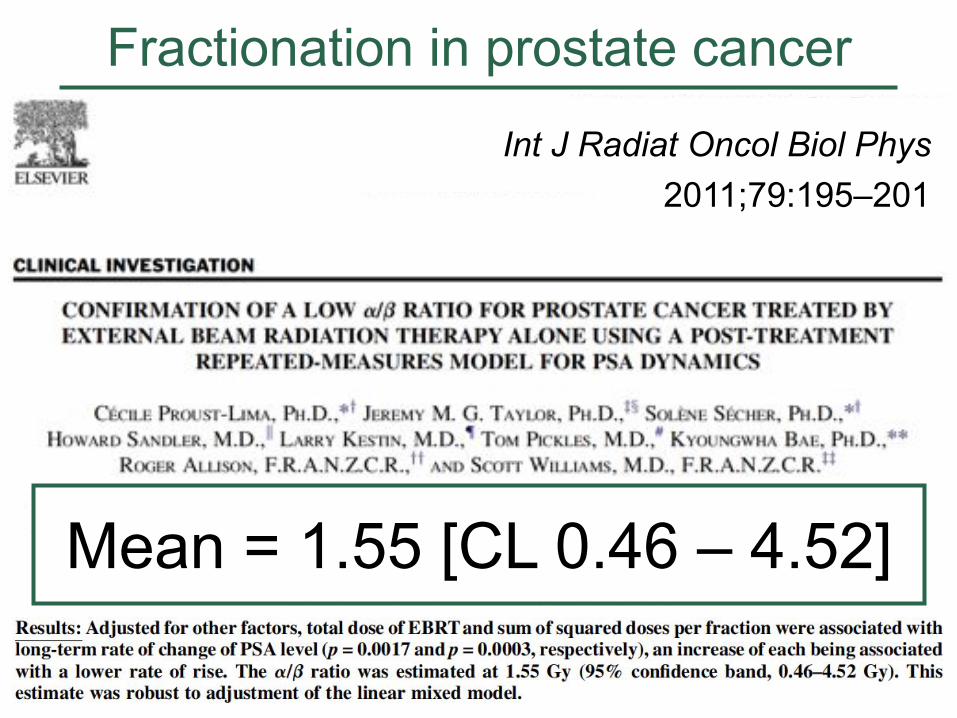
Fractionation in prostate cancer
Mean = 1.55 [CL 0.46 – 4.52]
Int J Radiat Oncol Biol Phys 2011;79:195–201
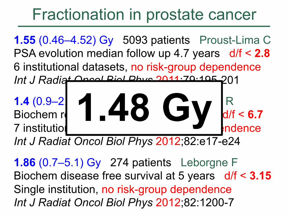
Fractionation in prostate cancer 1.55 (0.46–4.52) Gy 5093 patients Proust-Lima C PSA evolution median follow up 4.7 years d/f < 2.8 6 institutional datasets, no risk-group dependence Int J Radiat Oncol Biol Phys 2011;79:195-201
1.4 (0.9–2.2) Gy 5969 patients Miralbell R Biochem relapse free survival at 5 years d/f < 6.7 7 institutional datasets, no risk-group dependence Int J Radiat Oncol Biol Phys 2012;82:e17-e24
1.86 (0.7–5.1) Gy 274 patients Leborgne F Biochem disease free survival at 5 years d/f < 3.15 Single institution, no risk-group dependence Int J Radiat Oncol Biol Phys 2012;82:1200-7
1.48 Gy
MCJ Aug 12 42
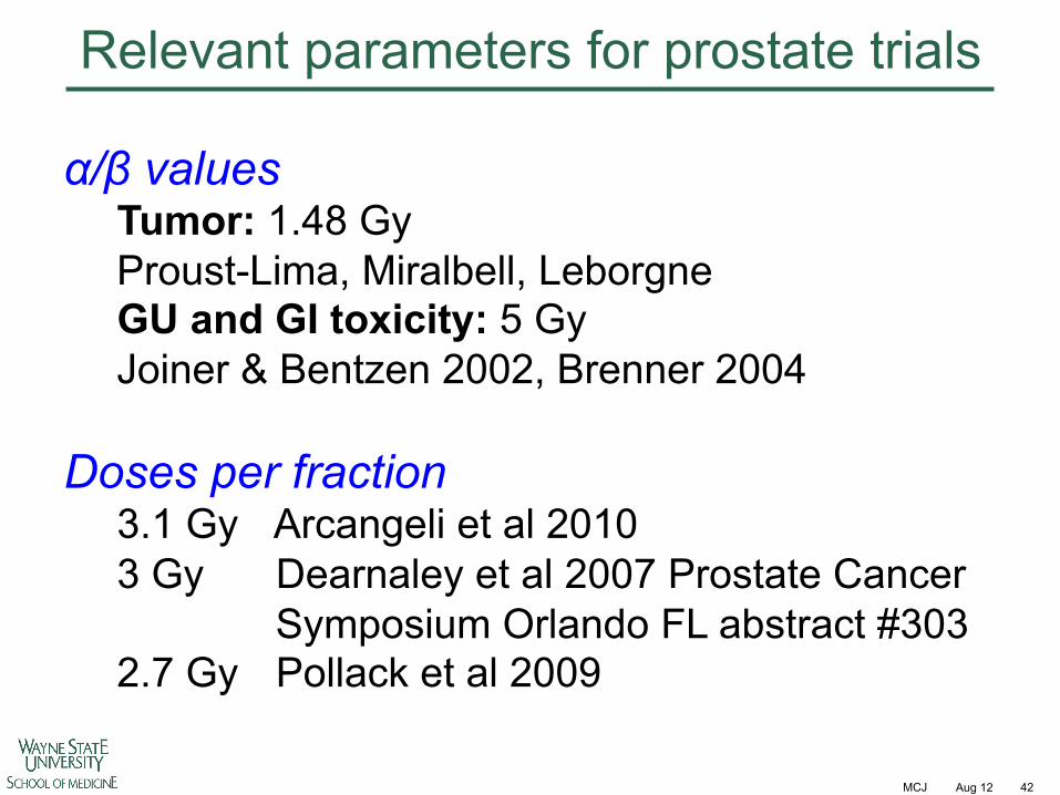
Relevant parameters for prostate trials
!/" values Tumor: 1.48 Gy Proust-Lima, Miralbell, Leborgne GU and GI toxicity: 5 Gy Joiner & Bentzen 2002, Brenner 2004
Doses per fraction
3.1 Gy Arcangeli et al 2010 3 Gy Dearnaley et al 2007 Prostate Cancer
Symposium Orlando FL abstract #303 2.7 Gy Pollack et al 2009
MCJ Aug 12 43
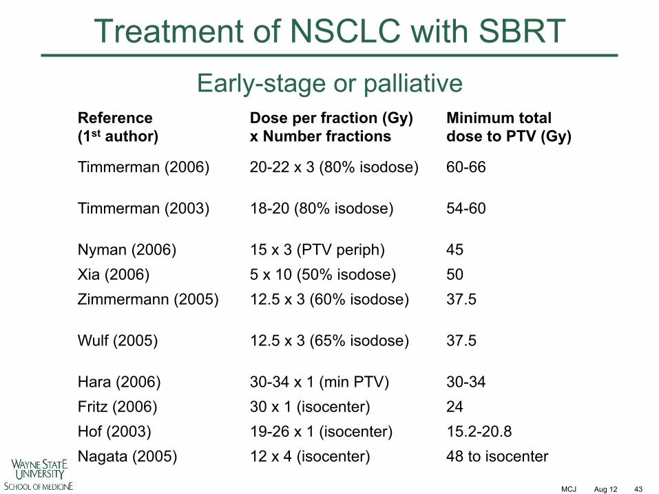
Treatment of NSCLC with SBRT
Reference (1st author)
Dose per fraction (Gy) x Number fractions
Minimum total dose to PTV (Gy)
Timmerman (2006) 20-22 x 3 (80% isodose) 60-66
Timmerman (2003) 18-20 (80% isodose) 54-60
Nyman (2006) 15 x 3 (PTV periph) 45 Xia (2006) 5 x 10 (50% isodose) 50 Zimmermann (2005) 12.5 x 3 (60% isodose) 37.5
Wulf (2005) 12.5 x 3 (65% isodose) 37.5
Hara (2006) 30-34 x 1 (min PTV) 30-34 Fritz (2006) 30 x 1 (isocenter) 24 Hof (2003) 19-26 x 1 (isocenter) 15.2-20.8 Nagata (2005) 12 x 4 (isocenter) 48 to isocenter
Early-stage or palliative
MCJ Aug 12 44
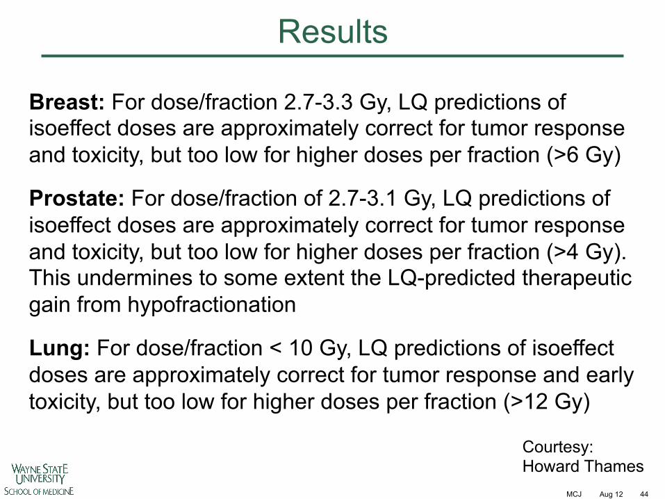
Results
Breast: For dose/fraction 2.7-3.3 Gy, LQ predictions of isoeffect doses are approximately correct for tumor response and toxicity, but too low for higher doses per fraction (>6 Gy)
Prostate: For dose/fraction of 2.7-3.1 Gy, LQ predictions of isoeffect doses are approximately correct for tumor response and toxicity, but too low for higher doses per fraction (>4 Gy). This undermines to some extent the LQ-predicted therapeutic gain from hypofractionation
Lung: For dose/fraction < 10 Gy, LQ predictions of isoeffect doses are approximately correct for tumor response and early toxicity, but too low for higher doses per fraction (>12 Gy)
Courtesy: Howard Thames
MCJ Aug 12 45
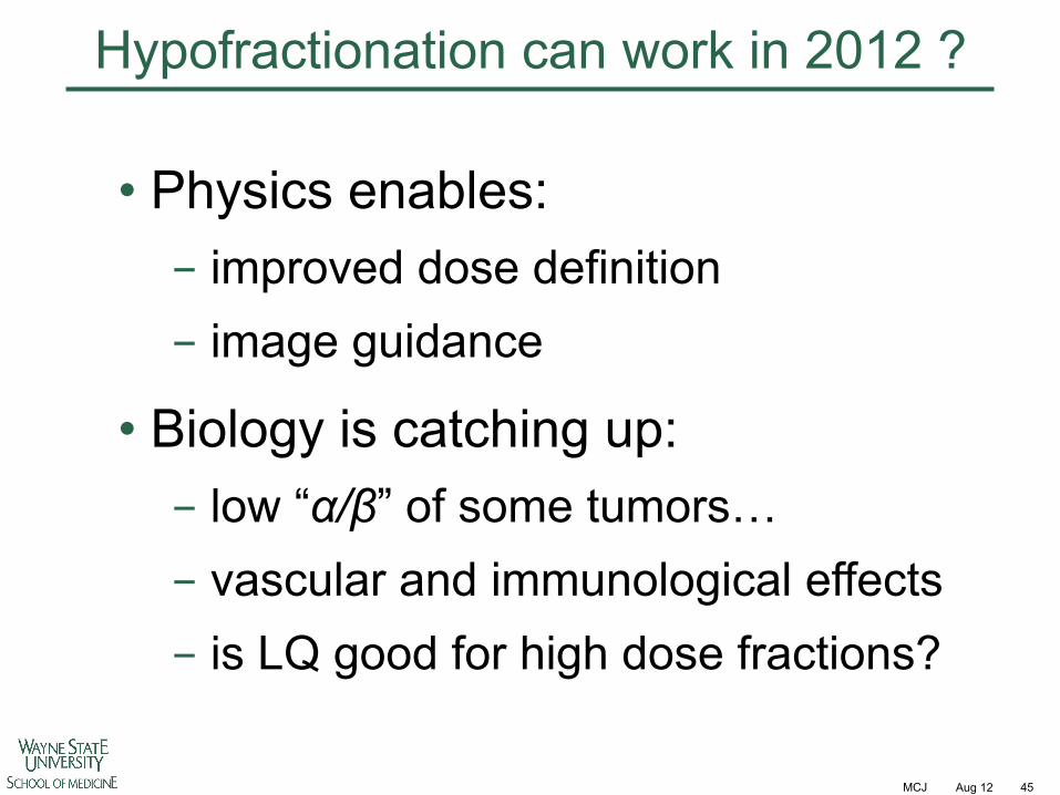
Hypofractionation can work in 2012 ?
• Physics enables: - improved dose definition - image guidance
• Biology is catching up: - low “!/"” of some tumors… - vascular and immunological effects - is LQ good for high dose fractions?