2: Ultrasound imaging and x-rays - Moodle 2017-2018 · 2: Ultrasound imaging and x-rays 1. How does...
7
Fund BioImag 2019 2-1 2: Ultrasound imaging and x-rays 1. How does ultrasound imaging work ? 2. What is ionizing electromagnetic radiation ? Definition of ionizing radiation 3. How are x-rays produced ? Bremsstrahlung Auger electron After this course you 1. understand the basic principle of ultrasound imaging 2. Are able to estimate the influence of frequency on resolution and penetration. 3. are capable of calculating echo amplitudes based on acoustic impedance; 4. know which parts of the electromagnetic spectrum are used in bio-imaging 5. know the definition of ionizing radiation; 6. understand the principle of generation of ionizing radiation and control of energy and intensity of x-ray production; Fund BioImag 2019 2-3 2 Reflection (echo formation) is key to imaging 2-1. What are the main fates of US waves in matter ? Sound wave travels through the substance but loses energy I(x) 1. Attenuation 2. Refraction 3. Scatter Sound wave dispersed in all directions 4. Reflection Sound wave bounces back to probe kxf e I x I D 0 ) ( Attenuation coefficient D [dB/(cm Mhz)] D is usually given in dB: dB=10logI(x)/I 0 [3dB=2fold increase in I(x): 10 0.3 =2 Unit conversion: k=ln10/10] Typically D~0.5dB/(cm MHz) ĺ 0+] VLJQDO ZLOO ORVH G% SHU FP RI WUDYHO (2 fold loss in wave energy) Material D [dB/cm MHz] Water 0.002 Blood 0.2 Tissue 0.7 Bone 15 Lung 40 Sound wave bends as it hits an interface at an oblique angle
Transcript of 2: Ultrasound imaging and x-rays - Moodle 2017-2018 · 2: Ultrasound imaging and x-rays 1. How does...
Fund BioImag 20192-1
2: Ultrasound imaging and x-rays
1. How does ultrasound imaging work ?2. What is ionizing electromagnetic radiation ?
Definition of ionizing radiation
3. How are x-rays produced ?BremsstrahlungAuger electron
After this course you1. understand the basic principle of ultrasound imaging2. Are able to estimate the influence of frequency on resolution and penetration.3. are capable of calculating echo amplitudes based on acoustic impedance;4. know which parts of the electromagnetic spectrum are used in bio-imaging5. know the definition of ionizing radiation;6. understand the principle of generation of ionizing radiation and control of
energy and intensity of x-ray production;
Fund BioImag 20192-32
Reflection (echo formation) is key to imaging
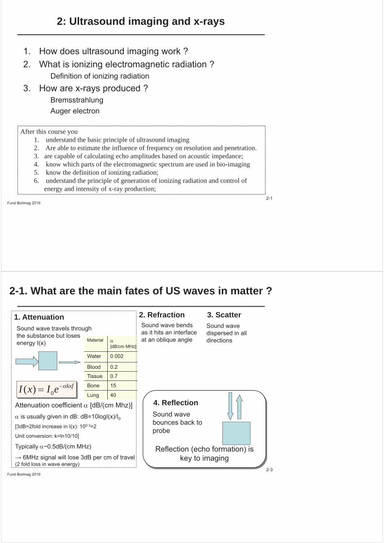
2-1. What are the main fates of US waves in matter ?
Sound wave travels through the substance but loses energy I(x)
1. Attenuation 2. Refraction 3. ScatterSound wave dispersed in all directions
4. ReflectionSound wave bounces back to probe
kxfeIxI 0)(Attenuation coefficient [dB/(cm Mhz)]
is usually given in dB: dB=10logI(x)/I0[3dB=2fold increase in I(x): 100.3=2
Unit conversion: k=ln10/10]
Typically ~0.5dB/(cm MHz)
(2 fold loss in wave energy)
Material [dB/cm MHz]
Water 0.002
Blood 0.2
Tissue 0.7
Bone 15
Lung 40
Sound wave bends as it hits an interface at an oblique angle
Fund BioImag 20192-4
What is the basic principle of US imaging ?
UItrasound: frequency f=1-20MHz (not 20kHz)
Sound wave propagation velocity c [c= f]~330m/s (air) = 0.33 mm/μs~1.45-1.6 mm/μs (tissue) (1cm~7μs)
(increases with density bone ~ 4 mm/μs)
The basic principle of imaging using sound waves :1. Emit sound pulse
(length [1-5 μs] is a multiple of cycle time 1/f)2. Measure time and intensity of echo3. Reconstruct using known wave propagation velocity c
Distance of tissue boundary from probe (transducer)
Distance=speed x time/2
transducer
Fund BioImag 20192-5
What determines the resolution in US imaging ?
Pulse duration t = N/f
Wavelength determines minimal resolution
1. To have defined frequency:Pulse length = N/f
2. Separation of return echoes, e.g.T > 2 pulse length
1. Resolution
increases with f
2. Penetration (cf. attenuation)
decreases with f
min. echo separation, e.g., T 2 t
T1=2x1/c
T2=2x2/cT=T1-T2=2 x/c
Overlap:No Gap,No separate echoes
Gap: Separate echoes
x
Fund BioImag 20192-6
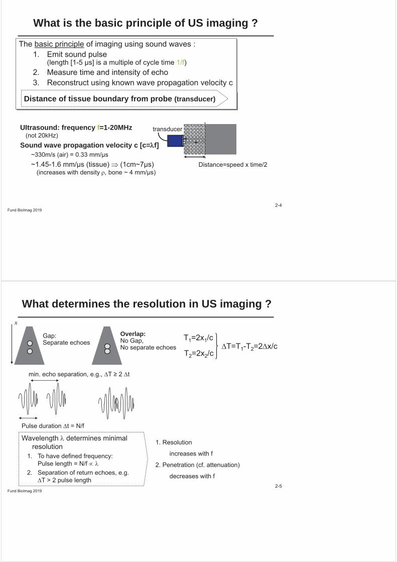
When does an acoustic echo occur ? Acoustic impedance and reflection ratio
Definition: Z= c [kg/m2s=rayls]
Amount of reflected wave energy Iref=I0RI
At interface between objects with different acoustical properties
Acoustic impedance Z
2
21
21
ZZZZRI
Z1 Z2
Reflection coefficient Transmission II RT 1
Probability of reflection + transmission is = 1:
2-7
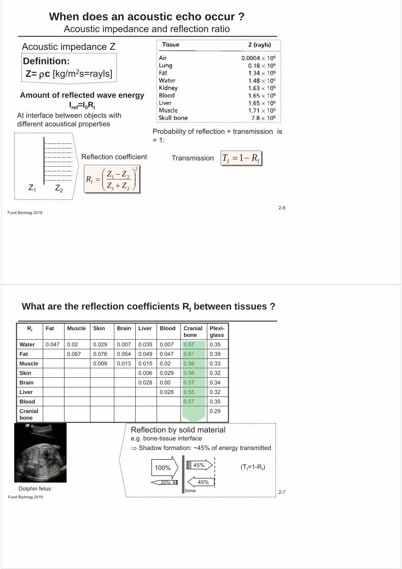
What are the reflection coefficients RI between tissues ?
RI Fat Muscle Skin Brain Liver Blood Cranial bone
Plexi-glass
Water 0.047 0.02 0.029 0.007 0.035 0.007 0.57 0.35
Fat 0.067 0.076 0.054 0.049 0.047 0.61 0.39
Muscle 0.009 0.013 0.015 0.02 0.56 0.33
Skin 0.006 0.029 0.56 0.32
Brain 0.028 0.00 0.57 0.34
Liver 0.028 0.55 0.32
Blood 0.57 0.35
Cranial bone
0.29
0.57
0.61
0.56
0.56
0.57
0.55
0.57
Reflection by solid material e.g. bone-tissue interface
Shadow formation: ~45% of energy transmitted
Dolphin fetusFund BioImag 2019
100% 45%
45%20%
bone
(TI=1-RI)
Fund BioImag 20192-8
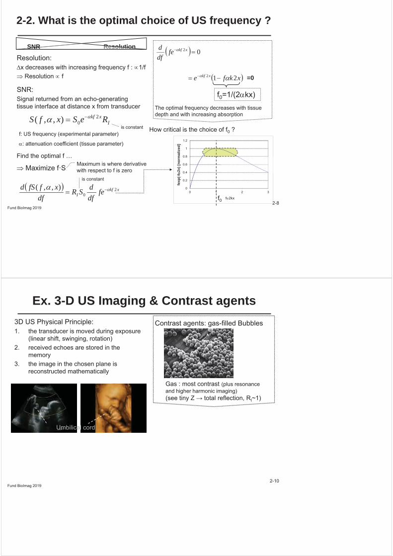
2-2. What is the optimal choice of US frequency ?
SNR:Signal returned from an echo-generating tissue interface at distance x from transducer
Ixkf ReSxfS 2
0),,(
xkfI fe
dfdSR
dfxffSd 2
0),,(
Maximum is where derivative with respect to f is zero
02xkffedfd
xkfe xkf 212
f0=1/(2 kx)The optimal frequency decreases with tissue depth and with increasing absorption
0
0.2
0.4
0.6
0.8
1
1.2
0 1 2 3
fexp
(-f2x
) [no
rmal
ized
]
f 2kx
How critical is the choice of f0 ?
f0
=0
f: US frequency (experimental parameter)
: attenuation coefficient (tissue parameter)
Resolution Resolution SNR
Resolution:x decreases with increasing frequency f : 1/f
Resolution f
Find the optimal f …
Maximize f·S
is constant
is constant
Fund BioImag 20192-10
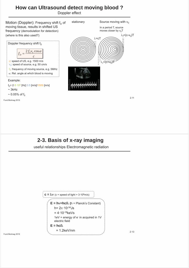
Ex. 3-D US Imaging & Contrast agents3D US Physical Principle:1. the transducer is moved during exposure
(linear shift, swinging, rotation)2. received echoes are stored in the
memory3. the image in the chosen plane is
reconstructed mathematically
Contrast agents: gas-filled Bubbles
Gas : most contrast (plus resonance and higher harmonic imaging)
I~1)
Umbilical cord
2-11
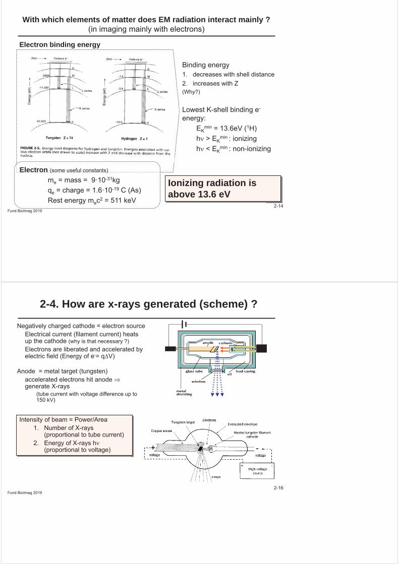
How can Ultrasound detect moving blood ?Doppler effect
Motion (Doppler): Frequency shift fD of moving tissue, results in shifted US frequency (demodulation for detection)(where is this also used?)
cvffD
cos2 00
f=(c-v0)T
r=(c+v0)T
=cT
v0: speed of source, e.g. 50 cm/s
f0: frequency of moving source, e.g. 5MHz
Doppler frequency shift fD
Example: fD= 2·5·106 [Hz] 0.5 [m/s]/1500 [m/s]
~ 3kHz ~ 0.05% of f0
Fund BioImag 2019
c: speed of US, e.g. 1500 m/s
In a period T, source moves closer by v0T
stationary Source moving with v0
: Rel. angle at which blood is moving
Fund BioImag 20192-13
2-3. Basis of x-ray imaginguseful relationships Electromagnetic radiation
c = 8m/s)
E = h =hc/ (h = Planck’s Constant)h= 2 ·10-34Js= 4·10-18keVs 1eV = energy of e- in acquired in 1V electric field
E = hc/= 1.2keV/nm
Fund BioImag 20192-14
With which elements of matter does EM radiation interact mainly ? (in imaging mainly with electrons)
Electron binding energy
Binding energy 1. decreases with shell distance2. increases with Z(Why?)
Lowest K-shell binding e-
energy:EK
min = 13.6eV (1H)h > EK
min : ionizingh < EK
min : non-ionizing
Ionizing radiation is above 13.6 eV
Electron (some useful constants)
me = mass = 9·10-31kg qe = charge = 1.6·10-19 C (As)Rest energy mec2 = 511 keV
Fund BioImag 20192-16
2-4. How are x-rays generated (scheme) ?
Negatively charged cathode = electron sourceElectrical current (filament current) heats up the cathode (why is that necessary ?)Electrons are liberated and accelerated by electric field (Energy of e-= q V)
Anode = metal target (tungsten)accelerated electrons hit anode generate X-rays
(tube current with voltage difference up to 150 kV)
Intensity of beam = Power/Area 1. Number of X-rays
(proportional to tube current)2. Energy of X-rays h
(proportional to voltage)

Fund BioImag 20192-17
Emission of x-rays I: What is Bremsstrahlung ?
Elastic scattering: Probability ~ Z2/Ee-
2
Inelastic scattering: releaseProbability ~ Z2
Max. Energy: Eei
Consider the interaction of e- with stationary atom as collision :
pi=pf+pphoton
pi
pfpphoton
Decreasing energy
Coulomb:a ~ qeZ/mer2 PBrems = qe
2a2/6 0c3
No info on directionality of radiation(but maximum energy is defined, how?)
High Z: Tungsten is a good target
Fund BioImag 20192-18
Emission of x-rays II: What are Characteristic (fluorescent) X-rays ?
Impacting e- liberates inner shell e-
1. Atom is excited (higher energy state)
2. Vacancy
3. Filled by outer shell electron (cascading)
4. Emission of characteristic x-ray
Auger emission
The excited atom can also reduce energy by liberating an additional e- (Auger e-):