Laporan Pendahuluan Dan Asuhan Keperawatan CA Cervix Dan CA Endometrium
Upload
virgil-oconnorCategory
view
215download
3


1.Uterus..2.Falopian tubes..
3.Ovaries..4.Cervix..5.Vagina..
6.Endometrium..


The basic workup to evaluate male factors :
Is semen analysis
Semen sample should be collected after 48 h of abstinence and examined within 2 to 3 h of collection. .

volume 2 ml or more
pH7,2-8,0
sperm conc.20x106 sperm/ml or more
total sperm count40x106 sperm per ejaculate or more
motility > 50% mobile sperm at 6 h
morphology30% or more with normal forms
vitality75% or more live,i.e.,excluding dye
white blood cellsfewer than 1x10⁶/ml
Normal values of semen variables: Standard Tests ..


What’s needed to Conceive?The right Time, The right Situation :
1- Hypothalamic- pituitary- gonadal axis intact.
2- Reproductive systems of both intact, physiologically & anatomically.
3- Juxtaposition of male & female gametes, at the ampulla.
4- At the optimal stage of maturation, normal preovulatory oocyte & normal enough sperms.
5- following, Transportation to the uterine cavity.
6- when the Endometrium is supportive to develop & implant.
7- Coital frequency is enough to let semen be deposited in close temporal relationship to release of oocyte.

*Fertility:The capacity to reproduce.
*Fecundability:
The probability of achieving a pregnancy each month. Rate : 0.22/mo
*Fecundity: The ability to achieve a live birth within one menstrual cycle. Rate : 0.15-0.18/mo

When to conceive?
90%18 months
75%9 months
60%6 months
25%1st month
80-90%
Of couples
1 yr
After that there’s low monthly conception rate without treatment

Probability of conception (fecundability) :
Factors influncing :
1.Age :Maximum at 24 y/o.Slight decrease : 24-30 y/o.Rapid decrease : 30+ y/o.
2.Frequency :4-5 times at week : maximum chance.
3.Age of man :Maximum fertility at 24 y/o.Slight decrease after 40 y/o.Spermatogenesis upto age 70.

The following ADVERSELY affect fertility:Menstrual FSH > 10.Oligospermia.No previous conception.


What’s infertility?
Sterility: absolute inability to conceive .
Infertility: inability of couple of reproductive age to achieve conception after 1 y of sexual intercourse without contraception .
1ry infertility: infertility that occurs without any prior pregnancies .
2ry infertility: infertility that occurs after previous conception .
Subfertility: relative state of decrease capacity to conceive .
70% of fertilization fails .
So, 10-15% of couples experience infertility
23% of them without treatment conceive within 2 years, 10% more conceive within 4 years
End of spectrum… Sterility


40% Multiple causes…
Causes
Female32%
Male18%
(30% undiagnosed)
Both18.5%
Idiopathic11.1%

MALE FACTORS
• 90% is related to sperm production and function•Coital factor is 40 %

• Pretesticular causes of infertility Congenital or acquired
- Hypothalamus No GnRH release> No Gonadotrpin>
hypogonadotropic hypogonadism. 1. Idiopathic hypogonadotropic hypogonadism:
may be due to defective migration of GnRH neurons> isolated or Kallmann syndrome
2. Prader-Willi syndrome3. Laurence-Moon-Biedl syndrome4. Other: as CNS tumors, temporal lobe seizures,
and many drugs (eg, dopamine antagonists)

Pituitary congenital or acquiredcaused by tumor (functional or nonfunctional)*, infarction,
radiation, infection, or granulomatous disease: 1. Prolactinoma: Adenoma> most common functional
pituitary tumor, gynecomastia and galactorrhea, bilateral hemanopia.
2. Isolated LH deficiency (fertile eunuch): eunuchoidal body habitus, large testis, and a low ejaculatory volume.
3. Isolated FSH deficiency: This is a very rare cause. oligospermia.
4. Thalassemia: Excess iron in the pituitary gland and testis
5. Cushing disease: negative feedback on the hypothalamus

Peripheral organs tumors or exogenous
1. Cortisol excess: by adrenal hyperplasia, adenomas, carcinoma, lung tumors or cushing syndrome> negative feedback on pitutary
2. Congenital adrenal hyperplasia (CAH)*: inefficient cortisol production with short stature, precocious puberty, small testis, and occasional bilateral testicular rests.
3. High Estrogen: due to Sertoli cell tumors, Leydig tumors, liver failure, or massive obesity> negative feedback on the pituitary.
4. Iatrogenic: High cortisol due to steroid therapy for ulcerative colitis, asthma, arthritis, or organ transplant> inhibition of GnRH release.

Primary testicular causes of infertility
1. Chromosomal abnormalities
1.Klinefelter syndrome*: most common, 1 per 500-1000 male births, Classically 47,XXY, most are azoospermic with 20% show presence of residual spermatogenesis, many of 2ry spermatocytes & spermatids with normal patterns.
Increased gonadotropin, while 60% have decreased testosterone 2.XX male (sex reversal syndrome)*: often short, small firm testis
and gynecomastia, normal-sized penis. Seminiferous tubules show sclerosis.

XYY male: 0.1-0.4% of newborn.often tall and severely oligospermic or azoospermic, aggression, maturation arrest or germ cell aplasia. Might have some normal functional sperm.
Noonan syndrome (46,XY): as male Turner syndrome, webbed neck, short stature, low-set ears, ptosis, shieldlike chest, lymphedema of hands and feet, cardiovascular abnormalities, and cubitus valgus. Leydig cell function is impaired.
Mixed gonadal dysgenesis (45,X/46,XY): ambiguous genitalia, a testis on one side, and a streaked gonad on the other.
Down syndrome: mild testicular dysfunction, varying
degrees of reduction in germ cell. LH and FSH are usually elevated.
Myotonic dystrophy: 75% testicular atrophy
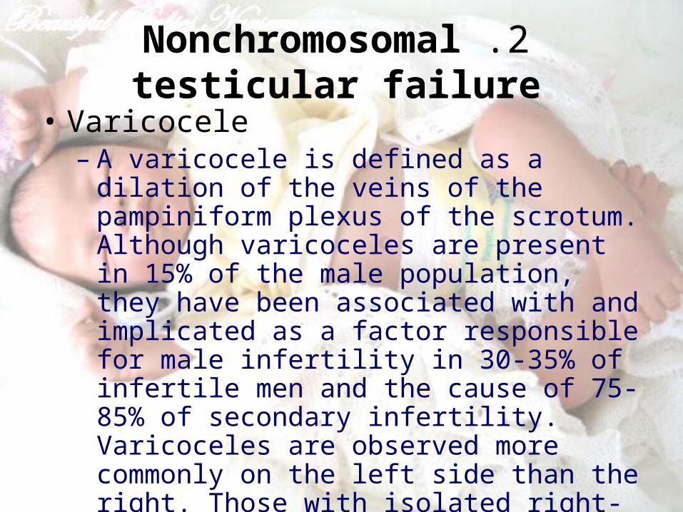
2 .Nonchromosomal testicular failure
• Varicocele– A varicocele is defined as a dilation of the
veins of the pampiniform plexus of the scrotum. Although varicoceles are present in 15% of the male population, they have been associated with and implicated as a factor responsible for male infertility in 30-35% of infertile men and the cause of 75-85% of secondary infertility. Varicoceles are observed more commonly on the left side than the right. Those with isolated right-sided varicoceles should be evaluated for retroperitoneal pathology.

• Cryptorchidism: • An estimated 3-4% of full-term males are born with an
undescended testicle; however, less than 1% remain undescended by the age of 1 year. Risks for cryptorchidism include family history and prematurity. It may be observed as part of syndromes such as prune belly syndrome. Patients have an increased risk of infertility. The higher and longer the testicle resides outside the scrotum, the greater the likelihood of damage to the seminiferous tubules.
• Trauma:• Testicular trauma is the second most common acquired
cause of infertility. The testes are at risk for both thermal and physical trauma because of their exposed position

Sertoli-cell-only syndrome (germinal cell aplasia): Patients with germinal cell aplasia have LH and testosterone levels within the reference range but have an increased FSH level. The etiology is unknown but is probably multifactorial. small- to normal-sized testes and azoospermia. Secondary sex characteristics are normal.
• Chemotherapy Such as cyclophosphamide
• Radiation therapy

• Orchitis The most common cause of acquired
testicular failure in adults is viral orchitis, usually caused by the mumps virus, echovirus, or group B arbovirus
• Granulomatous disease leprosy & sarcoidosis
• Sickle cell disease microinfarction & secondary testicular failure

• Posttesticular causes of infertility
• Posttesticular causes of infertility include problems with sperm transportation through the ductal system (congenital or acquired). it is observed in 7% of infertile patients. Additionally, the sperm may be unable to cross the cervical mucus or may have ultrastructural abnormalities.

• Congenital blockage of the ductal system: observed in children of mothers who were exposed to DES during pregnancy. called segmental dysplasia
• Cystic fibrosis: congenital bilateral absence of the vas deferens.
• Acquired blockage of the ductal system:• infections, such as chlamydia, gonorrhea,
tuberculosis, and smallpox. • Trauma, previous attempts at sperm aspiration, and
inguinal surgery may also result in ductal blockage. • Small calculi may block the ejaculatory ducts, or
prostatic cysts may extrinsically block the ducts. • Scrotal surgery

• Antisperm antibodies: Antisperm antibodies bind to sperm and impair motility.
• Immotile cilia syndrome: Patients experience sinusitis, bronchiectasis, and infertility.
• Retrograde ejaculation: This is caused by an open bladder neck during ejaculation. Retrograde ejaculation may be due to causes such as diabetes, bladder neck surgery, alpha-antagonists, transurethral prostatectomy )TURP(, colon or rectal surgery, multiple sclerosis, or spinal cord injury.

Other Factors
A. Environmental & Occupational1. toxins like: tobacco & marijuana,
heroin, cocaine, and crack cocaine, alchol, radiation, exposure to lead, other heavy metals, and pesticides
2. Social trends IUD, sexual revolution & salpingitis!!
3. Psychological effects stresses have No significant effect on fertility

B. Age decreases coital frequency & affects fertility,
For females, in 1/3 of female between 35-45yrs old(Chromosomal abnormalities and poor oocyte quality, poor
embryonic quality, low implantation rate, increased miscarriage, and low birth rates)
For males, (Testosterone decrease, gonadotropin increase, sperm
concentration and semen volume change, libido decreases, and incidence of birth defects increases)
Despite this, male fertility is not as much affected as female… It only takes them longer…

C. Exercise
Compulsive exercise>excessive endorphins> interferes with FSH and LH production> ovulatory
disorders and LPD. In males, oligospermia.
D. Extreme weight loss or gain
hypothalamic-pituitary-ovarian axis
*Anorexia nervosa or bulimia> hypothalamic amenorrhea, *Weight gain> a low FSH level, and low LH secretion> tolerated better
F. Systemic diseases
e.g. *CRF, SCAAffecting both hormones & gonadal tissue

Female Causes
Ovulatory20-25%
Cervical5-10%
Uterine-Tubal30%
Peritoneal40%
Pelvic Immunologic

1. Ovulation Factors
Ovulatory dysfunction:
alteration in the frequency and duration of the menstrual cycle.
Pathophysiology:
The most common problem is the absence of ovulation (irregular or delayed)
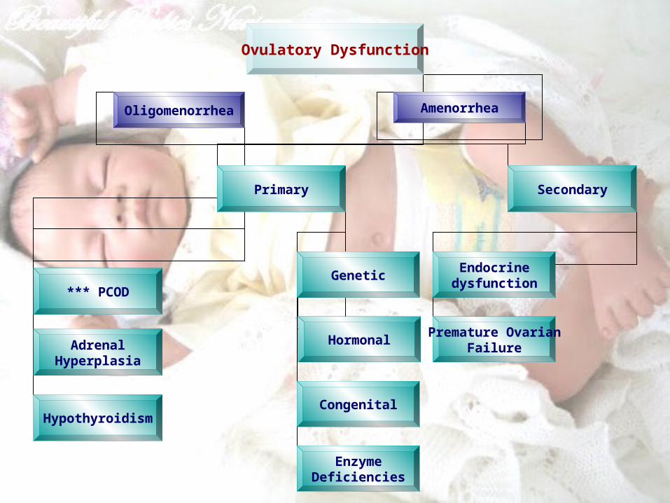
Ovulatory Dysfunction
Oligomenorrhea Amenorrhea
PCOD***
AdrenalHyperplasia
Hypothyroidism
Primary Secondary
Genetic
Hormonal
Congenital
EnzymeDeficiencies
Endocrinedysfunction
Premature OvarianFailure

Classification of premature ovarian failure Ovarian follicle depletion
Pure gonadal dysgenesis Idiopathic Congenital ovarian torsion Turner syndrome Fragile X chromosome Galactosemia Autoimmune Viral oophoritis
Ovarian follicle dysfunction 17,20-desmolase deficiency Gonadotropin-receptor blocking immunoglobulins Antibodies to gonadotropins
Idiopathic - Resistant ovary syndrome

Endometrial & Ovulation factors
LPD (luteal phase dysfunction) -Ovulatory dysfunction due to,
At time of ovulation, follicle size is small < 23-24mm in diameter
Preovulatory LH surge is rather blunt, high enough to induce resumption of myosis of the oocyte but not to induce follicular rupture and normal corpus luteum function
Decreased sensitivity of progestrone receptors at endometrial level
(Soules, 1989- Saracoglu, 1985.)

2.Tubal Factors
A. Congenital: - absence of the fallopian tube(s) can be due to spontaneous torsion in utero followed by necrosis and reabsorption.
B. Acquired: - salpingitis: chlamydia, gonorrhea
- Elective tubal ligation and salpingectomy

3.Uterine Factors:
A. Congenital: -Müllerian duct abnormalities: 1. total absence of the uterus and vagina (ie, Rokitansky-Küster-Hauser syndrome) 2. minor defects such as arcuate uterus, blind horn and vaginal septa (transverse or longitudinal). .

B. Acquired: Endometritis: due to a traumatic delivery, D&C, IUD, or
any instrumentation (eg, myomectomy, hysteroscopy) of the endometrial cavity intrauterine adhesions or synechiae (ie, Asherman syndrome), partial or total obliteration of the endometrial cavity.
Placental polyps: from placental remains.
Intramural and submucous fibroids:
1. distortion of the cavity; compromise the blood supply; a lack of embryo implantation,
2. Early miscarriages, premature delivery, and abruptio placentae.

4.Cervical Factors
-1- Bacterial infection (cervicitis)
- 2- Autoimmune antibodies
- 3- previous surgery for abnormal pap smears and
- 4- Congenital, narrow cervical opening.

5.Pelvic or Peritoneal Factors
Inflammation, adhesions encapsulating ovaries, reducing tubal motility, blockage of cul-de-sac result from:
1. abdominal or pelvic surgery,
2. appendicitis,
3. pelvic infections such as STDs, leading to pelvic inflammatory disease (PID): mostly Chlamydia, gonorrhea

4. Endometriosis*: intrauterine tissue grows outside of the uterus> pelvic pain & reproductive failure or incidental finding
*The incidence of endometriosis in primary infertility is 26%, and secondary infertility 13%
*7 folds increased risk of endometriosis with family history
Mechanism:*Severe endometriosis mechanical or adhesions
*mild to moderate “immune abnormalities & embryo toxic serum”

5. Adenexal mass
6. Ovarian cyst rupture
7. Large myoma or pelvic masses

6.Immunologic Factors
loss of eggs, early menopause, spontaneous abortion, Abs attacking the semen
-by affecting the ovulation factor
-Autoimmune conditions of ovaries or thyroid
-Antiphospholipid syndrome


History
1. Evaluation should be done for both partner.
2. Personal data.
3. Chief complain.
4. Past obstetric history.

5. Gynecological Hx. (female)
Menstruation: age of menarche lmp
regularity duration
Contraceptive: type – duration
Sexual: STD - post coital bleeding
vaginal discharge
superficial or deep dyspareunia
frequent of intercourse
use of (K-Y)gel

6. Systemic review:( female) W. change – hirestism – acnes – frontal
bolding.( male ) impotence – premature ejaculation change in libido – testicular truma sign of infection or swelling

• You should ask according to factor discussed before.
• Ask about previous marriage with offspring.
7. Past medical Hx.
-medical -surgical – blood transfusion – allergies –
8. Family Hx.
9. Social Hx.


Examination
1. Vital sign.2. Height – weight – BMI3. General: hand - eye – face – neck4. Breast : 5. Abdomen.6. Gynecological. 7. PV8. Bimanual.9. Extremities.


• usually if the age of female above 35yrs or there's history of gynoclogical abnormality, it is better to start investigation earlier.
• However, a reasonable compromise is to prolong somewhat the testing & treatment to the 2nd yr of infertility.
• Start with the least invasive )for the first 6-8 months( like Blood work for hormones, female & male as indicated US, Semen analysis, Hystrosalpingiogram that sometimes maybe even therupeutic.
• Laparsopy should be the last resort in a female only needed in 5-10% after 18-24 mo!

What to investigate?
MaleI. Lab Studies
1.Semen analysis :A specimen is collected by a clean, dry, sterile container or
during coitus using special condoms must be evaluated to assess for variations in sperm number and quality. A variety of parameters is measured, such as ejaculate volume and sperm density, quality, motility, and morphology.

AnalysisFindingConclusion
Ejaculate volume
Low 1,5 mlPostejaculation urine )retrograde ejaculation(
TRUS )absence of vas deferens(Hormonal evaluation )hypogonadism
High5 mlLikely contaminant
Semen quality
Dos not coagulateTRUS )ejaculatory duct obstruction(
Dose not liquifyHormonal analysis
Sperm density
Oligospermia (<20 million per mL)
Severe oligospermia (<5 million per mL)
TRUS )partial ejaculatory duct obstruction(
Antisperm antibody evaluationHormonal analysis
Physical examination for varicocele
Azoospermia Sperm centrifuged to verify azoospermia
Postejaculation urine )retrograde ejaculation(
Hormonal evaluationTesticular biopsy )testicular failure(TRUS )ejaculatory duct obstruction(
motilityDecreaseAntisperm antibodiesPhysical examination for varicocele

Computer-aided semen analysis )CASA(:uses a video camera and computer to visualize and
analyze sperm concentration and movement.
Infection:An increased number of round cells are often observed in
patients with infectious or inflammatory processes.
Other tests:Semen may be analyzed for levels of zinc, citric acid, acid
phosphates, and alpha-glucosidase. These tests are used to determine gland failure or obstruction.

2.Antisperm antibody test :Antisperm antibodies may form when sperm are exposed to
the body's defense outside of the blood-testis barrier. These antibodies bind to sperm and may lead to infertility due to a decreased ability to penetrate the cervical mucus, premature acrosome reaction, and decreased ability to bind to the zona pellucida. Known causes include ductal obstruction, infection, testicular torsion, cryptorchidism, testicular or spermatic cord trauma, or varicocele.

Several methods are available to detect antisperm antibodies:
1. radioimmunoassay (RIA) .
2. enzyme-linked immunosorbent assay (ELISA).
3. but the most specific test is the immunobead test.

3. Hormonal analysis :Fewer than 3% of cases of male infertility are estimated
to be due primarily to a hormonal cause.
A routine part of the initial evaluation is testing of specific serum hormone levels, which usually includes FSH, LH, testosterone, and prolactin.
These 4 hormones are closely related and have an impact on sperm production.
Abnormalities may be a sign of a primary hypothalamic, pituitary, or testicular problem.

II. Imaging Studies:
• Transrectal ultrasound
• Scrotal ultrasound
• Vasogram

• Postcoital test
III. Other Tests

femaleA complete evaluation of the female
reproductive tract must include:• cervical.• uterine.• endometrial• tubal• peritoneal• ovarian factors.

Ovarian factors
1-Basal Body Temoerature (BBT): assess the duration luteal function, done on awakening
before any activity.
The test must be done for at least 3 months.
Normal BBT: is biphasic, there should be raise of (0.4‘F) at ovulation last for at least 11 days.
A monophasic BBT is associated with anovulatory cycles, although this is not a reliable finding in 20% of patients.

2. Serum Progesterone:• Normally >5 ng/ml indicates activity• In the cycles appropriate for conception. Mid luteal conc.
Usually exceed 10 ng//ml• In PCOD it might be below 2 ng/ml
3. In patients with PCOD, the gonadotropin assay reveals a normal to slightly low FSH concentration and a slight or very elevated LH level.
Prolactin, thyroid, and adrenal hormones can be elevated if a combination of glandular dysfunction (pluriglandular syndrome) is present.

4. Pelvic ultrasonography:
• PCOD: enlarged ovaries with multiple small (<10 mm) follicles at the periphery of the ovarian cortex.

5. Endometreal Biopsy:
1-3 days prior menses or 12-13 days after LH surge to avoid false postives.
• Taken from upper anterior aspect of utrine fundus histological development must be dated. Retrospectively, it's realted to the onset of next menses: If more than 2 days lag in at least 2 cycles LPD.
• NB: Endometrial biopsy: finding that shows a proliferative pattern,
pelvic ultrasonography results that show absence of a follicle and/or a corpus luteum are better criteria for diagnosing lack of ovulation.

Cervical factors
• The cervical factor accounts for 5-10% of infertility.
• Cervical mucus production, amount, and characteristics change according to the estrogen concentration during the late follicular phase.
• PCT test

PCT test
• An abnormal PCT result may indicate • (1) poor timing,• (2) hostile cervical mucus related to cervicitis (eg, bacteria/yeast),• (3) low pH,• (4) sperm antibodies,• (5) poor technique during intercourse (eg, premature ejaculation), • (6) undiagnosed male factor infertility, • (7) anatomical defects (eg, due to DES exposure in utero, cervical
cone).
• An abnormal PCT result must be repeated in subsequent cycles before a final diagnosis is made. Note that an abnormal PCT result does not preclude the possibility of a spontaneous pregnancy; therefore, be cautious to avoid a premature final diagnosis.

Uterine factors
• Uterine factors can be congenital or acquired. They may affect the endometrium or the myometrium and are responsible for 2-5% of infertility cases

Endometrial factors
• Some defects can be detected during the pelvic examination. These include absence of the vagina and uterus, vaginal septum, and the presence of fibroids.
• Detection of most defects requires ancillary studies such as HSG, pelvic ultrasonography, hysterosonogram, and MRI.
• Operative procedures, such as laparoscopy and hysteroscopy, are often necessary for confirmation of the final diagnosis.

Hysterosalpingogram
The HSG is the most frequently used diagnostic tool to evaluate the endometrial cavity.
provides accurate information about the 1. endocervical canal2. diameter and configuration of the internal endometrial
cavity 3. uterine/tubal junction (cornual ostium)4. diameter, location, and direction of the fallopian tube5. status of the fimbriae6. pelvic adhesions and uterine, ovarian, or adnexal
masses May use as theaputic.

Ultrasonography
• Use to evaluation of the position of the uterus within the pelvis and provides more information about its size and irregularities.
• Pelvic sonograms also help in the early detection of uterine fibroids and endometrial polyps and help demonstrate the presence of ovarian cysts, adnexal masses, and endometriomas.
• Ultrasonography helps in the diagnosis of anovulation, polycystic ovaries, and persistent corpus luteum cysts.

Magnetic resonance imaging
• diagnostic tool, although it should be limited to those patients in whom a definitive diagnosis cannot be done by HSG, ultrasonography, and hysteroscopy findings.
• MRI is useful in the diagnosis of complex pelvic masses and for conditions as congenital malformation related to cryptomenorrhea and absence of the cervix .
Hysteroscopy
• Hysteroscopy is a method of direct visualization of the endometrial cavity. The technology has changed substantially and now uses optical devices, videocamera-enhanced images, and television monitors, which allow more efficient participation and coordination of other members of the operating room team.

Tubal factors
• The 2 most frequent tests used for diagnosis of tubal pathology are HSG and laparoscopy

Peritoneal factors
• Laparoscopy Identifies previouslly unsuspected pathological conditions in 30% to 50% of women with unexplained infertility.
• Endometriosis is the most common finding.


MANEGMENT
• Male1. Medical Care : Endocrinopathies Antisperm antibodies Retrograde ejaculation Semen processing Lifestyle Infections

2. Surgical Care: ((treat the cause))
Varicocelectomy .
Vasovasostomy or vasoepididymostomy.
Transurethral resection of the ejaculatory duct.
Sperm retrieval techniques.
Assisted reproduction techniques (ART ).
Intracytoplasmic sperm injection .

3. Consultations: Geneticist
Endocrinologist
4. Diet
5. Activity

• Female((treat the cause))
1. Treatment of cervical factors
2. Treatment of uterine factors
3. Treatment of endometrial factors
4. Treatment of tubal and peritoneal factors
5. Treatment of ovarian factors


Prognosis
• Aim of treatments is to increase the rate as well as the likelihood of conception.
• Without therapy, spontaneous conception occurs at decrease rate in infertile couples as time progress.
• 50% to 60 % of infertile couples will conceive with a through evaluation and application of current treatments short of in vitro fertilization(IVF), embryo transfer or gamete intrafallopian transfer(GIFT)