1980 GALLIFORMES, SYLVIORNITHIDAE HOLOCENE NEW … · 297; Articular, juv., NCP 219, 315; Angular,...
14
19 DESCRIPTION OF THE SKULL OF THE GENUS SYLVIORNIS POPLIN, 1980 (AVES, GALLIFORMES, SYLVIORNITHIDAE NEW FAMILY), A GIANT EXTINCT BIRD FROM THE HOLOCENE OF NEW CALEDONIA Cécile MOURER-CHAUVIRÉ & Jean Christophe BALOUET MOUREH-CHAUVIRÉ, C. & BALOUET, l.C, zoos. Description of the skull of the genus Syluiornis Poplin, 1980 (Aves, Galliformes, Sylviornithidae new family), a giant extinct bird from the Holocene of New Caledonia. In ALCOVER, J.A. & BaVER, P. (eds.): Proceedings of the International Symposium "Insular Vertebrate Evolution: the Palaeontological Approach': Monografies de la Societat d'Història Natural de les Balears, lZ: Z05-118. Resum El crani de Sylviornismostra una articulació crania rostral completament mòbil, amb dos còndils articulars situats sobre el rostrum, el qual s'insereix al crani en dues superfícies articulars allargades. La presència de dos procesos rostropterigoideus sobre el basisfenoide del rostrum i la forma dels palatins permet confirmar que aquest gènere pertany als Galliformes, però les característiques altament derivades del crani justifiquen el seu emplaçament a una nova família, extingida, Sylviornithidae. El crani de Syluiornis està extremadament eixamplat i dorsoventralment aplanat, mentre que el rostrum és massís, lateralment comprimit, dorsoventralment aixecat i mostra unes cristae tomiales molt fondes. El rostrum exhibeix un ornament ossi gran. La mandíbula mostra una símfisi molt allargada, les branques laterals també presenten unes cristae tomiales fondes, i la part posterior de la mandíbula és molt gruixada. Es discuteix el possible origen i l'alimentació de Syluiornis. Paraules clau: Aves, Galliformes, Extinció, Holocè, Nova Caledònia. Abstract The skull of Syluiornis shows a completely mobile craniorostral articulation, with two articular condyles situated on the rostrum, which insert into two elongated articular surfaces on the cranium. The presence of two rostropterygoíd processes on the basisphenoid rostrum and the shape of the palatines make it possible to confirm that this genus belongs to the Galliformes, but the highly derived characteristics of the skull justify its placement in a new, extinct family Sylviornithidae. The cranium of Syluiornis is extremely widened and dorsoventrally flattened, while the rostrum is massive, laterally com pressed, dorsoventrally raised and displays very sharp cristae tomiales. The rostrum bears a large bony ornament. The mandible shows a very elongated symphysis, the lateral branches also show sharp cristae tomiales, and the posterior part of the mandible is very thick. The possible diet of Syluiornis is discussed. Key words: Aves, Galliformes, Extinction, Holocene, New Caledonia. INTRODUCTION The genus Syluiomis was described by F. Poplin (1980) from some very fragmentary postcranial remains, gathered by J. M. Dubois in a fossiliferous well situated at Ure, in the Kanumera bay, on Isle of Pines, to the south-east of the main island of New Caledonia. These remains were attri buted to a ratite. More complete material was collected by F. Poplin and J. C. Balouet in 1980 and made it possible to show that this bird was in fact a galliform, which was attri buted to the recent family Megapodiidae (Poplin et al., 1983; Poplin & Mourer-Chauviré, 1985). Later, J. C. Balouet disco vered several other fossiliferous localities on the main island and found very numerous remains of Syluiornis in one of the Pindai caves, in particular a cranium and a rostrum. These elements of the skull of Sylviorniswere described by Balouet in his doctoral dissertation, which has remained unpublished (Balouet, 1984), and were illustrated with a short description in two later papers (Balouet, 1986; 1991). We think that it is necessary to publish on this material in more details. Our paper will be limited to the description of the cranial material. The species Syluiornis neocaledoniae Poplin, 1980 was described from the material of Isle of Pines. The material from the main island differs from that of Isle of Pines by the absence of a pygostyle, whereas the pygostyle is present in the type-population. It is likely that the form from the main island represents a second species, but it wilinot be described here. The Pindai caves are situated on the Nepoui peninsula, on the western coast of the main island, The Syluiotnis remains described here come from the main Pindai cave, the description of which is given by Balouet and Olson (1989). Charcoal associated with Syluiornis or other extinct bird bones have given a radiocarbon age of 1750 ± 50 BP (Gif 6341). Syluiornis is the most abundant bird in this locality. DESCRIPTION OF THE SKULL OF SYLVIORNIS ==================- 205
Transcript of 1980 GALLIFORMES, SYLVIORNITHIDAE HOLOCENE NEW … · 297; Articular, juv., NCP 219, 315; Angular,...

19DESCRIPTION OF THE SKULL OF THE GENUS SYLVIORNIS POPLIN,1980 (AVES, GALLIFORMES, SYLVIORNITHIDAE NEW FAMILY),A GIANT EXTINCT BIRD FROM THE HOLOCENE OF NEW CALEDONIA
Cécile MOURER-CHAUVIRÉ & Jean Christophe BALOUET
MOUREH-CHAUVIRÉ, C. & BALOUET, l.C, zoos. Description of the skull of the genus Syluiornis Poplin, 1980 (Aves, Galliformes,Sylviornithidae new family), a giant extinct bird from the Holocene of New Caledonia. In ALCOVER, J.A. & BaVER, P. (eds.): Proceedings ofthe International Symposium "Insular Vertebrate Evolution: the Palaeontological Approach': Monografies de la Societat d'HistòriaNatural de les Balears, lZ: Z05-118.
ResumEl crani de Sylviornismostra una articulació crania rostral completament mòbil, amb dos còndils articulars situats sobre
el rostrum, el qual s'insereix al crani en dues superfícies articulars allargades. La presència de dos procesos rostropterigoideussobre el basisfenoide del rostrum i la forma dels palatins permet confirmar que aquest gènere pertany als Galliformes, peròles característiques altament derivades del crani justifiquen el seu emplaçament a una nova família, extingida,Sylviornithidae. El crani de Syluiornis està extremadament eixamplat i dorsoventralment aplanat, mentre que el rostrum és
massís, lateralment comprimit, dorsoventralment aixecat i mostra unes cristae tomiales molt fondes. El rostrum exhibeix un
ornament ossi gran. La mandíbula mostra una símfisi molt allargada, les branques laterals també presenten unes cristaetomiales fondes, i la part posterior de la mandíbula és molt gruixada. Es discuteix el possible origen i l'alimentació de
Syluiornis.Paraules clau: Aves, Galliformes, Extinció, Holocè, Nova Caledònia.
AbstractThe skull of Syluiornis shows a completely mobile craniorostral articulation, with two articular condyles situated on the
rostrum, which insert into two elongated articular surfaces on the cranium. The presence of two rostropterygoíd processeson the basisphenoid rostrum and the shape of the palatines make it possible to confirm that this genus belongs to the
Galliformes, but the highly derived characteristics of the skull justify its placement in a new, extinct family Sylviornithidae.The cranium of Syluiornis is extremely widened and dorsoventrally flattened, while the rostrum is massive, laterally com
pressed, dorsoventrally raised and displays very sharp cristae tomiales. The rostrum bears a large bony ornament. Themandible shows a very elongated symphysis, the lateral branches also show sharp cristae tomiales, and the posterior part ofthe mandible is very thick. The possible diet of Syluiornis is discussed.
Key words: Aves, Galliformes, Extinction, Holocene, New Caledonia.
INTRODUCTION
The genus Syluiomis was described by F. Poplin (1980)from some very fragmentary postcranial remains, gatheredby J. M. Dubois in a fossiliferous well situated at Ure, in theKanumera bay, on Isle of Pines, to the south-east of themain island of New Caledonia. These remains were attributed to a ratite. More complete material was collected by F.
Poplin and J. C. Balouet in 1980 and made it possible to
show that this bird was in fact a galliform, which was attributed to the recent family Megapodiidae (Poplin et al., 1983;Poplin & Mourer-Chauviré, 1985). Later, J. C. Balouet discovered several other fossiliferous localities on the main islandand found very numerous remains of Syluiornis in one ofthe Pindai caves, in particular a cranium and a rostrum.
These elements of the skull of Sylviorniswere described
by Balouet in his doctoral dissertation, which has remained
unpublished (Balouet, 1984), and were illustrated with a
short description in two later papers (Balouet, 1986; 1991).We think that it is necessary to publish on this material inmore details. Our paper will be limited to the description ofthe cranial material. The species Syluiornis neocaledoniae
Poplin, 1980 was described from the material of Isle ofPines. The material from the main island differs from that ofIsle of Pines by the absence of a pygostyle, whereas the
pygostyle is present in the type-population. It is likely thatthe form from the main island represents a second species,but it wilinot be described here.
The Pindai caves are situated on the Nepoui peninsula,on the western coast of the main island, The Syluiotnisremains described here come from the main Pindai cave,the description of which is given by Balouet and Olson(1989). Charcoal associated with Syluiornis or other extinctbird bones have given a radiocarbon age of 1750 ± 50 BP (Gif6341). Syluiornis is the most abundant bird in this locality.
DESCRIPTION OF THE SKULL OF SYLVIORNIS ==================- 205

The 23 other bird species, studied by Balouet and Olson
(1989) include 7 extinct species. The rest of the faunaincludes very numerous Chiroptera (at least 4 species), a
new family, genus and species of primitive crocodile,Mekosuchidae (Balouet & Buffetaut, 1987), the horned tor
toise Meiolania (Gaffney et al., 1984) and a monitor lizard,Varanus (Balouet, 1984; 1991).
SYSTEMATIC STUDY
Order Galliformes
Family Sylviornithidae nov. fam.
Type-genus: Syluiornis Poplin, 1980, the only included
genus.Remark: The genus Syluiornis was placed in the recent
family Megapodiidae based mainly on characteristics of the
postcranial skeleton. The study of the skull shows that this
genus is highly derived. The characters that bring togetherSyluiornis and the Megapodiidae can be considered as plesiomorphic.
Family diagnosis: True mobile articulation betweenthe cranium and the rostrum, with two articular condyleson the rostrum, formed by the anterior part of the embryonic nasal bones, which are inserted into two articular sur
faces borne by the posterior parts of embryonic nasal
bones, later fused to frontals. Lacrimals fused to frontalsand forming a ventrally oriented lobe; this lobe bears a
small articular surface which comes into contact with a
tubercle situated on the rostrum. Cranium very flattenedand widened posteriorly, with a very strong development of
exoccipitals and squamosals. On the ventral surface, flexure of the posterior part of the cranium just posteriorly to
the basípterygoid processes; the basitemporal plate makesan angle of ca. 135 o relative to the plane of the basisphenoid rostrum. Occipital condyle situated almost in the cen
ter of the posterior surface of the cranium; its axis is parallel to the surface of the basisphenoid rostrum; occipitalforamen situated just ventral to the dorsal surface. Rostrum
very elevated and narrow and bearing a bony dorsal orna
ment; naris situated close to the posterior part in the adult;
rostrum ending anteriorly in a pointed hook, with cuttingtomial crests on the ventral surface. Quadratojugal with a
sigmoid shape in the adult; its anterior part articulates witha surface situated at mid-length of the posterior border of
the rostrum; posterior portion wide and paddle-shaped.Left and right palatines fused posteriorly in the adult.Vomer absent. Mandible with anterior part narrow and la
terally compressed; mandibular symphysis very long and
thick; posterior part very widened, with thick bone; longand thick retroarticular processes, not pointed posteriorlyand dorsally but rather with a rounded profile. These cha
racteristics are considered autapomorphic.Distribution: Holocene of New Caledonia.
Genus Sylviomis Poplin, 1980
Sylviornis cf. neocaledoniae Poplin, 1980
Description of the cranial material from the main
Pindai cave (the anatomical terminology generally follows
Weber, 1996).List of the material examined (this material is preserved
in the collection of the Paris Muséum national d'Histoire
naturelle):Cranium, adult, NCP 241; Cranium, juv., NCP 260 + 262;
Nasal, posterior part, juv., right, NCP 261; Frontals, juv., left,NCP 264-265; Squamosal, juv., left, NCP 262; Ethmoids, juv.,NCP 316-319; Quadrates, left, NCP 41-45, 222-240;Quadrates, right, NCP 46-64, 244, 268-270; Quadrates, juv.,NCP 272-276; Pterygoids, left, NCP 65-77, 245, 320-322, 325;Pterygoids, right, NCP 78-99; Pterygoids, juv., NCP 323-324;Palatines, right and left fused, NCP 121-136, 250, 277;Palatines, left, NCP 137-153; Palatines, right, NCP 154-168,251,278-279; Quadratojugals, left, complete, NCP 114-120;Quadratojugals, left, incomplete, NCP 169-177, 192-195,281-283; Quadratojugals, right, complete, NCP 106-113,246, 253-259; Quadratojugals, right, incomplete, NCP 178-
191; Quadratojugals, left, juv., NCP 203-210, 284;Quadratojugals, right, juv., NCP 198-202, 247-249, 285-286;Jugals, juv., NCP 211, 287-289; Quadrate + pterygoid +
quadratojugal, left, NCP 252, right, NCP 271; Rostrum,adult, NCP 242; Rostrum, juv., NCP 220, 263; Nasal, anterior
part, juv., NCP 221, 290-293; Mandible, symphyses and fragments of symphyses, NCP 213,215,243,294-295,299,310-312; Mandible, fragments of branches, NCP 216, 313;
Fig. 1. Sylviornis e]. neocaledoniae.Reconstruccio del crani, mostrant
la posició dels diferents elements,norma lateral esquerra.
naris
interorbital septum
processus postorbital is
Fig. 1. Syluiornis cf. neocaledoniae.Reconstruction of the skull, sho
wing the position of the differentelements, left lateral view.
premaxillary +
maxillary +
nasal
palatines
quadratojugal+ jugal
processuszygomaticus
quadrate
206 -�---- _, INSULAR VERTEBRATE EVOLUTION

Mandible, frarnents of articular parts, NCP 214, 217, 296-
297; Articular, juv., NCP 219, 315; Angular, juv., NCP 212;Supraangular, juv., NCP 218, 314; Articular + supraangular +
angular, right, juv., NCP 298.In addition, several fragments of mandible collected in
July 2003 byT. H. Worthy, A. Anderson, C. Sand, J. Jones, andR Pechey in the same cave, have been examined on photographs. This material is at present deposited in the Museumof New Zealand Te Papa Tongarewa, Wellington, but has not
been catalogued.A restoration of the different cranial elements is given
in figures 1 and 2.
Adult cranium (NCP 241, fig. 3)The skull of Syluiornis is prokinetic and characterized
by the presence of a true crania rostral hinge, with two partscompletely separated. According to Bock (1964, p. 4): "the
prokinetic skull is characterized by a hinge or region of
bending at the junction of the nasal and frontal bones;hence the entire jaw moves as a unit". However Bock indicates that this articulation does not necessarily correspondto the suture between the embryonic nasal and frontalbones. The juvenile cranium (NCP 260 + 262) shows that thefrontals are contiguous in the sagittal plane, but that their
junction leaves a wide triangular space between them at
their anterior end (fig. 4). This can also be seen on the juvenile cranium of other Galliformes, for example in the chicken (Jollie, 1957, fig. 2), or in a juvenile of the megapodeAlectura latharni (Weber, 1996, fig. 3). The posterior part ofthe nasal (NCP 261) inserts in this triangular space. The
juvenile material shows that the nasals consist of two different parts that articulated along a rectilinear hinge. The ante
rior part of the nasal is made up of two branches that laterfuse with the premaxillary and the maxillary. The posteriorpart of the nasal later fuses with the frontal. In the juvenilethe two parts of the nasal must have been held together byfibrous ligaments. From the juvenile elements it is possibleto state that the hinge between the cranium and the ros
trum was embryologically intranasal and not frontonasal.The features of the juvenile cranium also shows that a true
craniorostral articulation was already present in the earlystages of development of Syloiornis.
The adult cranium is extremely widened and flattened.This is largely due to the development of the exoccipitals indorsal and lateral directions. The crista nuchalis transversa
is rectilinear on the dorsal surface of the skull, then it con
tinues laterally, forming two wide, projecting occipital crests
at the level of the junction between the parietals and the
squamosals with the supraoccipital and the exoccipitals,and then continues on ventrally along the paroccipitalprocesses.
On the dorsal surface, the lacrimals are fused with theanterior part of the frontals. The fusion is still discernable bythe presence of a large foramen on the left side. In some
Galliformes, particularly in the Cracidae, the lacrimals are
extended by a long process that penetrates inside the orbital
cavity. In the Megapodiidae the lacrimals are very small,sometimes barely visible, and fused with the frontals andthe nasals at the level of the craniofacial flexion zone. Theydo not have an orbital process. In Syluiomis the orbital
process is also absent. The anterior part of the cranium, dorsal to the craniarostral hinge, is formed by the junction ofthe posterior part of the nasals with the ethmoid. This part
quadratojugal+ jugal
quadrate
Fig. 2. Syluiornis cf. neocaledoniae. Reconstruction of the skull showingthe position of the different elements, ventral view.
Fig. 2. Sylviornis cf. neocaledoniae. Reconstruccio del crani, mostrant la
posició dels diferents elements, norma ventral.
shows, in the sagittal plane, a median tubercle flanked bytwo depressions, indicating the presence of a synovial joint.On the posterior part of the supraorbital ridge, there is a
series of small foran1ina.The posterior surface is characterized by the very great
enlargment of the exoccipitals, which is continued by the
strongly ventrally projecting paroccipital processes. The
supraoccipital bears a smooth median ridge. The occipitalforamen is higher than wide and is situated just ventral to
the dorsal surface. The occipital condyle is situated almostat the center of the posterior surface, its dorsal surface is
slightly concave. On each side of the occipital condyle thereare two well-marked depressions containing foran1ina forblood-vessels and nerves.
The ventral surface is characterized by the presence oftwo processus rostropterygoideus (Weber, 1996). They are
oval in shape, slightly elevated relative to the surface of the
basisphenoid rostrum, oriented in parallel in an anteroposterior direction, and separated by a wide space, ca. 140 %wider than the adjacent width of the rostropterygoidprocesses. These processes are situated at the base of the
basisphenoid rostrum, which is triangular in shape, rela
tively short anteroposteriorly, and wide at its base. At theanterior end of the basisphenoid rostrum the interorbital
septum widens and fuses with the ethmoid, Usually in birdsthe ventral part of the cranium is a relatively flat surface andthe basitemporal plate (or basiparasphenoid plate in
Ericson, 1996) is a horizontal plane in the prolongation of
DESCRIPTION OF THE SKULL OF SYLVIORNIS..,.-----------.-------- 207
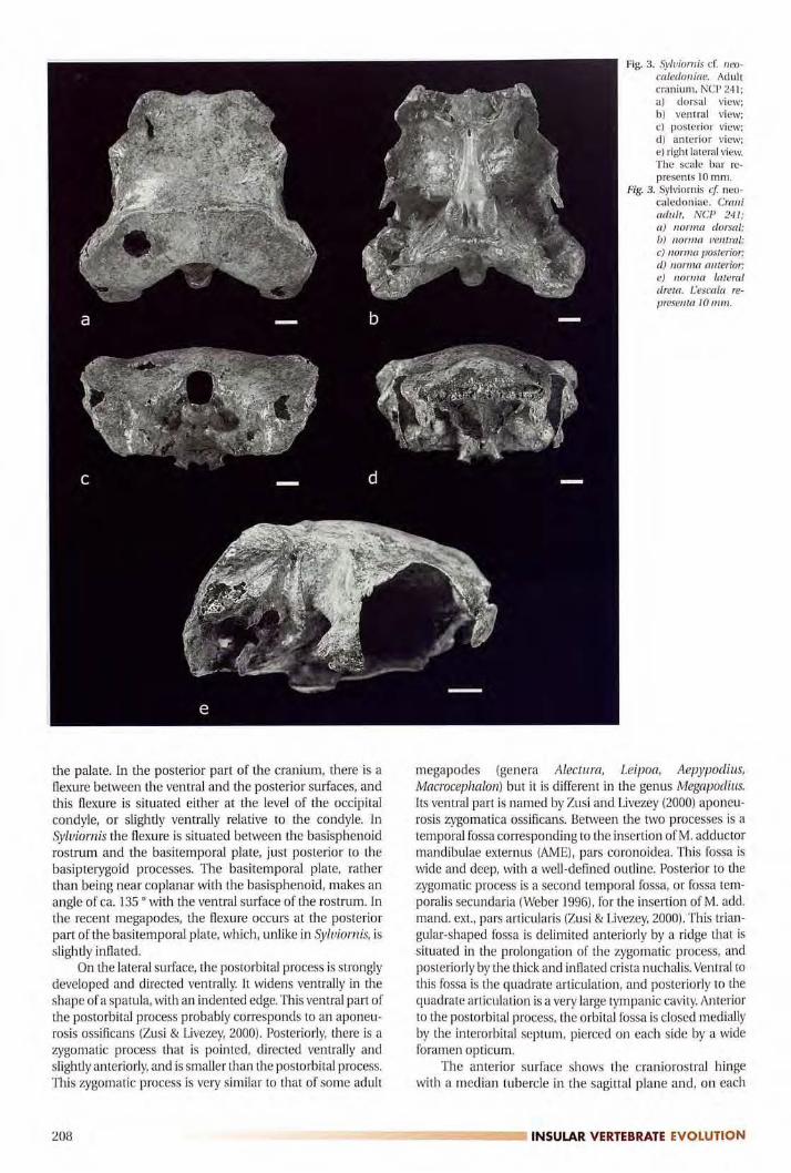
the palate. In the posterior part of the cranium, there is a
flexure between the ventral and the posterior surfaces, andthis flexure is situated either at the level of the occipitalcondyle, or slightly ventrally relative to the condyle. In
Syluiornis the flexure is situated between the basisphenoidrostrum and the basitemporal plate, just posterior to the
basipterygoid processes. The basitemporal plate, ratherthan being near coplanar with the basisphenoid, makes an
angle of ca. 135 o with the ventral surface of the rostrum. In
the recent megapodes, the flexure occurs at the posteriorpart of the basitemporal plate, which, unlike in Syiuiornis, is
slightly inflated.On the lateral surface, me postorbital process is strongly
developed and directed ventrally. It widens ventrally in me
shape of a spatula, with an indented edge. This ventral part ofme postorbital process probably corresponds to an aponeurosis ossificans (Zusi & Livezey, 2000). Posteriorly, mere is a
zygomatic process mat is pointed, directed ventrally and
slightly anteriorly, and is smaller man me postorbital process.This zygomatic process is very similar to that of some adult
Fig. 3. Syluiornis cf. neo
caledoniae. Adultcranium, NCP 241;al dorsal view;b) ventral view;el posterior view;dl anterior view;el right lateral view.The scale bar re
presents 10 mm.
Fig. 3. Sylviornis cf neo
caledoniae. Craniadult, NCP 241;a) norma dorsal;b) norma ventral;e) norma posterior;d) norma anterior;e) norma lateraldreta. L'escala re
presenta 10 mm.
megapodes (genera Alectura, Leipoa, Aepypodius,Macrocephaion) but it is different in me genus Megapodius.Its ventral part is named by Zusi and Livezey (2000) aponeurosis zygomatica ossificans. Between the two processes is a
temporal fossa corresponding to me insertion ofM. adductormandibulae externus (AME), pars coronoidea. This fossa is
wide and deep, with a well-defined outline. Posterior to me
zygomatic process is a second temporal fossa, or fossa tem
poralis secundaria (Weber 1996), for me insertion of M. add.mand. ext., pars articularis (Zusi & Livezey, 2000). This trian
gular-shaped fossa is delimited anteriorly by a ridge mat is
situated in me prolongation of the zygomatic process, and
posteriorly by the thick and inflated crista nuchalis. Ventral to
this fossa is me quadrate articulation, and posteriorly to the
quadrate articulation is a very large tympanic cavity. Anteriorto me postorbital process, me orbital fossa is closed mediallyby me interorbital septum, pierced on each side by a wideforamen opticum.
The anterior surface shows the craniorostral hingewith a median tubercle in the sagittal plane and, on each
208 ---- INSULAR VERTEBRATE EVOLUTION
side, two media-laterally oriented articular surfaces. Oneach side the lacrimals form anteriorly a lobe situated ven
trally relative to the hinge and ventrally oriented. On themedial side of the lacrimals there is a small, rounded, articular facet, which comes into contact with a process of the
rostrum, situated ventrally relative to the articular condylesof the hinge. Then, more ventrally, and medially situatedthere is the ethmoid, with a median ridge, and the two alaeethmoidales. On each side of the ethmoid there is a wideorbitonasal foramen, and ventral to the foramen a largearticular surface, oval in shape and slightly concave. On a
cast of the cranium it is possible to see that the right ala ethmoidale was extended in lateroventral direction by a thin,sharp process. This process was subsequently broken on
the specimen NCP 241.
Juvenile cranium (NCP 260, 261, 262, fig. 4)This juvenile cranium has been reconstructed from iso
lated unfused bones, based on matching sizes and shapes.As for the adult, its general shape is very wide and flat. The
supraorbital region is much narrower than in the adultbecause the lacrimals are not yet fused with the frontals.The crista nuchalis transversa is much less developed andthe cristae occipitales are less projecting in lateral and dorsal directions. The squamosal is anteroposteriorly elongated and does not show the enormous swelling of its posterior part that is seen in the adult.
Several juvenile ethmoids have been identified. Theyshow a wide, flat part at their anterior side, and a ventral keel.On each side of the keel is the sulcus olfactorius. Dorsally theanterior part shows articular surfaces which in the adult are
fused with the ventral surface of the nasals (fig. 5, g).
Rostrum (NCP 242, fig. 5, a-c))The adult rostrum NCP 242 does not fit with the crani
um NCP 241 and comes from a slightly larger individual.This rostrum is formed by the fusion of the anterior parts ofthe nasals, the premaxillaries, the maxillaries and the ma
xilla-palatines. It shows a true hinge, with two large articular
condyles, which project ca. 1 em from the lateral facies
inlmediately in front of them and which insert into thearticular surfaces of the posterior parts of the nasals. Its dorsal part bears in the adult a bony ornament, made of verythin cancellated bone, and is incompletely preserved. At theanterior part and at the base of this ornament, there is a
groove and a flattened surface. Ventral to the articular
condyle, there is a small process [3 mm by 3 mm] that articulates with the small articular surface situated on the ven
tral process of the lacrimal.The beak is extremely high and narrow. Its surface has a
shagreened aspect that indicates the presence, in adults, of a
thick ramphotheca. On the juvenile specimens the surface ofthe beak is rather smooth. The nostril is rounded and situated almost at the posterior part of the rostrum. The anteriormost part of the beak is a narrow point, ventrally directed.The external tomial crest forms a dorsally oriented sinus,then a ventrally oriented lobe. In ventral view one can see
two external tomial crests, two internal ones that continueonto the posteroventral angle of the beak, and one mediancrest. Anteriorly the adoral surface is formed by the ventralfusion of the premaxillaries. Posteriorly there is a secondarypalate formed by the ventral fusion of the maxillaries.
In posterior view, ventral to the craniarostral hinge,there are the two processes that articulate with the lacrimal
processes, then two large openings of the internal nares. On
Fig. 4. Syluiotnis cf. neocaledoniae. Reassembled juvenile cranium, Nep260 + 262; a) dorsal view (the posterior part of the right nasal Nep261 has been placed in the anterior opening of the frontals); b)ventral view without the nasal Nep 261. The arrows indicate thearticular surfaces for the quadrates. The scale bar represents 10mm.
Fig. 4. Sylviornis et neocaledoniae. Crani juvenil remuntat, NCP 260 +
262; a) norma dorsal (la part posterior del nasal dret NCP 261 s'hasituat a l'obertura anterior dels frontals); b) norma ventral sense elnasal NCP 261. Les fletxes indiquen les superficies articulats per als
quadrats. Eescala representa 10 mm.
DESCRIPTION OF THE SKULL OF SYLVIORNIS _m:::::===============:..: 209

Fig. 5. Sylviomis cf. neocaledoniae. Adult rostrum, Nep 242; a) right lateral view; b) posterior view (for a and b the thick arrow indicates thetubercle which corresponds to the articular surface found on theventral process of the lacrimal, and the thin arrow indicates theindentation for the articulation with the anterior part of the
quadratojugal); c) ventral view. Juvenile rostrum and left nasal,Nep 263, and right nasal, Nep 290; d) right lateral view (the thickarrow indicates the tubercle which later becomes the articular
condyle of the adult and the thin arrow indicates the tuberclewhich corresponds to the articular surface found on the ventral
process of the lacrimal); e) dorsal view. Right juvenile nasal, ante
ríorpart, Nep 221; f) medial view. Juvenile ethmoid, Nep 316; g)right lateral view (one can see the sulcus olfactorius). The scale bar
represents 10 mm.
each side there is a small indentation that corresponds to
the articulation of the anterior part of the quadratojugal.In the juvenile specimens, the anterior part of the
nasal is not yet fused with the premaxillary (NCP 263 + 290,fig. 5, d-e). Anteriorly it shows two branches, a dorsal one,which inserts into a groove situated on the posterior part ofthe premaxillary (NCP 263), and a maxillary one, which
probably rested against the maxillary (fig. 5, fl. Posteriorlyit presents, on each lateral side, a large tubercle that, in the
adult, becomes the articular condyle, and ventrally a verysmall point that bears the articular surface for the lacrimal
(fig. 5, d).
Fig.5. Sylviornis ct neocaledoniae. Rostre adult, NCP 242; a) norma lateral dreta; b) norma posterior (per a a i b lajlexta gruixada indica eltubercle que correspon a la superficie articular que es troba sobre el
procés ventral del lacrimal, i lafletxa prima indica la indentaci6 pera l'articulaci6 amb la part anterior del quadratojugal); c) norma
ventral. Rostre juvenil i nasal esquerre, NCP 263, i nasal dret, NCP290; d) norma lateral dreta (lafletxa gruixada indica el tubercle queposteriorment es transforma en el còndil articular de l'adult i la
fletxa prima indica el tubercle que correspon a la superficie articular que es troba sobre el procés ventral del lacrimal); e) norma dorsal. Nasal juvenil dret, part anterior. NCP 221; f) norma medial.Etmoidejuvenil, NCP316; g) norma lateral dreta (es pot veure el sulcus olfactorius). Eescala representa 10 mm.
Quadrate (fig. 6, e-f)The processus oticus ends dorsally in two articular
condyles, the squamosal and the prootic ones, which are
contiguous but distinct in the adults. The squamosalcondyle is about twice as large as the prootic one. On theanterior surface of the processus oticus, the eminentiaarticularis is a poorly delimited tubercle, projecting dorsally. The processus orbitalis, triangular in shape, is very elon
gated, slightly incurved medially, and ends in a roundedlobe. The articular surface for the quadratojugal is a deepsocket, surrounded by a thick rim. The mandibular articulation is made up of two obliquely elongated condyles,
210 -v-; ..=����,..,...._.�..".,..,.-� INSULAR VERTEBRATE EVOLUTION

Fig. 6. Syluiornis cf. neocaledoniae. Right and left fused palatines, NCP
250; a) dorsal view; b) ventral view. Right quadrate, NCP 244;c) medial view; d) lateral view; e) dorsal view; f) ventral view. Rightquadratojugal, fused with the jugal, NCP 246; g) lateral view;h) dorsal view; i) medial view. Juvenile right palatine, unfused, NCP
251; j) ventral view. Adult left pterygoid, NCP 245; k) dorsal view;I) ventral view. Juvenile left pterygoid, NCP 323; m) ventral view.Three juvenile stages of right quadratojugals, still unfused with the
jugals, medial views; n) NCP 247, juvenile; o) NCP 248, more juvenile; p) NCP 249, still more juvenile. The scale bar represents 10¡TIm.
Fig.6. Sylviomis et neoealedoniae. Palatins dret i esquerrefusionats, NCP250; a) norma dorsal; b) norma ventral. Quadrat dret, NCP 244;c) norma medial; d) norma lateral; e) norma dorsai.f) norma ven
tral. Ouadratojugal dret fusionat amb el jugal, NCP 246; gJ norma
lateral; h) norma dorsal; i) norma medial. Palati dret juvenil, no
fusionat, NCP 251 ;j) norma ventral. Pterigoide adult esquerre, NCP245; k) norma dorsal; I) norma ventral. Pterigoide juvenil esquerre,NCP 323; m) norma ventral. Tres estadis juvenils de quadratojugalsdrets, encara no fusionats amb els jugals, normes medials; n) NCP
247,juvenil; o) NCP 248, més juuenil; p) NCP 249, encara més juuenil. Eescala.representa 10 mm.
DESCRIPTION OF THE SKULL OF SYLVIORNIS _mm:::::¡:================-- 211

a b e d
SylviornísNCP 66 NCP 245
Fig. 7. Dibuixos de pterigoides esquerres,norma dorsal, a dos diferents individusde Sylviornis el neoealedoniae (a, b) ia tres gèneres recents de megàpocles (e,d, e). No a escala.
Fig. 7. Drawings of left pterygoids, dorsalview, in two different individuals of
Syluiornis cf. neocaledoniae (a, b) andin three recent genera of megapodes(c. d, el. Not to scale.
e
Leipoa ocellataUSNM 345086
Megapodius cumingii Alectura lathamiMNHN Paris 1884·379 l'SL 1379
medialis and lateralis, separated by a shallow groove. Thelateral condyle is much larger than the medial one. At theanterior part of the medial condyle there is an almost he
mispheric, convex, articular surface for the pterygoid (inrecent megapodes this surface is more dorsally situatedand clearly separated from the medial condyle). The articular surface for the pterygoid continues dorsally along theventral border of the orbital process, on its medial side. Onthe medial surface, at the posterior end, dorsally relative to
the condylus lateralis, there is a flattened surface for theretroarticular process of the mandible. A pneumatic foramen is present, in the middle of the medial surface of the
bone, at the base of the otic process.
Pterygoid (fig. 6, k-m)This bone shows a large, oval, articular surface for the
processus rostropterygoideus, a surface that is oriented at
about 45 o in relation to the long axis of the bone. The ratiobetween the length of the articular surface and the lengthof the bone is highly variable. Sume pterygoíds are longwith a small articular surface (NCP 245), others are shortwith a large articular surface (NCP 66; see fig. 7). At theanterior end of the pterygoid is the articular surface for the
palatine, which is made up of an elongated surface on theventral side, and a point on the dorsal side. The posteriorend, widened and crescent-shaped, bears a rounded articular surface for the quadrate on the medial side, and a
hook-shaped extension on the lateral side.The articulation with the processus rostropterygoideus
is not situated at the anterior end of the bone, but rather at
its anterior third and this characteristic is also found in therecent megapodes that we have been able to study (see fig.7), whereas in the Cracidae and Phasianidae, as well as the
Anatidae, the articulation is situated at the anterior end.This characteristic cannot be considered as a synapomorphy of Syluiornis and the megapodes, however, because it isalso present in the primitive Anseriformes Anhimidae andAnseranatidae (Dzerzhinsky, 1995), as well as in the EoceneAnseranatidae Anatalavis (Olson, 1999). This characteristicis therefore considered as plesiomorphic.
Palatine (fig. 6, a-b, j)The palatine shows the charateristic shape of the
Galliformes, with a narrow posterior part and a very elon
gated anterior branch. It is different form the anseriformes
palatine, which has a wide, wing-like posterior part and a
212
short anterior branch. On some specimens the right andleft palatines are fused posteriorly, but this fusion probablyoccurred relatively late in development because in theavailable material, the number of individuals with fused vs.
unfused palatines is about equal. Most unfused palatinescome from small, juvenile, individuals, but there are also
large-sized, unfused palatines. When the palatines are
unfused, the medial surface is rough, indicating the presence of a fibrous joint.
The fused palatines show two branches that end cra
nially by a flattened paddle, with a rough and fibrous sur
face. These branches probably fit into the cavity betweenthe primary and secondary palates, and were probablyattached in this cavity by ligaments. The symphyseal part ofthe palatines shows, on its dorsal side, a point oriented dor
sally and a median ridge. At its posterior part there are the
two, clearly separated, rounded articular surfaces for the
pterygoids. On its ventral side there are two elongate,slightly concave, muscle impressions. There is no contact
for a possible vomer and no piece that could be interpretedas a vomer has ever been found.
Quadratojugal (fig. 6, g-i, n-p)It is formed by the fusion of the quadratojugal sensu
stricto with the jugal. Here also the fusion occurs late, as in
some adult-sized individuals the jugal is still unfused. Theadult quadratojugal has a very sinuous shape. It is dorso
ventrally flattened on its anterior part, then latero-mediallyflattened on its posterior part, and it ends posteriorly in an
oval paddle, with a strong dorsal tubercle on its medial sidefor the quadrate articulation. This shape changes accordingto age. Young individuals do not have a posterior paddle but
only a tubercle for the quadrate (NCP 249, fig. 6, p), afterwhich the paddle develops progressively (NCP 248, 247, fig.6, o-n). Likewise young individuals have a much more rectilinear quadratojugal, and it becomes more sinuous as it
grows. The anterior part of the quadratojugal is made up ofa rough articular surface on the medial side, and a point on
the lateral side. This articular surface and point rest againstan indentation on the posterior side of the rostrum, where
they must have been tied by ligaments, in such a way thatthe rostrum could move around the crania rostral hinge.
Mandible (fig. 8)No complete mandible is known. The fragments indi
cate that the mandibular symphysis was very long (NCP
---�---.., INSULAR VERTEBRATE EVOLUTION
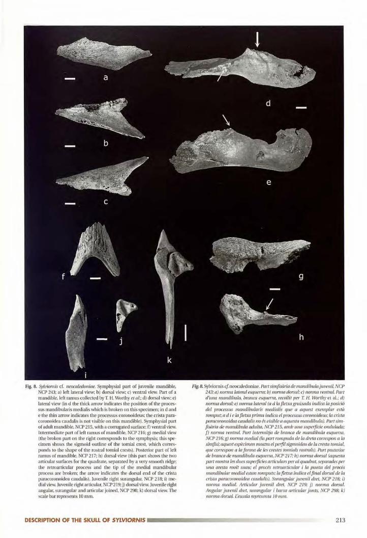
Fig.8. Sylviornis cf. neocaledoniae. Symphysial part of juvenile mandible,NCP 243; a) left lateral view; b) dorsal view; c) ventral view. Part of a
mandible, left ramus collected by T H. Worthy et a!.; d) darsal view; e)lateral view (in d the thick arrow indicates the position of the processus mandibularis medialis which is broken on this specimen; in d ande the thin arrow indicates the processus coronoideus; the crista paracoronoidea caudal is is not visible on this mandible). Symphysial partof adult mandible, NCP 215, with a corrugated surface; f) ventral view.Intermediate part of left ramus of mandible, NCP 21G; g) medial view
(the broken part on the right corresponds to the symphysis; this specimen shows the sigmoid outline of the rornial crest, which corres
ponds to the shape of the rostral tomial crests). Posterior part of leftramus of mandible, NCP 217; h) dorsal view (this part shows the two
articular surfaces far the quadrate, separated by a very smooth ridge;the retroarticular process and the tip of the medial mandibular
process are broken; the arrow indicates the dorsal end of the crista
paracoronoidea caudalis). luvenile right surangular, NCP 218; i) me
dial view. Juvenile right articular. NCP 219; j) dorsal view. Juvenile rightangular, surangular and articular joined, NCP 298; k) dorsal view. Thescale bar represents 10 mm.
Fig. 8. Sylviornis cI neocaledoniae. Partsimfisària de mandlbulajuvenil, NCP
243; a) norma lateral esquerra; b) norma dorsal; c) norma ventral. Partd'una mandibula, brancà esquerra, recollit per T H. Worthy et al.; d)norma dorsal; e) norma lateral (a d Ia fletxa gruixada indica la posiciódel processus mandibularis medialis que a aquest exemplar està
romput; a el i e lafletxa prima indica el processus coronoideus; la crista
patacoronoidea caudalis no és uisio!« a aquesta mandibuia). Parr sim
[isària de mandíbula adulta, NCP 215, amb Llna superficie ondulada;f) norma ventral. Part intermitja de branca de mandíbula esquerra,NCP 216; g) norma medial (la part rompuda de la dreta correspon a la
símfisi; aquest espècimen mostra el perfil sigmoideu de la cresta tomial,que coitespon a la forma de les crestes tomials rostrals). Part posteriorde branca de mandibula esquerra, NCP 217; h) norma dorsal (aquestapart niosua les dues superficies aniculars per al quadrat, separades perII./Ja aresta molt suau; el procés retroarticular i la punta del procésmandibular medial estan romputs; la ftetxa indica el final dorsal de lacrista. paracoronoidea caudalis). Surangular juvenil dret, NCP 218; i)norma medial. Articular juvenil dret, NCP 219; j) norma dorsal.
Angular juvenil dret, surangular i barro articular junts, NCP 298; k)norma dorsal. l'escala representa 10 mm.
DESCRIPTION OF THE SKULL OF SYLVIORNIS ..--------���--�-- 213
243, minirnallength 64.5 mm, fig. 8, a-c), and that the bonewas very thick at the level of the symphysis (NCP 213, mi
nimal depth of the symphysis 18.3 mm). The fragments ofmandibular symphysis from adults, particularly NCP 215
(fig. 8, f), show a strongly corrugated ventral surface, with
anteroposteriorly oriented ridges. On the fragment ofmandibular ramus NCP 216 (fig. 8, g), the dorsal part formsa sharp edge, with a sinuous line that articulates with theexternal tomial crest of the rostrum. Both internal and
external surfaces of this mandibular ramus show a sha
greened ornamentation.The articular part of the mandible (NCP 217, fig. 8, h)
shows two articular cotylae for the two mandibular
condyles of the quadrate, separated by a smooth ridge.This articulation is not situated in a depression. The medial mandibular process is short and wide, with a pneumatic foramen on its lateral side, ventral to its tip. On the dor
sal side of the mandibular ramus, close to the articulation,there is a processus corona ideus and, on the lateral side, a
dorsoventrally oriented crista paracoronoidea caudalis
(Weber, 1996). This crista has a tubercle at its dorsal part,and is not incurved but quite straight.
The posterior part of a left mandible collected by T. H.
Worthy et al. (fig. 8, d-e) shows that, in the adult, this area
is extremely thick in a mediolateral direction. The retroar
ticular process of this mandible is very wide, both medio
laterally and anteroposteriorly. The dorsalmost part of thisretroarticular process is incompletely preserved but it
seems to have a rounded outline, while in the recent
megapodes it ends by a dorsally, or dorsoposteriorly o
riented point.In most birds in which both are present, the medial
process and the retroarticular process of the mandible are
approximately the same size, but in Syluiomis, the retroar
ticular process is much more developed than the medialmandibular one.
The development of the posterior part of themandible can be correlated with the strong development, both in lateral and posterior directions, of the posterior part of the cranium. There is a strong contrast
between the anterior part of the mandible, which formstwo cutting blades, and the posterior part which is verythick and massive.
Among the juvenile material there are numerous
unfused mandibular bones, such as angulars, articulars,and supraangulars (fig. 8, i-k). This indicates that fusion ofthe mandibular bones probably occurred late during the
development of the animal.
MEASUREMENTS (IN MM)
Adult cranium (NCP 241)Total length from the lacrimal process to the posterior
most part of the paroccipital process: 100; maximum width
at the level ofthe exoccipitals: 108; maximum dorsoventral
height from the top of the cranial vault to the sphenoid,between the rostropterygoid processes: 56.0; minimum
width of the frontals at the level of the orbits: 65.6; mini
mum width of the parietals and squamosals at the level of
the temporal fossa: 73.4; width at the level of the cranioros
tral hinge: 57.0; anteroposterior length from the tuberclesituated in the middle of the crania rostral hinge to the
occipital condyle: 93.4; anteroposterior length from the
same point to the posteriormost part of the paroccipitalprocess: 106.6; anteroposterior length from the same pointto the crista nuchalis transversa, in the sagittal plane: 67.0;anteroposterior length of the supraoccipital, from the crista
nuchalis transversa to the top of the occipital foramen, in
the sagittal plane: 25.0; dorsoventral height of the posteriorand lateral part of the cranium, from the top of the crista
occipitalis to the base of the paroccipital process: 53.0;internal width of the occipital foramen: 12.0; internal
height of the occipital foramen: 16.0; width of the occipitalcondyle: 12.0; height of the occipital condyle: 9.0; antero
posterior length of the rostropterygoid processes: 16.2 and
16.2; width of the rostrapterygoid processes: 7.5 and 7.2
Adult rostrum (NCP 242)Maximum length from the anterior point of the beak to
the articular condyle of the craniarostral hinge: 148.0;dorsoventral height of the rostrum from the top of the arti
cular condyle of the hinge to the posterior angle: ca. 69.0;length from the point of the beak to the posterior angle: ca.
102.0; maximum width between the tips of the two articu
lar condyles: 60.0; length of the bony ornament at its base:
52.0; width of the bony ornament at its base: 41.2; width ofthe premaxillaries at the level of the external tomial crests:
26.6; width of the rostrum at the level of the posterior angle:22.2 ; at the level of the articular surfaces for the quadratojugals: 30.0; dorsoventral height from the flattened surfacesituated at the base of the bony ornament, to the internaltomial crest: 64.0; dorsoventral and anteroposterior diameter of the external naris: 12.5 and 12.0
MandiblesAnterior parts: see table 1
Cranial parts of mandibles NCP213 NCP243 NCP295 NCP294adult juvenile juvenile juvenile
Length of the almost complete- - 47.8mandibular symphysis
-
Length of the mandibular symphysis 57.0 64.5 54.0as preserved
-
Maximum dorsoventral heigth of- 16.2 13.6 12.0the complete symphysis
Maximum dorsoventral heigth of18.0 -
the symphysis as preserved- -
Table 1. Syluiornis ef. neocaledoniae, Pindai Cave, New Caledonia.Measurements (mm) of the anterior parts of mandible.
214
Taula 1. Sylviornis ef neocaledoniae, Pindai Cave, New Caledonia.Mesures (en mm) de les parts anteriors de la mandíbula.
�----__---I INSULAR VERTEBRATE EVOLUTION

Posterior part (specimen collected by T. H. Worthy et
al.): dorsoventral height at the level of the articular surfacefor the quadrate: 23.0; height from the top of the retroarticular process to the ventralrnost part of the mandible:estim. 42.5; anteroposterior length of the retroarticular
process at the level of the articular surface for the quadrate:33.6; mediolateral width of the mandible at the level of thearticular surface for the quadrate: estim. 46.0 (based on the
specimen collected by T. H. Worthy et al. and on NCP 217)
Quadrates, pterygoids, palatines, and quadratojugalsSee tables 2 to 5.
JUSTIFICATION FOR THE CREATION OF A NEW FAMILY
The presence of two well-developed basipterygoidprocesses makes it possible to state that Syluiornis belongsto Galliformes or to Anseriformes (Weber, 1993;Dzerzhinsky, 1995; Ericson, 1996; Livezey, 1997), and the
presence of palatines with very elongated anterior branchesand posterior parts (partes laterales) that are narrow andnot wing-like shows that it belongs to Galliformes.
Among the Galliformes, the postcranial skeleton showsmore similarities with Megapodiidae than with other families (Poplin et al., 1983; Poplin & Mourer-Chauviré, 1985),but these similarities seem to be symplesiomorphic. Thecranial characteristics also present some similarities with
Megapodiidae, as for example the slightly developedlacrimal, devoid of orbital process; the position of the articular surface for the rostrapterygoid process at the fust thirdof the pterygoid and not at its anterior end; the presence ofwell developed alae ethmoidales. The first two characteristics are symplesiomorphies and the third one is variablewithin the Galliformes (Ericson, 1996).
The creation of a new family is justified by the presence
ofnumerous autapomorphic characteristics in the skull andmandible, characteristics that are indicated in the diagnosis. To our knowledge Syluiornis is the only bird to show a
true diarthrosis, of ginglymus type, between the skull andthe beak, and to show a cranial flexure situated just posterior to the basiperygoid processes.
COMPARISON WITH OTHER EXTINCT GALLIFORM
FAMIUES
Three extinct families have been described within the
Galliformes, the Gallinuloididae Lucas, 1900, the
Quercymegapodiidae Mourer-Chauviré, 1992, and the
Paraortygidae Mourer-Chauviré, 1992. These three families
display primitive characteristics compared with the recent
families of Galliformes (Mourer-Chauviré, 1992; Mayr, 2000;Dyke & Gulas, 2002; Mayr & Weidig, 2004). These characteristics are mainly the presence of a hollow, cup-like, scapular facet on the coracoid, and the absence of a transverse
ridge at the beginning of the incisura capitis on thehumerus. These characteristics are absent in Syluiorniswhich on the contrary displays the derived character states.
DISCUSSION
It is possible to think that the the ancestor of Syluiorniswas a galliform comparable in its osteological features to
the recent megapodes, which would have reached NewCaledonia at an unknown date, and would have evolvedthere in insular isolation, where it became flightless and
acquired its highly derived cranial features. This ancestralform could have reached New Caledonia when it was still
Quadrates Extremes Mean sd n V
Dorsoventral h� from the tip of processus 34.0-38.8 36.15 1.25 35 3.45oticus to the ventr ast part of condylus medialis
Craniacaudal length from the cranial tip of processus 46.0-51.3 47.90 1.89 9 3.95orbitalis to the caudal part of processus mandibularis
Mediolateral width of the top of processus oticus 16.7-20.2 18.35 0.84 35 4.58
Craniacaudal length of the mandibular articular surface 28.0-35.8 30.59 1.71 33 5.59
Table 2. Sylviornis cf. neoealedoniae, Pindai Cave, New Caledonia.Measurements (mm) of the quadrates.
Taula 2. Sylviornis et neocaledoniae, Pindai Cave, New Caledonia.Mesures (en mm) dels quadrats.
Pterygoids Extremes Mean sd n V
Maximum craniacaudal length 31.4-40.8 36.77 2.12 35 5.77
Maximum length of the articular facet12.4-17.4 14.66 1.28 34 8.73for the processus rostropterygoideus
Width of this articular facet 6.6-8.6 7.66 0.49 34 6.40
Table 3. Syloiornis cf. neocaledoniae, Pindai Cave, New Caledonia.Measurements (mm) of the pterygoids.
Taula 3. Sylviornis et neocaledoniae, Pindai Cave, New Caledonia.Mesures (en mm) dels pterigoides.
DESCRIPTION OF THE SKULL OF SYLVIORNIS mmz::::================= 215
able to fly. Alternatively it may have colonized New
Caledonia during the Eocene, via the now submergedRennell ridge (Balonet, 1984). Olson (1980) has demonstra
ted that megapodes are able to cross large expanses of sea
water and to colonize islands. The recent discoveries of
many extinct megapodes in South Pacific islands (Jones et
al., 1995; Steadman, 1999) clearly indicates that megapodeswere much more widespread in the past, and that theirrecent distribution is relictual. A large, extinct, form of the
genus Megapodius, M. molistructor, was also present in theHolocene of New Caledonia (Balouet & Olson, 1989).
The oldest representative of the family Megapodiidae,the genus Ngauiupodius, has been described from the late
Oligocene of South Australia (Boles & Ivison, 1999), and it is
therefore possible to suppose that a comparable form mayhave reached New Caledonia at an indeterminate period,between the late Oligocene and the Holocene.
An instance of convergent evolution possibly occurredin the Fiji Islands where Worthy (2000) has described a giantflightless megapode, Megauitiornis, from Viti Levu island. In
the dimensions of its postcranial skeleton the Pindai
Syluiornis is on average 25 % larger than Megauitiornis, but
the head of Syluiornis is proportionately much larger, beingabout twice as large as the estimated length for
Megauitiorn is. In Megauitiornis the rostrum is very high,and relatively narrow, but not so narrow as in Syluiornis. The
associated cranial fragments show that in Megauitiorrüs thecraniorostral hinge was unfused, but there was not a true
diarthrosis, with two articular condyles and two concave
articular surfaces. The nasal shows a robust maxillaryprocess, indicating that it was not completely fused with the
maxillary and the premaxillary to form a massive rostrum as
in Syluiornis. The anterior part of the mandible of
Megauitiomis also shows convergence with that of
Sylviornis. It is very high dorsoventrally and very robust, and
its symphysis occupies ca. 45 % of the mandibular length.But its posterior part is different. In Megauitiornis the
processus mandibulare lateral is is a very robust, rounded
prominence, and the retroarticular process is prominent,narrow and deep, whereas in Sylviornis the mandibular
ramus is so thick at its posterior part that the processusmandibulare lateralis is not visible, and the retroarticular
process is very long anteroposteriorly, wide at its base, and
seems to have a rounded outline at its top. In the postcranialskeleton the main differences between Megauitiornis and
Syluiamis are the shape of the tarsometatarsus, which is
proportionally more robust in Megavitio rn is, and the presence, on one of the two tarsometatarsi described, of a sul
cus extensorius connected by a groove to the distal foramen
(Worthy,2000).In conclusion, Megauitiornis shows some convergent
evolution with Sylviornis, but is less advanced. It is possibleto think that several different forms of megapodes, or
megapode-like galliforms, colonized the South Pacificislands at different periods of time, and have given rise to
somewhat convergent but unrelated forms.
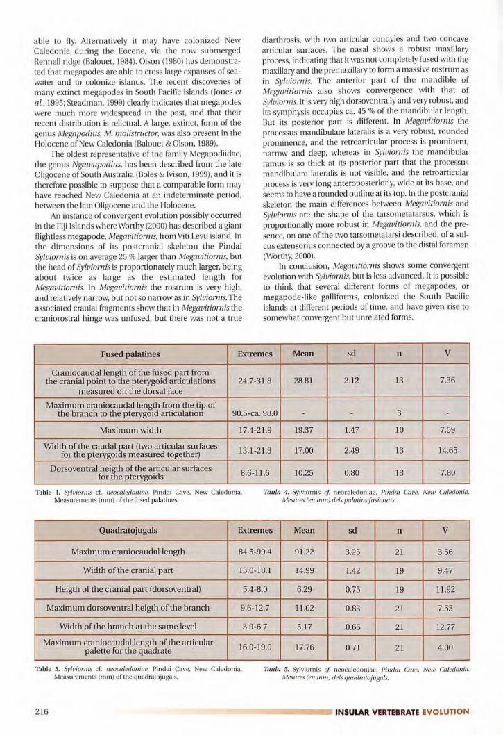
Fused palatines Extremes Mean sd n V
Craniacaudal length of the fused part fromthe cranial point to the pterygoid articulations 24.7-31.8 28.81 2.12 13 7.36
measured on the dorsal face
Maximum craniacaudal length from the tip ofthe branch to the pterygoid articulation 90.5-ca. 98.0 - - 3 -
Maximum width 17.4-21.9 19.37 1.47 10 7.59
Width of the caudal �art (two articular surfaces13.1-21.3 17.00 2.49 13 14.65for the pterygoi s measured together)
Dorsoventral hei� of the articular surfaces8.6-H.6 10.25 0.80 13 7.80for e pterygoids
Table 4. Sylviornis cf. neocaledoniae, Pindai Cave, New Caledonia.Measurements (mm) of the fused palatines.
Taula 4. Sylviornis et neocaledoniae, Pirulai Cave, New Caledonia.Mesures (en mm) dels palatins fusionats.
Quadratojugals Extremes Mean sd n V
Maximum craniacaudal length 84.5-99.4 91.22 3.25 21 3.56
Width of the cranial part 13.0-18.1 14.99 1.42 19 9.47
Heigth of the cranial part (dorsoventral) 5.4-8.0 6.29 0.75 19 11.92
Maximum dorsoventral heigth of the branch 9.6-12.7 11.02 0.83 21 7.53
Width of the branch at the same level 3.9-6.7 5.17 0.66 21 12.77
Maximum craniacaudal length of the articular16.0-19.0 17.76 0.71 21 4.00palette for the quadrate
Table 5. Sylviornis cf. neocaledoniae, Pindai Cave, New Caledonia.Measurements (mm) of the quadratojugals.
Taula 5. Sylviornis et neocaledoniae, Pindai Cave, New Caledonia.Mesures (en mm) dels quadratojugals.
216 --=-==========::::IINSULAR VERTEBRATE EVOLUTION
DIET OF SYLVIORNIS
There is some similarity between the skull of Syluiomisand that of the early Tertiary bird Diatryma which alsoshows a narrow, cutting beak, a mobile crania rostral articu
lation, and a very complex articulation between the quadratojugal and the posterior part of the rostrum (Andors, 1988).Witmer and Rose (1991) have put forward the hypothesisthat Diairymavees a carnivorous predator but Andars (1988;1992), using other arguments, has shown that it was vegetarian.
The Australian Dromornithidae, previously consideredas ratites, have been studied again by Murray & Megirian(1998) who have shown that, according to their cranial cha
racteristics, they must be classified within the Anseriformes.Some of these Dromornithidae have a very high, narrow
beak, ending anteriorly in a hook, and a completely mobilecraniorostral articulation, as in Sylviornis (Murray &
Vickers-Rich, 2004). The mandible was very high dorsoven
trally. However, according to Murray & Megirian (1998, p.78), the rostrum was round-tipped rather than pointed, andthe hooked tip differed markedly from the slender, sharplypointed hook in raptors and carrion-eating birds. Their con
clusion is that the Dromornithidae were specialized herbi
vores, able to shear tough plant material. In Syluiornis the
shape of the rostrum is very pointed and the hooked' tip'resembles that of raptors, or of birds widely recognized as
carnivorous such as the Phorusrhacidae. In posterior viewthe shape of the cranium, in the Dromornithidae, is very different in its dorsoventral elongation from that of Sylviornis.
The Mauritian Dodo, Raphus cucullatus, also had a
beak ending in a powerful hook. But this hook was mainlyformed by the ramphotheca and the bony beak shows a
much less pronounced point (Strickland & Melville, 1848).In the Dodo the anterior part of the rostrum is wide andbecomes narrower in its middle part. In the RodriguesSolitaire, Pezophaps solitaria, the rostrum is elongated andnarrow (Newton & Newton, 1870), but in these two forms,unlike Sylviornis, the rostrum is not massive. Its dorsal partconsists of a rather thin bony blade formed by the fusion ofthe processus frontalis of the premaxillary with the processus premaxillaris of the nasal. Ventrally there is a slot-likeexternal naris that is very elongated anteroposteriorly. Thereis no articulation between the cranium and the rostrum,
just a flexion zone. The ventral surface of the rostrum doesnot show cutting tomial crests. The mandible has narrow
and elongated branches and a very short symphysis. It isclear that the diet of Sylviorniswas different from that of the
Raphidae, which were primarily vegetarian.In the islands devoid of terrestrial mammals, the eco
logical niche of large herbivores is often filled by large landtortoises. This is the case for example, in the recent gianttortoises of Aldabra and the Galapagos, and of the recentlyextinct tortoises of Madagascar and the Mascarenes. In allthese forms the masticatory apparatus is not a single cuttingblade but a wide triturating surface. The most extreme case
is that of the Mascarenian tortoises, which have several
ridges, each of them bearing a row of small bony tubercles(Bour, 1979-80).
In the Hawaiian Islands there were no giant land-tortoises and their ecological niche was probably occupied bythe flightless ducks called moa-nalos (Olson & James, 1991).
In these forms the premaxillaries and the dentaries were
very short, massive, and generally presented blunt toothlike projections.
Most of the cranial characteristics of Syluiotnis are different from those of the typically vegetarian forms such as
the weird anseriforms, the Australian Dromornithidae, theHawaiian moa-nalos, pigeons in the Raphidae, or theMascarenian tortoises.
Balouet (1986) proposed the hypothesis that Syluiorniswas vegetarian and fed on roots and tubercles. This hypothesis was significantly supported by the very high numbers of
Sylviornisin the fossil sample, which implies that it could not
have been a carnivorous predator, hunting and feeding on
other large vertebrates. However it is also possible to propose that Syluiornis fed on invertebrates. In this case, theavailable alimentary resources, such as marine organisms or
terrestrial gastropods, for example, could have been abundant and varied enough to sustain a large population of this
species. We think that the skull of Syluiomis has evolved as an
adaptation to a particular and highly specialized diet, but itis not possible at this stage to be more precise.
SYLVIORNIS AND THE ORAL TRADITION
Sylviorniswas contemporaneous with the first arrival ofman in New Caledonia and its disappearance is certainlydue to overhunting (Balouet, 1986; 1987). The oral traditionhas retained the memory of a vanished bird, called Du
(Griscelli, 1976). According to tradition this bird was giantand flightless. It laid a single egg and did not incubate it.This egg took four months to hatch (from November to
April). The Du moved along on the ground very rapidly, withits wings spread out. It had a red feathering and a bonyornament on the head. P. Griscelli writes: "It seems that theDu had on its head a kind of bony, solid, casque, in the
shape of a star" (1976, p 5, our translation). But the word"Ghi" which designates this ornament should rather betranslated as helmet. According to the inhabitants ofHouaïlou the Du laid an egg on the top of a lizard shelteredin the hollow of a banyan, then went away, leaving the lizardto incubate it for four months and to break the shell with its
jaw. Lastly P. Griscelli states that "tradition attributes greataggressiveness to this bird, in connection with totemic ritesand cannibalism" (1976, p. 5, our translation).
ACKNOWLEDGEMENTS
The authors want to express their thanks to the ParisNational Museum of Natural History, L. Ginsburg, P. Janvier,E Poplin, S. Tillier, and A. Tillier; to the National Museum ofNatural History, Smithsonian Institution, Washington, S.Olson, H. James, A. Ross, J. Dean and B. Schmidt; to theOffice de la Recherche Scientifique et Technique d'OutreMer, D. Frimigacci and B. Vienne; to the Service des Eaux et
Forêts, Nouméa, E Boulet and Y. Letocart; to the Musée
Néocalédonien, P. Gaudin and E. Kasarherou; to the tribesof Isle of Pines and Nepoui, and their authorities, as well as
DESCRIPTION OF THE SKULL OF SYLVIORNIS E!::================ 217

to F Hannecart, and to P. Millener from New Zealand. Wethank an anonymous reviewer for his improvements anduseful comments, and T. Worthy for sharing his informationwith us. Photographs are by N. Podevigne (UniversitéClaude Bernard - Lyon 1).
REFERENCES
Andors, A.V 1988. Ciant groundbirds of North America (Aves,Diatrymidae). Ph. D. dissertation, Columbia University, New
York, xvi + 577 pp. (unpublished).Andors, A.V 1992. Reappraisal of the eocene groundbird Diatryma
(Aves: Anseromorphae). Natural History Museum of Los
Angeles County, Science Series, 36: 109-125.
Balouet, I.C, 1984. Paléontologie des Vertébrés de Nouvelle Calédonieet Paléobiogéographie du Pacifique Sud Ouest. Thèse de 3'""
cycle, Université Pierre et Marie Curie - Paris 6, no 84-39, 84 pp.
(unpublished).Balouet, J.C. 1986. Une actualité millénaire. Premiers colons de
Nouvelle-Calédonie. LUnivers du Vivant, Paris, 7: 35-47.
Balouet, I.e. 1987. Extinctions des vertébrés terrestres de NouvelleCalédonie, in Les Extinctions dans l'histoire des Vertébrés.Mémoires de la Société géologique de France, 150: 177-183.
Balouet, J.C. 1991. The fossil vertebrate record of New Caledonia. In
Vickers-Rich, P.; Monaghan, J.M.; Baird, R.F & Rich, T.H. (eds.),Vertebrate Palaeontology of Australasia: 1383-1409. Pioneer
Design Studio and Monash University PublicationsCommittee, Melbourne.
Balouet, J.e. & Buffetaut, E. 1987. Mekosuchus inexpectatus, n. g., n.
sp., Crocodilien nouveau de I'Holocène de NouvelleCalédonie. Comptes Rendus de l'Académie des Sciences de
Paris, t. 304, Sér. II, 14: 853-856.
Balouet, J. e. & Olson, S.L. 1989. Fossil Birds from Late Quaternarydeposits in New Caledonia. Smithsonian Contributions to
Zoology, 469: iv + 38 pp.Bock, w.J., 1964. Kinetics of the Avian Skull. Journal ofMorphology,
114: 1-42.
Boles, W.E. & Ivison, T]. 1999. A New Genus of Dwarf Megapode(Galliformes: Megapodiidae) from the Late Oligocene ofCentral Australia. In Olson, S.L. (ed.), Avian Paleontology at theclose of the 20" Century: Proceedings of the 4'" International
Meeting of the Society for Avian Paleontolgy and Evolution,Washington, D.C., 4-7June 1996. Smithsonian Contributions to
Paleobiology, 89: 199-206.
Bour, R. 1979-80. Histoire de la tortue terrestre de Bourbon. Bulletinde l'Académie de l'Ile de la Réunion, 25: 97-147.
Dyke, G.J. & Gulas, B.E .. 2002. The Fossil Galliform Bird
Paraortygoides from the Lower Eocene of the United
Kingdom. American Museum Novitates, 3360: 14 pp.Dzerzhinsky, FY. 1995. Evidence for common ancestry of the
Galliformes and Anseriformes. Courier ForschungsinstitutSenckenberg, 181: 325-336.
Ericson, P.G.P. 1996. The skeletal evidence for a sister-group rela
tionship of anseriform and galliform birds - a critical evaluation. Journal ofAvian Biology, 27: 195-202.
Gaffney, E.S.; Balouet, J.e. & Broin, F de. 1984. New occurrences ofExtinct Meiolaniid Turtles in New Caledonia. AmericanMuseum Novitates, 2800: 6 pp.
Griscelli, P. 1976. Deux oiseaux fossiles de Nouvelle-Calédonie.Bulletin de la Société d'Etudes historiques de la NouvelleCalédonie, 29: 3-6.
Jollie, M.T. 1957. The head skeleton of the chicken and remarks on
the anatomy of this region in other birds. Journal ofMorphology, 100 (3): 389-436.
Jones, D.N.; Dekker, R.w.J & Roselaar, CS. 1995. Bird Families of theWorld. The Megapodes (Megapodiidae). Oxford UniversityPress, Oxford. NewYork and Tokyo. 262 pp.
Livezey, B.e. 1997. A phylogenetic analysis of basal Anseriformes, thefossil Presbyornis, and the interordinal relationships of water
fowl. Zoological Journal of the Linnean Society, 121: 361-428.
Mayr, G. 2000. A new basal gaUiform bird from the Middle Eoceneof Messel (Hessen, Germany). Senckenbergiana Lethaea, 80
(1): 45-57.
Mayr, G. & Weidig, l. 2004. The Early Eocene bird Gallinuloides
ioyomingensis - a stem group representative of Galliformes.Acta Palaeontoiogica Polonica, 49 (2): 211-217.
Mourer-Chauviré, e. 1992. The Galliformes (Aves) from the
Phosphorites du Quercy (France): Systematics and
Biostratigraphy. Natural History Museum of Los AngelesCounty, Science Series, 36: 67-95.
Murray, P.F & Megirian, D. 1998. The skull of Dromornithid birds:Anatomical evidence for their relationship to Anseriformes.Records of the South Australian Museum, 31: 51-97.
Murray, P.F & Vickers-Rich, P. (2004). Magnificent Mihirungs. TheColossal Flightless Birds of the Australian Dreamtime. Indiana
University Press. Bloomington. 416 pp.Newton, A. & Newton, E. 1870. On the osteology of the Solitaire or
Didine Bird of the Island of Rodriguez, Pezophaps solitaria
(Gmel.). Philosophical Transactions of the Royal Society,London, 159: 327-362.
Olson, S.L. 1980. The significance of the distribution of the
Megapodiidae. Emu, 80: 21-24.
Olson, S.L. 1999. The Anseriform Relationships ofAnataiauis Olsonand Parris (Anseranatidae), with a new species from the LowerEocene London Clay. In Olson, S.L. (ed.), Avian Paleontologyat the close of the 20'" Century: Proceedings of the 4'"International Meeting of the Society for Avian Paleontolgy andEvolution, Washington, D.C., 4-7 June 1996. SmithsonianContributions to Paleobiology, 89: 231-243.
Olson, S.L. & James, H.F 1991. Description of Thirty-two New
Species of Birds from the Hawaiian Islands: Part l. NonPasseriformes. Ornithological Monographs, 45: 1-88.
Poplin, F 1980. Syluiornis neocaledoniae, n. g., n. sp. (Aves), Ratiteéteint de la Nouvelle Calédonie. Comptes Rendus del'Académie des Sciences de Paris, t. 290, Sér. D: 691-694.
Poplin, F & Mourer-Chauviré, e. 1985. Syluiornis neocaledoniae(Aves, Galliformes, Megapodiidae), oiseau éteint de I'Ile desPins (Nouvelle Calédonie). Geobios, 18 (1): 73-97.
Poplin, F; Mourer-Chauviré, e. & Evin, J. 1983. Position systématique et datation de Syluiornis neocaledoniae, Mégapode géant(Aves, Galliformes, Megapodiidaei éteint de la NouvelleCalédonie. Comptes Rendus de l'Académie des Sciences de
Paris, t. 297, Sér. II: 301-304.
Steadman, D.W. 1999. The biogeography and extinction of
megapodes in Oceania. In Dekker et al., Proceedings of theThird International Megapode Symposium. Zoologischevemandeiingen, 327: 7-21.
Strickland, H.E. & Melville, A.G. 1848. The Dodo and its Kindred, or
the history, affinities and osteology of the Dodo, Solitaire, andother extinct birds of the islands Mauritius, Rodriguez, andBourbon. Reeve, Benham, and Reeve. London. 141 pp.
Weber, E. 1993. Zur Evolution basicraníaler Gelenke bei Vògeln,insbesondere bei Hillmer- und Entenvogeln (Galloanseres).Zeitschrift fiir zoologische Systematik und Evolutions
[orschung, 31: 300-317.
Weber, E. 1996. Das Skelet-Muskel-System des Kieferapparatus von
Aepypodius arjakianus (Salvadori, 1877) (Aves, Megapodiidae). Courier Forschungsinstitut Senckenberg, 189: 132 pp.
Witmer, L.M. & Rose, K.D. 1991. Biomechanics and the jaw apparatus of the gigantic Eocene bird Diatryma: implications for dietand mode of life. Paleobiology, 17(2): 95-120.
Worthy, T.H. 2000. The fossil megapodes (Aves: Megapodiidae) of
Fiji with description of a new genus and two new species.Journal ofThe Royal Society ofNew Zealand, 30 (4): 337-364.
Zusi, R.L. & Livezey, B.e. 2000. Homology and phylogenetic implications of some enigmatic cranial features in Galliform andAnseriform birds. Annals ofCarnegie Museum, 69 (3): 157-193.
218 �-�-----------, INSULAR VERTEBRATE EVOLUTION