39 malignant soft tissue masses on magnetic resonance imaging
Upload
muhammad-bin-zulfiqarCategory
view
190download
0
15 Cerebellopontine Angle Masses on Magnetic Resonance
Imaging

CLINICAL IMAGAGINGAN ATLAS OF DIFFERENTIAL DAIGNOSIS
EISENBERG
DR. Muhammad Bin Zulfiqar PGR-FCPS III SIMS/SHL

• Fig SK 15-1 Acoustic neuroma. Marked contrast enhancement of the left-sided lesion (arrowheads). Note the normal neural structures on the right.

• Fig SK 15-2 Acoustic neuroma. Bilateral tumors (n) are seen in this coronal scan of a patient with neurofibromatosis.

• Fig SK 15-3 Meningioma. Note that the mass (arrows) has a relatively broad base along the petrous bone.

• Fig SK 15-4 Epidermoid. (A) Axial T1-weighted and (B) axial T2-weighted scans show an oblong mass (arrows) of slightly higher signal than CSF enveloping the basilar artery and extending around the pons.

• Fig SK 15-5 Lipoma. Axial T1-weighted image shows that the lesion has signal intensity similar to that of subcutaneous fat.25

• Fig SK 15-6 Metastasis. (A) Axial T2-weighted image shows a metastasis of the right cerebellopontine angle (from lung carcinoma) that mimics an acoustic neuroma but is associated with unusual middle ear retention (*). (B) Contrast-enhanced axial T1-weighted scan shows intense enhancement of the lesion, which extends into the cochlea (arrow). Note the presence of another enhancing lesion at the tip of the right petrous bone (arrowhead).25

• Fig SK 15-7 Arachnoid cyst. (A) Axial T1-weighted image shows the CSF-intensity cyst stretching the left 7th and 8th cranial nerve complex (arrow). (B) Axial T2-weighted scan shows the cyst displacing the vascular structures of the cerebellopontine angle (arrowheads).25

• Fig SK 15-8 Aneurysm of the left posterior inferior cerebellar artery. Axial T2-weighted image shows the typical lack of signal (arrow) within the aneurysm. Note the lymphoma in the right pterygopalatine fossa (arrowheads), which explained the patient's neuralgia.25

• Fig SK 15-9 Thrombosed aneurysm of the right posterior inferior cerebellar artery. (A) Axial T2-weighted image shows a focal area of calcification (arrowhead). Note the normal right hypoglossal canal (arrow), a finding inconsistent with an acoustic neuroma. (B) Contrast coronal T1-weighted scan shows homogeneous enhancement of the organized thrombus, which completely fills the aneurysm.25

• Fig SK 15-10 Glomus jugulare tumor with intracranial involvement and bony erosion. (A) Axial T1-weighted image shows the mass (arrowheads) in the left temporal bone and posterior fossa extending across the midline. Note the large veins on the surface of the tumor. (B) Coronal contrast-enhanced image shows displacement of the brainstem, bony erosion from the large enhanced mass, and parotid involvement.26

• Fig SK 15-11 Brainstem glioma. A contrast axial T1-weighted image shows central enhancement in an unusual round grade III glioma located in front of the porus.25

• Fig SK 15-12 Hemangioblastoma. (A) Axial T2-weighted image in a patient with von Hippel-Lindau disease shows a solid tumor in the left cerebellopontine angle. Note the vascular pedicle (arrowhead), which appears as a flow void with all sequences. (B) Contrast T1-weighted axial image shows homogeneous enhancement of the tumor (arrowhead).25

• Fig SK 15-13 Choroid plexus papilloma. (A) Axial T2-weighted image shows a right cerebellopontine angle papilloma extending through the foramen of Luschka. The tumor contains massive hypointense calcification (arrowhead). (B) Contrast T1-weighted axial scan shows intense enhancement of the hypervascularized tumor. Note the normal choroid plexus in the left foramen of Luschka.25
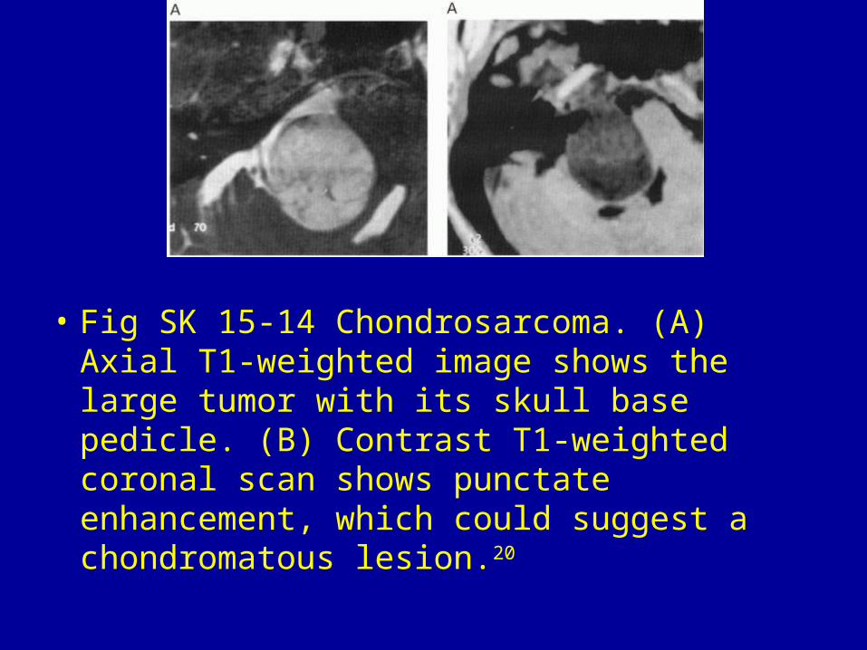
• Fig SK 15-14 Chondrosarcoma. (A) Axial T1-weighted image shows the large tumor with its skull base pedicle. (B) Contrast T1-weighted coronal scan shows punctate enhancement, which could suggest a chondromatous lesion.20

• Fig SK 15-15 Trigeminal neuroma. Isointense mass on the right (arrow). Note the normal 5th cranial nerve on the left (arrowhead).