14 Unit 1 Chapter 14. 14 Unit 1 3 components of the cardiovascular system = blood, heart, blood...
50
14 Unit 1 The Cardiovascular The Cardiovascular System: Blood System: Blood Chapter 14
-
Upload
edward-dickerson -
Category
Documents
-
view
214 -
download
0
Transcript of 14 Unit 1 Chapter 14. 14 Unit 1 3 components of the cardiovascular system = blood, heart, blood...

14
Uni
t 1
The Cardiovascular The Cardiovascular System: BloodSystem: Blood
The Cardiovascular The Cardiovascular System: BloodSystem: Blood
Chapter 14
14
Uni
t 1
FunctionFunctionFunctionFunction
• 3 components of the cardiovascular system = blood, heart, blood vessels
• Function of CV system = transports substances to and from body cells
• Hematology = branch of science that deals with study of blood, blood-forming tissues and disorders associated with them
14
Uni
t 1
Functions of BloodFunctions of BloodFunctions of BloodFunctions of Blood
• Is a type of connective tissue
• Transportation- gasses (O2 & CO2), nutrients (GI tract), heat & waste, hormones (endocrine system)
• Regulation- pH, temperature, water balance in cells
• Protection- loss of blood via clotting, WBC vs. disease; production of interferons and complement

14
Uni
t 1
Components Components Components Components
• 5 facts about blood1.Blood is thicker than water 2.Temp is 38° C3.Slightly alkaline 7.35 – 7.454.8% of total body weight5.Volume = males – 5-6 liters females – 4-5 liters

14
Uni
t 1
CompositionCompositionCompositionComposition
• Connective tissue-Two parts• Plasma = straw-colored liquid
with dissolved materials (~55%)
• Formed Elements = cells and cells fragments (~45%) 99% of the formed elements are RBCs

14
Uni
t 1
CompositionCompositionCompositionComposition
• hematocrit (Hct) = Percent of blood volume occupied by red blood cells (RBC)
• Buffy coat – part made up by white blood cells (WBC) and plateletes ~1%
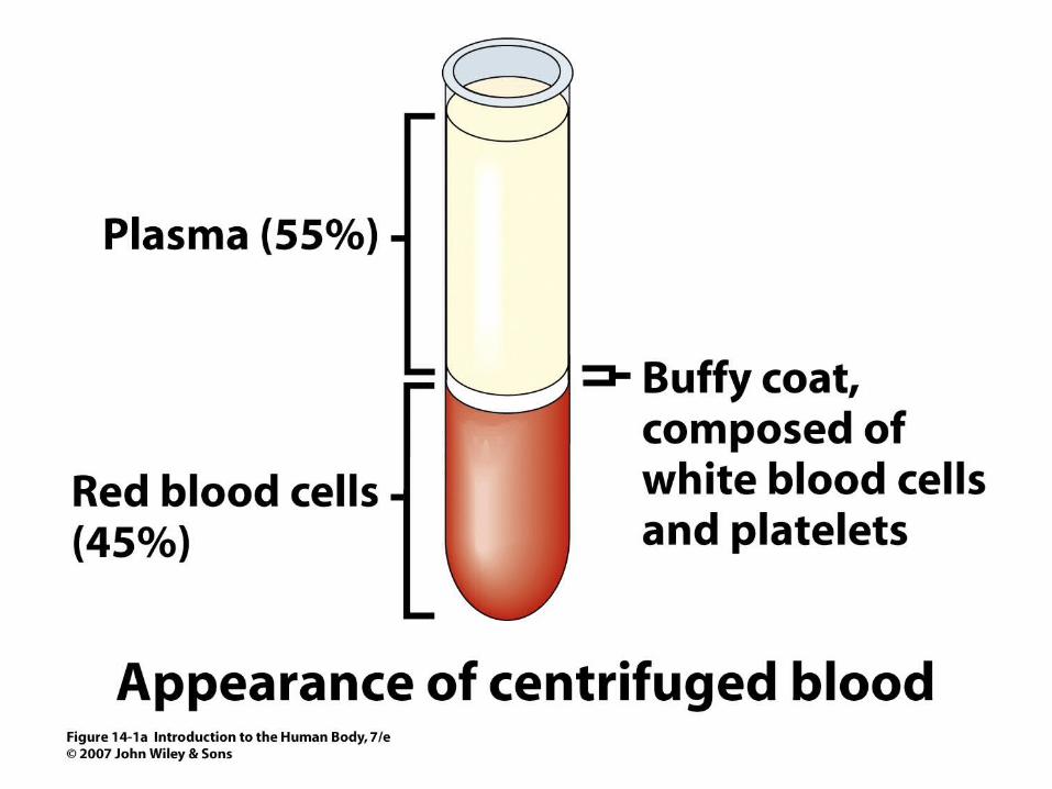
Figure 14.1aFigure 14.1a

Figure 14.1bFigure 14.1b
14
Uni
t 1
PlasmaPlasmaPlasmaPlasma
• ~91% water, 7% proteins, 1.5 % other solutes
• Proteins: Albumin (54%)- maintains osmotic pressure;
• Globulins (38%)- antibodies for defense
• Fibrinogen (7%)- clotting• Other solutes in plasma: Electrolytes ,
nutrients, gases, hormones, vitamins & waste products
14
Uni
t 1
Formed ElementsFormed ElementsFormed ElementsFormed Elements
I. Red Blood CellsII. White blood cells
A. granular Leukocytes1. Neutrophils2. Eosinophils3. Basophils
B. Agranular leukocytes1. T & B lymphocytes & natural Killer cells2. monocytes
III Platelets

14
Uni
t 1
Formation of Red Blood Formation of Red Blood CellsCells
Formation of Red Blood Formation of Red Blood CellsCells
• Hemopoiesis – production of formed elements
• 3 months before birth and throughout life occurs in red bone marrow
• Contains pluripotent stem cells• In response to specific hormones
these develop through a series of changes to form all of the blood cells
14
Uni
t 1
Formation of Red Blood Formation of Red Blood CellsCells
Formation of Red Blood Formation of Red Blood CellsCells
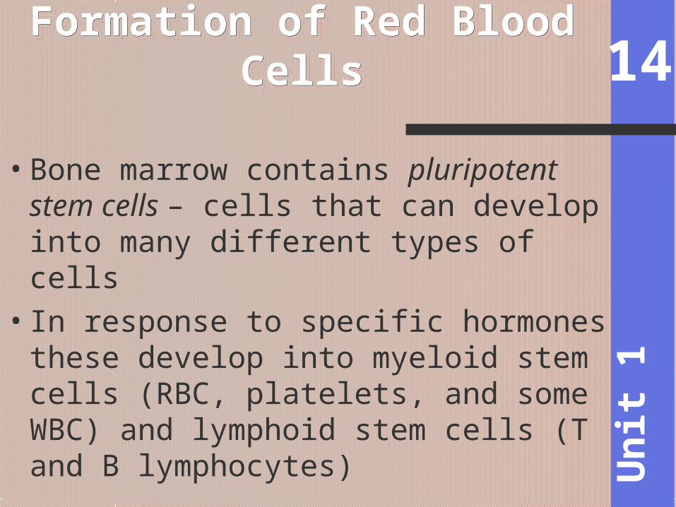
• Bone marrow contains pluripotent stem cells – cells that can develop into many different types of cells
• In response to specific hormones these develop into myeloid stem cells (RBC, platelets, and some WBC) and lymphoid stem cells (T and B lymphocytes)
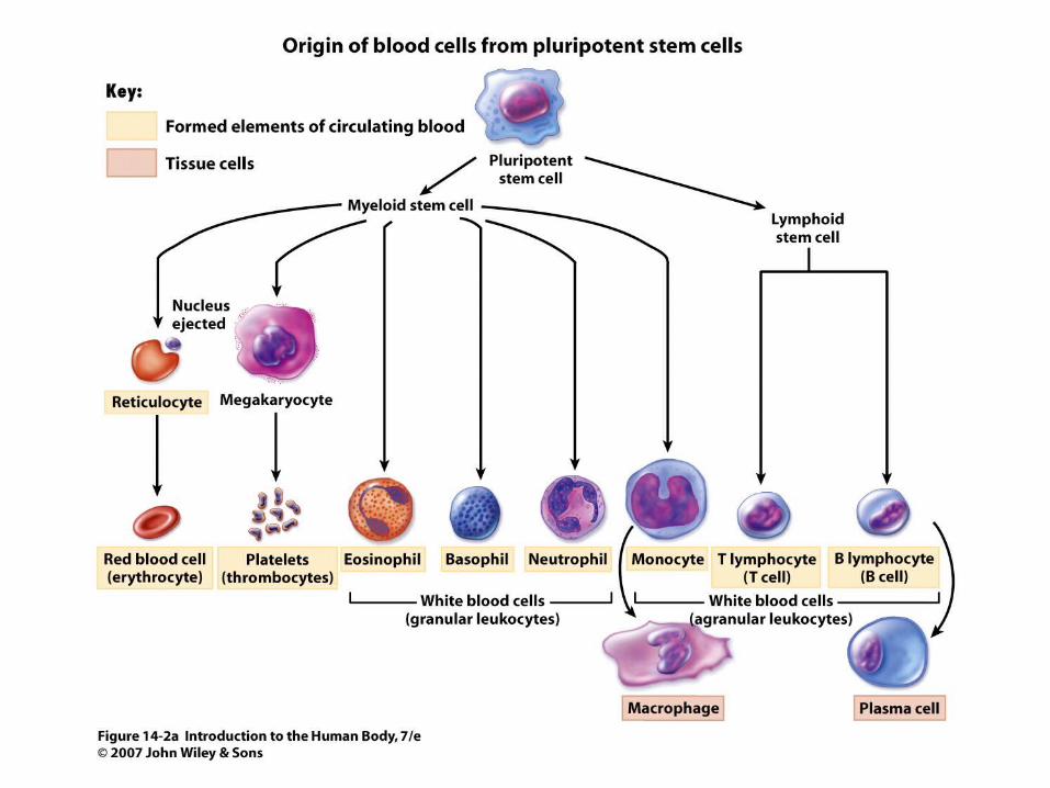
Figure 14.2aFigure 14.2a
Figure 14.2bFigure 14.2b
14
Uni
t 1
Red Blood CellsRed Blood Cells Red Blood CellsRed Blood Cells
•AKA = Erythrocytes•Contains - hemoglobin
protein- carries oxygenAlso carries some CO2
•Male has ~ 5.4 million cells/µl; Female has ~4.8 million
14
Uni
t 1
Erythrocytes (RBCs)Erythrocytes (RBCs)Erythrocytes (RBCs)Erythrocytes (RBCs)
• Production rate = 2 million/sec• Contain no nucleus & other
organelles• Consists of membrane, cytosol,
hemoglobin• Biconcave shape provides for
greater surface area for the diffusion of gas molecules into/out of a RBC

14
Uni
t 1
RBC CyclingRBC CyclingRBC CyclingRBC Cycling
• Wear out fast- live ~120 days due to wear and tear as they squeeze through capillaries
• Worn out and ruptured cells are cleared by macrophages (liver spleen, red bone marrow)
• Fe- recycled in bone marrowCarried in blood on transferrin – plasma
protein acts as a transporter for iron

14
Uni
t 1
RBC CyclingRBC CyclingRBC CyclingRBC Cycling
• biliverdin bilirubin – what the non-iron portion of heme is converted into and excreted (bile)
• Urobilinogen and stercobilin – by products of bilirubin; some absorbed in large intestine; ultimately excreted in urine or feces

Figure 14.3Figure 14.3
14
Uni
t 1
RBC ProductionRBC ProductionRBC ProductionRBC Production
•called erythropoiesis•Released as reticulocytes
(almost a RBC)Mature to erythrocytes in 1-2 days
•Production & destruction is balanced

14
Uni
t 1
RBC SynthesisRBC SynthesisRBC SynthesisRBC Synthesis
•If O2 carrying capacity falls, RBC productions is inc. by a negative feedback loop
•Low O2 delivery (hypoxia)
•erythropoietin (EPO)- kidney hormone stimulates erythropoiesis

14
Uni
t 1
RBC SynthesisRBC SynthesisRBC SynthesisRBC Synthesis
•Cyanosis – life threatening condition caused by prolonged hypoxia; bluish-purple skin coloration
•Anemia – reduced O2 carrying capacity – reduced # of RBC or ↓ amount of hemoglobin in blood; pale skin

Figure 14.4Figure 14.4

14
Uni
t 1
White Blood CellsWhite Blood CellsWhite Blood CellsWhite Blood Cells
• aka = leukocytes• Classified as either 1. granular
which contains a chemical filled cytoplasmic granule/vesicle (neutrophils, eosinophils, basophils) or 2. agranular (lymphocytes, monocytes)
• Function in defense: phagocytosis or antibody production

14
Uni
t 1
White Blood CellsWhite Blood CellsWhite Blood CellsWhite Blood Cells
• Phagocytes:Neutrophil- first responders; release
lysozomes that destroy bacteriaMonocytes slower response; become
wandering macrophages (big eaters compared to neutrophils)
Eosinophil- work in interstitial fluid; Involved in suppressing allergic responses; phagocitize antibody-antigen complexes and are effective against some parasitic worms
Basophil- intensify allergic reactions

14
Uni
t 1
White Blood CellsWhite Blood CellsWhite Blood CellsWhite Blood Cells
•Phagocytes:a. Neutrophil- first responders;
release lysozomes that destroy bacteria
b. Monocytes slower response; become wandering macrophages (big eaters compared to neutrophils)

14
Uni
t 1
White Blood CellsWhite Blood CellsWhite Blood CellsWhite Blood Cells
• Phagocytes:c. Eosinophil- phagocitize antibody-antigen complexes involved in suppressing allergic responses
d. Basophil- release heparin, histamines, serotonin; intensify allergic reactions

14
Uni
t 1
White Blood CellsWhite Blood CellsWhite Blood CellsWhite Blood Cells
• Producing antibodies (Immune response):B-cells – plasma cells – produce
antibodies to help destroy bacteria/inactivate toxins
T-cells – attack viruses, fungi, cancer cells, transplanted cells, bacterial cells
Natural killer (NK) cells – attack infectious microbes and some types of tumor cells

14
Uni
t 1
White Blood CellsWhite Blood CellsWhite Blood CellsWhite Blood Cells
• Major histocompatibility (MHC) antigens – proteins on the surface of a cells that act as “cell identity markers” that are unique for each person; used to type tissue for transplants
14
Uni
t 1
WBC Life SpanWBC Life SpanWBC Life SpanWBC Life Span
• Number of RBC vs. WBC is 700:1; 5000-10,000 WBC /µl blood
• Life span is a few days normally; a few hour during an active infection; some B and T cells live for years
14
Uni
t 1
WBC Life SpanWBC Life SpanWBC Life SpanWBC Life Span
•Leukocytosis= normal increase in WBC #’s response to stresses; indicates inflammtion/infection
• Differential white blood cell count is the measure of the different types of WBC per 100 WBC (helps in diagnosis)
•Leukopenia = abnormally low WBC numbers; never good; radiation, shock, some chemotherapeutic agents
14
Uni
t 1
PlateletsPlateletsPlateletsPlatelets
• Myeloid stem cells megakaryoblasts megakaryocytes 2000 -3000 fragments = platelets
• Plug damaged blood vessels• Promote blood clotting• Life span 5-9 days

14
Uni
t 1
HemostasisHemostasisHemostasisHemostasis
• Hemostasis = stationary blood – a series of responses that stop bleeding when blood vessels are injured
1. Vascular reactions (spasm) 2. platelet plug formation3. blood clotting (coagulation)
• Hemorrhage - loss of large amounts of blood

14
Uni
t 1
HemostasisHemostasisHemostasisHemostasis
• 1. Vascular spasm – contraction of smooth muscle in the walls of a damaged blood vessel; effective for several minutes to several hours; platelets at the site release chemicals to enhance vasoconstriction maintaining vascular spasmQuick reduction of blood loss

14
Uni
t 1
HemostasisHemostasisHemostasisHemostasis
• 2. platelet plug formation – platelets stick to parts of a damaged blood vessel; they release chemcials to make other platelets sticky; eventually forms a platelet plug
14
Uni
t 1
HemostasisHemostasisHemostasisHemostasis
• 3. blood clotting –when blood is withdrawn it separates into serum and a gel called fibrin (protein fibers that make up a clots)
• Clotting (coagulation) –process of clot formation; series of chemical reactions involving clotting factors
• Thrombosis – clotting in an unbroken blood vessel

14
Uni
t 1
3 Stages of Clotting 3 Stages of Clotting 3 Stages of Clotting 3 Stages of Clotting
• 1. prothrombinase is formed• 2. prothrombinase converts
prothrombin (a protein) thrombin (an enzyme)
• 3. thrombin converts fibrinogen (another protein) fibrin (fibers that form the threads of a clot) clot

14
Uni
t 1
Prothrombinase Prothrombinase FormationFormation
Prothrombinase Prothrombinase FormationFormation
• Extrinsic pathway happens quickly;tissue factor(TF) from damaged
cells makes prothrombinase
• Intrinsic Pathway takes several minutes Materials “intrinsic” to blood make
prothrombinase

14
Uni
t 1
Clot Retraction & Vessel Clot Retraction & Vessel RepairRepair
Clot Retraction & Vessel Clot Retraction & Vessel RepairRepair
• Clot retraction- consolidation or tightening of the fibrin clot
• As the clot retracts damaged edges of blood vessels pull closer together
• Permanent repairs take place when fibroblasts form connective tissue and endothelial cells repair the vessel lining

14
Uni
t 1
Control MechanismsControl MechanismsControl MechanismsControl Mechanisms
•Fibrinolysis: dissolving of unnecessary clots by activated plasmin which digests fibrin threads in a clot

14
Uni
t 1
Clotting in Blood VesselsClotting in Blood VesselsClotting in Blood VesselsClotting in Blood Vessels
• Clots can be triggered by 1. Roughness on vessel wall as a
result of atherosclerosis, trauma, infection
2. If the flow of blood becomes too slow clotting factors accumulate enough to trigger a clot

14
Uni
t 1
Control MechanismsControl MechanismsControl MechanismsControl Mechanisms
•Thrombosis - clotting in an unbroken vessel (thrombus = clot)
• Blood clot, air bubble, or fat from a broken bone transported by blood= embolus and can block a small blood vessel
•Pulmonary embolism – blockage of blood vessel in the lungs

Figure 14.5Figure 14.5
14
Uni
t 1
Blood Group and TypesBlood Group and TypesBlood Group and TypesBlood Group and Types
• Agglutinogens – antigens composed of glycolipids/proteins on the surface of RBC
• Blood groups are categorized based on the presence or absence of various antigens
• >24 blood groups and >100 antigens
14
Uni
t 1
Blood TypesBlood TypesBlood TypesBlood Types
• We will deal with ABO and Rh groups
• ABO blood group has two antigens A and B
• Your blood type is determined by which you have (if you have the A antigen you have blood type A; AB blood has both the A and B antigen; O has no antigen)

14
Uni
t 1
ABO GroupABO GroupABO GroupABO Group
• Blood usually has antibodies or agglutinins that can react with antigense.g. anti-A antibody or anti-B
antibody• You don’t react with your own
antigensThus: type A has anti-B and vice
versa
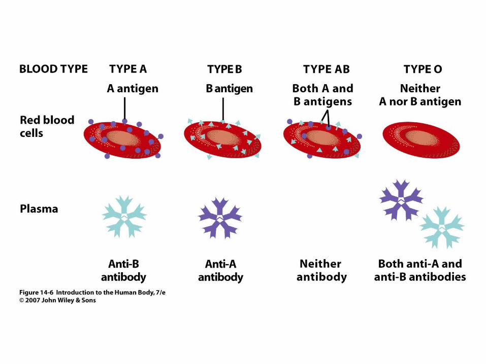
Figure 14.6Figure 14.6

14
Uni
t 1
Rh Blood GroupRh Blood GroupRh Blood GroupRh Blood Group
• The Rh blood group is named after the rhesus monkey where the Rh antigen was discovered
• If have antigen you are Rh+
• Normally we don’t have anti-Rh antibodies they develop after the first exposure from transfusion
14
Uni
t 1
TransfusionsTransfusionsTransfusionsTransfusions
• Transfusion – transfer of whole blood or blood components (plasma)
• If mismatched blood is given antibodies in the recipient's blood bind to the antigens on the donated RBC and causes hemolysis (rupture of RBC)

14
Uni
t 1
TransfusionsTransfusionsTransfusionsTransfusions
• Universal recipients - Type AB has no AB antibodies so can receive any ABO type blood
• Universal donors - Type O have neither antigen so can donate to any other ABO type
• Misleading because of many other blood groups that must be matched