14 Lymphocytes and Pulsing Magnetic Fieldsandrewamarino.com/PDFs/MB/MB_Ch14.pdf · partial...
32
327 14 Lymphocytes and Pulsing Magnetic Fields RUGGERO CADOSSI, GIOVANNI EMILIA GIOVANNI CECHERELLI, GIUSEPPE TORELLI Center for Experimental Haematology, Department of Internal Medicine II, University of Modena Modena, Italy INTRODUCTION Pulsing electromagnetic fields (PEMFs) are presently used in centers around the world to promote the healing of congenital and acquired pseudarthroses (1-6). Despite the large number of clinical studies, very little is known about the mechanism of action of PEMFs at the cellular level. Several biological models have been used to investigate PEMF effects at the cellular level. Luben et al. (7) showed that PEMF exposure inhibited parathyroid hormone (PTH) action on cultured bone cells, but did not interfere with the action of vitamin D. It is interesting to observe that, while PTH binds to a receptor on the cell membrane, vitamin D acts directly within the cell in the cytoplasm. Dixey and Rein (8) observed that noradrenaline release by cultured nerve cells was significantly increased after 30 minutes of PEMF exposure. In an attempt to explain this result, they pointed out that cytoplasmic secretory phenomena are dependent on Ca ++ influx, and they suggested that the observed phenomenon might be due to a PEMF effect on Ca ++ influx across the cell membrane. Rodan et al. (9) found that cartilage cells exposed to 5 Hz electric fields exhibited increased DNA synthesis. They were able to inhibit the electric field effect by means of verapamil, a calcium antagonist. Liboff et al. (10) reported enhance d DNA synthesis when cultured fibroblasts were exposed to an electromagnetic field during that phase of the cell cycle in which the transition from G 1 to S occurred. Goodman et al. (11) showed increased RNA transcription in dipterian salivary gland cells in cell cultures exposed to PEMFs. Ottani et al. (12) demonstrated that PEMF exposure increased the regeneration rate in rat liver after partial haepatectomy. Grattarola et al. (13) and Conti et al. (14) employed different signals, but both reported that PEMF exposure decreased the response of normal human lymphocytes to lectin stimulation. Conversely, Hellman et al. (15) and Emilia et al. (16) reported that lectin-stimulated lymphocytes exposed to electromagnetic radiation exhibited increased
Transcript of 14 Lymphocytes and Pulsing Magnetic Fieldsandrewamarino.com/PDFs/MB/MB_Ch14.pdf · partial...
327
14
Lymphocytes and Pulsing Magnetic Fields
RUGGERO CADOSSI, GIOVANNI EMILIA GIOVANNI CECHERELLI, GIUSEPPE TORELLI
Center for Experimental Haematology, Department of Internal Medicine II, University of Modena
Modena, Italy
INTRODUCTION
Pulsing electromagnetic fields (PEMFs) are presently used in centers around the world to promote the healing of congenital and acquired pseudarthroses (1-6). Despite the large number of clinical studies, very little is known about the mechanism of action of PEMFs at the cellular level.
Several biological models have been used to investigate PEMF effects at the cellular level. Luben et al. (7) showed that PEMF exposure inhibited parathyroid hormone (PTH) action on cultured bone cells, but did not interfere with the action of vitamin D. It is interesting to observe that, while PTH binds to a receptor on the cell membrane, vitamin D acts directly within the cell in the cytoplasm. Dixey and Rein (8) observed that noradrenaline release by cultured nerve cells was significantly increased after 30 minutes of PEMF exposure. In an attempt to explain this result, they pointed out that cytoplasmic secretory phenomena are dependent on Ca++ influx, and they suggested that the observed phenomenon might be due to a PEMF effect on Ca++ influx across the cell membrane. Rodan et al. (9) found that cartilage cells exposed to 5 Hz electric fields exhibited increased DNA synthesis. They were able to inhibit the electric field effect by means of verapamil, a calcium antagonist.
Liboff et al. (10) reported enhance d DNA synthesis when cultured fibroblasts were exposed to an electromagnetic field during that phase of the cell cycle in which the transition from G1 to S occurred. Goodman et al. (11) showed increased RNA transcription in dipterian salivary gland cells in cell cultures exposed to PEMFs. Ottani et al. (12) demonstrated that PEMF exposure increased the regeneration rate in rat liver after partial haepatectomy.
Grattarola et al. (13) and Conti et al. (14) employed different signals, but both reported that PEMF exposure decreased the response of normal human lymphocytes to lectin stimulation. Conversely, Hellman et al. (15) and Emilia et al. (16) reported that lectin-stimulated lymphocytes exposed to electromagnetic radiation exhibited increased
328 CADOSSI ET AL.
DNA synthesis.
THE CELLULAR TARGETS OF PEMFs
The review of the published data (7-47) allows us to draw some general conclusions about PEMF mechanisms of action. PEMFs may act by interfering with those membrane processes that govern the interaction between an active substance and its receptor. The cell membrane is probably one example of a PEMF target. PEMFs also appear to produce an effect on Ca++ flux across the cell membrane, and they seem capable of triggering an increase in the pool of free intracellular Ca++. In the light of these considerations, the present interest in the lymphocyte system is understandable, because this biological model is particularly helpful in the study of all the possible sites of action of PEMFs.
Chiabrera et al. used the lymphocyte system to study the PEMF effect on the kinetics of the reaction between the lectin and the membrane receptor (48). Cadossi et al. (49) and Conti et al. (43) estimated the effects produced by PEMF exposure on 45Ca++- influx into the lymphocyte. Hellman et al. (50), Emilia et al. (16), and Conti et al. (14) used this system to evaluate the effect on DNA synthesis.
Before describing the details of lymphocyte cultures, we will briefly summarize the physiology of the lymphocytes and the changes that occur when lymphocytes are cultured in the presence of mitogens; we will focus on lectin stimulation.
THE LYMPHOCYTE-LECTIN MODEL
LYMPHOCYTE STIMULATION BY LECTINS
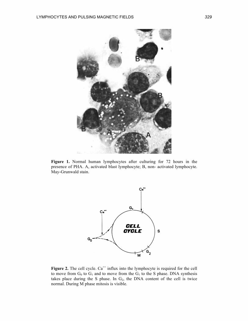
When cultured in the presence of a lectin such as phytohemagglutinin (PHA) (a glycoprotein extracted from Phaseolus vulgaris), human normal lymphocytes obtained from peripheral blood undergo a series of morphological changes. The small, relatively inactive lymphocyte becomes transformed into a lymphoblast, a larger, metabolically active cell (51-54) (Figure 1). This morphological change is coupled with a cell transition from the resting phase, G0, to the G1 phase of the cell cycle.
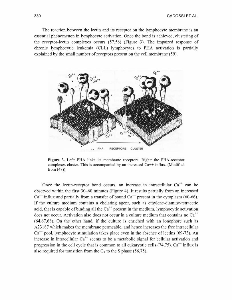
A lymphocyte in G1 does not necessarily enter the S phase. Some cells never begin DNA synthesis (55,56) even when they are morphologically transformed into blasts. In late G1, there is a point that the cell must cross to enter the S phase of DNA synthesis. The crossing point is dependent on Ca++ influx. Once the cell has entered the S phase, it usually completes the cycle and doubles. It can then either proceed in the cycle or come out of it (Figure 2).
LYMPHOCYTES AND PULSING MAGNETIC FIELDS 329
Figure 1. Normal human lymphocytes after culturing for 72 hours in the presence of PHA. A, activated blast lymphocyte; B, non- activated lymphocyte. May-Grunwald stain.
Figure 2. The cell cycle. Ca++ influx into the lymphocyte is required for the cell to move from G0 to G1 and to move from the G1 to the S phase. DNA synthesis takes place during the S phase. In G2, the DNA content of the cell is twice normal. During M phase mitosis is visible.
330 CADOSSI ET AL.
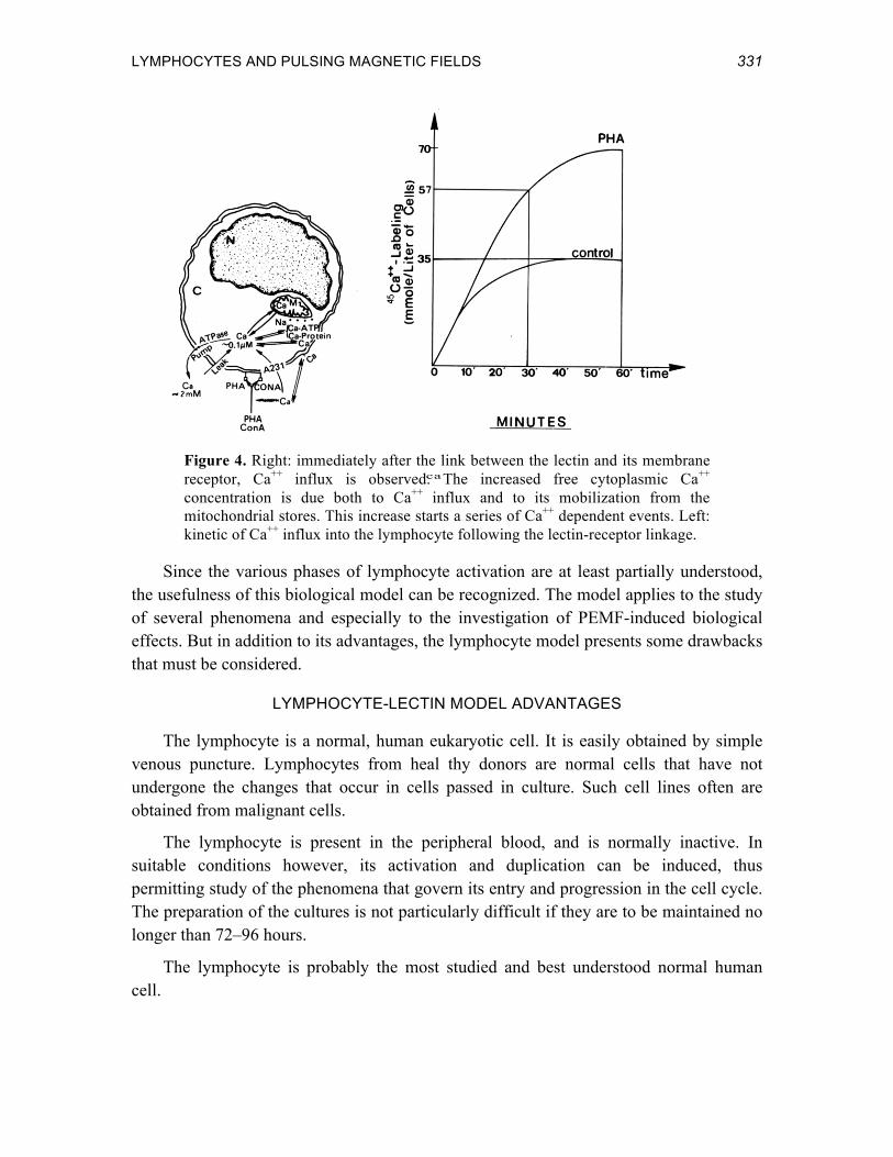
The reaction between the lectin and its receptor on the lymphocyte membrane is an essential phenomenon in lymphocyte activation. Once the bond is achieved, clustering of the receptor-lectin complexes occurs (57,58) (Figure 3). The impaired response of chronic lymphocytic leukemia (CLL) lymphocytes to PHA activation is partially explained by the small number of receptors present on the cell membrane (59).
Figure 3. Left: PHA links its membrane receptors. Right: the PHA-receptor complexes cluster. This is accompanied by an increased Ca++ influx. (Modified from (48)).
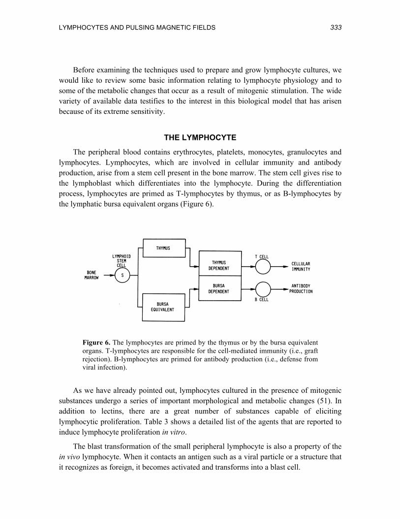
Once the lectin-receptor bond occurs, an increase in intracellular Ca++ can be observed within the first 30–60 minutes (Figure 4). It results partially from an increased Ca++ influx and partially from a transfer of bound Ca++ present in the cytoplasm (60-66). If the culture medium contains a chelating agent, such as ethylene-diamine-tetracetic acid, that is capable of binding all the Ca++ present in the medium, lymphocytic activation does not occur. Activation also does not occur in a culture medium that contains no Ca++ (64,67,68). On the other hand, if the culture is enriched with an ionophore such as A23187 which makes the membrane permeable, and hence increases the free intracellular Ca++ pool, lymphocyte stimulation takes place even in the absence of lectins (69-73). An increase in intracellular Ca++ seems to be a metabolic signal for cellular activation and progression in the cell cycle that is common to all eukaryotic cells (74,75). Ca++ influx is also required for transition from the G1 to the S phase (56,75).
LYMPHOCYTES AND PULSING MAGNETIC FIELDS 331
·
ca
Figure 4. Right: immediately after the link between the lectin and its membrane receptor, Ca++ influx is observed. The increased free cytoplasmic Ca++ concentration is due both to Ca++ influx and to its mobilization from the mitochondrial stores. This increase starts a series of Ca++ dependent events. Left: kinetic of Ca++ influx into the lymphocyte following the lectin-receptor linkage.
Since the various phases of lymphocyte activation are at least partially understood, the usefulness of this biological model can be recognized. The model applies to the study of several phenomena and especially to the investigation of PEMF-induced biological effects. But in addition to its advantages, the lymphocyte model presents some drawbacks that must be considered.
LYMPHOCYTE-LECTIN MODEL ADVANTAGES
The lymphocyte is a normal, human eukaryotic cell. It is easily obtained by simple venous puncture. Lymphocytes from heal thy donors are normal cells that have not undergone the changes that occur in cells passed in culture. Such cell lines often are obtained from malignant cells.
The lymphocyte is present in the peripheral blood, and is normally inactive. In suitable conditions however, its activation and duplication can be induced, thus permitting study of the phenomena that govern its entry and progression in the cell cycle. The preparation of the cultures is not particularly difficult if they are to be maintained no longer than 72–96 hours.
The lymphocyte is probably the most studied and best understood normal human cell.

332 CADOSSI ET AL.
LYMPHOCYTE-LECTIN MODEL DRAWBACKS
The amount of stimulation by lectins varies among different donors, and it may vary within the same donor depending on immunological status. The lymphocyte cell population is not synchronized, and cannot be made synchronous.
The obtainable quantity of lymphocytes is a limiting factor. The lymphocyte population lacks homogeneity in that several subpopulations can be delineated. Table 1 lists some of the most common surface markers used to identify B- and T- lymphocytes. The main categories recognized are B- and T- lymphocytes, whose responses to mitogenic stimuli induced by lectins are different as shown in Table 2. The lymphocytic response to lectins is also regulated by the release of growth factors that modulate the response of the different lymphocyte subpopulations (Figure 5).
Table 1. Lymphocyte Identification by Surface Markers
Cell Type Surface Markers S Ig C3 Fc ME SE IAg
T – + + – + + B + + + + – +
Mitogen Lymphocytes
Human Mouse T B T B
Phytohemagglutinin (PHA) + ? + – Concanavalin A (ConA) + – + – Pokeweed (PWM) + + + + Insoluble PHA, ConA, PWM + + + +
Figure 5. Schematic representation of the interaction between T- and B-lymphocytes responding to PHA stimulus.
LYMPHOCYTES AND PULSING MAGNETIC FIELDS 333
Before examining the techniques used to prepare and grow lymphocyte cultures, we would like to review some basic information relating to lymphocyte physiology and to some of the metabolic changes that occur as a result of mitogenic stimulation. The wide variety of available data testifies to the interest in this biological model that has arisen because of its extreme sensitivity.
THE LYMPHOCYTE
The peripheral blood contains erythrocytes, platelets, monocytes, granulocytes and lymphocytes. Lymphocytes, which are involved in cellular immunity and antibody production, arise from a stem cell present in the bone marrow. The stem cell gives rise to the lymphoblast which differentiates into the lymphocyte. During the differentiation process, lymphocytes are primed as T-lymphocytes by thymus, or as B-lymphocytes by the lymphatic bursa equivalent organs (Figure 6).
Figure 6. The lymphocytes are primed by the thymus or by the bursa equivalent organs. T-lymphocytes are responsible for the cell-mediated immunity (i.e., graft rejection). B-lymphocytes are primed for antibody production (i.e., defense from viral infection).
As we have already pointed out, lymphocytes cultured in the presence of mitogenic substances undergo a series of important morphological and metabolic changes (51). In addition to lectins, there are a great number of substances capable of eliciting lymphocytic proliferation. Table 3 shows a detailed list of the agents that are reported to induce lymphocyte proliferation in vitro.
The blast transformation of the small peripheral lymphocyte is also a property of the in vivo lymphocyte. When it contacts an antigen such as a viral particle or a structure that it recognizes as foreign, it becomes activated and transforms into a blast cell.

334 CADOSSI ET AL.
Within the first two hours after the lymphocyte-lectin interaction, an enhanced incorporation of precursors occurs in proteins (76), RNA (77), and in lipids (78). The formation of lactate and pyruvate also increases (79). DNA synthesis begins 24 hours after PHA addition (80). The significant change in RNA metabolism associated with blast formation is shown in Figure 7. If a culture is maintained for 48–72 hours, mitosis can be observed.
Table 3. Agents Reported to Induce Lymphocyte Proliferation
Non-Specific Mitogens Specific Antigens Chymotripsin Ag-Ab complexes Yeast zymosan Dilantin Lectins Diphtheria toxoid Microwave irradiation Histoplasm Papain Penicillin Staphylococcal filtrate Polio vaccine Streptolysin Purified protein derivate Trypsin Ragweed pollen Some endotoxins Tissue Antigens Typhoid-paratyphoid vaccine Cerebrospinal fluid Vaccinia vaccine Extract of white blood cells Varidase Fetal calf serum Homologous macrophages Antisera Leukocyte supernatant Monkey antihuman Ig Lymphocyte homologous cultures Rabbit antihuman leukocyte Platelets Some antisera Sera
Some recent techniques of in situ hybridization, making use of messenger RNAs whose transcription is specifically related to the phases of the cell cycle, allow us to show the cell transition through the different phases of the cell cycle from a molecular standpoint (81) (Figure 8).
CHRONIC LYMPHOCYTIC LEUKEMIA LYMPHOCYTES
Chronic lymphocytic leukemia (CLL) is a pathological condition characterized bythe presence in the patients’ peripheral blood of a large number of small lymphocytes (very often over 30,000/mm3), whose metabolic activity is poor. These are generally extremely homogeneous populations (90%). CLL lymphocytes often share the membrane antigens that are specific for B-lymphocytes and, much more rarely, for T-lymphocytes. It is therefore the B-lymphocyte populations that show an impaired response to lectin stimulation (82,83). These characteristics make CLL lymphocytes very useful for lectin studies.
LYMPHOCYTES AND PULSING MAGNETIC FIELDS 335
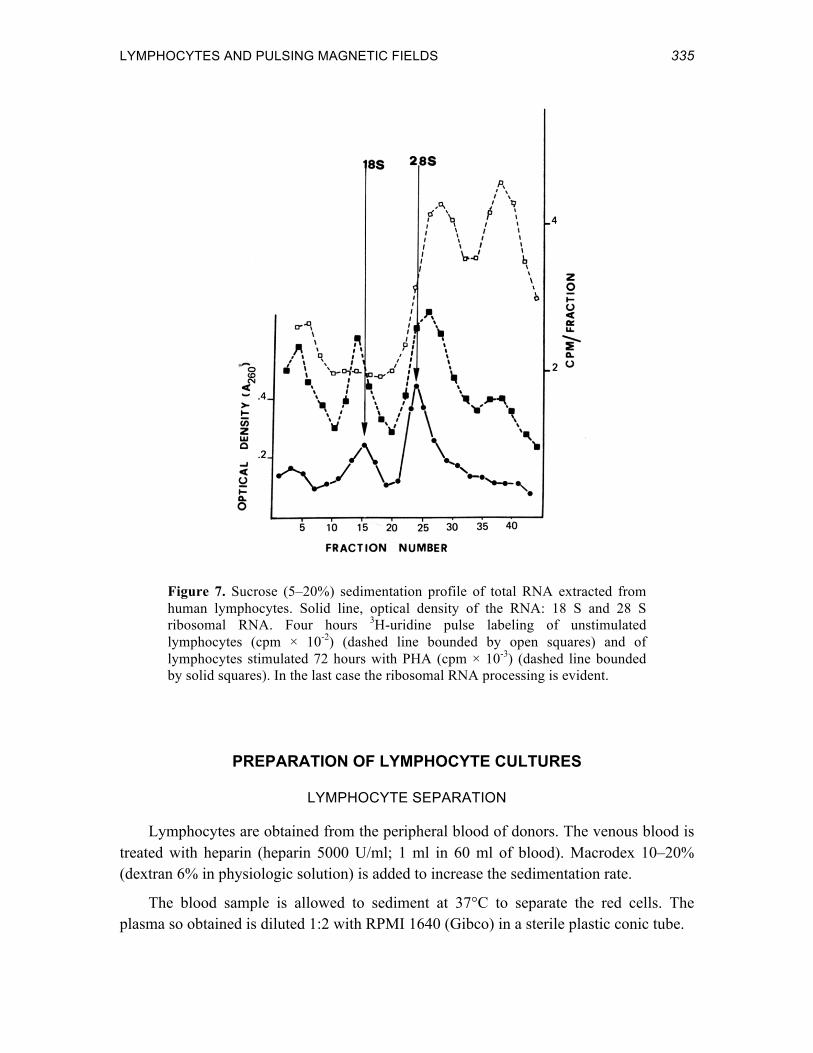
Figure 7. Sucrose (5–20%) sedimentation profile of total RNA extracted from human lymphocytes. Solid line, optical density of the RNA: 18 S and 28 S ribosomal RNA. Four hours 3H-uridine pulse labeling of unstimulated lymphocytes (cpm × 10-2) (dashed line bounded by open squares) and of lymphocytes stimulated 72 hours with PHA (cpm × 10-3) (dashed line bounded by solid squares). In the last case the ribosomal RNA processing is evident.
PREPARATION OF LYMPHOCYTE CULTURES
LYMPHOCYTE SEPARATION
Lymphocytes are obtained from the peripheral blood of donors. The venous blood is treated with heparin (heparin 5000 U/ml; 1 ml in 60 ml of blood). Macrodex 10–20% (dextran 6% in physiologic solution) is added to increase the sedimentation rate.
The blood sample is allowed to sediment at 37°C to separate the red cells. The plasma so obtained is diluted 1:2 with RPMI 1640 (Gibco) in a sterile plastic conic tube.
336 CADOSSI ET AL.
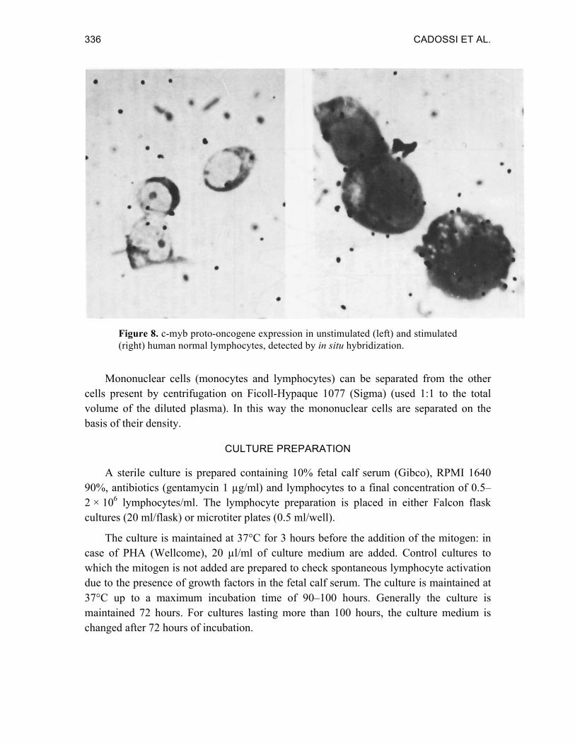
Figure 8. c-myb proto-oncogene expression in unstimulated (left) and stimulated (right) human normal lymphocytes, detected by in situ hybridization.
Mononuclear cells (monocytes and lymphocytes) can be separated from the other cells present by centrifugation on Ficoll-Hypaque 1077 (Sigma) (used 1:1 to the total volume of the diluted plasma). In this way the mononuclear cells are separated on the basis of their density.
CULTURE PREPARATION
A sterile culture is prepared containing 10% fetal calf serum (Gibco), RPMI 1640 90%, antibiotics (gentamycin 1 µg/ml) and lymphocytes to a final concentration of 0.5–2 × 10⁶ lymphocytes/ml. The lymphocyte preparation is placed in either Falcon flask cultures (20 ml/flask) or microtiter plates (0.5 ml/well).
The culture is maintained at 37°C for 3 hours before the addition of the mitogen: in case of PHA (Wellcome), 20 µl/ml of culture medium are added. Control cultures to which the mitogen is not added are prepared to check spontaneous lymphocyte activation due to the presence of growth factors in the fetal calf serum. The culture is maintained at 37°C up to a maximum incubation time of 90–100 hours. Generally the culture is maintained 72 hours. For cultures lasting more than 100 hours, the culture medium is changed after 72 hours of incubation.
LYMPHOCYTES AND PULSING MAGNETIC FIELDS 337
THE EFFECT OF PEMFs ON LYMPHOCYTES
The lymphocyte-lectin experimental model has been used by many authors, both to study the metabolic effect of PEMFs used clinically, and to evaluate possible side-effects due to chronic exposure to the electric fields of the high-voltage powerlines. All the studies that we will consider deal with non-thermal effects.
GENOVA GROUP
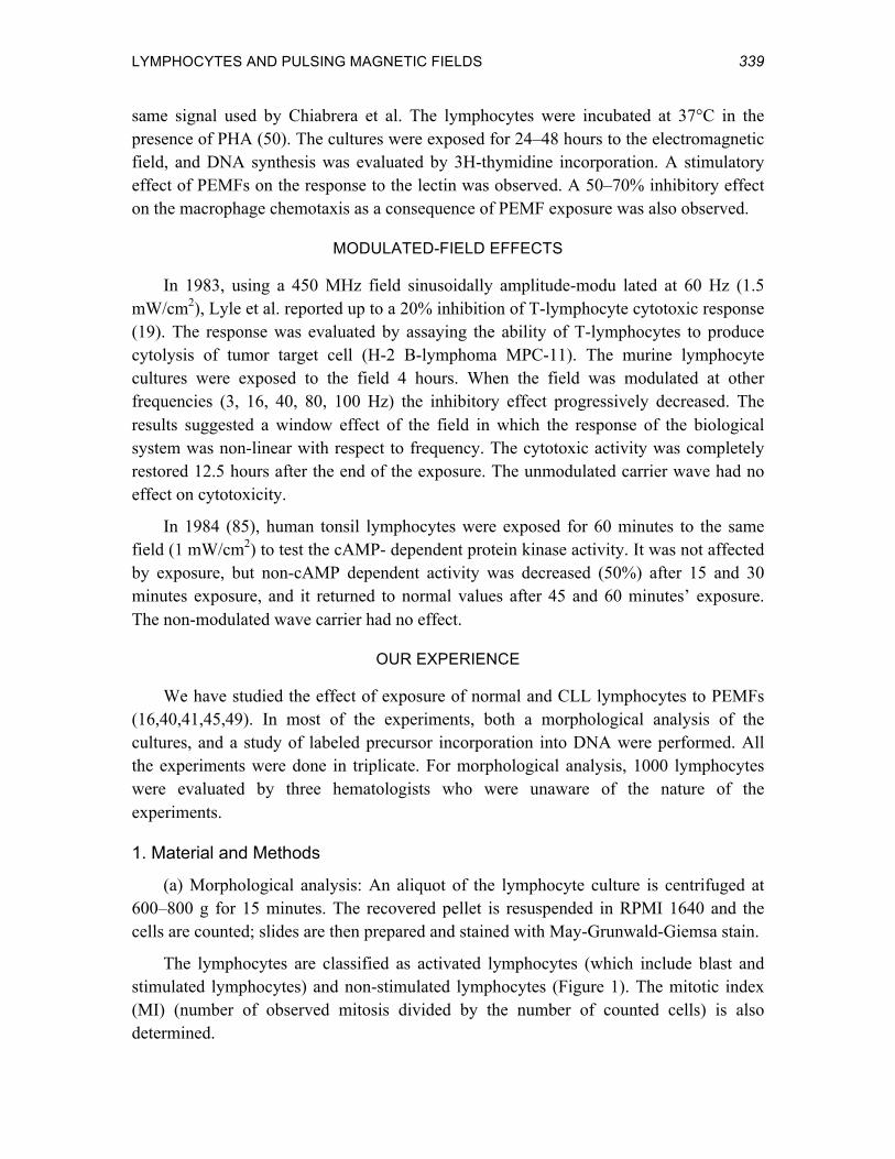
The investigators (13,34,46,48) studied the effect of PEMFs developed at Columbia University on normal human lymphocytes. Their PEMF generator supplied a pair of air-core coils with either a single-pulse signal or a burst signal. Both are used clinically for different indications. The signal employed is shown in Figure 9.
Figure 9. Waveform of the electrical signal used by the Genova group, as it is induced in a calibrated coil probe.
Lymphocyte cultures containing sub-optimal concentrations of PHA (2.5–10 µl/ml) were prepared from a sample of peripheral blood and incubated at 37°C for 72 hours. Four different samples were prepared: (a) control cultures; (b) PHA-containing cultures; (c) PEMF-exposed cultures; and (d) PHA-containing plus PEMF-exposed cultures. The samples were exposed to PEMFs for 72 hours. A careful and detailed analysis of the exposure system was given.
Both the change in lymphocyte volume corresponding to lymphocyte activation, and the number of cells stained by acridine orange (a measure of the DNA content of the cell) were evaluated. The results showed that PEMF exposure decreased the response of the lymphocytes to sub-optimum PHA concentrations.
On the bases of these and other observations, the authors developed a mathematical model to fit the effect of PEMFs on the interaction between PHA and its membrane receptor. They concluded that PEMF exposure decreased the time spent by PHA in the proximity of its cellular receptor, thereby decreasing the response to the mitogenic

338 CADOSSI ET AL.
stimulus.
The computerized model developed allowed a prediction that PEMF exposure increased the availability of free Ca++ near the membrane and the Ca++ influx via pre-existing channels.
L’AQUILA GROUP
A wide series of studies were performed on the effect of PEMFs on mitogen-stimulated lymphocytes (14,43,44). Normal lymphocyte cultures were prepared and DNA synthesis was evaluated by 3H-thymidine incorporation, using the microtiter-plate technique. The lymphocytes were incubated for 72 hours at 37°C, and at the 66th hour the labeled precursor was added to the cultures. DNA synthesis was evaluated by tricloracetic-acid precipitation and counting.
The electromagnetic-field generator supplied a pair of coils with a “train of pulses of variable form and intensity;” the field value was 23–65 Gauss. The signal waveform and the actual field intensity are not given.
The effect of different frequencies on the response of lymphocytes to PHA, pokeweed mitogen (PWM) and concanavalin A (ConA) was tested. A window effect was observed , and the most inhibitory response occurred at 3 Hz. Table 4 shows the results obtained at different frequencies with the three lectins.
Table 4. Inhibitory Effect of PEMFs on the Lymphocyte Response to Lectins
Mitogen Inhibitory Effect Frequency 1 Hz 3 Hz 50 Hz 200 Hz
PHA Yes Yes Yes Yes ConA No Yes Yes No PWM No Yes No No
To test the hypothesis that PEMFs could interfere with 3H-thymidine transport across the cell membrane, the cultures were exposed to PEMFs in the last 6 hours of incubation: no effect was observed as a consequence of PEMF exposure. The authors suggested that PEMFs could prevent the link between the receptor and the mitogen, and could decrease Ca++ influx into the lymphocyte. The authors have recently shown stimulating effect of PEMFs on the lymphocyte response to the mitogens when suboptimal concentration of lectins are used.
WASHINGTON DC GROUP
Spleen lymphocytes obtained from adult female BALB/c mice were exposed to the
LYMPHOCYTES AND PULSING MAGNETIC FIELDS 339
same signal used by Chiabrera et al. The lymphocytes were incubated at 37°C in the presence of PHA (50). The cultures were exposed for 24–48 hours to the electromagnetic field, and DNA synthesis was evaluated by 3H-thymidine incorporation. A stimulatory effect of PEMFs on the response to the lectin was observed. A 50–70% inhibitory effect on the macrophage chemotaxis as a consequence of PEMF exposure was also observed.
MODULATED-FIELD EFFECTS
In 1983, using a 450 MHz field sinusoidally amplitude-modu lated at 60 Hz (1.5 mW/cm2), Lyle et al. reported up to a 20% inhibition of T-lymphocyte cytotoxic response (19). The response was evaluated by assaying the ability of T-lymphocytes to produce cytolysis of tumor target cell (H-2 B-lymphoma MPC-11). The murine lymphocyte cultures were exposed to the field 4 hours. When the field was modulated at other frequencies (3, 16, 40, 80, 100 Hz) the inhibitory effect progressively decreased. The results suggested a window effect of the field in which the response of the biological system was non-linear with respect to frequency. The cytotoxic activity was completely restored 12.5 hours after the end of the exposure. The unmodulated carrier wave had no effect on cytotoxicity.
In 1984 (85), human tonsil lymphocytes were exposed for 60 minutes to the same field (1 mW/cm2) to test the cAMP- dependent protein kinase activity. It was not affected by exposure, but non-cAMP dependent activity was decreased (50%) after 15 and 30 minutes exposure, and it returned to normal values after 45 and 60 minutes’ exposure. The non-modulated wave carrier had no effect.
OUR EXPERIENCE
We have studied the effect of exposure of normal and CLL lymphocytes to PEMFs (16,40,41,45,49). In most of the experiments, both a morphological analysis of the cultures, and a study of labeled precursor incorporation into DNA were performed. All the experiments were done in triplicate. For morphological analysis, 1000 lymphocytes were evaluated by three hematologists who were unaware of the nature of the experiments.
1. Material and Methods
(a) Morphological analysis: An aliquot of the lymphocyte culture is centrifuged at 600–800 g for 15 minutes. The recovered pellet is resuspended in RPMI 1640 and the cells are counted; slides are then prepared and stained with May-Grunwald-Giemsa stain.
The lymphocytes are classified as activated lymphocytes (which include blast and stimulated lymphocytes) and non-stimulated lymphocytes (Figure 1). The mitotic index (MI) (number of observed mitosis divided by the number of counted cells) is also determined.
340 CADOSSI ET AL.
(b) DNA synthesis assay: 3H-thymidine, 1 µCi/ml of culture medium (New England Nuclear; 21.5 Ci/mM), is added (usually after the initial 48 hours of incubation). At the 72nd hour of incubation the cells are collected, and aliquots are used for assaying the labeled precursor incorporation into the DNA. Ten milliliters of 10% tricloracetic acid (TCA) is added to the lymphocyte sample and, after 10 minutes at 4°C, the sample is filtered on Whatmann fiber-glass filters. The filters are washed twice with 10 ml of 10% TCA, dried, and then counted in Toluene-Permafluor (24:1) in a liquid scintillator (Packard).
(c) Karyotype preparation: Chromosomes are prepared according to the Summer ASG-technique for G band analysis (84,85). The technique yields evidence of translocations, deletions, and other damage that occurs as a consequence of exposure.
(d) Ca++ influx evaluation: In some experiments the cultured lymphocytes were used to test the effect of PEMFs on 45Ca++-influx. One milliliter cultures containing 2 × 10⁶ lymphocytes are prepared and maintained at 37°C for 3 hours before the addition of 10 µCi/ml of 45Ca++ (Amersham Int., 45CaC12 10 mCi/Mg-Ca). When the experiments are performed with PHA-stimulated cultures, 45Ca++ is added immediately after PHA addition. Half of the cultures are used as controls, and the others are exposed to PEMFs.
After 1 hour incubation at 37°C in the presence of the 45Ca++, 5 ml of cold NaCl (0.15 M) is added and the cultures are centrifuged at 800 g for 5 minutes. The lymphocytes are washed twice and then 1 ml of Soluene 350 (Packard) is added; the lymphocyte pellet is incubated overnight, and then 10 ml of Toluene-Permafluor is added and the sample is counted in a liquid scintillation counter.
(e) T-lymphocyte removal: T-lymphocytes can be removed from the culture medium because of the presence on their surface of specific receptors (86,87). Once the lymphocytes have been separated, 0.5 ml of culture medium containing 5 × 10⁶ lymphocytes are added to 0.25 ml of fetal calf serum and to an equal volume of 10% sheep red blood cells sensitized with AET (Sigma), the mixture is centrifuged at 100 g per 10 minutes at 4°C and incubated overnight at 4°C. The sheep erythrocytes link to the T-lymphocytes and form rosettes (Figure 10).
By centrifugation on Ficoll-Hypaque 1077 at 4°C, rosetted T-lymphocytes can be removed (found on the bottom of the gradient). With this technique about 95–98% of the T-lymphocytes can be removed.
(f) The exposure system: For PEMF exposure, the cultures are placed between two coils as shown in Figure 11. The control and PEMF-exposed cultures are maintained in two separate incubators placed in different rooms. Control cultures are also maintained in the same incubator as the exposed cultures, but in a position where no current is induced in a coil probe. There is no significant difference between these control cultures and those maintained in the separate incubators. No difference is found in the temperature of the
LYMPHOCYTES AND PULSING MAGNETIC FIELDS 341
medium between the control and exposed cultures.
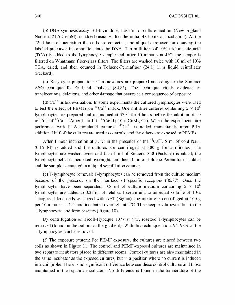
Figure 10. T-lymphocyte rosette. The lymphocyte in the center is surrounded by sensitized sheep erythrocytes.
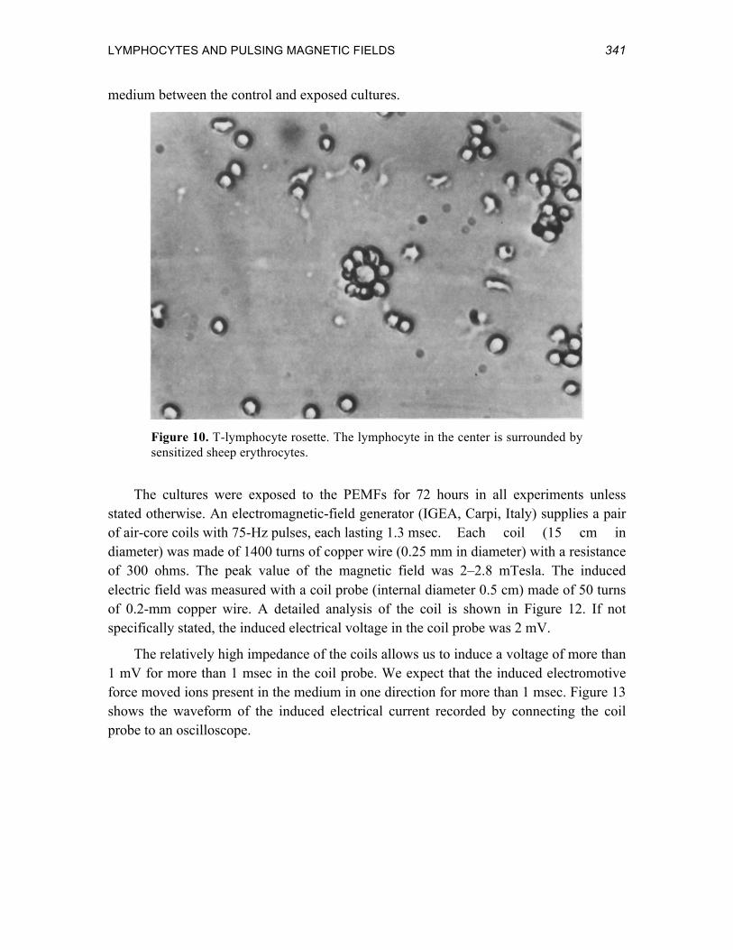
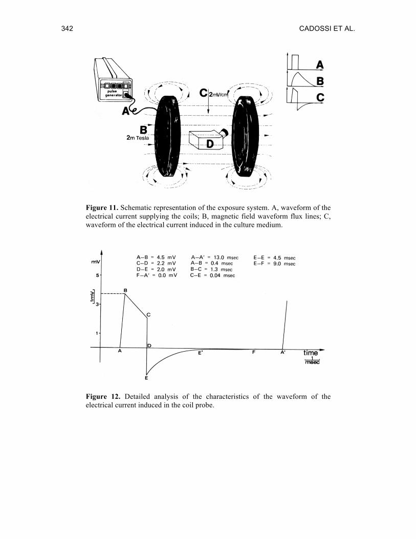
The cultures were exposed to the PEMFs for 72 hours in all experiments unless stated otherwise. An electromagnetic-field generator (IGEA, Carpi, Italy) supplies a pair of air-core coils with 75-Hz pulses, each lasting 1.3 msec. Each coil (15 cm in diameter) was made of 1400 turns of copper wire (0.25 mm in diameter) with a resistance of 300 ohms. The peak value of the magnetic field was 2–2.8 mTesla. The induced electric field was measured with a coil probe (internal diameter 0.5 cm) made of 50 turns of 0.2-mm copper wire. A detailed analysis of the coil is shown in Figure 12. If not specifically stated, the induced electrical voltage in the coil probe was 2 mV.
The relatively high impedance of the coils allows us to induce a voltage of more than 1 mV for more than 1 msec in the coil probe. We expect that the induced electromotive force moved ions present in the medium in one direction for more than 1 msec. Figure 13 shows the waveform of the induced electrical current recorded by connecting the coil probe to an oscilloscope.
342 CADOSSI ET AL.
Figure 11. Schematic representation of the exposure system. A, waveform of the electrical current supplying the coils; B, magnetic field waveform flux lines; C, waveform of the electrical current induced in the culture medium.
Figure 12. Detailed analysis of the characteristics of the waveform of the electrical current induced in the coil probe.

LYMPHOCYTES AND PULSING MAGNETIC FIELDS 343
Figure 13. Actual oscilloscope recording of the voltage induced in the coil probe. 1 msec/cm time division and 1 mV/cm amplitude division.
2. Results
The cultured human lymphocytes exhibited some spontaneous stimulation because of growth factors contained in fetal calf serum. The amount of such stimulation was 4–8% in the case of normal lymphocytes, and even lower for CLL lymphocytes. This spontaneous lymphocyte stimulation was not modified by exposure to PEMFs.
When PHA or PWM were present in the culture medium, the percentage of activated normal and CLL lymphocytes increased significantly in the PEMF-exposed cultures compared to the controls. The same increase was observed when evaluating both DNA synthesis and mitotic index. The PEMF stimulatory effect was particularly evident in the CLL cultures. The increase in mitotic index suggested that PEMFs raised not only the number of lymphocytes activated by lectins, but also favored the entry into the S phase of the cell cycle of both lymphocytes stimulated by PHA alone, and of PHA + PEMFs stimulated lymphocytes.
Table 5 shows the average values obtained after stimulating 14 CLL and 10 normal cultures with PHA. The statistical analysis showed a stimulatory effect of PEMFs over all the tested parameters. In all the experiments performed, the mitotic index was always increased in the exposed cultures. PEMFs in association with PHA induced almost all the lymphocytes to enter the cell cycle.
Table 6 shows the results obtained using PWM. The same stimulatory effect of PEMFs was observed, indicating that it was not dependent on the lectin used.
Because of the importance of Ca++ fluxes for lymphocyte activation by lectins, we
344 CADOSSI ET AL.
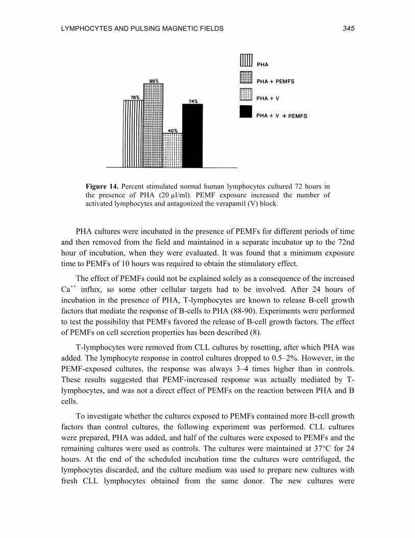
++ evaluated the effects of PEMF exposure on calcium. After 1 hour of exposure, the amount of 45Ca++ present in the lymphocyte cytoplasm was increased both when PHA was present in the culture medium, as well· as when it was not added. However the increased Ca++ influx in the case of cultures to which PHA had not been added was not enough to induce lymphocyte proliferation. Further experiments were conducted in the presence in the culture medium of a Ca++ antagonist, 10-6 M verapamil. Verapamil inhibited the lymphocyte response to PHA, but when the cultures were exposed to PEMFs, the verapamil block was antagonized so that the number of responsive cells increased to the value obtained with PHA alone (Figure 14). These data suggested that the PEMF effect on the cell could be explained on the basis of an effect on Ca++ fluxes across the membrane. If this was the case, short exposure times (1–2 hours) should be sufficient because Ca++ influx is an early event in lymphocyte activation ( the G0-G1 transition phase).
Table 5. Effect of PEMFs on the Response of Normal (N = 10) and CLL (N = 14) Lymphocytes to PHA Stimulation
Parameter Evaluated PHA PHA + PEMFs Normal Lymphocytes: % Activated 81% 98%* 3H-thymidine (cpm × 10-3) 140 203* Mitotic Index 0.065 0.085* CLL Lymphocytes: % Activated 32% 48%* 3H-thymidine (cpm × 10-3) 20 38* Mitotic Index 0.01 0.03* *P < 0.01, Student’s t test
Table 6. Effect of PEMFs on the Response of Normal (N = 3) and CLL (N = 3) Lymphocytes to PWM Stimulation
Parameter Evaluated PWM PWM + PEMFs Normal Lymphocytes: % Activated 65% 97% Mitotic Index 0.05 0.07 CLL Lymphocytes: % Activated 16% 31% Mitotic Index 0.01 0.02
LYMPHOCYTES AND PULSING MAGNETIC FIELDS 345
Figure 14. Percent stimulated normal human lymphocytes cultured 72 hours in the presence of PHA (20 µl/ml). PEMF exposure increased the number of activated lymphocytes and antagonized the verapamil (V) block.
PHA cultures were incubated in the presence of PEMFs for different periods of time and then removed from the field and maintained in a separate incubator up to the 72nd hour of incubation, when they were evaluated. It was found that a minimum exposure time to PEMFs of 10 hours was required to obtain the stimulatory effect.
The effect of PEMFs could not be explained solely as a consequence of the increased Ca++ influx, so some other cellular targets had to be involved. After 24 hours of incubation in the presence of PHA, T-lymphocytes are known to release B-cell growth factors that mediate the response of B-cells to PHA (88-90). Experiments were performed to test the possibility that PEMFs favored the release of B-cell growth factors. The effect of PEMFs on cell secretion properties has been described (8).
T-lymphocytes were removed from CLL cultures by rosetting, after which PHA was added. The lymphocyte response in control cultures dropped to 0.5–2%. However, in the PEMF-exposed cultures, the response was always 3–4 times higher than in controls. These results suggested that PEMF-increased response was actually mediated by T-lymphocytes, and was not a direct effect of PEMFs on the reaction between PHA and B cells.
To investigate whether the cultures exposed to PEMFs contained more B-cell growth factors than control cultures, the following experiment was performed. CLL cultures were prepared, PHA was added, and half of the cultures were exposed to PEMFs and the remaining cultures were used as controls. The cultures were maintained at 37°C for 24 hours. At the end of the scheduled incubation time the cultures were centrifuged, the lymphocytes discarded, and the culture medium was used to prepare new cultures with fresh CLL lymphocytes obtained from the same donor. The new cultures were
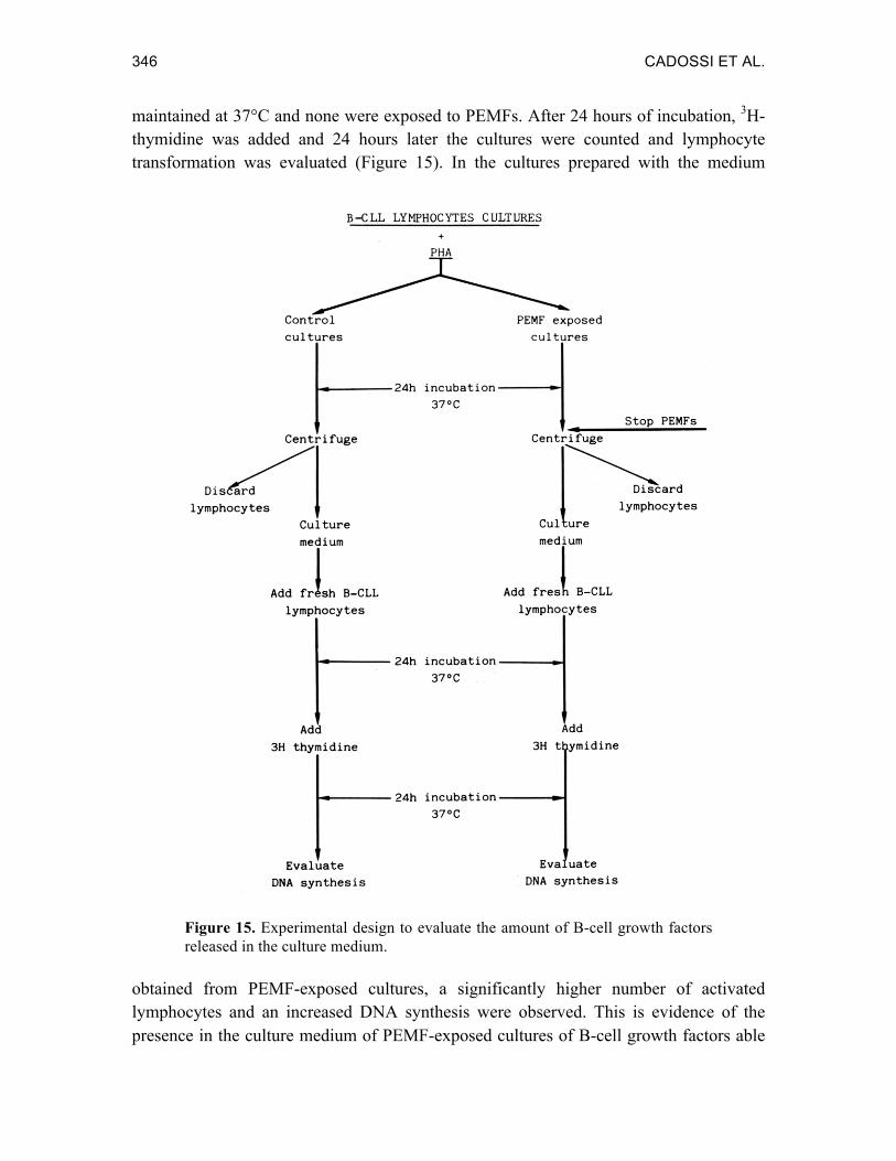
346 CADOSSI ET AL.
maintained at 37°C and none were exposed to PEMFs. After 24 hours of incubation, 3H-thymidine was added and 24 hours later the cultures were counted and lymphocyte transformation was evaluated (Figure 15). In the cultures prepared with the medium
Figure 15. Experimental design to evaluate the amount of B-cell growth factors released in the culture medium.
obtained from PEMF-exposed cultures, a significantly higher number of activated lymphocytes and an increased DNA synthesis were observed. This is evidence of the presence in the culture medium of PEMF-exposed cultures of B-cell growth factors able

LYMPHOCYTES AND PULSING MAGNETIC FIELDS 347
to increase lymphocyte response to PHA. We do not know whether the increased B-cell growth-factor content in PEMF-exposed cultures can be attributed to an increased synthesis or to an augmented release from the T-lymphocytes. The latter effect could be dependent on increased cytoskeleton contraction, which requires Ca++.
When lectins bind to membrane receptors, they cluster on the cell membrane. The lectin-receptor clustering event is necessary for Ca++ influx and lymphocyte activation (48,91,92). We evaluated whether PEMF exposure could increase lymphocyte response in the presence of sub-optimal PHA concentrations as a consequence of an effect on Ca++ influx. Cultures containing different concentrations of PHA were prepared, and half were exposed to PEMFs. Exposure to PEMFs increased the lymphocyte response in the presence of low PHA concentrations, and a greater effect was observed at intermediate PHA concentrations (Figure 16). The same effect has recently been described by Conti (93)and Cantini (94).
Figure 16. Stimulatory effect of PEMFs in the presence of sub-optimal concentration of PHA in the culture medium. The experiment was performed with normal lymphocytes.
348 CADOSSI ET AL.
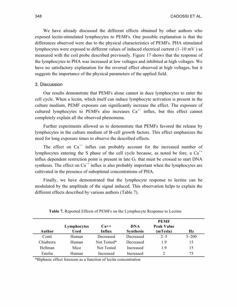
We have already discussed the different effects obtained by other authors who exposed lectin-stimulated lymphocytes to PEMFs. One possible explanation is that the differences observed were due to the physical characteristics of PEMFs. PHA stimulated lymphocytes were exposed to different values of induced electrical current (1–10 mV ) as measured with the coil probe described previously. Figure 17 shows that the response of the lymphocytes to PHA was increased at low voltages and inhibited at high voltages. We have no satisfactory explanation for the reversal effect observed at high voltages, but it suggests the importance of the physical parameters of the applied field.
3. Discussion
Our results demonstrate that PEMFs alone cannot in duce lymphocytes to enter the cell cycle. When a lectin, which itself can induce lymphocyte activation is present in the culture medium, PEMF exposure can significantly increase the effect. The exposure of cultured lymphocytes to PEMFs also increases Ca++ influx, but this effect cannot completely explain all the observed phenomena.
Further experiments allowed us to demonstrate that PEMFs favored the release by lymphocytes in the culture medium of B-cell growth factors. This effect emphasizes the need for long exposure times to observe the described effects.
The effect on Ca++ influx can probably account for the increased number of lymphocytes entering the S phase of the cell cycle because, as noted be fore, a Ca++ influx dependent restriction point is present in late G1 that must be crossed to start DNA synthesis. The effect on Ca++ influx is also probably important when the lymphocytes are cultivated in the presence of suboptimal concentrations of PHA.
Finally, we have demonstrated that the lymphocyte response to lectins can be modulated by the amplitude of the signal induced. This observation helps to explain the different effects described by various authors (Table 7).
Table 7. Reported Effects of PEMFs on the Lymphocyte Response to Lectins
Author Lymphocytes
Used Ca++ Influx
DNA Synthesis
PEMF Peak Value
(mTesla) Hz Conti Human Decreased Decreased 2–5 3–200
Chiabrera Human Not Tested* Decreased 1.9 15 Hellman Mice Not Tested Increased 1.9 15 Emilia Human Increased Increased 2 75
*Biphasic effect foreseen as a function of lectin concentration
LYMPHOCYTES AND PULSING MAGNETIC FIELDS 349
ASSESSMENT OF HEALTH RISK
The lymphocyte model has been used for evaluation of possible damage induced by high-voltage powerlines. We will briefly mention some recent results obtained by exposing lymphocyte cultures to high electric fields.
Winters et al. (95) showed that the exposure to 60-Hz electric fields (300 mA/m2) or magnetic fields (1.0 Gauss) had no effect on human lymphocyte response to a lectin mitogenic stimulus. However, they reported that a significantly increased response was observed in some exposed cultures. They concluded that there was no consistent or significant pattern in the observed responses.
Smith et al. (96) reported that 2-µsec electric-field pulses at up to 3.5 kV/cm did not affect DNA synthesis in mouse spleen lymphocytes incubated in the presence of ConA, PHA or LPS. One 10-µsec pulse, 2.4–3.5 kV/cm, produced a significant reduction in the response of lymphocytes to LPS, which was attributed to cell death.
Nordenson et al. (97), in an in vivo investigation, found that 17 of 20 switchyard workers exhibited chromosomal anomalies. The rate of chromatid and chromosome breaks was significantly higher than in controls. They also showed that 1 mA/cm2 at 50 Hz did not induce any chromosome damage, but that 3-µsec spark discharges (peak strength of 3.5 kV/cm) caused chromosome breaks with a frequency similar to that induced by ionizing radiation.
CONCLUSIONS
The biological model considered here is of great interest, and will probably be used in future studies. For this reason, some points must be further discussed from a physical and a biological point of view. There are several techniques for preparing lymphocyte cultures, and we have shown that small differences in the amplitude of the induced electrical current can significantly change the results. Because of this, the shape of the container in which the lymphocytes are cultivated will play an important role, and should be taken into consideration. One of the most used techniques is the microtiter plate because it is convenient and allows testing of several small samples at the same time. Attention should be paid to magnetic-field homogeneity. If it is not uniform, as often happens in the peripheral wells, different amounts of current will flow in the wells giving rise to misleading results. The exposure conditions must be carefully controlled, and a clear and exhaustive description of the electromagnetic field used should always be given, to allow different authors to exactly reproduce the experimental conditions.
As we pointed out at the beginning, among the drawbacks of the lymphocyte–lectin biological model is the extremely wide range of variability which makes it necessary to repeat the experiments several times. The lymphocyte response to the mitogenic stimulus of the lectins can change at different times even within the same donor. The response
350 CADOSSI ET AL.
among different donors varies significantly.
Finally, the response depends on immunological functional conditions that cannot be foreseen. The immediate consequence is that the amount of the response not only to lectins, but also to PEMFs can vary significantly.
EXPERIMENTS SUGGESTED BY IN VITRO RESULTS
In spite of the extreme variability of the biological response, the lectin-lymphocyte system has allowed us to obtain important information. However we are far from a satisfactory explanation for the mechanisms of action of PEMFs. The available data allow us to propose further experiments and suggest fascinating hypotheses, both for the interpretation of the obtained results in other areas of research (1,4,98-100), and for possible future clinical applications.
Since the initial results, interest has focused on the effect of PEMFs on ion-transport across the cell membrane. It was shown that PEMFs could interfere with Ca++ transport across the membrane, and most authors described an increased Ca++ influx with low-frequency fields (8,9,16,18,21). This PEMF effect is important because Ca++ is involved in nearly all the metabolic events of the cell (101,102). It has been observed that Ca++ increase in the cytoplasm can be considered a non-specific signal for proliferation that is common to all eukaryotic cells (74). For these reasons, the study of the effect of PEMFs on the transport of the ions across the cell membrane in the presence, in the culture medium, of specific inhibitors of ion transport will probably be one of the most interesting areas of investigation, and will probably give an enormous amount of information both on the mechanism of action of PEMFs, and on the mechanisms of action of ion-transport inhibitors.
Another interesting area of research will be the study of the relative effect of the different parameters characterizing the electromagnetic field on the same biological target. We have already shown that a change in the amplitude of induced electrical current can change a stimulatory effect to an inhibitory one. Thus, depending on the exposure condition chosen, we can modulate the same cellular function by inhibiting or stimulating it.
IN VITRO AND IN VIVO RESULTS
We have seen that the final result of exposing lymphocytes cultured in the presence of lectins to PEMFs is an increased number of lymphocytes that are activated by lectin: 100% in the case of normallymphocytes, and 40% in the case of CLL lymphocytes. We have also shown that PEMF exposure increases lymphocyte duplication. A similar effect of PEMFs was described by Ottani et al. (12) studying the regenerating liver in rats. She observed that the number of cells involved in the repair process in the exposed animals was higher than in controls. The healing end-point remains the same for both control and
LYMPHOCYTES AND PULSING MAGNETIC FIELDS 351
experimental animals.
The same effect probably accounts for the results obtained by Law et al. (103) who treated sheep osteotomies with electromagnetic stimulation, and by Wahlstrom (104) who treated fresh rib fractures with PEMFs. Both the authors found that, while the final healing time was not affected by stimulation, at intermediate times the healing process was more advanced in PEMF-exposed bones. They observed a higher metabolic activity in the PEMF-exposed group. See also the chapter by Dal Monte.
PROSPECTIVE CLINICAL APPLICATIONS OF THE RESULTS OBTAINED IN VITRO
The data suggest that PEMFs can actually induce cells to enter both the cell cycle and the S phase of the cycle, but that they do not change the rate that the cells move through the cycle. It is interesting to observe that recently Barbiroli et al. (105), studying regenerating liver in the animals exposed to PEMF, described an increased expression of the c-myc proto-oncogene, which is transcribed by cells preparing to enter the cell cycle.
Zecca et al. (106) described a protective effect of PEMFs on the mortality of mice exposed to lethal doses of X-rays. Bone-marrow aplasia has been treated with moderate success using PHA; these experiments might be reconsidered in the light of our in vitro experiments (107-109). It should be interesting to determine whether PEMF exposure could shorten the recovery time of bone marrow after a therapeutically induced aplasia, by favoring a quicker cellular trigger. The possibility of having in the same period of time more cells entering into the S phase of the cell cycle can be used when trying to synchronize the cells. We are now planning to expose leukemic cells obtained from the peripheral blood of patients to PEMFs and to alkalating antitumoral agents to evaluate their combined effect.
Finally, we wish to stress that the study of the mechanism of action of PEMFs on lymphocytes gives us an opportunity to examine lymphocyte physiology from a completely original point of view. The increased response we obtained with CLL lymphocytes can allow an easier study of the karyotype of these malignant cells. Our results show that the impaired response of 20% of the lymphocyte population cannot be simply attributed to the lack or insufficient presence of PHA receptors. Otherwise they would have responded when exposed to PEMFs in the absence of the lectins.
Even though these studies do not allow a complete understanding of the mechanism of action of PEMFs, they do show the importance of the model with respect to a wide spectrum of possible clinical applications.
352 CADOSSI ET AL.
REFERENCES 1. Bassett, C.A.L. The development of pulsed electromagnetic fields (PEMFs) for ununited
fractures and arthrodeses. Orthop. Clin. N. Am. 15:61-88, 1984.
2. Dal Monte, A. and Poli, G. Treatment of congenital pseudarthrosis with endomedullary nail and low frequency pulsing electromagnetic fields: a controlled study. J. Bioelectricity 4:195-209, 1985.
3. DeHaas, W.G., Watson, J. and Morrison, D.M. Non-invasive treatment of ununited fracture of the tibia using electrical stimulation. J. Bone Joint Surg. 62:465-470, 1980.
4. Fontanesi, G., Dal Monte, A., Rinaldi, E., Traina, G.C., Mammi, G.I., Giancecchi, F., Rotini, R., Poli, G., Negri, V., Virgili, B. and Cadossi, R. The effect of low frequency pulsing electromagnetic fields for the treatment of congenital and acquired pseudarthroses. J. Bioelectricity 3:155-175, 1984.
5. Sedel, L., Christel, P., Duriez, J., Duriez, R., Eard, J., Ficat, C., Cauchoix, J. and Witvoet, J. Resultats de la stimulation par champ electromagnetique de la consolidation des pseudarthroses. Rev. Chir. Orthop. 67:11-23, 1981.
6. Sharrard, W.J.W. Treatment of congenital and infantile pseudarthrosis of the tibia with pulsing electromagnetic fields. Orthop. Clin. N. Am. 15:143-161, 1984.
7. Luben, R.A., Cain, C.D., Chen, M.C., Rosen, D.M. and Adey, W.R. Effects of electromagnetic stimuli on bone and bone cells in vitro. Proc. Natl. Acad. Sci. USA 79:4180-4184, 1982.
8. Dixey, R. and Rein, G. 3H-noradrenaline release potentiated in a clonal nerve cell line by low-intensity pulsed magnetic fields. Nature 296:253-256, 1982.
9. Rodan, G.A., Bourret, L.A. and Norton, L.A. DNA synthesis in cartilage cells is stimulated by oscillating electric fields. Science 199:690-692, 1978.
10. Liboff, A.R., William Jr., T., Strong, D.M. and Wistar Jr., R. Time-varying magnetic fields: effect on DNA synthesis. Science 223:818-820, 1984.
11. Goodman, R., Bassett, C.A.L. and Henderson, A.S. Pulsing electromagnetic fields induce cellular transcription. Science 220:1283-1285, 1983.
12. Ottani, V., Monti, M.G., Piccinini, G., Pernecco, L., Zaniol, P., Ruggeri, A. and Barbiroli, B. Pulsed electromagnetic fields increase the rate of rat liver regeneration after partial hepatectomy. Proc. Soc. Exp. Biol. Med. 176:371-377, 1984.
13. Grattarola, M., Viviani, R. and Chiabrera, A. Modeling of the perturbation induced by low-frequency electromagnetic fields on the membrane receptors of stimulated human lymphocytes. I. Influence of the fields on the system's fields. Studia Biophysica 91:117-124, 1982.
14. Conti, P., Gigante, G.E., Cifone, M.G., Alesse, E., Ianni, G., Reale, M. and Angeleletti, P.U. Reduced mitogenic stimulation of human lymphocytes by ELF electromagnetic fields. FEBS Lett. 162:156-160, 1983.
15. Hellman, K.B., Hellman, A., Strickland, A.G. and Fowler, A.K. Effects of pulsed electromagnetic fields on the cells of the immune system. 3rd Annual BRAGS. San Francisco, 1983.
16. Emilia, G., Torelli, G., Ceccherelli, G., Donelli, A., Ferrari, S., Zucchini, P. and Cadossi, R. Effect of low-frequency low-energy pulsing electromagnetic fields on the response to

LYMPHOCYTES AND PULSING MAGNETIC FIELDS 353
lectine stimulation of human normal and chronic lymphocytic leukemia lymphocytes. J. Bioelectricity 4:145-161, 1985.
17. Chiabrera, A., Hinsenkamp, M., Pilla, A., Ryaby, J., Ponta, D., Belmont, A., Beltrame, F., Grattarola, M. and Nicolini, C. Cytofluorometry of electromagnetically controlled cell differentiation. J. Histochem. Cytochem. 27:375-381, 1979.
18. Dihel, L.E., Sonneborn, J.S. and Middaugh, C.R. Effect of an extremely low-frequency electromagnetic field on the cell division rate and plasma membrane of Paramecium tetraurelia. Bioelectromagnetics 6:61-71, 1985.
19. Lyle, D.B., Schechter, P., Adey, W.R. and Lundak, R.L. Suppression of T-lymphocyte cytotoxicity following exposure to sinusoidally amplitude-modulated fields. Bioelectromagnetics 4:281-292, 1983.
20. Murray, J.C. and Farndale, R.W. Modulation of collagen production in cultured fibroblasts by PEMFs. Biochim. Biophys. Acta 838:98-105, 1985.
21. Bawin, S.M., Adey, W.R. and Sabbot, I.M. Ionic factors in release of 45Ca2+ from chick cerebral tissue by electromagnetic fields. Proc. Natl. Acad. Sci. USA 75:6314-6318, 1978.
22. Blackman, C.F., Benane, S.G., House, D.E. and Joines, W.T. Effects of ELF (1-120 Hz) and modulated (50 Hz) RF fields on the efflux of calcium ions from brain tissue in vitro. Bioelectromagnetics 6:1-11, 1985.
23. Colacicco, G. and Pilla, A.A. Transduction of electromagnetic signals into biological effects. Accounts of kinetics and energetics. Bioelectrochem. Bioenerg. 12:259-265, 1984.
24. Colacicco, G. and Pilla, A.A. Electromagnetic modulation of biological processes: influence of culture media and significance of methodology in the Ca-uptake by embryonal chick tibia in vitro. Calcif. Tissue Int. 36:167-174, 1984.
25. Fitton-Jackson, S., Hickman, J. and Murray, C. Effects of pulsed magnetic fields on acute tendon injuries. BEMS. San Francisco, CA, 1985.
26. Fitton-Jackson, S., Marsland, T.P., Farndale, R.W. and Boutle, A.R. Physical factors involved in biological rsponses to pulsed magnetic fields. 2nd Annual BRAGS. Oxford, 1982.
27. Korenstein, R., Somjen, D., Fischler, H. and Binderman, I. Capacitative pulsed electric stimulation of bone cells. Induction of cyclic-AMP changes and DNA synthesis. Biochim. Biophys. Acta 803:302-307, 1984.
28. Laub, F. and Korenstein, R. Actin polymerization induced by pulsed electric stimulation on bone cells in vitro. Biochim. Biophys. Acta 803:308-313, 1984.
29. Lerner, E.J. Biological effects of electromagnetic fields: new findings linking changes in organisms to irradiation by weak fields encourage researchers to posit theories, although research is incomplete. IEEE Spectrum 21:57-69, 1984.
30. Norton, L., Pilla, A., Geller, S. and Tansman, L. Pulsating elecromagnetically induced current (PEMIC) augment chemoimmunotherapy in experimental cancer. 4th Annual BRAGS. Kyoto, Japan, 1984.
31. Norton, L.A. Effects of a pulsed electromagnetic field on a mixed chondroblastic tissue culture. Clin. Orthop. 167:280-290, 1982.
354 CADOSSI ET AL.
32. Pino, A., Ricci, R. and Piombo, G. Absence of DNA damage in liver, spleen and kidney of rats after exposure to therapeutic magnetic fields. IRCS Med. Sci. Cell and Molecular Biology 13:257-258, 1985.
33. Rein, G. and Pilla, A. Cell surface electrochemistry and the electromagnetic modulation of basic cell functions: application of cell adhesion. 8th Symposium on Bioelectricity & Bioenergetics. Bologna, Italy, 1985.
34. Beltrame, F., Chiabrera, A., Gliozzi, A., Grattarola, M., Parodi, G., Vecchio, D., Ponta, D., Vernazza, G. and Viviani, R. Electromagnetic control of cell reactivation. Electromagnetic Waves and Biology Symposium:33-39, 1980.
35. Chiabrera, A., Grattarola, M., Viviani, R. and Braccini, C. Modeling of the perturbation induced by low-frequency electromagnetic fields on the membrane receptors of stimulated human lymphocytes. II. Influence of the fields of the mean lifetimes of the aggregation process. Studia Biophysica 91:125-131, 1982.
36. Joley, W.B., Hinshaw, D.B., Knierim, K. and Hinshaw, D.B. Magnetic field effects on calcium efflux and insulin secretion in isolated rabbit islets of Langerhans. Bioelectromagnetics 4:103-106, 1983.
37. Markov, M.S. and Todorov, N.G. Electromagnetic field stimulation of some physiological processes. Studia Biophysica 99:151-156, 1984.
38. Stevenson, A.P. and Tobey, R. Potassium ion influx measurements on cultured Chinese hamster cells exposed to 60-Hertz elecdtromagnetic fields. Bioelectromagnetics 6:189-198, 1985.
39. Takashima, S. and Asakura, T. Desickling of sickled erythrocytes by pulsed radio-frequency field. Science 220:411-413, 1983.
40. Cadossi, R., Emilia, G., Torelli, G., Ceccherelli, G., Ferrari, S. and Ruggieri, P. The effect of low-frequency pulsing electromagnetic fields on the response of human normal lymphocytes to phytohaemagglutinin. Bioelectrochem. Bioenerg. 3:115-119, 1985.
41. Cadossi, R., Emilia, G., Torelli, G., Ferrari, S., Donelli, A., Zucchini, P. and Ceccherelli, G. Effect of low-frequency low-energy pulsing electromagnetic fields on the blastic transformation of human normal and chronic lymphocytic leukemia lymphocytes by lectins. Proceedings of the 3rd Southern Biomedical Engineering Conference. Birmingham, AL, 1984.
42. Cifone, M.G., Boidi, E., Alesse, E., Reale, M., Gigante, G.E. and Conti, P. Electromagnetic fields modify thromboxane B2 release by ionophore-activated neutrophils. IRCS Med. Sci. 12:719-720, 1984.
43. Conti, P., Gigante, G.E., Alesse, E., Cifone, M.G., Fieschi, C., Reale, M. and Angeleletti, P.U. A role for Ca2+ in the effect of very low-frequency electromagnetic field on the blastogenesis of human lymphocytes. FEBS Lett. 181:28, 1985.
44. Conti, P., Gigante, G.E., Cifone, M.G., Alesse, E., Fieschi, C. and Angeleletti, P.U. Effect of electromagnetic fields on two calcium dependent biological systems. J. Bioelectricity 4:227-236, 1985.
45. Emilia, G., Cadossi, R., Torelli, G., Ferrari, S., Donelli, A. and Zucchini, P. Low-frequency, low-energy pulsing electromagnetic fields: stimulating effect on human lymphocytes of chronic lymphocytic leukemia (CLL). 3rd Annual BRAGS. San Francisco, CA, 1983.

LYMPHOCYTES AND PULSING MAGNETIC FIELDS 355
46. Grattarola, M., Chiabrera, A., Bonanno, G., Viviani, R. and Raveane, A. Electromagnetic field effects on phytohemagglutinin (PHA) induced lymphocyte reactivation. 5th ISPAB Course "Interactions between Electromagnetic Fields and Cells". Erice, Italy, 1984.
47. Grattarola, M., Chiabrera, A., Viviani, R. and Parodi, G. Interactions between weak electromagnetic fields and biosystems: a summary of nine years of research. J. Bioelectricity 4:211-225, 1985.
48. Chiabrera, A., Grattarola, M. and Viviani, R. Interaction between electromagnetic fields and cells: microelectrophoretic effects of ligands and surface receptors. Bioelectromagnetics 5:173-191, 1984.
49. Cadossi, R., Emilia, G. and Torelli, G. Effect of low-frequency pulsing electromagnetic fields on the response to phytohemagglutinin of human normal and chronic lymphocytic leukemia lymphocytes. in Grandolfo, M. and Michaelson, S.M., Eds., Biological Effects and Dosimetry of Non-Ionizing Radiation: Static and ELF Electromagnetic Fields. New York:Plenum Press, 1985.
50. Hellman, K.B., Brewer, P.P., Fowler, A.K., Hellman, A. and Swicord, M.L. The effect of electromagnetic fields on lymphocyte function: enhancement of mitogenic stimulation. BEMS. San Francisco, CA, 1985.
51. Henry, P.H. Composition and biochemistry of lymphocytes and plasma cells. in Williams, W.J., Ed., Hematology. New York:McGraw Hill, 1977, pp. 895-899.
52. Inman, D.R. and Cooper, E.H. Electron microscopy of human lymphocytes stimulated by phytohemagglutinin. J. Cell Biol. 19:441, 1963.
53. Nowell, P.C. Phytohemagglutinin: an initiator of mitosis in cultures of normal human leucocytes. Cancer Res. 20:462, 1960.
54. Tanaka, Y., Epstein, L.B., Brecher, G. and Stohlman Jr., F. Transformation of lymphocytes in cultrues of human peripheral blood. Blood 22:614, 1963.
55. Baserga, R. The Biology of Cell Reproduction. Cambridge: Harvard University Press, 1985.
56. Maizel, A., Metha, S.R., Hauft, S., Franzini, D., Lachman, L.B. and Ford, R.J. Human T lymphocyte/monocyte interaction in response to lectin: kinetics of entry into the S-phase. J. Immunol. 127:1058-1064, 1981.
57. Cone, R.B. The Lymphocyte: Structure and Functions. New York: Marcel Dekker, 1977.
58. Purrello, F., Burnham, D.B. and Goldfine, I.D. Insulin receptor antiserum and plant lectins mimic the direct effects of insulin on nuclear envelope phosphorylation. Science 221:462-464, 1983.
59. Speckart, S.F., Boldt, D.H. and MacDermott, R.P. Chronic lymphatic leukemia (CLL): cell surface changes detected by lecting binding and their relation to altered glycosyltransferase activity. Blood 52:681-695, 1978.
60. Freedman, M.H. and Khan, N.R. A rapid technique for measuring calcium uptake in mitogen-induced T and B lymphocytes. Can. J. Biochem. 57:1344-1350, 1979.
61. Freedman, M.H., Raff, M.C. and Gomperts, B. Induction of increased calcium uptake in mouse T lymphocytes by concanavalin A and its modulation by cyclic nucleotides. Nature 255:378-382, 1975.

356 CADOSSI ET AL.
62. Hesketh, T.R., Smith, G.A., Houslay, M.D., Warren, G.B. and Metcalfe, J.C. Is an early calcium flux necessary to stimulate lymphocytes? Nature 267:490-494, 1977.
63. Lichtman, A.H., Segel, G.B. and Lichtman, M.A. Effects of trifluoperazine and mitogenic lectins on calcium ATPase activity and calcium transport by human lymphocyte plasma membrane vesicles. J. Cell. Physiol. 111:213-217, 1982.
64. Lichtman, A.H., Segel, G.B. and Lichtman, M.A. The role of calcium in lymphocyte proliferation. Blood 61:413-422, 1983.
65. Maino, V.C., Green, N.M. and Crumpton, M.J. The role of calcium ions in initiating transformation of lymphocytes. Nature 251:324-327, 1974.
66. Tsien, R.Y., Pozzan, T. and Rink, T.J. T-cell mitogens cause early changes in cytoplasmic free Ca++ and membrane potential in lymphocytes. Nature 295:68-70, 1982.
67. Lee, K.S. and Tsien, R.W. Mechanism of calcium channel blockade by verapamil, D600, diltiazem and nitrendipine in single dialysed heart cells. Nature 302:790-794, 1983.
68. Whitney, R.B. and Sutherland, R.M. Requirement for calcium ions by phytohemagglutinin. J. Cell. Physiol. 80:329-338, 1972.
69. Hovi, T., Allison, A.C. and Williams, S.C. Proliferation of human peripheral blood lymphocytes induced by A23187, a streptomyces antibiotic. Exp. Cell Res. 96:92-100, 1976.
70. Jensen, P. and Rasmussen, H. The effect of A23187 upon calcium metabolism in the human lymphocyte. Biochim. Biophys. Acta 468:146-156, 1977.
71. Lichtman, A.H., Segel, G.B. and Lichtman, M.A. Total and exchangeable calcium in lymphocytes: effects of PHA and A23187. J. Supramol. Struct. 14:65-75, 1980.
72. Resch, K., Bouillon, D. and Gemsa, D. The activation of lymphocytes by the ionophore A23187. J. Immunol. 120:1514-1520, 1978.
73. Truneh, A., Albert, F., Golstein, P. and Schmitt-Verhulst, A.M. Early steps of lymphocyte activation bypassed by synergy between calcium ionophores and phorbol ester. Nature 313:318-320, 1985.
74. Hesketh, T.R., Moore, J.P., Moore, J.D.H., Taylor, M.V., Rogers, J., Smith, J.A. and Metcalfe, J.C. A common sequence of calcium and pH signals in the mitogenic stimulation of eukaryotic cells. Nature 313:481-484, 1985.
75. Poenie, M., Alderton, J., Tsien, J.Y. and Steinhardt, R.A. Changes of free calcium levels with stages of the cell division cycle. Nature 315:147-149, 1985.
76. Sell, S., Rowe, D.S. and Gell, P.G.H. Studies on rabbit lymphocytes in vitro. III. Protein, RNA, and DNA synthesis by lymphocyte culture after stimulation with phytohemagglutinin, with staphylococcal filtrate, with antiallotype serum, and with heterologous antiserum to rabbit whole serum. J. Exp. Med. 122:823-839, 1965.
77. Torelli, U.L., Henry, P.H. and Weissman, S.M. Characteristics of the RNA synthesized in vitro by the normal human small lymphocyte and the changes induced by phytohemagglutinin stimulation. J. Clin. Invest. 47:1083-1095, 1968.
78. Huber, H., Strieder, N., Winnler, H., Reiser, G. and Koppelstaetter, K. Studies on the incorporation of 14C-sodium acetate into the phospholipids of phytohaemagglutinin-stimulated and unstimulated lymphocytes. Br. J. Haematol. 15:203-209, 1968.

LYMPHOCYTES AND PULSING MAGNETIC FIELDS 357
79. Packman, L.M. The carbohydrate metabolism and respiration of isolated small lymphocytes. In vitro studies of normal and phytohemagglutinin stimulated cells. Blood 30:691-706, 1967.
80. Loeb, L.A., Agarwal, S.S. and Woodside, A.M. Induction of DNA polymerase in human lymphocytes by phytohemagglutinin. Proc. Natl. Acad. Sci. USA 61:827-834, 1968.
81. Emilia, G., Donelli, A., Torelli, G., Ceccherelli, G., Zucchini, P. and Torelli, U. Cellular levels of mRNA from c-myc, c-myb and c-fes onc-genes in normal myeloid and erythroid precursors of human bone marrow: an in-situ hybridization study. Br. J. Haematol. 62:287-292, 1986.
82. Craddock, C.G. The response of llymphatic tissue to antigens and mitogens. in Williams, W.J., Ed., Hematology. New York:McGraw-Hill, 1972, pp. 820-827.
83. Rundles, R.W. Chronic lymphocytic leukemia. in Williams, W.J., Ed., Hematology. New York:McGraw-Hill, 1972, pp. 880-896.
84. Moorhead, P.S., Nowell, P.C., Mellman, W.J., Battips, D.M. and Hungerford, D.A. Chromosome preparations of leucocytes form human peripheral blood. Exp. Cell Res. 20:613-618, 1960.
85. Sumner, A.T., Evans, H.J. and Buckland, R.A. New technique for distinguishing between human chromosomes. Nature 232:31-32, 1971.
86. Bach, J.F. Evaluation of T-cells and thymic serum factors in man using the rosette technique. Immunol. Rev. 16:196-217, 1973.
87. Froland, S.S. and Natvig, J.B. Identification of three different human lymphocyte publications by surface markers. Immunol. Rev. 16:114-162, 1973.
88. Craddock, C.G., Longmire, R.L. and McMillan, R. Function of lymphocytes and plasma cells in immunity. in Williams, W.J., Ed., Hematology. New York:McGraw-Hill, 1977, pp. 920-936.
89. Greaves, M., Janossy, J. and Doenhoff, M. Selective triggering of human T and B lymphocytes in vitro by polyclonal mitogens. J. Exp. Med. 140:1-18, 1974.
90. Mingari, M.C., Gerosa, F., Carra, G., Accolla, R.S., Moretta, A., Zubler, R.H., Waldmann, T.A. and Moretta, L. Human interleukin-2 promotes proliferation of activated B cells via surface receptors similar to those of activated T cells. Nature 312:641-643, 1984.
91. Lin-Liu, S., Adey, W.R. and Poo, M.M. Migration of cell surface concanavalin A receptors in pulsed electric fields. Biophys. J. 45:1211-1217, 1984.
92. McLaughlin, S. and Poo, M.M. The role of electro-osmosis in the electric field-induced movement of charge macromolecules on the surfaces of cells. Biophys. J. 34:85-93, 1981.
93. Conti, P., Giganti, G.E., Cifone, M.G., Alesse, E., Fieschi, C., Bologna, M. and Angeletti, P.U. Mitogen dose-dependent effect of weak pulsed electromagneti field on lymphocyte blastogenesis. FEBS Lett. 199:130-134, 1986.
94. Cantini, M., Cossarizza, A., Bersani, F., Cadossi, R., Ceccherelli, G., Tenconi, R., Gatti, G. and Franceschi, C. Enhancing effect of low-frequency pulsed electromagnetic fields on lectin-induced human lymphocyte proliferatio. J. Bioelectricity 5:91-104, 1986.
358 CADOSSI ET AL.
95. Winters, W.D., Guest, G.F., Winters, B.T. and Phillips, J.R. Human leukocyte responses after exposure to 60 Hz electromagnetic fields in vitro. Proceedings of the Seventh Annual Meeting of the Bioelectromagnetics Society. San Francisco, 1985.
96. Smith, G.K. and Cleary, S.F. Effects of pulsed electric fields on mouse spleen lymphocytes in vitro. Biochim. Biophys. Acta 763:325-331, 1983.
97. Nordenson, I., Hansson MIld, K., Nordstrom, S., Sweins, A. and Birke, E. Clastogenic effects in human lymphocytes of power frequency electric fields: in vivo and in vitro studies. Radiat. Environ. Biophys. 23:191-201, 1984.
98. Cacciatore, E., Cadossi, R. and Caselli, G. Campos electromagnéticos pulsantes de baja frequencia: su empleo en las enfermedades vasculares periféricas. Angiologia 32:212-216, 1980.
99. Orgel, M.G., O'Brien, W.J. and Murray, H.M. Pulsing electromagnetic field therapy in nerve regeneration: an experimental study in the cat. Plast. Reconstr. Surg. 73:173-183, 1984.
100. Zecca, L., Costi, P. and Dal Conte, G. Immune response and antiinflammatory activity of pulsed MHz waves. 2nd Annual Meeting of the Bioelectrical Repair and Growth Society. Oxford, 1982.
101. Robinson, K.R. and McCaig, C. Electrical fields, calcium graidents, and cell growth. Ann. N.Y. Acad. Sci. 339:132-138, 1980.
102. Whitfield, J.F., Boynton, A.L., MacManus, J.P., Rixon, R.H., Sikorska, M., Tsang, B., Walker, P.R. and Swierenga, S.H. The roles of calcium and cyclic AMP in cell p{Robinson, 1980 #921}roliferation. Ann. N.Y. Acad. Sci. 339:216-240, 1980.
103. Law, H.T., Annan, I., McCarthy, I.D., Hughes, S.P., Stead, A.C., Camburn, M.A. and Montgomery, H. The effect of induced electric currents on bone after experimental osteotomy in sheep. J. Bone Joint Surg. Br. 67:463-469, 1985.
104. Wahlstrom, O. Electromagnetic fields used in the treatment of fresh fractures of the radius. Proceedings of the 2nd Annual Meeting of the Bioelectrical Repair and Growth Society. Oxford, 1982.
105. Barbiroli, B., Ferrari, S., Battini, R., Monti, M.G. and Zaniol, P. Pulsed ELF magnetic fields influence the expression of oncogenes during liver regeneration following partial hepatectomy. Proceedings of the Seventh Annual Meeting of the Bioelectromagnetics Society. San Francisco, 1985.
106. Zecca, L., Cal Conte, G., Furia, G. and Ferrario, P. Effects of ELF magnetic fields on immune response and inflammation. Association for Biomedical Applications of Electromagnetism. Venezia, 1984.
107. Aksoy, M. and et al. The results obtained from a triple treatment with androgens, corticosteroids and phytohaemagglutinins in aplastic anaemia of various aetiology. Med. Bull. Istanbul Med. Faculty 3-4:56, 1970-1971.
108. Hayes, D.M. and Spurr, C.L. Use of phytohemagglutinin to stimulate hematopoiesis in humans. Blood 27:78-84, 1966.
109. Humble, J.G. The treatment of plastic anaemia with phytohemagglutinin. Lancet 1:1345, 1964.