14 hilar and mediastinal lymph node enlargement
13
14 Hilar and Mediastinal Lymph Node Enlargement
-
Upload
dr-muhammad-bin-zulfiqar -
Category
Education
-
view
710 -
download
0
Transcript of 14 hilar and mediastinal lymph node enlargement
14 Hilar and Mediastinal Lymph Node Enlargement
CLINICAL IMAGAGINGAN ATLAS OF DIFFERENTIAL DAIGNOSIS
EISENBERG
DR. Muhammad Bin Zulfiqar PGR-FCPS III SIMS/SHL
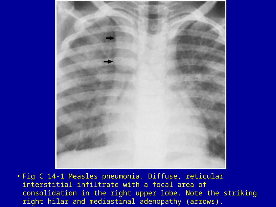
• Fig C 14-1 Measles pneumonia. Diffuse, reticular interstitial infiltrate with a focal area of consolidation in the right upper lobe. Note the striking right hilar and mediastinal adenopathy (arrows).
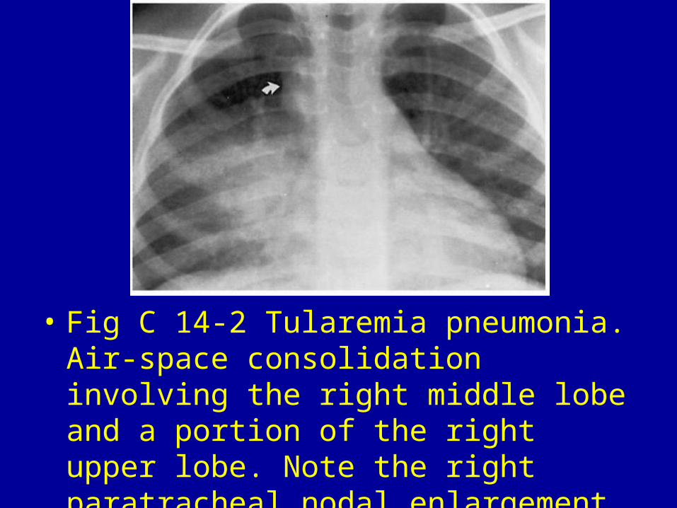
• Fig C 14-2 Tularemia pneumonia. Air-space consolidation involving the right middle lobe and a portion of the right upper lobe. Note the right paratracheal nodal enlargement (arrow).
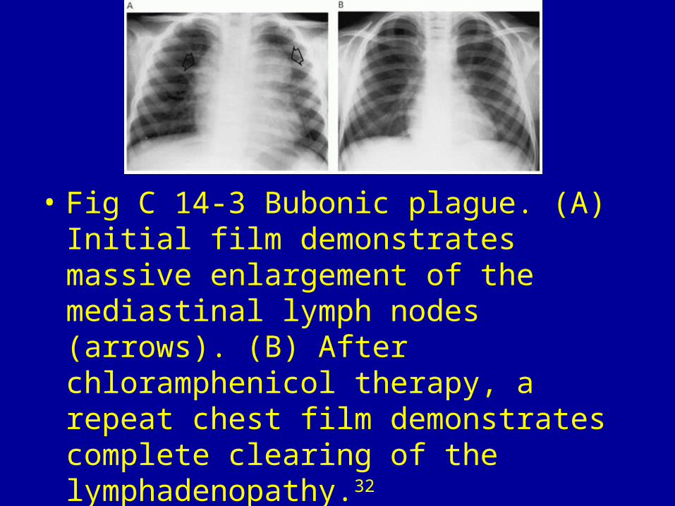
• Fig C 14-3 Bubonic plague. (A) Initial film demonstrates massive enlargement of the mediastinal lymph nodes (arrows). (B) After chloramphenicol therapy, a repeat chest film demonstrates complete clearing of the lymphadenopathy.32
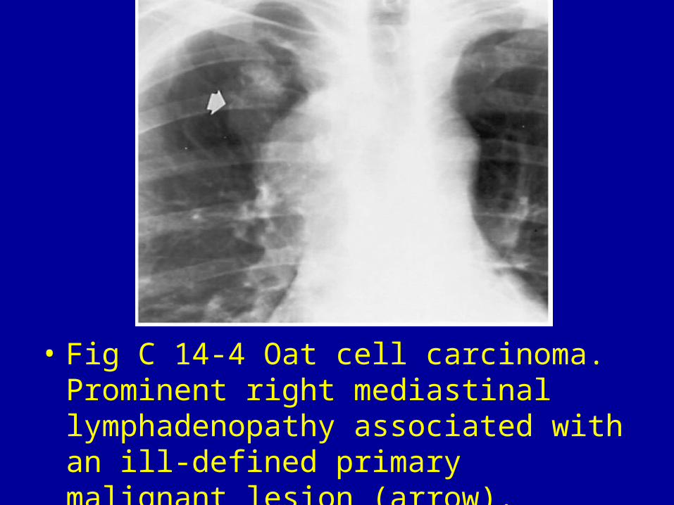
• Fig C 14-4 Oat cell carcinoma. Prominent right mediastinal lymphadenopathy associated with an ill-defined primary malignant lesion (arrow).
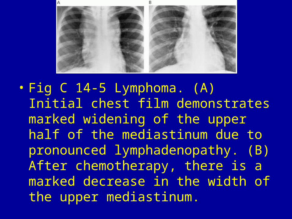
• Fig C 14-5 Lymphoma. (A) Initial chest film demonstrates marked widening of the upper half of the mediastinum due to pronounced lymphadenopathy. (B) After chemotherapy, there is a marked decrease in the width of the upper mediastinum.
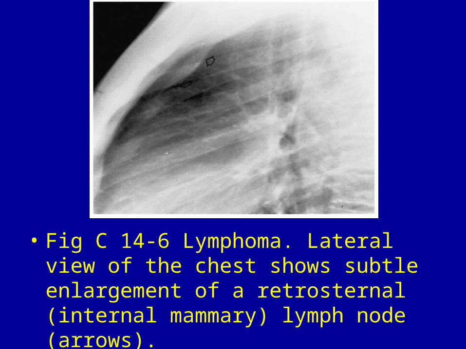
• Fig C 14-6 Lymphoma. Lateral view of the chest shows subtle enlargement of a retrosternal (internal mammary) lymph node (arrows).
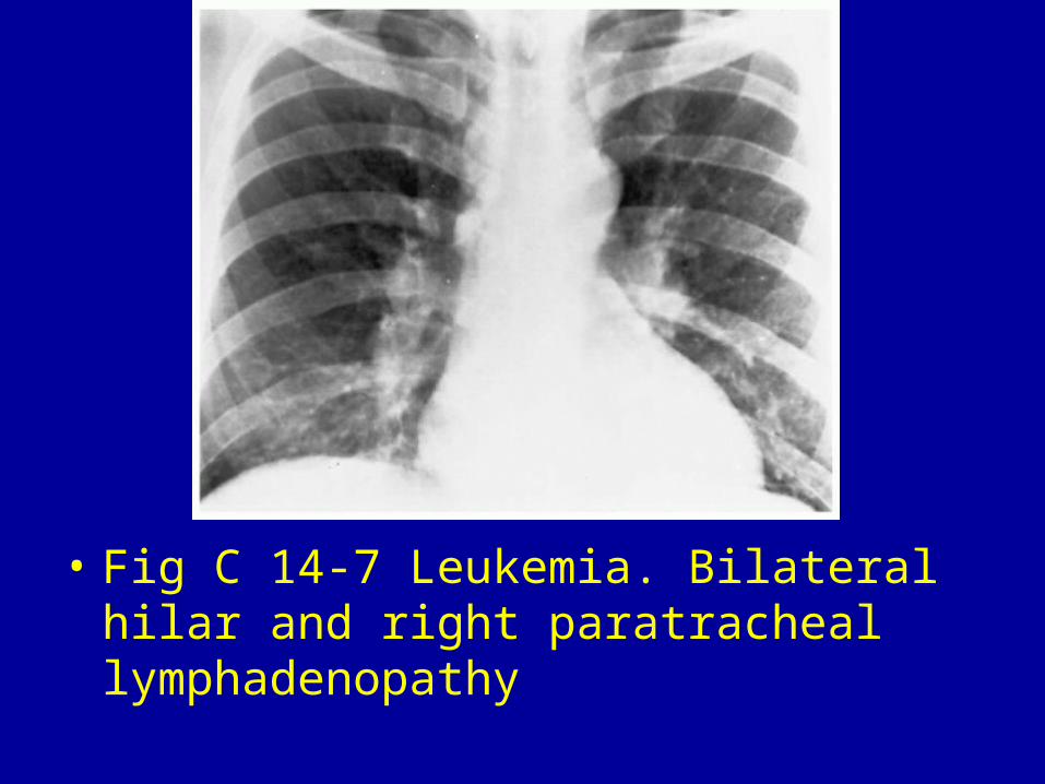
• Fig C 14-7 Leukemia. Bilateral hilar and right paratracheal lymphadenopathy
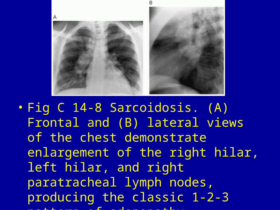
• Fig C 14-8 Sarcoidosis. (A) Frontal and (B) lateral views of the chest demonstrate enlargement of the right hilar, left hilar, and right paratracheal lymph nodes, producing the classic 1-2-3 pattern of adenopathy.