14 2008 ACVIM Abstracts -...
55
ABSTRACT #1 EVALUATION OF FOUR DNA EXTRACTION METHODS FOR THE DETECTION OF TRITRICHOMONAS FOETUS IN FELINE STOOL SPECIMENS BY POLYMERASE CHAIN RE- ACTION. SH Stauffer, AJ Birkenheuer, MG Levy, H Marr, JL Gookin. College of Veterinary Medicine, North Carolina State University, Raleigh, NC. Feces are increasingly recognized as practical samples for molec- ular diagnosis of infectious disease. Extraction of PCR-quality DNA from feces can be challenging due to co-extraction of PCR inhibitors. Accordingly, we examined the effect of four commercial- ly-available DNA extraction methods on sensitivity of PCR for detection of Tritrichomonas foetus (TF) in naturally-infected and TF-spiked feline stool. Kits evaluated included ExtractMaster Fecal DNA Extraction Kit, Epicentre Biotechnologies (Kit A); QIAamp DNA Stool Mini Kit, Qiagen (Kit B); UltraClean Fecal DNA Kit, MoBio (Kit C); and ZR Fecal DNA Kit, Zymo Research (Kit D). In accordance with manufacturer instructions, DNA was extracted from 180 mg (A,B), 50 mg (C), 100 & 150 mg (D) aliquots of feline feces to which was added 20 ml volumes containing 0–10,000 cultured feline TF. Each kit was also used to extract DNA from the feces of each of 10 naturally infected and 10 uninfected cats. DNA was eluted in 300 ml (A), 200 ml (B), 50 ml (C), or 100 ml (D) of respective elution buffer. Endogenous PCR inhibitors in extracted DNA was examined by PCR amplification of an 876 bp gene fragment of bacterial 16S rRNA. DNA was then tested by single tube nested PCR for ampli- fication of partial ITS1, 5.8S and ITS2 rRNA genes of TF. Kit D provided the most sensitive detection of TF DNA as ex- pressed by both organisms per DNA extraction and organisms per PCR reaction. To account for differences in DNA concentrations between kits (i.e. fecal sample size and elution volumes), the limit of detection for each kit as expressed by the number of TF per PCR reaction was as follows: Kit B 5 250, Kit A 5 167, Kit C 5 100, Kit D (150 mg fecal sample) 5 5, and Kit D (100 mg fecal sample) 5 0.5. PCR performed on DNA extracted from cultured TF (no feces) or TF-spiked feces (100 mg) using Kit D was positive with as few as 10 TF per extraction. Further, DNA extraction using Kit D could be completed in the shortest time of all kits tested. These studies identify the ZR Fecal DNA Kit as superior to the other kits tested for extraction of PCR-quality DNA from feline feces. ABSTRACT #2 INVESTIGATION OF ENTEROBACTER CLOACAE INFEC- TIONS AT A SMALL ANIMAL VETERINARY TEACHING HOSPITAL. JS Weese. University of Guelph, Guelph, Ontario. A wide range of pathogens can cause hospital-associated (HA) infections in small animal hospitals. Among these is Enterobacter cloacae, which is one of the most clinically relevant Enterobacter spp and a common cause of HA infection in humans. Recently, multi- drug resistance has become a concern, particularly with emergence of extended-spectrum beta-lactamase and extended spectrum ce- phalosporinase producing strains. An infection control investi- gation was initiated at the Ontario Veterinary College Teaching Hospital (OVCTH) in the fall of 2007 in response to anecdotal con- cerns about Enterobacter cloacae infections in hospitalized animals. Enterobacter cloacae was isolated from 45/36719 animals from January 1, 2005 to October 31, 2007, for an overall incidence of 1.2/ 1000 admissions. The monthly incidence rate ranged from 0 to 4.3/ 1000 admissions. Twenty-one (47%) cases were classified as com- munity-associated, while 17 (38%) were hospital associated. Seven (15%) were community-onset but hospital associated, with three of these associated with other veterinary hospitals. There was no in- crease in the incidence of overall or hospital-associated infections during the study period. The urinary tract was the most common site of infection (n511, 24%). Wound infections (excluding surgical site infections) ac- counted for 8 (18%) of infections, with superficial and deep surgical site infections accounting for 7 (16%) and organ/space sur- gical site infections accounting for another 2 cases. Urinary tract infections were most common among animals with CA infection, accounting for 8/21 (38%) cases with wound infections accounting for 4 (19%) cases. Of the 24 cases associated with the OVCTH, 17 (71%) had sur- gery, 15 (63%) were hospitalized in the intensive care unit, 10 (42%) had indwelling urinary catheters placed, and 20 (83%) had received antimicrobials prior to onset of infection. Risk factors for E. cloacae infection could not be determined because a noninfected control group was not evaluated. Surgical site infections accounted for 9 (38%) HA cases. Overall, only 2/11 (18%) urinary tract infections were associated with prior placement of a urinary catheter. Nine (20%) animals died or were euthanized and E. cloacae was impli- cated as a causative or contributing factor in 5 (56%) of those cases. Two main antimicrobial phenotype patterns were identified. One (n525) was characterized by susceptibility to fluoroquinolones, tet- racycline, and trimethoprim with variable susceptibility to cefoxitin while the other (n514) was characterized by resistance to these an- timicrobials. Prior administration of antimicrobials was associated with presence of the more resistant phenotype (P50.044) but there was no association between this phenotype and origin of infection (P50.74) and no increase in the prevalence of this phenotype from 2005 to 2007 (P50.97). Infections with this phenotype were not associated with nonsurvival (P50.74). There was no evidence of a, HA outbreak or increase in preva- lence, yet identification of multidrug resistant E. cloacae in both CA and HA infections is concerning and requires ongoing surveillance. ABSTRACT #3 STAPHYLOCOCCUS PSEUDINTERMEDIUS: A NEWLY RECOGNIZED PATHOGEN IN DOGS AND CATS. MC Faires 1 , D Slavic 2 , JS Weese 1 . 1 Ontario Veterinary College, 2 Animal Health Laboratory, University of Guelph, Guelph, Ontario. Staphylococcus intermedius has typically been regarded as the predominant pathogenic Staphylococcus spp in dogs and cats, and a leading cause of skin and soft tissue infections. In 2005, a novel Staphylococcus species, Staphylococcus pseudintermedius, was iden- tified. This organism is closely related to, but distinct from, S. intermedius. Gene-sequence based methods are required to differ- entiate these two species; however, these techniques are rarely performed in clinical laboratories, and as a result the prevalence and characteristics of S. pseudintermedius are poorly understood. Recent evidence suggests that S. pseudintermedius may actually be the predominant Staphylococcus spp in dogs and cats but misiden- tified as S. intermedius by diagnostic laboratories. The objective of this study was to use sequence based methods to identify putative S. intermedius isolates from dogs and cats and to evaluate antimi- crobial resistance and virulence factors among S. pseudintermedius isolates. Isolates from dogs and cats identified as S. intermedius by con- ventional laboratory methods were obtained from the University of Guelph Animal Health Laboratory. Isolates were collected in a se- rial manner without selection. DNA was extracted, sequencing of the sodA gene was performed, and isolates were identified via se- quence alignment with reference staphylococcal strains through GenBank (www.ncbi.nlm.nih.gov/blast/BLAST.cgi). Antimicrobial susceptibility testing was performed and PCR was used to identify various virulence factors and antimicrobial genes. A total of 25 isolates were obtained from 21 dogs and 2 cats. Medical records were not available for 2 of the isolates. 25/25 (100%) were identified as S. pseudintermedius Severity of infection ranged from superficial dermatitis to rapidly fatal necrotizing fascii- tis with the majority of isolates from otitis externa 9/23 (39.1%) and urinary tract infections 6/23 (26.1%). Antimicrobial susceptibility was as follows: amoxicillin/clavulanate 23/23 (100%), ampicillin 7/ 23 (30.4%), cephalothin 23/23 (100%), clindamycin 18/23 (78.3%), gentamicin 23/23 (100%), tetracycline 18/23 (78.3%), and trimetho- prim/sulfa 19/23 (82.6%). Not all antimicrobials were tested for all isolates, based on laboratory protocols regarding antimicrobial panel and site of infection. Inducible resistance to clindamycin was detected by D-test in 1 isolate reported as clindamycin-susceptible (5.6%). Detection of virulence factors and antimicrobial resistance genes is ongoing. This study identified S. pseudintermedius as an important patho- gen in dogs and cats, and suggests that S. intermedius may not be a major concern in these species. Further studies are required to eval- uate clinically relevant virulence factors to assist in understanding the pathogenesis of disease caused by S. pseudintermedius. 14 2008 ACVIM Abstracts
Transcript of 14 2008 ACVIM Abstracts -...

ABSTRACT #1EVALUATION OF FOUR DNA EXTRACTION METHODSFOR THE DETECTION OF TRITRICHOMONAS FOETUS INFELINE STOOL SPECIMENS BY POLYMERASE CHAIN RE-ACTION. SH Stauffer, AJ Birkenheuer, MG Levy, H Marr, JLGookin. College of Veterinary Medicine, North Carolina StateUniversity, Raleigh, NC.
Feces are increasingly recognized as practical samples for molec-ular diagnosis of infectious disease. Extraction of PCR-qualityDNA from feces can be challenging due to co-extraction of PCRinhibitors. Accordingly, we examined the effect of four commercial-ly-available DNA extraction methods on sensitivity of PCR fordetection of Tritrichomonas foetus (TF) in naturally-infected andTF-spiked feline stool.
Kits evaluated included ExtractMaster Fecal DNA ExtractionKit, Epicentre Biotechnologies (Kit A); QIAamp DNA Stool MiniKit, Qiagen (Kit B); UltraClean Fecal DNA Kit, MoBio (Kit C);and ZR Fecal DNA Kit, Zymo Research (Kit D). In accordancewith manufacturer instructions, DNA was extracted from 180mg(A,B), 50mg (C), 100 & 150mg (D) aliquots of feline feces to whichwas added 20ml volumes containing 0–10,000 cultured feline TF.Each kit was also used to extract DNA from the feces of each of 10naturally infected and 10 uninfected cats. DNA was eluted in 300ml(A), 200ml (B), 50ml (C), or 100ml (D) of respective elution buffer.Endogenous PCR inhibitors in extracted DNA was examined byPCR amplification of an 876 bp gene fragment of bacterial 16SrRNA. DNA was then tested by single tube nested PCR for ampli-fication of partial ITS1, 5.8S and ITS2 rRNA genes of TF.
Kit D provided the most sensitive detection of TF DNA as ex-pressed by both organisms per DNA extraction and organisms perPCR reaction. To account for differences in DNA concentrationsbetween kits (i.e. fecal sample size and elution volumes), the limit ofdetection for each kit as expressed by the number of TF per PCRreaction was as follows: Kit B 5 250, Kit A 5 167, Kit C 5 100, KitD (150mg fecal sample) 5 5, and Kit D (100mg fecal sample) 5 0.5.PCR performed on DNA extracted from cultured TF (no feces) orTF-spiked feces (100mg) using Kit D was positive with as few as 10TF per extraction. Further, DNA extraction using Kit D could becompleted in the shortest time of all kits tested.
These studies identify the ZR Fecal DNA Kit as superior to theother kits tested for extraction of PCR-qualityDNA from feline feces.
ABSTRACT #2INVESTIGATION OF ENTEROBACTER CLOACAE INFEC-TIONS AT A SMALL ANIMAL VETERINARY TEACHINGHOSPITAL. JS Weese. University of Guelph, Guelph, Ontario.
A wide range of pathogens can cause hospital-associated (HA)infections in small animal hospitals. Among these is Enterobactercloacae, which is one of the most clinically relevant Enterobacter sppand a common cause of HA infection in humans. Recently, multi-drug resistance has become a concern, particularly with emergenceof extended-spectrum beta-lactamase and extended spectrum ce-phalosporinase producing strains. An infection control investi-gation was initiated at the Ontario Veterinary College TeachingHospital (OVCTH) in the fall of 2007 in response to anecdotal con-cerns about Enterobacter cloacae infections in hospitalized animals.
Enterobacter cloacae was isolated from 45/36719 animals fromJanuary 1, 2005 to October 31, 2007, for an overall incidence of 1.2/1000 admissions. The monthly incidence rate ranged from 0 to 4.3/1000 admissions. Twenty-one (47%) cases were classified as com-munity-associated, while 17 (38%) were hospital associated. Seven(15%) were community-onset but hospital associated, with three ofthese associated with other veterinary hospitals. There was no in-crease in the incidence of overall or hospital-associated infectionsduring the study period.
The urinary tract was the most common site of infection (n511,24%). Wound infections (excluding surgical site infections) ac-counted for 8 (18%) of infections, with superficial and deepsurgical site infections accounting for 7 (16%) and organ/space sur-gical site infections accounting for another 2 cases. Urinary tractinfections were most common among animals with CA infection,accounting for 8/21 (38%) cases with wound infections accountingfor 4 (19%) cases.
Of the 24 cases associated with the OVCTH, 17 (71%) had sur-gery, 15 (63%) were hospitalized in the intensive care unit, 10 (42%)had indwelling urinary catheters placed, and 20 (83%) had receivedantimicrobials prior to onset of infection. Risk factors for E. cloacaeinfection could not be determined because a noninfected controlgroup was not evaluated. Surgical site infections accounted for 9(38%) HA cases. Overall, only 2/11 (18%) urinary tract infectionswere associated with prior placement of a urinary catheter. Nine(20%) animals died or were euthanized and E. cloacae was impli-cated as a causative or contributing factor in 5 (56%) of those cases.
Two main antimicrobial phenotype patterns were identified. One(n525) was characterized by susceptibility to fluoroquinolones, tet-racycline, and trimethoprim with variable susceptibility to cefoxitinwhile the other (n514) was characterized by resistance to these an-timicrobials. Prior administration of antimicrobials was associatedwith presence of the more resistant phenotype (P50.044) but therewas no association between this phenotype and origin of infection(P50.74) and no increase in the prevalence of this phenotype from2005 to 2007 (P50.97). Infections with this phenotype were notassociated with nonsurvival (P50.74).
There was no evidence of a, HA outbreak or increase in preva-lence, yet identification of multidrug resistant E. cloacae in both CAand HA infections is concerning and requires ongoing surveillance.
ABSTRACT #3STAPHYLOCOCCUS PSEUDINTERMEDIUS: A NEWLYRECOGNIZED PATHOGEN IN DOGS AND CATS. MCFaires1, D Slavic2, JS Weese1. 1Ontario Veterinary College,2Animal Health Laboratory, University of Guelph, Guelph,Ontario.
Staphylococcus intermedius has typically been regarded as thepredominant pathogenic Staphylococcus spp in dogs and cats, and aleading cause of skin and soft tissue infections. In 2005, a novelStaphylococcus species, Staphylococcus pseudintermedius, was iden-tified. This organism is closely related to, but distinct from,S. intermedius. Gene-sequence based methods are required to differ-entiate these two species; however, these techniques are rarelyperformed in clinical laboratories, and as a result the prevalenceand characteristics of S. pseudintermedius are poorly understood.Recent evidence suggests that S. pseudintermedius may actually bethe predominant Staphylococcus spp in dogs and cats but misiden-tified as S. intermedius by diagnostic laboratories. The objectiveof this study was to use sequence based methods to identify putativeS. intermedius isolates from dogs and cats and to evaluate antimi-crobial resistance and virulence factors among S. pseudintermediusisolates.
Isolates from dogs and cats identified as S. intermedius by con-ventional laboratory methods were obtained from the University ofGuelph Animal Health Laboratory. Isolates were collected in a se-rial manner without selection. DNA was extracted, sequencing ofthe sodA gene was performed, and isolates were identified via se-quence alignment with reference staphylococcal strains throughGenBank (www.ncbi.nlm.nih.gov/blast/BLAST.cgi). Antimicrobialsusceptibility testing was performed and PCR was used to identifyvarious virulence factors and antimicrobial genes.
A total of 25 isolates were obtained from 21 dogs and 2 cats.Medical records were not available for 2 of the isolates. 25/25(100%) were identified as S. pseudintermedius Severity of infectionranged from superficial dermatitis to rapidly fatal necrotizing fascii-tis with the majority of isolates from otitis externa 9/23 (39.1%) andurinary tract infections 6/23 (26.1%). Antimicrobial susceptibilitywas as follows: amoxicillin/clavulanate 23/23 (100%), ampicillin 7/23 (30.4%), cephalothin 23/23 (100%), clindamycin 18/23 (78.3%),gentamicin 23/23 (100%), tetracycline 18/23 (78.3%), and trimetho-prim/sulfa 19/23 (82.6%). Not all antimicrobials were tested for allisolates, based on laboratory protocols regarding antimicrobialpanel and site of infection. Inducible resistance to clindamycin wasdetected by D-test in 1 isolate reported as clindamycin-susceptible(5.6%). Detection of virulence factors and antimicrobial resistancegenes is ongoing.
This study identified S. pseudintermedius as an important patho-gen in dogs and cats, and suggests that S. intermedius may not be amajor concern in these species. Further studies are required to eval-uate clinically relevant virulence factors to assist in understandingthe pathogenesis of disease caused by S. pseudintermedius.
14 2008 ACVIM Abstracts

ABSTRACT #4PREVALENCE OF STAPHYLOCOCCUS AUREUS ANDMRSA CARRIAGE IN THREE POPULATIONS. S Kottler1, JRMiddleton1, JS Weese2, LA Cohn1. 1University of Missouri Collegeof Veterinary Medicine, Columbia, MO. 2Ontario VeterinaryCollege, University of Guelph, Guelph, Ontario, Canada.
Considered primarily a human pathogen, methicillin-resistantStaphylococcus aureus (MRSA) can colonize animals. Prior studieshave demonstrated a higher prevalence of MRSA colonization inhealthcare workers (human and veterinary) than in the people withnon-medically related occupations. We hypothesized that the prev-alence of MRSA colonization would be higher in people and petswho reside in households with veterinary or human healthcareworkers than in households without healthcare workers. Onehealthy volunteer (nasal swab) and a pet from the same household(nasal and rectal swabs) were sampled. Swabs were placed in en-richment broth and coagulase positive staphylococci (CPS) wereidentified by standard methods. CPS were classified as S. aureus us-ing a latex agglutination test (LAT) and polymyxin B susceptibility.Staphylococcus aureus isolates were screened for methicillin resis-tance via PBP2a LAT and mecA PCR. Proportional data werecompared using the Chi square test (P o 0.05).
Of the 601 sample sets (1,803 samples) obtained, 202 sets werefrom households with human healthcare workers (group 1), 223from veterinary healthcare workers (group 2), and 176 from non-healthcare workers (group 3). And 103/601 (17.1%) pets sampledwere cats and 498/601 (82.9%) were dogs. Overall, S. aureus wasisolated from 166/601 (27.6%) of humans and 79/601 (13.1%) of petnasal and rectal samples. For 26/245 (10.6%) S. aureus isolates,both a human and animal in the same household were colonized.Shared colonization was equally likely in all groups (10 from group1, 10 group 2, and 6 from group 3).
Of 245 S. aureus isolates, 49 were MRSA (20%). Among S.aureus isolates, 31 of 166 human isolates (18.7%) wereMRSA while18/79 (22.8%) pet isolates were MRSA (Table). MRSA was presentin 31/601 (5.2%) of humans sampled and 18/601 (3.0%) pets. Therewere no differences in prevalence of MRSA between groups of hu-mans (9/202 group 1, 13/223 group 2, 9/176 group 3) or pets (6/202group 1, 4/223 group 2, and 8/176 group 3). For 6/49 (12.2%) of theMRSA isolates, both an animal and human in the same householdwere colonized representing two households per group.
Group 1 Group 2 Group 3 Total
MRSA among human SA isolates 9/60 (15%) 13/60 (22%) 9/46 (20%) 31/166 (19%)
MRSA among pet SA isolates 6/22 (27%) 4/34 (12%) 8/23 (35%) 18/79 (23%)
We were unable to demonstrate that colonization with S. aureusor MRSA was more common in households with healthcare work-ers. However, we found a greater prevalence of both S. aureus andMRSA in pet animals than has been previously demonstrated, anddemonstrated that MRSA is found as commonly among S. aureusisolates from pets as from human isolates.
ABSTRACT #5
EFFECTS OF ZAFIRLUKAST, A CYSTEINYL-LEUKO-TRIENE RECEPTOR ANTAGONIST, IN A LOW DOSE ENDO-TOXIN INFUSION MODEL OF FELINE SEPSIS. Sharp CR,DeClue AE, Reinero CR. University of Missouri, College ofVeterinary Medicine, Columbia, MO.
Cysteinyl–leukotrienes (cys-LT) are important inflammatory me-diators contributing to systemic inflammation and hemodynamicinstability during sepsis and there is experimental evidence that in-hibiting the effects of leukotrienes ameliorates these sequelae. Wehypothesized that the cys-LT receptor antagonist, zafirlukast, aninexpensive and readily available cys-LT receptor antagonist, wouldattenuate systemic inflammation and hemodynamic derangement ina low-dose endotoxin infusion model of feline sepsis.
This was a randomized, blinded, placebo-controlled crossoverdesign, utilizing 6 adult cats. The cats were given a priming dose oflipopolysaccharide (LPS) (1mcg/kg, IV) at time 0 and then eitherplacebo (flour) or zafirlukast 10mg orally at 6 and 12 hours. Thecats were challenged at 14 hours with LPS (2mcg/kg/h � 6 h, IV).There was a 30 day washout period between evaluations. Rectaltemperature, heart rate (HR), systolic arterial blood pressure (BP),
plasma interleukin-6 (IL-6) concentration, and tumor necrosis fac-tor (TNF) activity were evaluated. Repeated measures ANOVAwith post-hoc Tukey’s test or paired t-test were used for statisticalanalysis with a p-valueo 0.05 considered significant.
LPS administration induced significant systemic inflammationand hemodynamic derangement as indicated by increasedmean�SD rectal temperature at 4, 5, and 6 hours (baseline,39.1�0.5; 4 h, 39.9�0.4; 5 h, 40.0�0.3; 6 h, 39.8�0.4 1C; po0.001),increased IL-6 concentrations at 3 hours (baseline, 68.8�194.8; 3 h,883.7�492.2 pg/mL; po0.004), increased TNF activity at 1.5 hours(baseline, ND; 1.5 h, 151�49 ng/ml; po0.001), and relative hypo-tension at 6 hours (baseline, 145.7�19.8; 6 h, 110�27.54mmHg;p50.03), when compared to baseline regardless of treatment group.There was no significant difference between the placebo andzafirlukast treatment groups in regards to rectal temperature, HR,BP, IL-6 concentrations, or TNF activity at any time point.
These data indicate that antagonism of cys-LT with zafirlukasthas neither beneficial nor deleterious effects in this model of felinesepsis.
ABSTRACT #6FACTORS ASSOCIATED WITH ORGAN DYSFUNCTIONAND DEATH IN DOGS WITH SEPSIS. JM Burkitt, K Hopper.University of California, Davis, School of Veterinary Medicine,Davis, CA.
Sepsis is the systemic response to infection, and is associated witha high mortality rate in dogs. Literature regarding the clinicalcourse, distant organ dysfunction, and factors associated with mor-tality in dogs with naturally occurring sepsis is limited. The purposeof this investigation was to describe factors associated with distantorgan dysfunction and death in a heterogeneous group of septicdogs admitted to an intensive care unit.
Post-hoc analysis was completed on prospectively collected datafrom septic dogs admitted to an intensive care unit from October2004 to October 2005. Animals were included only if they fit criteriafor the systemic inflammatory response syndrome due to a confirmedinfectious etiology. Signalment, infected anatomic site(s), infectiousagent(s), clinical course, and outcome were collected on a standard-ized data collection sheet. Categorical variables were analyzed usingPearson’s chi-square test, and Po 0.05 was considered significant.
Forty-seven dogs were included in the investigation. Twenty dogs(42%) were discharged from the hospital alive. The most commonlyinfected anatomic sites were the peritoneal cavity (n 5 14; 30%), thesubcutaneous tissues (11; 23%), and the lung (9; 19%). Forty-five ofthe infections were bacterial (96%) and the remaining two were fun-gal. Septic shock occurred in 10/47 dogs (21%). Distant organdysfunction occurred in 21/47 dogs (45%) and was associated with asurgical disease process (P 5 0.03), hypoglycemia (P 5 0.011), thepresence of septic shock (P 5 0.002), and death (P 5 0.019). Four-teen of 21 dogs (67%) of dogs with distant organ dysfunction diedor were euthanized for grave prognosis. Hypoglycemia (P 5 0.045),total number of failing organs (P 5 0.003), and presence of septicshock (Po 0.001) were each associated with death or euthanasia forsevere illness. All dogs that developed septic shock died.
Distant organ dysfunction occurs frequently in dogs with sepsisand is associated with death. Distant organ dysfunction may be seenmore commonly in dogs with hypoglycemia, a surgical disease pro-cess, or septic shock. Septic shock in the dog carries a particularlypoor prognosis.
ABSTRACT #7EFFECTS OF ALLOANTIGEN EXPOSURE AND CELL-ASSOCIATED MUCOSAL FIV CHALLENGE ON FELINETOLL-LIKE RECEPTOR GENE EXPRESSION. K Cairns,S Kumar, S Leavell, BD Assogba, MJ Burkhard. Department ofVeterinary Biosciences, College of Veterinary Medicine, Center forRetrovirus Research, The Ohio State University, Columbus, OH.
Toll-like receptors (TLRs) are important players in innate viralimmunity. Endosomal TLRs (3,7,8,9) recognize viral nucleic acidwhile cell surface TLRs (2,4) sense viral protein. Many viruses ac-tivate TLRs, including cytomegalovirus, respiratory syncytial virus,and HIV. Although often protective, there are circumstances in
152008 ACVIM Abstracts

which TLR activation augments viral pathogenicity. Nine felineTLRs exist and in vitro FIV infection alters TLR expression. Allo-antigen exposure also modulates anti-viral immunity. In cats,mucosal exposure to alloantigen induces cellular alloimmune re-sponses that are associated with reduced viral burden after mucosalFIV challenge.
The goal of this study was to investigate TLR gene expression infeline peripheral blood mononuclear cells (PBMC), intra-epithelial(IEL) and lamina propria (LPL) lymphocytes and iliac (ILN), mes-enteric (MLN) and popliteal (PLN) lymph nodes to determine ifalloantigen exposure and cell-associated mucosal FIV challengemodulate TLR gene expression.
Specific pathogen free (SPF) cats were vaginally exposed weeklyfor 12 weeks to lymphocyte media (n 5 7) or allogeneic PBMC inmedia (n 5 7). Blood samples were obtained prior to exposure.Twelve weeks post-exposure, 3 cats per group were euthanized tocollect blood and tissues. Remaining animals (n 5 4 per group) werevaginally challenged with cell-associated FIV. Blood and tissuesamples were obtained 12 weeks post-challenge at euthanasia.RNA was extracted and TLR expression was analyzed by real-timeRT-PCR. Standard curves were generated from plasmids contain-ing each TLR and GAPDH and TLR copy number was normalizedto GAPDH.
Basal TLR expression was variable based upon tissue type. Allo-antigen exposure did not alter TLR expression in PBMC, IEL orLPL. However, it did modulate TLR expression in lymph nodes,particularly the MLN where TLR 1,4,5,6 were decreased and TLR7,9 were increased. TLR expression was also decreased in ILN(TLR9) and PLN (TLR 2,3,8). These findings suggest diffuse nodalalloantigen processing, which impacts innate immunity via TLRsignaling pathways.
FIV challenge significantly increased TLR 7 expression, de-creased TLR 2 expression, and had variable effects on TLR 6,8,9expression in most tissues. This is notable given the endosomal lo-cation of TLR 7, 8 and 9 and the importance of these TLRs in anti-viral immunity. FIV exposure had variable effects on the otherTLRs. These viral effects on TLR expression were also modulatedby prior exposure to alloantigen.
These data indicate a role for TLRs in both alloantigen process-ing and host response to FIV. Previous data show allogeneicimmune responses increase the threshold for susceptibility to muco-sal FIV infection. These data suggest this effect may occur via TLRpathways. Additional studies are indicated to further characterizethe role of both TLRs and alloantigen exposure in FIV.
ABSTRACT #8DETECTION OF FELINE CALICIVIRUS RNA, FELINE HER-PESVIRUS-1 DNA, AND BARTONELLA SPP. DNA INTISSUES OF CATS WITH AND WITHOUT GINGIVOSTO-MATITIS. K Dowers, N Wilkerson, JR Hawley, M Brewer, MRLappin. Department of Clinical Sciences, Colorado StateUniversity, Ft. Collins, CO.
Gingivostomatitis (GS) is a common syndrome in cats; feline ca-licivirus (FCV), feline herpesvirus 1 (FHV-1), and Bartonella spp.are common differential diagnoses. Currently, there is minimal in-formation available concerning the presence of nucleic acids of thesemicrobes in the oral tissues of cats with or without GS. The objec-tive of this study was to report the prevalence rates of FCV RNA,FHV-1 DNA, and Bartonella spp. DNA in gingival tissues of cats.
Fresh tissue biopsies from affected areas of cats with GS (n 5 42)were obtained and submitted on ice by practicing veterinariansthroughout the United States. A 6mm skin biopsy punch was usedto collect a full thickness biopsy from the right palatoglossal arch ofnormal cats housed in a Humane Society (n 5 19) in Colorado aftereuthanasia for reasons unrelated to the study. The normal cats hadlived in the Humane Society for 1 to 33 days (median 5 7 days) and14 of 19 had been administered an intranasal, modified live FCV,FHV-1, and panleukopenia on admission. The majority of cats withGS had been vaccinated but it is unknown when the last inoculationwas given. DNA and RNA were extracted from the fresh tissues byuse of a commercially available kit and previously described PCRassays (FHV-1 and Bartonella spp.) or reverse transcriptase PCRassay (FCV) was used to amplify the target DNA or RNA.
FCV RNA was amplified from a statistically greater number ofcats with GS than control cats (Fischer’s exact test; p 5 0.001). Bar-
tonella spp. DNAwas amplified only from cats with GS and all wereB. clarridgeiae but the significance of these findings is unknown asBartonella spp. are regionally defined by flea risk and the controlcats came from a low flea risk state.
Organisms
Cat group FCV FHV-1 Bartonella spp.
GS 17/42 (40.5%) 2/42 (4.8%) 3/34 (8.8%)
Control 0/19 (0%) 3/19 (15.8%) 0/19 (0%)
The results suggest that FCV was associated with stomatitis insome of the cats with GS. In addition, administration of an intra-nasal modified live FCV containing vaccine approximately 1–33days prior to oral tissue biopsy is unlikely to result in a positive re-sult in the reverse transcriptase PCR assay for FCV used here.
ABSTRACT #9
PREVALENCE OF HEMOTROPIC MYCOPLASMA INFEC-TION IN THE FELINE SHELTER AND CLIENT-OWNEDPOPULATIONS OF SASKATOON AND EVALUATION OFTHE PRECISION OF TWO FELINE HEMOTROPIC MYCO-PLASMA QUALITATIVE PCR. BD Nibblett1, E Snead1,C Waldner2, S Taylor1, M Jackson3. 1Department of Small AnimalClinical Sciences, 2Department of Large Animal Clinical Sciences,3Department of Pathology, Western College of VeterinaryMedicine, University of Saskatchewan, Saskatoon, SK, CANADA.
The objective of part A of this study was to evaluate the preva-lence of subclinical hemotropic Mycoplasma (HM) infection in twodistinct feline populations: a local shelter population (SPCA) and aclient-owned population. The objective of Part B of this study wasto evaluate the inter- and intratest variability of two independentqualitative PCR assays used for the diagnosis of feline HM infec-tions.
For part A, an EDTA and serum blood sample was collectedfrom 58 SPCA and 57 client-owned cats determined to be healthybased on physical examination. The packed cell volume, total pro-tein, and retroviral status were determined for each cat. In addition,each cat was screened for a subclinical HM infection using a) aqualitative polymerase chain reaction (PCR) assay for the 16SrRNA of Mycoplasma haemofelis and ‘‘Candidatus M. ha-emominutum’’, and b) cytologic evaluation of a blood smearlooking for evidence of hemoplasma organisms.
In part B, blood samples from 44 cats were submitted to a secondlaboratory for determination of HM infection using another qual-itative PCR assay to evaluate intertest variability. The bloodsamples included 26 samples from part A (6 positives and 20 nega-tives) and 18 samples from cats with historical clinical or subclinicalHM infections. Where sample volume permitted, the PCR testingwas repeated (16 of 44 samples at the first laboratory and 40 of 44samples at the second laboratory) to assess the intratest variabilityfor both PCR assays.
For part A, the prevalence of subclinical HM infection was 12%(7/58) in the SPCA population and 4% (2/57) in the client-ownedpopulation. Within the SPCA population,M. haemofelis was foundin 5% (3/58) and ‘‘Candidatus M. haemominutum’’ in 7% (4/58) ofcats. The two client-owned cats were infected with M. haemofelis.There was no statistically significant difference in the infection ratebetween the two populations (p50.16). Across both populations, norisk factors for HM infection were identified (sex, age, neuter status,breed, retroviral status, external parasitism).
For part B, there was substantial agreement between the two in-dependent PCR assays for M. haemofelis (k50.601 95%CI 0.32–0.89) as well as for ‘‘Candidatus M. haemominutum’’ (k50.7095%CI 0.40–0.99). Both PCRs performed well with zero intratestvariability respectively.
There is no difference in the subclinical HM infection rates be-tween the SPCA and client-owned cat populations evaluated. Inusing PCR testing for HM infection, results between laboratoriesshow substantial agreement, but not perfect agreement. This high-lights the need for continued attention to standardization of PCRtesting.
16 2008 ACVIM Abstracts
ABSTRACT #10DETECTION OF CANINE DISTEMPER VIRUS RNA FROMBLOOD AND CONJUNCTIVAL SWABS COLLECTED FROMHEALTHY PUPPIES AFTERADMINISTRATIONOFAMOD-IFIED LIVE VACCINE. JH Burton, JK Veir, L Pearce, JRHawley, MR Lappin. Department of Clinical Sciences, ColoradoState University, Fort Collins, CO.
Reverse transcriptase polymerase chain reaction (RT-PCR) as-says are now being used to detect canine distemper virus (CDV)RNA in samples from dogs to aid in the clinical diagnosis of infec-tion. However, little information exists on whether administrationof modified live CDV containing vaccines affects the test results. Theobjective of this study was to determine the duration of detectablelevels of CDV RNA in blood and conjunctival cells from healthypuppies vaccinated with a modified-live CDV containing vaccine.
Of the 12 six week old puppies selected for study, six were from abitch with a high CDV antibody titer (1:512) against CDV and sixwere from a bitch with a low CDV antibody titer (1:64). On day 0,the puppies from the bitch with the high CDV titer were seropositiveand the puppies from the bitch with the low CDV titer were sero-negative. Amodified-live CDV containing vaccine was administeredSQ on day 0 and blood and conjunctival swabs were collected ondays 0, 2, 5, 7, 10, and 14. Fluorescent antibody (FA) staining ofsmears made from the conjunctival swabs and a conventional CDVRT-PCR on RNA extracted from whole blood and conjunctivalswabs were performed on samples from all time points.
Positive FA test results were detected in one seronegative puppy onday 0, five seronegative puppies and five seropositive puppies on day2, one seronegative puppy on day 5, and five seronegative puppiesand one seropositive puppy on day 10. CDVRNA was not amplifiedfrom conjunctival swabs of any puppy on any day. CDV RNA wasamplified from blood of three seronegative puppies on day 7.
Current CDV diagnostic test results are not specific for the dis-ease and so positive CDV FA results on conjunctival smears andconventional CDV RT-PCR results on blood should be cautiouslyinterpreted, especially in recently vaccinated, previously naıve dogs.
ABSTRACT #11DETECTION OF CANINE PARVOVIRUS DNA FROMBLOOD AND FECES COLLECTED FROM HEALTHY PUP-PIES AFTER ADMINISTRATION OF MODIFIED LIVEVACCINE. JH Burton, JK Veir, AK Morris, JR Hawley, MRLappin. Department of Clinical Sciences, Colorado StateUniversity, Ft. Collins, CO.
Polymerase chain reaction (PCR) assays are now being used todetect canine parvovirus (CPV) DNA in samples from dogs to aid inthe clinical diagnosis of infection. However, little information existson whether administration of modified live CPV containing vac-cines affects the test results. The objective of this study was todetermine the duration of detectable levels of CPV DNA in bloodand CPV antigen in feces from healthy puppies vaccinated with amodified-live CPV containing vaccine.
Of the 12 six week old puppies selected for study, six were from abitch with a high CPV antibody titer (1:640) and six were from abitch with a low CPV antibody titer (1:10). On day 0, all puppieswere seronegative, regardless of maternal antibody status. A mod-ified-live CPV containing vaccine was administered SQ on day 0 andblood and feces were collected on days 0, 2, 5, 7, 10, and 14. Con-ventional CPV PCRwas performed onDNA extracted from each ofthe whole blood samples. Two commercially available CPV antigenELISAs were performed on each of the fecal samples.
CPV antigen was detected in feces of one dog by one of the ELI-SAs on day 5. CPVDNAwas amplified from blood of some dogs onday 2 (8 dogs), day 5 (7 dogs), day 7 (8 dogs), day 10 (6 dogs), andday 14 (12 dogs). CPV antibody titers and CPV PCR results wereavailable for all dogs on days 7, 10, and 14. Of the 26 samples withpositive CPV PCR results, 21 were concurrently positive for CPVantibodies (titer range 64–16,384; median titer 5 16,384).
The modified live vaccine used here was unlikely to result in pos-itive results in the fecal antigen ELISAs studied. However, the CPVPCR assay could detect CPV DNA in many vaccinated puppies re-gardless of antibody titer. Positive CPV PCR assay results fromblood need to be interpreted cautiously, especially in recently vacci-nated, previously naıve dogs.
ABSTRACT #12DURATIONOF INFECTION AND EFFICACYOF DOXYCYC-LINE TREATMENT IN DOGS EXPERIMENTALLY CO-INFECTED WITH ANAPLASMA PLATYS AND EHRLICHIACANIS. MJ Beall1, SD Gaunt2, R Chandrashekar1,K DeBisceglie1, B Thatcher1, PPVP Diniz3, EB Breitschwerdt3.1IDEXX Laboratories Inc., Westbrook, ME; 2Louisiana StateUniversity School of Veterinary Medicine, Baton Rouge, LA;3North Carolina State University College of Veterinary Medicine,Raleigh, NC.
Rhipicephalus sanguineus is a ubiquitous tick responsible fortransmitting Ehrlichia canis and most likely Anaplasma platys todogs, as either single or co-infections. The occurrence of co-infec-tion may be difficult to diagnose since thrombocytopenia andanemia result from either infection. The purpose of this study wasto evaluate the clinical significance of A. platys and E. canis co-infection in dogs. Results from the initial phase of the study werereported previously and revealed a more severe thrombocytopeniaand anemia in co-infected dogs as compared to single infectionswith either organism (ACVIM 2007). Results from the chronicphase of the study, which evaluated the duration of A. platys andE. canis infection and treatment efficacy, are reported here. Six monthold female hound-type dogs were inoculated intravenously withLouisiana isolates of A. platys and/or E. canis. Six groups of sixdogs each were evaluated: non-infected controls, A. platys infected,E. canis infected, A. platys and E. canis co-infected, A. platys infect-ed administered E. canis 112 days later, and E. canis infectedadministered A. platys 112 days later. Doxycycline treatment(10mg/kg PO daily � 28d) was initiated at 211 days post-infection(PI), followed by immunosuppression (dexamethasone 0.3mg/kgIM daily � 5 d) beginning 410 days PI. Molecular evidence of infec-tion was assessed by two independent laboratories using eitherconventional or real-time PCR on whole blood collected during thestudy, as well as bone marrow and lymph node aspirates collected atthe time of immunosuppression. Thrombocyte counts in dogs in-fected only with A. platys returned to normal by 115 days PI andPCR results were consistently negative after 110 days PI, prior todoxycycline treatment. Most A. platys/E. canis co-infected dogs (8/9) were also PCR negative in blood for A. platys prior to doxycyc-line treatment. In contrast, all untreated E. canis infected dogs (12/12), whether single or co-infected, remainedE. canis PCR positive inblood for more than a year and had reduced platelet concentrations(mean 125,600/uL). Most of these dogs also tested positive by PCRfor E. canis DNA in bone marrow (8/12) and lymph node (12/12)following immunosuppression. All E. canis infected dogs, both sin-gle and co-infected, receiving doxycycline (12/12) were E. canis PCRnegative in blood and lymph node prior to immunosuppression andhad normal thrombocyte concentrations (mean 308,600/uL). De-spite immunosuppression, neither A. platys nor E. canis DNA wasdetected in doxycycline-treated dogs. Although co-infection ofA. platys and E. canismay result in more severe hematologic abnor-malities, these experimental infections can be successfully treatedwith doxycycline.
ABSTRACT #13ATOVAQUONE AND AZITHROMYCIN FOR THE TREAT-MENT OF CYTAUXZOON FELIS. AJ Birkenheuer1, LA Cohn2,MG Levy1, EB Breitschwerdt1, HS Marr1. 1North Carolina StateUniversity—College of Veterinary Medicine, Raleigh, NC.2University of Missouri—College of Veterinary Medicine,Columbia, MO.
Cytauxzoonosis is an emerging infectious disease of domestic catsin the United States. Historically Cytauxzoon felis was believed tobe uniformly fatal in domestic cats. However, over the past decadethere have been scattered reports of cats with and without anti-pro-tozoal drug therapy surviving C. felis infections. Atovaquone andazithromycin has recently been demonstrated to be the most effec-tive therapy for the treatment of canine babesiosis caused byBabesia gibsoni, and organism related to C. felis. The purpose ofthis study is to describe the survival rate of cats with cytauxzoonosisthat were treated with atovaquone and azithromycin combinationtherapy.
Twenty-two cats fromAR, NC, andMOwith naturally occurringcytauxzoonosis (infections confirmed by microscopy and/or poly-
172008 ACVIM Abstracts

merase chain reaction) were treated with atovaquone (15mg/kg POTID) and azithromycin (10mg/kg PO Q24). Additional supportivecare varied between cases, but all cases were treated with intrave-nous fluids and most with heparin. Sixty-four percent (14/22) of thecats survived to discharge and 36% (8/22) died or were euthanized.At least 6 of the cats that died did so within several hours of admis-sion. Detailed clinical and laboratory data were not available for allcases. Long-term follow-up information was available for six catsand they all are alive and clinically normal at least 19 months afterdischarge. Each of the veterinarians that participated in treatmentof cats for this study had prior experience in treatment ofcytauxzoonosis with either supportive care alone or supportive careplus imidocarb dipropionate. Prior to the beginning of this study,survival of C. felis infected cats from their practices was a rare oc-currence.
Survival of these cases cannot clearly be attributed to the anti-protozooal therapy, but atovaquone and azithromycin combinationtherapy appears to be a promising treatment option forcytauxzoonosis. Cats that survive cytauxzoonosis can have com-plete resolution of signs and excellent long-term prognosis. Acontrolled study comparing the efficacy of atovaquone and azithro-mycin to imidocarb dipropionate is ongoing.
ABSTRACT #14COMPARISON OF TWO DRUG PROTOCOLS FOR CLEAR-ANCE OF CYTAUXZOON FELIS INFECTIONS. LA Cohn1,AJ Birkenheuer2, E Ratcliff3. 1University of Missouri – College ofVeterinary Medicine, Columbia, MO2. North Carolina StateUniversity—College of Veterinary Medicine, Raleigh, NC3.Fairgrove Veterinary Hospital, Fairgrove, MO.
Although infection of domestic cats withCytauxzoon felis is usualfatal, some cats survive. The piroplasms stage of the protozoalpathogen may persist in the surviving cats indefinitely, as it does inthe bobcat reservoir host. The purpose of this prospective, random-ized clinical trial was to compare the efficacy of two treatmentprotocols for the eradication of parasites in persistently infectedcats. A colony of cats with multiple survivors of C. felis wasidentified. Nine cats positive for C. felis by PCR were randomlydivided into two groups. All cats were treated topically withfipronil q 3 weeks throughout the study period. Imidocarb dipropi-onate was administered to the first group (4mg/kg IM twice,2 weeks apart) and atovaquone (15mg/kg PO q 8 hr) and azithro-mycin (10mg/kg PO q 24 hours) were administered to the secondgroup for 10 days. Blood was obtained from each cat immediatelybefore treatment and again at 1, 6, and 8 weeks after treatment.Stained smears were examined microscopically, and samples under-went PCR testing for C. felis DNA using previously describedmethods. Cats that failed to clear the infection at either 6 or 8 weekswere ‘‘crossed over’’ and the alternative treatment administeredwith identical follow-up.
Four cats were initially treated with imidocarb and 5 withatovaquone/azithromycin. All cats treated with imidocarb remainedpersistently infected at all 3 time periods afterwards. Of the catstreated with atovaquone/azithromycin, 2 cats tested negative at 1,6, and 8 weeks; 1 cat was positive at 1 week only, another at6 weeks only, and yet another at 8 weeks only. After the 4 catsinitially treated with imidocarb were then treated with atovaquone/azithromycin, 1 cat tested negative at 1, 6, and 8 weeks, 1 cattested negative at 1 week and was lost to follow-up, and 2 catstested positive at 1 week but negative at weeks 6 and 8. The twocats that initially ‘‘failed’’ treatment with atovaquone/azithromycinwent on to receive imidocarb, and one cat tested positive at 1, 6, and8 weeks while the other tested negative on all 3 post-treatmentdates.
Our results demonstrate that although neither treatmentcompletely eliminates parasitemia due to C. felis infection, theparasite burden is greatly decreased by treatment with atovaquone/azithromycin as compared to imidocarb dipropionate. It isunclear if persistently infected cats can serve as competent reser-voirs for infection when bitten by a tick. If so, a marked reductionin the circulating parasite load might reduce the likelihood thatsurviving cats infect naıve ticks. Certainly, imidocarb dipropionatedoes not effectively reduce parasitemia in persistently infectedcats.
ABSTRACT #15SERUMTHYMIDINE KINASE CONCENTRATIONS IN NOR-MAL VERSUS TUMOR-BEARING DOGS. Selting KA1 andThamm DH2. 1University of Missouri, Columbia, MO.2AnimalCancer Center, Colorado State University (ACC-CSU), FortCollins, CO.
Proliferation indices have been histochemically evaluated in tu-mor tissue and can predict biologic behavior more precisely thantumor grade in some cases. Thymidine kinase (TK) is a soluble bio-marker present in S-phase of a salvage pathway for DNA synthesis,and can be measured in serum. TK activity correlates with stage,prognosis, and relapse in dogs and humans with lymphoma. Thepurpose of this study was to compare TK concentrations amongdogs with various tumors and to healthy dogs.
Serum samples previously stored at �80 1C at the ACC-CSUwere assayed: transitional cell carcinoma (TCC, n518), osteosarco-ma (n515), lymphoma (n514, with 9 B-cell, 3 T-cell, and 2 nullcell), and hemangiosarcoma (n518). Sera from age-matched clini-cally normal dogs comprised a control population (n533). AnELISA using AZT as a TK1 substrate was used. Comparisonsamong groups were made using 1- and 2-tailed student T-tests asappropriate.
TK activity in normal canine serum ranged from 0 to 5.6U/L(mean, 2.8U/L). An upper limit of normal (mean12SD) was estab-lished at 6.16U/L. TK activity was significantly higher than control(po0.0001) in dogs with hemangiosarcoma and lymphoma(mean1/�SD 5 34.21/�36.5 and 59.01/�43.4, respectively).There was a significant difference between B- and T- cell lymphoma(77 vs. 1.2U/L, p50.0035). In 22%, 13%, 71%, and 80% of dogswith TCC, osteosarcoma, lymphoma, and hemangiosarcoma, re-spectively, TK activity was above the normal range.
TK activity may be a useful for the early detection of canine lym-phoma and hemangiosarcoma, and may help differentiate betweenlymphoma immunophenotypes in dogs.
Originally presented at the Veterinary Cancer Society AnnualConference, Fort Lauderdale, FL, November 2007.
ABSTRACT #16CORRELATION OF SURVIVIN AND KI-67 IMMUNOREAC-TIVITY IN CANINE URINARY BLADDER TISSUES. WVelando Rankin, CJ Henry, SE Turnquist, JR Turk, JW Tyler,ME Beissenherz, JA Green. University of Missouri-Columbia,College of Veterinary Medicine, Columbia, MO.
Survivin is an inhibitor of apoptosis that we have previouslyidentified in canine urinary bladder transitional cell carcinoma(TCC), normal bladder, and cystitis tissues. The purpose of thisstudy was to correlate survivin expression with cell proliferation(assessed by Ki-67 immunoreactivity) in canine urinary bladder tis-sues.
Murine anti-Ki-67 antibody was used on formalin-fixed, paraffin-embedded TCC, normal urinary bladder, and cystitis samples thathad been previously evaluated for nuclear and cytoplasmic survivinimmunoreactivity. The total number of epithelial cells and cells pos-itive for Ki-67 were manually counted in ten 400� fields per sample;counts were expressed as a percentage of total cells.
TCC samples without nuclear survivin (n58) had an average Ki-67 score of 18% compared to 31.2% for those samples with nuclearsurvivin (n511) (P50.02). Cystitis tissues without nuclear survivin(n511) had an average Ki-67 score of 7.1% compared to 44.6% inthose with nuclear survivin (n512) (Po0.001). Normal bladder tis-sues lacked nuclear survivin immunoreactivity. Tumor sampleswithout (n512) cytoplasmic survivin and those with cytoplasmicsurvivin (n57) had no significant difference in average Ki-67 scores(23% versus 27.2%, P50.5). Normal tissues without (n526)and with (n516) cytoplasmic survivin had average Ki-67 scores of2.2% and 2.7% (P50.4). Only one cystitis sample had cytoplasmicsurvivin.
Urinary bladder samples with nuclear survivin have higherKi-67 immunoreactivity compared to those without nuclear sur-vivin. Prospective studies are warranted to determine if nuclearsurvivin is associated with more aggressive disease and its potentialas a prognostic tool or therapeutic target in canine urinary bladderTCC.
18 2008 ACVIM Abstracts

ABSTRACT #17EXPRESSION OF GALECTIN-1, 3, AND 7 IN CANINE HEM-ANGIOSARCOMA. L Parshley, M Kiupel, D Sledge, T Allen, EMcNiel.Michigan State University, East Lansing, MI.
Galectins are a family of carbohydrate binding proteins that havebeen shown to have diverse biological activities. Recent evidencepoints to a role for galectins in tumorigenesis through multiplemechanisms including angiogenesis, metastasis, and immune systemevasion. To date there are limited data available for galectins andtheir role in canine cancers. The purpose of this study was to exam-ine the expression levels and the patterns of expression of galectin-1,3, and 7 in canine hemangiosarcoma (HSA).
Expressions of galectin-1, 3, and 7 were elevated in normal caninespleen and splenic hemangiosarcoma using western blot analysisand immunohistochemistry.
Semi-quantitative analysis of the western blots did not indicatesignificant expressional differences for galectin 1 or 3 in HSA andnormal spleen. Expression of galectin 7 was variable but withoutsignificant pattern between tumors and normal spleen. Immunohis-tochemistry indicated no staining of tumor cells for galectin-1 and 7,whereas 15–20% of tumor and normal cells had weak cytoplasmicstaining for galectin-3. There was no difference between individualtumors in galectin-3 staining. Inflammatory cells, particularly hist-iocytic cells, and reactive fibroblasts within in normal tissues andtumors had strong cytoplasmic signals for galectin-3. The stroma oftumor and normal tissues had weakly positive staining for galectin-1without appreciable difference between individual tumors or nor-mal tissue. Our data do not support a role for these galectins incanine hemangiosarcoma tumorigenesis, which is in contrast to ourobservations in canine epithelial malignancies.
ABSTRACT #18URINE PROTEIN CREATININE RATIO IN BERNESEMOUNTAIN DOGS OF HIGH OR LOW RISK OF DEVELOP-ING MALIGNANT HISTIOCYTOSIS. L Nielsen, M Aronsson,E Persson, F McEvoy, A Lundorff Jensen, A-T Kristensen.Department of Small Animal Clinical Studies, LIFE, University ofCopenhagen, Denmark.
Neoplastic conditions can result in proteinuria due to cancer re-lated antigenic stimulation and subsequent glomerulonephropathy.
The aim of this study was to establish the normal urine proteincreatinine ratio (UPC) in a population of healthy Bernese mountaindogs (BMD) and to assess if there was a difference in UPC in dogsof higher risk of developing malignant histiocytosis (MH) comparedto dogs with a low risk of developing this neoplasia. Thirty-onehealthy BMD aged 4–6 years, half of which came from families witha history of neoplasia, were screened for MH. The dogs were exam-ined by blood work, urinalysis including culture and sensitivity,thoracic radiography, and abdominal ultrasonography. The kid-neys were examined specifically by UPC and by the resistance index.The UPC was performed by the enzymatic method.
Parameters were investigated using a multivariate analysis andlinear correlations in SAS 9.0.
None of the dogs were diagnosed with renal disease or MH andthere was no correlation between a family history of neoplasia andan elevated UPC. However most of the dogs had a UPC between 0.6and 1 with the mean and standard deviation being 0.8�/1 0.41,which is higher than that observed in published studies, includingstudies in other BMD.
The higher UPC could be explained by different methodology inthe creatinine measurements or be due to slow progressive age re-lated changes in the kidneys. Repeated UPC measurement in thesame dogs to confirm proteinuria and further investigations of UPCin BMD affected by MH are warranted.
ABSTRACT #19CANINE CUTANEOUS MAST CELL TUMORS: ASSOCIA-TIONS WITH SEX, NEUTER STATUS, AND BREED. CRWhite1, E Procter-Gray2, AE Hohenhaus1, J Kelsey2. 1The AnimalMedical Center, New York, NY, 2University of MassachusettsMedical School, Worcester, MA.
This study evaluated dogs with cutaneous mast cell tumors(MCT) seen at the Animal Medical Center (AMC) with regard tosignalment and American Kennel Club (AKC) breed group.
This was a case-control study of dogs with grade 2 or 3 MCT(cases) diagnosed histologically at the AMC in 1997–1998 and2005–2006. Cases (N5252) were identified by searching the AMCPathologyDepartment database. Controls (N51608) were random-ly selected from all dogs seen at the AMC during the 2 time periods.
Intact female (FI) dogs had a significantly lower relative risk fordeveloping MCT when compared to spayed females (FS)(OR56.36, 95%C.I [3.46–11.72], po.001), castrated males (MC)(OR54.23, 95%C.I. [2.26–7.92], po.001), and intact males (MI)(OR52.22, 95%C.I. [1.14–4.32], p5.02). Pairwise comparisons us-ing the Scheffe adjustment for multiple comparisons showed thatthe 4 categories fell into 3 groups with respect to odds of MCT:FS4 MC 4 intact dogs of both sexes. The odds ratio (OR) ofhaving a MCT according to breed was examined for the 25most popular breeds at the AMC relative to mixed breed dogs.Adjusting for age, sex, and year, Boxers had the highest OR ofMCT. Yorkshire Terriers (OR50.28, 95%C.I. [0.08–0.92], p5.04)
Breed Odds Ratio 95% Confidence Interval P-value
Boxer 5.26 2.55–10.87 o.001
Labrador 3.64 2.18–6.08 o.001
Pug 2.73 1.29–5.79 .009
Golden Retriever 2.14 1.15–4.00 .017
had significantly lower odds ofMCT than mixed breeds. When oddsratios for MCT were examined according to AKC grouping ofbreeds, the sporting (OR52.50, 95% C.I. [1.64–3.79], po.001) andworking (OR51.89, 95% C.I. [1.15–3.11], p5.01) groups werefound to have a significantly increased OR for developing MCT.Logistic regression showed that within the AMC canine populationthe odds of MCT in dogs 6–10 years of age (OR55.16, 95% C.I.[3.57–7.45], po.001) and dogs 410 years old (OR53.34, 95% C.I.[2.24–4.97], po.001) were significantly higher than among dogs 0–5years of age.
This study identifies a previously unreported association betweenneuter status and MCT. It also confirms previously reported breedpredilections for the development of MCT and identifies specificAKC breed groups with higher odds of this tumor.
ABSTRACT #20
Abstract withdrawn.
ABSTRACT #21HYDROPULSION TO BIOPSY AND DEBULK NASAL TU-MORS. EA Ashbaugh1, BC McKiernan2, CJ Miller1, B Powers3.1Wheat Ridge Veterinary Specialists, Wheat Ridge, CO; 2SouthernOregon Veterinary Specialty Clinic, Medford, OR; 3Colorado StateUniversity, Fort Collins, CO.
Intranasal neoplasms of dogs and cats account for approximately1% of all neoplasia in these species. Rhinoscopy accompanied withhistopathology is the most definitive method to confirm a diagnosisof nasal neoplasia. It has been shown that rhinoscopic obtained bi-opsies can miss the diagnosis of nasal tumor up to almost 17% ofthe time. A relatively noninvasive technique to obtain a larger por-tion of mass tissue would help to increase the specificity of nasalbiopsy. A technique for nasal hydropulsion is described here thatallows for collection of a large portion of nasal tissue mass. Thistechnique also has value by immediately improving clinical signswhile awaiting histology results and treatment options.
Between January 2006 and January 2007, 24 patients (17 dogsand 7 cats) were evaluated with rhinoscopy and a nasal mass con-firmed at Wheat Ridge Veterinary Specialists. Rhinoscopy of theanterior and posterior nasal cavities using both a 2.7-mm multi-purpose rigid telescope and a 5.0-mm flexible bronchoscope is per-formed. Once a nasal mass is confirmed, hydropulsion is performedas described: One side of the nares is digitally occluded. A 20–60 ccsyringe containing room temperature sterile saline is inserted in thecontralateral past the alar fold and sealing it with digital pressure.Typically a large catheter tip syringe is used except for small cats
192008 ACVIM Abstracts

and dogs where a regular leur tip syringe could only be fitted into thenares. The saline (20–60cc volume) is forcefully infused, or hydro-pulsed, into the nasal cavity. This process is repeated on thecontralateral nares. Tissue is collected from the wet laboratory ta-ble or the oropharynx and preserved in 10% buffered formalin forhistopathological evaluation. The oropharynx is suctioned andcleaned thoroughly after the procedures The patient is then recov-ered routinely.
Hydropulsion was attempted in all 24 patients with a visible nasalmass. A diagnostic sample was successfully dislodged from the nasalcavity in 22 of 24 nasal tumors (92% success rate). Cats had a 100%success rate of diagnostic specimen obtained whereas dogs successrate was slightly lower at 88%. Nasal tumors that were identified in-clude adenocarcinoma (5), other carcinomas (8), lymphoma (2),osteosarcoma (1), other sarcomas (5), and one spindle cell tumor.Two cats and three dogs had hydropulsion performed multiple timesin order to alleviate recurring clinical signs of obstructive nasalbreathing and epistaxis. The symptom free interval ranged from fourmonths to fifteen months for these patients. In the 2 cases wherehydropulsion was unsuccessful, endoscopic pinch biopsies or true cutbiopsies (muti-lobulated osteosarcoma) were obtained. Postoperativecomplications included sneezing, mild postoperative epistaxis, andrarely, temporary retrobulbar swelling with subsequent bupthalmia.
Hydropulsion allows for minimally invasive debulking of sometumors, providing clinical relief and biopsy collection to direct fur-ther therapies.
ABSTRACT #22
SUBCUTANEOUS VERSUS MUCOSAL (INTRANASAL)ALLERGEN-SPECIFIC RUSH IMMUNOTHERAPY IN EX-PERIMENTAL FELINE ASTHMA. TM Lee-Fowler, LA Cohn,AE DeClue, CR Reinero. University of Missouri, College ofVeterinary Medicine, Columbia, MO.
Allergen specific immunotherapy addresses a dysregulated (Th2)immune response to aeroallergens. Previously, subcutaneous rushimmunotherapy (SC RIT) was shown to dampen eosinophilic air-way inflammation in experimental feline asthma; however, sideeffects were noted. In humans, mucosal immunotherapy has an im-proved safety and efficacy profile compared with SC RIT. In thisstudy, we hypothesized that IN RIT would be as efficacious as andsafer than SC RIT.
Twelve cats were sensitized and challenged with bermuda grassallergen (BGA) and randomly received SC or IN RIT. RIT was giv-en over 2 days (doses of 20–200mcg of BGA) followed by 200mcgBGA weekly as maintenance. Adverse reactions were recorded.Bronchoalveolar lavage fluid (BALF) % eosinophils and BALFIL-4 and IFN-g concentrations were measured before RIT (Day 1)and at months 1, 3, and 6 (M1, M3, M6).
Twelve adverse reactions were seen with SC RIT vs. 6 with INRIT; however, all were mild and self-limiting. BALF% eosinophilsdecreased after RIT in both groups (mean�SEM, SC RIT D162�12, M6 9�4; IN RIT D1 54�9, M6 14�6). The BALF IL-4:IFNg ratio decreased in both groups (mean�SEM, SC RIT D11.6�.4, M6 1.2�.3; IN RIT D1 2.4�.2, M6 1.0�.2).
Both protocols dampened airway eosinophilia and altered theTh2:Th1 cytokine profile locally; IN RIT had fewer adverse events.Either could be considered for treating allergic asthma.
ABSTRACT #23BERMUDA GRASS ALLERGEN IMMUNOTHERAPY EX-ERTS CROSS PROTECTION IN HOUSE DUST MITESENSITIZED CATS WITH EXPERIMENTAL ASTHMA. CRReinero, TM Lee, LA Cohn, R Cohen, AE DeClue. University ofMissouri, College of Veterinary Medicine, Columbia MO.
Allergen-specific immunotherapy relies on identification of thespecific allergen(s) to which animals have been sensitized. Adminis-tration of Bermuda grass allergen-specific rush immunotherapy(BGA-RIT) in cats sensitized and challenged with BGA has previ-ously been shown to confer protection (i.e., dampen eosinophilicairway inflammation). Since it may be difficult to determine whichspecific allergens are responsible for inducing the asthmatic pheno-type in cats with naturally developing asthma, we undertook a study
to evaluate if administration of RIT using a different allergen towhich the animal has been sensitized would have beneficial effectson the asthmatic phenotype. We hypothesized that administrationof BGA-RIT to cats sensitized to house dust mite allergen (HDMA)would at least in part blunt eosinophilic airway inflammation. Ninecats were enrolled: group 1 (n53) sensitized to BGA, group 2 (n54)sensitized to HDMA, group 3 (n52) placebo (saline) sensitized.Cats received weekly aerosol challenges of BGA, HDMA, and sa-line, respectively, for the duration of the study. All cats receivedsubcutaneous BGA RIT over a 2 day period (dose escalation to200mcg BGA), followed by weekly sc injections of BGA (200mcg).Bronchoalveolar lavage fluid (BALF) was collected prior to BGA-RIT (D0) and at month 1 (M1). Cytospins of the BALF were madeand differential counts performed. Results showed that the BALFeosinophil % decreased in cats sensitized to both BGA and toHDMA (mean�SEM, group 1 D0 26�12, M1 4�1; group 2D047�15, M1 16�3; group 3, D0 1�1, M1 1�1). Additional catsare being enrolled into this study. Preliminary results suggest thatimmunotherapy using one allergen exerts beneficial cross protectionto asthmatic cats sensitized to an unrelated allergen.
ABSTRACT #24DOSE EFFECTS OF FLUTICASONE PROPIONATE IN ANEXPERIMENTAL MODEL OF FELINE ASTHMA. LA Cohn,AE DeClue, RL Cohen, CR. Reinero. University of Missouri –College of Veterinary Medicine, Columbia, MO.
Feline asthma is an inflammatory lower airway disorder affectingup to 1% of cats. Treatment relies on administration of corticoste-roids; inhaled corticosteroids may result in control of airwayinflammation with fewer systemic effects than oral or injectable prep-arations. Currently, there is no evidence as to the appropriate dose ofinhaled corticosteroid to control airway inflammation in cats. Thegoal of this research was to compare efficacy of 44mg, 110mg, or220mg fluticasone propionate delivered by inhalation twice daily for21 days on eosinophilic airway inflammation in cats with experimen-tally-induced asthma. We hypothesized that the 44mg would controlairway inflammation equally as well as higher dosage formulations.
Six kittens were sensitized to Bermuda grass allergen to create anasthmatic phenotype; a minimum of 415% eosinophils on airwaylavage was documented prior to treatment. Cats were randomized toone of three treatment groups, and then received the fluticasone doseappropriate for the group (44mg, 110mg, or 220mg) twice daily for 21days. Evaluation included bronchoalveolar lavage (BAL) with enu-meration of eosinophils at baseline and after completion of the 21 daytreatment. After a four week wash-out period, cats were crossed-overto another dose group, and the procedures repeated until each cathad received all three dose regimens. Results were expressed as means�SD. Base line treatments were compared by ANOVA. Pre and posttreatment eosinophil percentage was compared via paired T-test.
Usable data were obtained for only 5 cats for the 44 mg and 220mgdoses due to either inadequate baseline eosinophilia or poor sam-pling. Airway eosinophils were not different between dose treatmentgroups at baseline (p50.225; 44 mg dose 25.5� 14.4% eos; 110mgdose 41.0� 26.8% eos; 220mg dose 22.3� 12.7% eos). After treat-ment, airway eosinophilia was reduced in all treatment groups ascompared to pretreatment values (44mg dose 6.6�4.6% p 5 0.021;110mg dose 8.3�9.2% p 5 0.018; 220mg dose 4.8�4.1% p 5 0.01).In conclusion, it appears that all three dosages (44, 110, and 220 mg/cat BID) of inhaled fluticasone were able to significantly diminishairway eosinophilia. Studies should be performed to evaluate clin-ical efficacy of 44 mg/cat BID fluticasone in cats with naturallyoccurring feline bronchopulmonary disease.
ABSTRACT #25COMPARISON OF CULTURE AND POLYMERASE CHAINREACTION FOR THE DETECTION OFMYCOPLASMA SPE-CIES IN CANINE AND FELINE RESPIRATORY TRACTSAMPLES. A Cruse, W Ratterree, S Sanchez, A Koenig. Uni-versity of Georgia, Athens, GA.
Mycoplasma species commonly colonize the respiratory tract ofnormal dogs and cats and have also been shown to be a respiratorypathogen. Culture is the gold standard for identifying Mycoplasma
20 2008 ACVIM Abstracts

sp; however, the organism is notoriously difficult to grow. PCR isfaster and does not rely on organism viability for identification. Thepurpose of this study was to compare results of PCR and culture forthe detection ofMycoplasma species in respiratory tract samples.
Records of the University of Georgia Veterinary Teaching Hos-pital and the Athens State Veterinary Diagnostic Lab were searchedfor dogs and cats with respiratory disease that had a MycoplasmaPCR and Mycoplasma culture (MC) performed on the same dayfrom the same respiratory specimen. Data collected included siteand method of sample collection and results of PCR, MC, othercultures, and cytology.
Results showed moderate agreement between PCR and culturefor the detection of Mycoplasma (24 of 30 samples; kappa coeffi-cient 0.59). Sensitivity and specificity of PCR were 81.8% and78.9%, respectively, using MC as the gold standard. There were nosignificant differences between proportion of false positives (13%)or false negatives (7%) (p50.4142). The six discordant results werefurther characterized as four PCR1/MC� and two PCR�/MC1samples. Sixty-seven percent (4/6) of the discordant results were ob-tained on mailed samples.
PCR compared favorably to culture for the identification of My-coplasma spp. in respiratory tract samples obtained from dogs andcats with respiratory disease. Discordant results occurred morecommonly in mailed samples.
ABSTRACT #26RADIOLOGICAL CHANGES IN FOXES (VULPES VULPES)EXPERIMENTALLY INFECTEDWITH ANGIOSTRONGYLUSVASORUM. Willesen JL1, McEvoy FJ1, Webster P2, Monrad J2,Jensen AT3, Svalastoga EL1, Koch J1. 1Dept of Small Anim. Clin.Sci. 2Centre of Exp. Parasitol. 3Dept. of Nat. Sci., University ofCopenhagen, Copenhagen, Denmark.
Clinical signs of canine pulmonary angiostrongylosis (CPA) areprimarily from the respiratory tract and are caused by the presenceand migration of adult worms and the first-stage larvae of An-giostrongylus vasorum in the pulmonary arteries and parenchyma.The clinical assessment is often supported by thoracic radiographs.The objective of the study was to investigate the effect of age andworm burden on the development of radiological manifestations ofCPA and develop an objective radiological score of pulmonarychanges in CPA using an experimental fox model.
Thirty-four foxes were grouped after age (young and adult), in-fective dose (50 and 200 third-stage larvae) and controls. Ten weeksafter inoculation, the radiographic studies were performed. Subse-quently, the foxes were euthanized and burdens of adult wormsestablished. Radiographs were assessed blinded by two of the au-thors following a predefined scoring system. Also, the type of lungpattern and anatomic location in the lung field were recorded. Basedon these parameters, the radiologists scored each set of radiographsin one of four categories according to severity. Mean and 95% con-fidence interval (CI) of worm count were calculated. A Fisher’sexact test was performed to determine differences in variables acrossdose and age groups. Inter- and intra-observer agreement was testedusing kappa statistics. Worm count and radiological score were an-alyzed using linear regression, and one way ANOVA tested therelationship between worm burden and age or dose. The study wasconducted under a Danish experimental animal licence.
The score for pulmonary changes differed significantly betweencontrol and infection groups. Radiological differentiation betweenhigh and low-dose groups was possible if data from the accessoryand cranial lung only were considered. A linear relationship couldbe shown between the radiological score versus worm count. Themean worm count of the young, high-dose foxes was significantlyhigher than in all the other infection groups (po.0001). Inter-ob-server variation in classifying the pattern of pulmonary changes waspoor to moderate (k-values of 0.12 to 0.53). Despite these inter-ob-server differences in the pattern of pulmonary changes, the overallradiological score for lung pathology was in agreement with inter-observer k-values of 0.62.
In conclusion, an objective, radiological scoring system was de-veloped that may allow reliable, reproducible assessment of theseverity of pulmonary changes in an experimental A. vasorum foxmodel. The anatomical distribution of lesions is more severe andmore frequent in the peripheral and caudal areas of the lung, andsupports previous data. This is similar to the changes reported in
dogs with spontaneous CPA. Finally, the finding of a relationshipbetween age and severity of lesions and number of adult worms re-covered seems to indicate an age related immunity in thedevelopment of CPA.
ABSTRACT #27BALLOON-EXPANDABLEMETALLIC STENT PLACEMENTFOR BENIGN NASOPHARYNGEAL STENOSIS IN 4 DOGSAND 3 CATS. A Berent, C Weisse, M Rondeau, A Reiter.Matthew J. Ryan Veterinary Hospital of The University ofPennsylvania, Philadelphia, PA.
Nasopharyngeal stenosis (NPS) is a pathologic narrowing withinthe nasopharynx caudal to the choanae, resulting in a variable de-gree of inspiratory stertor. This can occur as a congenital anomalyor be secondary to an inflammatory condition, surgery, trauma, or aspace-occupying lesion. Traditional therapy involves surgery or se-rial balloon dilatation procedures. The purpose of the present studywas to describe a novel, minimally invasive technique and clinicaloutcomes following balloon-expandable metallic stent (BEMS)placement for the treatment of benign NPS in veterinary patients.
Four dogs and three cats were diagnosed via computed tomogra-phy and rhinoscopy. Using fluoroscopy and retroflex rhinoscopy, aBEMS was advanced over a guidewire through the nares. Thestenotic lesion was then dilated to restore patency.
All seven patients presented with severe inspiratory stertor andevidence of upper airflow obstruction. All patients had immediateresolution of signs after stent placement. The procedure took a me-dian of 42 minutes (range 22–70 minutes). One patient had stricturein-growth into the stent resulting in stenosis recurrence; another pa-tient with a very caudal stenosis needed the caudal aspect of thestent trimmed because of hairball entrapment and exaggerated swal-lowing four months after stent placement. All animals lacked signsof discomfort, had immediate resolution of stertor, and 6/7 werebreathing normally at the time of this report (1–20 months afterstent placement).
Transnasal BEMS placement represents a short, safe, non-invasiveand effective treatment in animals with nasopharyngeal stenosis.
ABSTRACT #28THE UTILITY OF NT-proBNP TO DIFFERENTIATE CARDI-AC AND RESPIRATORY CAUSES OF DYSPNEA INCATS. G Wess, P Daisenberger, J Hirschberger. Clinic forSmall Animal Internal Medicine, LMU University of Munich,Germany.
In clinical practice it is necessary to differentiate cardiac and re-spiratory causes of dyspnea, as treatment of cardiac and respiratorycases is very different. This differentiation however is often not easyand expensive, but important for further decisions on diagnosticsand therapy. Measurement of B-type Natriuretic Peptide concen-trations (BNP) is helpful in distinguishing cardiac from noncardiaccauses of dyspnea in dogs and humans. Previous canine studies haveshown elevated BNP and NT-proBNP in dogs with congestive heartfailure. BNP is synthesized as a prohormone proBNP, secreted intothe blood stream, and cleaved into N-Terminal ProBNP (Nt-pro-BNP) and BNP. Nt-proBNP has no physiological activity, but ismore stable than BNP and therefore easier to measure.
The purpose of this prospective study was to evaluate the utilityof Nt-proBNP to differentiate cardiac and respiratory causes ofdyspnea in cats. A healthy control group was used to obtain refer-ence values for Nt-proBNP.
Nt-proBNP was measured in plasma samples from 74 cats usingan ELISA antibody assay (VETSIGN Feline CardioSCREEN Nt-proBNP, Guildhay Ltd, UK). The cats were classified according toechocardiography and thoracic x-rays into one of the followinggroups: clinical healthy (control) group (n533, mean age 4.6 years),cats with dyspnea due to respiratory causes (n521, mean age 8.7years) and dyspnea due to cardiac causes (n520, mean age 8.7years), Cardiac causes were decompensated hypertrophic card-iomyopathy (n514), restrictive cardiomyopathy (n54), and DCM(n52).
There was no significant difference (po 0.001) between the controlgroup (mean Nt-proBNP 1201/� 107pmol/l) and the respiratory
212008 ACVIM Abstracts

group (mean Nt-proBNP 1701/� 143pmol/l). Nt-proBNP values ofcats with dyspnea due to cardiac causes were significantly (po 0.001)higher than in the other groups (meanNt-proBNP 6861/� 368pmol/l). Using a cut-off value of 277pmol/l Nt-proBNP had a sensitivity of95.0% and a specificity of 84.6% for the differentiation between car-diac and respiratory causes of dyspnea in cats.
In conclusion, this feline ELISA Nt-proBNP assay was helpful inthe diagnosis of CHF in cats and had a very high sensitivity andgood specificity to differentiate between cardiac and respiratorycauses of dyspnea.
ABSTRACT #29
DOPAMINE ANTAGONIST STIMULATION TEST IN HORS-ES (EFFECT OF DOSE AND TESTING INTERVAL ONPLASMA ENDOGENOUS ACTH). LP Jackson, JE Sojka, GEMoore, BD Denton, MA Miller. Purdue University, WestLafayette, IN.
Equine pituitary pars intermedia dysfunction (PPID) is a chronicprogressive disease that occurs secondary to lack of dompaminergicinhibition on pituitary melanotrophs. It was our hypothesis that ad-ministration of a dopamine antagonist, domperidone, would result inan exaggerated increase in endogenous adrenocorticotropic hormone(ACTH). A pilot study demonstrated that ACTH levels are signifi-cantly elevated after domperidone administration in PPID horses, butthe appropriate test dosage and interval have not been determined.
The response of ACTH to different doses of domperidone wasevaluated in 8 horses (5 PPID and 3 control). While horses were ini-tially classified by clinical signs, definitive classification was made bypituitary histopathology. Blood was collected immediately prior toan 8 AM dose of domperidone and every 2 hours for 8 hours fol-lowing one of 4 doses (control, 0.5mg/kg, 1.25mg/kg, and 5mg/kgPO). Blood was collected in silicon coated EDTA tubes. Endoge-nous ACTH concentration (pg/ml) was determined using achemiluminescent immunoassay. Receiver operating characteristic(ROC) curves were generated to quantify test accuracy, and area-under-the-curve (AUC) was calculated for the different domperi-done doses and time points.
There was an increase ACTH in the PPID horses 2 and 4 hourspost domperidone administration. The median (range) baselineACTH in PPID horses was 53.8 pg/ml (15, 528) and 25.6 pg/ml(15.5, 30.9) in control horses. Following an oral 1.25mg/kg dose ofdomperidone the median (range) ACTH was 145 pg/ml (29.7, 303)in horses with PPID versus 23.4 pg/ml (12.7, 105) in control horsesat 2 hours, and 150.5 pg/ml (48.1, 365) in horses with PPID versus33 pg/ml (26, 38.8) in control horses at 4 hours. After an oral 5mg/kg dose of domperidone the median (range) ACTHwas 144.5 pg/ml(80.9, 662) in horses with PPID versus 28.9 pg/ml (12.1, 63.4) incontrol horses at 2 hours and 170.5 pg/ml (43.6, 808) in horses withPPID and 29pg/ml (13.5, 57.9) in control horses at 4 hours (Table 1).
In conclusion, the changes in ACTH in response to the medium(1.25mg/kg) and high dose (5mg/kg) domperidone were able to
differentiate PPID horses from control horses at both 2 and 4 hourspost domperidone administration.
ABSTRACT #30THE RELATIONSHIP OF INFILTRATIVE LEUKOCYTES TOADIPOSE TISSUE CHEMOKINE EXPRESSION AND INSU-LIN RESISTANCE IN DIET-INDUCED OBESITY INHORSES. TA Burns1, R Carter2, J McCutcheon2, R Geor2, JK
Belknap1. 1The Ohio State University, Columbus, OH. 2VirginiaTech University, Blacksburg, VA.
Equine metabolic syndrome is characterized by obesity (general-ized or regional adiposity), insulin resistance, and increased risk oflaminitis. In obesity in other species, fat depots function as ‘‘inflam-matory organs’’ in which the production of chemokines such asmonocyte chemoattractant protein (MCP) isoforms results in infil-tration of the adipose tissue by macrophages. This abnormal milieuof macrophages and adipocytes is reportedly responsible for theproduction of inflammatory molecules including proinflammatorycytokines and plasminogen activator inhibitor-1 (PAI-1); whereas asthe inflammatory molecules can be produced by both adipocytes andmacrophages, the majority of PAI-1 is reported to be produced bymacrophages. The release of these proinflammatory cytokines andPAI-1 into the circulation contributes to the decrease in insulin sen-sitivity observed in this syndrome. In a companion study of equinediet-induced obesity, 12 Arabian horses were documented to under-go an �20% increase in bodyweight and a marked decrease ininsulin sensitivity after being fed 200% of daily digestible energy re-quirements for 4 months. However, in samples of subcutaneousadipose tissue obtained before and after weight gain, we found min-imal evidence of abnormal proinflammatory cytokine expression inthe face of the marked decrease in insulin sensitivity. As these dataare in contrast to findings in human obesity studies, and the presenceor role of macrophages in adipose tissue in equine metabolic syn-drome has not been previously determined, the present studyinvestigated events concerning monocyte recruitment and macro-phage presence in adipose tissue in horses with obesity-relatedinsulin resistance. In biopsy samples of nuchal crest subcutaneousfat obtained from the 12 Arabian horses prior to and followingweight gain, mRNA concentrations of MCP-1, MCP-2, and PAI-1were quantified via real-time quantitative PCR. Formalin-fixed ad-ipose tissue was processed for immunohistochemistry using an anti-CD13 primary antibody (surface marker for cells of myeloid lineage)for quantification of leukocytes pre and post weight gain. No CD13-positive cells were noted in any of the adipose tissue samples. How-ever, the expression of MCP-2 and PAI-1 within adipose tissueincreased (Po0.05, Mann-Whitney U test) after weight gain, coinci-dent with the decrease in insulin sensitivity. The results of this studysuggest that the adipose tissue becomes a source of inflammatorymediators in the horse, but that unlike findings in human obesity,adipocytes may be the primary source of inflammatory mediator ex-pression, minimally influenced by monocyte infiltration. However,other fat depots, including visceral adipose tissue, must be examinedprior to generalizing these results to all fat depots in the horse.
ABSTRACT #31VALIDATION OF A COMMERCIAL ENZYME IMMUNOAS-SAY FOR DETECTION OF THE PRESENCE OFCLOSTRIDIUM DIFFICILE TOXINS IN FECES OF HORSESWITH ACUTE DIARRHEA. CE Medina-Torres, JS Weese, HStaempfli. Ontario Veterinary College, University of Guelph,Guelph, Ontario, Canada.
Clostridium difficile has been implicated as the causative agent ofacute diarrhea in adult horses and foals. A fast and accurate diag-nosis of C. difficile-associated disease (CDAD) is of the outmostimportance for an adequate therapeutic intervention. While the cellcytotoxicity assay (CTA) is considered the ‘‘Gold Standard’’, it isneither readily available nor conductive to routine testing, so en-zyme immunoassays are typically used. TheC. DIFFICILETOXA/B IITM ELISA (TECHLABs Inc; 2001 Kraft Drive, Blacksburg,VA 24060-6358) has been validated in humans, but has been report-ed to have low sensitivity and specificity in dogs. While this test iswidely used in horses, it performance on equine feces is unclear.
The objective of the present study was to determine the perfor-mance of the C. DIFFICILE TOX A/B IITM ELISA, and validateits use as a diagnostic tool for the detection of Clostridium difficiletoxins in feces of horses with acute diarrhea.
Fecal samples were collected and processed prospectively fromhospitalized horses with acute diarrhea for which the treating clini-cian requested C. difficile toxin testing. Samples were stored at 4 1Cwithin 2 hours of collection, and processed within the following 2weeks. The ELISA was performed in parallel with the CTA.
Table 1. Area under the ROC curve (95% CI) at 2 and 4 hrs
Time Dose AUC (95% CI)
2 hr 1.25mg/kg 0.899 (0.631–1.000)
5mg/kg 1.000 (1.000–1.000)
4 hr 1.25mg/kg 1.000 (1.000–1.000)
5mg/kg 0.944 (0.790–1.000)
22 2008 ACVIM Abstracts

Out of a total of 72 fecal samples tested, 19 (26.4%) were positiveusing the CTA. Sixteen (84.2%) CTA positive samples were positiveon the ELISA, and 2 (3.8%) CTA negative results were positive onthe ELISA. No significant difference was observed between theELISA test and the ‘‘gold standard’’ using the McNemar’s test [OR5 0.66 (95% confidence interval (CI) 5 0.08, 4.28); 2-tailed p 51.00], indicating that the probability of the ELISA being positive ornegative was the same as that of the gold standard. The ELISA hada sensitivity of 84% [(CI 5 61, 96%); median unbiased estimates(MUE) 5 84%], a specificity of 96% [(CI 5 87, 99%); MUE 596%] and positive and negative predictive values of 89% [(CI 5 67,98%); MUE 5 88%] and 94% [(CI 5 85, 94%); MUE 5 96%], re-spectively. A good agreement between the ELISA and the CTA wasobserved [Kappa 5 0.82 (CI 5 0.67, 0.97) (p-value under Ho of noagreement between tests iso0.0001)].
In conclusion, the overall performance of the test was adequate,being similar to that reported for human samples and superior tothat reported for canine samples. The high level of agreement be-tween the ELISA and the CTA indicates that the C. DIFFICILETOX A/B IITM ELISA test is an adequate diagnostic tool and a re-liable, practical test for the clinical diagnosis of CDAD in horses.
ABSTRACT #32EFFECTS OF INTRARECTALLY ADMINISTERED OM-EPRAZOLE PASTE ON GASTRIC JUICE PH IN ADULTHEALTHY HORSES. CL Rand, S Stanley, N Pusterla. School ofVeterinary Medicine, University of California, Davis, CA.
Gastric ulcers in horses have been associated with stress, pro-longed periods without feed, and low gastric fluid pH. Omeprazoleis a proven anti-ulcer medication used in veterinary medicine fortreatment of equine gastric ulcer syndrome. Horses have previouslybeen shown to have an increase in gastric fluid pH following admin-istration of both the oral and intravenous formulations ofomeprazole. However, use of the oral formulation of omeprazole isoften not possible in horses suffering from gastric reflux or neuro-logic conditions that prevent them from swallowing, and theintravenous formulation of omeprazole is expensive and can onlybe administered in a hospital environment. The goal of this studywas to evaluate the effects of a commercially available omeprazolepaste given intrarectally on the gastric juice pH of healthy adulthorses.
Ten healthy adult horses were randomly assigned to one of twogroups: 6 horses were assigned to the treatment group receiving4.4mg/kg omeprazole paste intrarectally for 5 consecutive daysand 4 horses were assigned to the control group. Horses were fastedfor 12 hours prior to gastric fluid collection. Gastric fluid was col-lected from each of the horses via nasogastric tube prior to (day 0)and 4 hours following the last day (day 5) of omeprazole adminis-tration. The gastric fluid pH was analyzed immediately followingcollection via a pH meter. Nonparametric analysis was used tocompare the two groups against one another, as well as againstbaseline (day 0) measurements. Levels of significance were set atpo0.05.
Gastric juice pH was low in all study horses on day 0 and variedfrom 1.46 to 4.52 (mean�SD53.45�1.3) for the treatment groupand 2.14 to 4.46 (3.52�1.0) for the control group. On day 5, thegastric juice pH remained low in 4 treated horses (range 1.8 to 2.66),while 2 horses showed an increase in gastric pH (pH45.0) that per-sisted for 24 hours following final intrarectal omeprazole pasteadministration. The gastric juice pH of all control horses remainedlow on day 5 (range 1.7 to 3.91, 3.0�1.0). No significant differences(p40.05) in gastric juice pH were found when groups were com-pared against one another on day 0 and day 5 as well as againstbaseline.
In conclusion, the study has shown that omeprazole paste,when administered intrarectally, causes an increase in gastricjuice pH in only 33% of the treated horses. Reasons for this vari-ability could be related to proximity of defecation following drugadministration, the formulation used in this study, or differingrates of absorption through the rectal mucosa. Further work is war-ranted at this time to determine if a different formulation ofomeprazole would result in enhanced ability to suppress gastric ac-id secretion and increase gastric juice pH when administered intra-rectally.
ABSTRACT #33DETERMINATION OF 24-HOUR URINE PROTEIN EXCRE-TION AND CORRELATION WITH RANDOM-SAMPLEURINE PROTEIN:CREATININE RATIO IN EQUIDS. B.Uberti, DB Eberle, GE Moore, BM Pressler, JE Sojka. PurdueUniversity, West Lafayette, IN.
Normal urine protein excretion has been determined in a varietyof species, including humans, dogs, and cats. Excess urine proteinloss of renal origin may occur due to primary renal diseases affect-ing the glomeruli, or secondary to systemic inflammatory diseases,presumptively secondary to inflammation. Proteinuria may be auseful early marker of inflammatory disease. The purpose of thisstudy was to determine 24-hr urine protein excretion in normalequids and determine how well this value correlates with random-sample urine protein:creatinine ratio (UPC).
Urine was collected from 6 random-source adult mares and 6adult female ponies. All animals were normal based on physical ex-amination at the time of enrollment in the study, barren, andremained healthy throughout the study period. For 24-hr urine col-lection periods, urine was collected via an indwelling Foley catheterattached to a disposable closed collection system. After placement ofthe catheter the bladder was completely emptied. Urine was thenevacuated at 4-hr intervals by means of a 1-way valve, volume wasmeasured, and aliquots were frozen at �20 1C until later analysis.Ten percent of the urine collected from each of the 4-hr time pointswas pooled to create a representative 24-hr pooled sample. Singlespot urine samples were also collected from the 6 mares on 3 differ-ent days using rigid mare urine collection catheters. Urine proteinand creatinine were measured by standard methodology.
All results obtained from horses and ponies were sufficiently sim-ilar such that the 12 animals were evaluated as a single group; 24-hrurine protein excretion ranged from 403.7 to 3144.2mg (mean1451.2mg; median 1513.5mg; SD 771.2mg). UPC in pooled sam-ples showed excellent correlation to 24-hr total protein excretionvalues (R250.913). UPC values measured from aliquots of each ofthe 4-hr time points likewise showed excellent correlation with 24-hrprotein excretion and minimal change over the 24-hr period, withvalues fluctuating maximally by 156.8% from the pooled UPC val-ue. UPC results from urine collected on different days likewiseremained relatively consistent, with results changing maximally by170.1% from the pooled UPC value; however, all values remainedwithin normal values reported in other species.
This study provides a preliminary reference range for 24-hr urineprotein excretion in equids, and establishes the reliability of UPC asa clinical estimator of 24-hr protein excretion. Future studies willincrease the power of the results reported here, and investigate theuse of the UPC as a diagnostic test and prognostic indicator in var-ious systemic inflammatory diseases of equids.
ABSTRACT #34THE PREVALENCE OF PLASMA TRANSFUSION REAC-TIONS AND THE PRESERVATION OF CLOTTINGFACTORS, ANTI-THROMBIN, AND PROTEIN C IN EQUINEPLASMA. EM Wilson1, SJ Holcombe1, JG Hauptman1, MBrooks2. 1Michigan State University College of VeterinaryMedicine, East Lansing, MI. 2Comparative CoagulationLaboratory, Comparative Coagulation Section—Animal HealthDiagnostic Laboratory, Cornell University, Ithaca, NY.
The hypothesis of this study was that acceptable levels of clottingfactors, anti-thrombin (AT), and Protein C would be preserved inequine plasma following collection and storage and that the preva-lence of plasma transfusion reactions would low and similar to otherspecies.
The method of plasma harvest is as follows. Ten horses wereanesthetized and blood was collected into sterile 5-liter fluid bagscontaining 250ml of sodium citrate 4%. Once 25 liters of bloodwere harvested the horse was humanely euthanized. The blood wasstored at 201 F for 48 hours in an upright position to allow separa-tion of red blood cells via gravity sedimentation. The plasma wasdecanted into sterile 3-liter bags and stored at �201 F.
Three plasma samples were collected from each horse; S0 wascollected prior to exsanguination and stored at�701 F for 72 hours,S1 was collected when the plasma was decanted and stored at �701F for 24 hours. Both sample were shipped to the Comparative
232008 ACVIM Abstracts

Coagulation Section of the Diagnostic Laboratory at Cornell Uni-versity. The S90 sample was collected at plasma decant and storedfor 90 days at �201 F and then shipped to the Comparative Coag-ulation Section of the Diagnostic Laboratory at Cornell University.Each plasma sample was analyzed for coagulation factors VIIthough XII, Protein C, and AT. Coagulation factors were measuredusing factor deficient substrate and modified APTT assay. Anti-thrombin and Protein C activity were analyzed using chromogenicsubstrate assay method. No equine reference values exist for ProteinC such that the assay was performed using a human standard. Datawere analyzed using a one-way ANOVA with repeated measures,po 0.05. Prevalence of plasma reactions was estimated by evaluatingmedical records from 46 consecutive horses that received plasmatransfusions. The activity of factors IX, X, AT, and Protein C sig-nificantly decreased from time S0 to time S1. The activity of factorXI significantly decreased from S1 to S90. However, the stability offactors VII, VIII, IX, XI, XII, Protein C, and AT was maintainedwithin the reference range following processing and 90 days of stor-age at �201 F. Only the activity of factor X significantly decreasedbelow the reference value. The prevalence of plasma transfusion re-actions was 11% (5/46 horses). Reactions included hives (n51),tachycardia and pyrexia (n52), tachypnea (n51), and severepruritus and swollen eyes (n51). None of the reactions was fataland all occurred during plasma administration.
Clotting factors, AT, and protein C were well preserved followingharvest and storage of plasma. Prevalence of reactions to fresh fro-zen plasma was low and similar to other species, with tachycardiaand pyrexia being the most common clinical signs.
ABSTRACT #35SYSTEMIC CALCINOSIS IN 5 HORSES. J Tan1, SJ Valberg1,M Sebastian2, G Davis3, L Goehring4, M Harland5, J Kelly6, LKuebelbeck6, J Newton5, J Reimer7, B Waldridge5. 1University ofMinnesota, St Paul, MN. 2Livestock Disease Diagnostic Center,Lexington, KY. 3Okotoks Animal Clinic, Okotoks, AB. 4ColoradoState University, Fort Collins, CO. 5Auburn University, Auburn,AL. 6Surgi-Care Center for Horses, Brandon, FL. 7Rood andRiddle, Lexington, KY.
Systemic calcinosis is a syndrome of calcium deposition in theconnective tissue of organs including lungs, kidneys, stomach,heart, and skin, not previously described in horses. The purpose ofthis study was to characterize the signalment, history, clinical signs,muscle biopsy findings, and postmortem findings of 5 horses withpotential systemic calcinosis. Records from the Neuromuscular Di-agnostic Laboratory at the University of Minnesota were searchedto identify horses with biopsy findings of calcified myofibers (VonKossa stains) and postmortem findings of dystrophic calcification inother organs.
Five Paint or Quarter Horses fit the criteria and ranged in agefrom 8 months to 6 yrs (median 3.5 yrs). Duration of clinical signsranged from 3 to 17 days, and in 3 of the 5 horses, respiratory illnesswas reported in conjunction with clinical signs. Common clinicalsigns were lethargy (5/5), inappetence (2/5), epaxial and glutealatrophy (3/5), and stiffness (3/5) or recumbency (2/5). Hyper-fibrinogenemia (5/5), hyperphosphatemia (5/5), a Ca�P product ofgreater than 70 (4/5), and high serum CK activity (1,880–950,000U/L) were consistently present. Treatment consisted of nonsteroidalantiinflammatories and antibiotics, and frequently included intra-venous fluids, dexamethasone, and furosemide. All horsesdeteriorated despite treatment and developed complications suchas colitis (2/5), thrombophlebitis (2/5), laminitis (1/5), respiratorydistress (1/5), or prolonged recumbency (1/5). Muscle biopsy find-ings included acute myofiber necrosis, macrophage infiltration,anguloid atrophy, centrally located myonuclei, rare multinucleatedgiant fibers, and scattered fibers with dense calcium deposition. Allhorses were euthanized. Postmortem findings revealed calcificationof muscle (5/5) and kidney cells (5/5). Calcification was also presentin the heart (3/5), lungs (3/5), liver (1/5), and vascular tunica intima(1/5). Two forms of systemic calcification occur in humans as a con-sequence of 11 or 21 hyperparathyroidism or elevated Ca�P product.In calciphylaxis, dystrophic calcification occurs in the tunica mediaof blood vessels, resulting in tissue ischemia and necrosis. In con-trast, systemic calcinosis results in calcification of connective tissueand cells within multiple organs. Corticosteroids are suggested toexacerbate calcification. We hypothesize that in some horses, hyper-
phosphatemia from rhabdomyolysis or hyperparathyroidismmay trigger systemic calcinosis. Prospective analysis of parathyroidhormone concentrations is indicated in horses with hype-rphosphatemia and calcified fibers in muscle biopsy. Until thiscondition is better understood, the prognosis remains guarded.
ABSTRACT #36PHARMACOKINETICS OF PERGOLIDE IN NORMALMARES. A Wright, L Beard, R Gehring, H Coetzee. College ofVeterinary Medicine, Kansas State University, Manhattan, KS.
Pars pituitary intermedia dysfunction (PPID) is a commonequine geriatric disease that affects approximately 14% of the agedequine population. Pergolide mesylate is the recommended treat-ment of choice for PPID. However, there are no pharmacokineticdata about the use of pergolide in horses. The objective of this studywas to determine the pharmacokinetics of oral pergolide in normalhorses.
Six healthy mares (with normal hair coats and a normal dexa-methasone suppression test) between the ages of 3–17 years of agewere included in the study. Mares were administered either placeboor pergolide (0.01mg/kg) in a randomized crossover design, with awashout period of approximately 14 days. Mares were fasted for 8hours prior to administration of the placebo/pergolide. Heparinizedblood samples were collected at baseline, and at 0.1, 0.25, 0.33, 0.5,0.75, 1, 1.5, 2, 3, 4, 5, 6, 8, 12, 24, and 48 hr after administration ofplacebo/pergolide. Plasma was separated by centrifugation and fro-zen at �80 1C until analysis. Pergolide concentrations weredetermined by HPLC coupled with electrospray ionization tandemmass spectroscopy. The lowest limit of quantitation (LLOQ) was0.5 ng/ml.
Results of noncompartmental pharmacokinetic analysis revealedthat pergolide is rapidly absorbed with a short time to peak concen-tration (median Tmax 5 0.415hr). The mean maximumconcentration (Cmax) was 4.05 ng/ml �2.02 ng/ml and the mean ter-minal half life (T1/2) was 5.86 � 3.42 hr. The area underneath thecurve (AUC) was 14.08 � 7.46 h�ng/ml. The calculated mean ap-parent volume of distribution (Vd/F) was 3,082 � 1,354ml/kg. Thecalculated mean apparent oral clearance (CL/F) was 1,204ml/kg/hr. The pharmacokinetic results indicate that pergolide is morerapidly absorbed and the peak plasma concentrations are muchhigher than reported in humans. The terminal half life of pergolidein horses (5.86 hr) is much shorter than reported in humans(21 hr). However, the rapid estimated rate of elimination in thisstudymay not a be true reflection of the elimination phase due to therelatively high LLOQ of this assay. Future recommended studiesare necessary to determine concentrations and efficacy at steadystate.
ABSTRACT #37VENOUS PLASMAL-LACTATE IS A PROGNOSTIC INDICA-TOR OF SURVIVAL IN NEONATAL FOALS. M Miskovic,M Levy, GE Moore. Purdue University, West Lafayette, IN.
Plasma L-lactate (lactate) can be used in the assessment of hy-dration, acid-base, and oxygenation status of critically ill neonatalfoals. While many factors contribute to the final outcome of surviv-al, earlier information about the overall systemic status of each foalmay help to formulate a more accurate prognosis on an individualbasis. Our objective was to assess the use of plasma L-lactate con-centration and the change in lactate over time as prognosticindicators for development of septicemia and survival to dischargein the population of neonatal foals seen at our referral facility.
Foals less than 2 weeks old admitted to Purdue University Veter-inary Teaching Hospital from January 2005 through July 2006 wereenrolled in this prospective, observational study. Information for aminimum database was collected at admission and a sepsis scorecalculated. Venous blood was drawn for plasma lactate concentra-tion measurement at admission, 12–16 hours later, and 36–40 hourslater. Foals were classified according to development of septicemiaand survival to discharge. The Wilcoxon rank sum test was used tocompare lactate concentrations between septicemic (sepsis score �11) and nonsepticemic (sepsis score o 11) foals and betweenfoals that survived and those that did not survive to discharge.
24 2008 ACVIM Abstracts

Multivariate logistic regression was used to create separate modelsfor predicting septicemia and nonsurvival. Receiver-operator char-acteristic curves were generated to suggest cut-off points for lactateconcentrations to predict nonsurvival.
Septicemic foals had higher lactate concentrations at admission(P o 0.001) and at 12–16 hours (P 5 0.007) than nonsepticemicfoals; there was no difference in the change in lactate concentrationsover time. Nonsurviving foals had higher lactate concentrations atadmission (P 5 0.001), 12–16 hours (P 5 0.001), and 36–40 hours (P5 0.005) than surviving foals; there was no difference in the changein lactate concentrations over time. Lactate concentration was not asignificant predictor of septicemia in a multivariate model, based onsepsis scores. In another multivariate model, increasing lactate con-centrations were associated with a 1.65-fold increase in the chanceof death.
A lactate concentration cut-off of � 5.3mmol/L is suggested topredict nonsurvival in neonatal foals, but a useful cut-off point forlactate could not be established for predicting septicemia. Venousblood L-lactate concentrations should be assessed early in the treat-ment of critically ill neonatal foals to assist in formulating aprognosis for survival.
ABSTRACT #38HYPOTHALAMIC-PITUITARY-ADRENAL AXIS DYSFUNC-TION IN CRITICALLY ILL NEONATAL FOALS. KA Hart1,NM Slovis2, MH Barton1. 1University of Georgia College ofVeterinary Medicine, Athens, GA. 2Hagyard Equine MedicalInstitute, Lexington, KY.
Dysfunction of the hypothalamic-pituitary-adrenal (HPA) axisoccurs frequently in critically ill humans, particularly patients withsepsis and septic shock, and has been shown to have a significantimpact on survival in human critical care. However, the prevalenceand significance of HPA axis dysfunction in critically ill neonatalfoals are not well characterized. The purpose of this study was toassess HPA axis function in foals o 7 days of age at the time ofadmission to tertiary referral centers and to determine correlationsbetween HPA axis dysfunction and indicators of disease severityand outcome. Basal plasma endogenous ACTH and total serumcortisol concentrations were measured with chemiluminescent as-says in 71 foals, and a synthetic ACTH stimulation test(measurement of serum total cortisol concentration before and 90minutes after intravenous administration of 100mg syntheticACTH) was performed in 59/71 foals. HPA axis dysfunction wasdefined as 1) an inappropriately low basal serum cortisol concen-tration and 2) an inadequate increase in serum cortisolconcentration (delta cortisol) after administration of syntheticACTH. Specifically, an inappropriately low basal cortisol concen-tration was defined as a basal cortisol concentration less than thelowest cortisol concentration (mean � 1 standard deviation)achieved after administration of a physiologic dose (10mg) of syn-thetic ACTH to healthy age-matched foals. An inadequate deltacortisol was defined as a delta cortisol less than the mean delta cor-tisol achieved in healthy age-matched foals using the same syntheticACTH stimulation protocol. Data were analyzed using chi-squareand Student’s t-tests with significance set at Po0.05.
Using these two criteria independently, HPA axis dysfunctionwas diagnosed in 49% (35/71) and 51% (30/59) of foals, respective-ly. Seventy-six percent of foals (54/71) met criteria for sepsis (sepsisscore�11 and/or positive blood culture); HPA axis dysfunction wasdiagnosed in 37% (20/54) and 41% (22/54) of septic foals, respec-tively, with the above criteria. In septic foals, low delta cortisol wascorrelated with nonsurvival (death or euthanasia for prognosticreasons during hospitalization; P50.04). In addition, 90% of theseptic foals with an increased basal serum cortisol concentrationand a low delta cortisol did not survive, as compared to a non-survival rate of only 9% in foals with a high basal cortisol concen-tration and an appropriate delta cortisol (P50.02). Finally, septicfoals with a low delta cortisol were more likely to have multiple or-gan failure (P50.03) and shock (P50.04) than were septic foals withan appropriate delta cortisol. These findings suggest that HPA axisdysfunction occurs with significant prevalence in critically ill foalsand that septic foals with concurrent HPA axis dysfunction aremore likely to have shock and multiple organ failure, and are lesslikely to survive.
ABSTRACT #39BACTEREMIA INEQUINENEONATALDIARRHEA. ARHollis,JE Palmer, PAWilkins.New Bolton Center, Kennett Square, PA.
Although studies have investigated bacteremia in the critically illneonatal foal, none specifically investigated bacteremia in neonatalfoals with diarrhea. These animals are likely to have a compromisedgastrointestinal tract and be at increased risk of bacteremia, per-haps with different organisms than in foals with other diseaseconditions.
Records of all neonatal (o30 d of age) foals presenting with di-arrhea between January 1990 and September 2007 were examined.Foals that developed diarrhea 4 12 hours after admission were ex-cluded. Thus 153 records were available for inspection with 133admission blood culture results recorded; 66 foals (50%) were ba-cteremic at admission, with 75 isolates. Blood culture from a further18 foals (14%) grew Corynebacterium spp., interpreted as skin con-tamination. Nine foals (14%) had two or more organisms isolated.One foal had 5 different organisms, interpreted as contamination.Forty-eight foals (36%) had no growth. ExcludingCorynebacteriumspp., 43 isolates (57%) were gram negative organisms, and 32 iso-lates (43%) were gram positive organisms. Enterococcus spp. (22isolates, 29%) were most common, many resistant to multiple anti-microbials, followed by Pantoea agglomerans (13 isolates, 17%).
The high prevalence of bacteremia, dominated by Enterococcusspp. and Pantoea agglomerans isolates, contrasts previous studies ofcritically ill foals. Bacteremia appears to be more common in neo-natal foals with diarrhea than other critically ill neonatal foals.Foals with diarrhea may be predisposed to gram positive or gramnegative bacteremia with a multiply resistant pathogen early in theclinical course. Decisions regarding antimicrobial selection shouldbe made with these differences in mind.
ABSTRACT #40SERUM IL-6 AND IL-10 CONCENTRATIONS IN NORMALAND SEPTIC NEONATAL FOALS. AJ Burtona, B Wagnerb,HN Erbb, DM Ainswortha. aDept of Clinical Sciences and bDeptof Population Medicine and Diagnostic Sciences, College ofVeterinary Medicine, Cornell University, Ithaca, NY.
Previously it was reported that compared to surviving septicfoals, nonsurviving foals had a 35-fold increase in IL-10 (Pusterlaet al., AJVR, 2006) and a 15-fold increase in IL-6 (Gold et al.,JVIM, 2007) expression in their PBMC. As gene expression profilescan be time-consuming, we sought to determine if serum [IL-6] and[IL-10] in foals would aid in the diagnosis and prognosis of septicemia.
A prospective study of septic neonatal foals admitted to the Cor-nell University (CU) Equine Hospital during the spring of 2007 wasperformed. Septicemia was confirmed in 7 foals using a combinationof blood culture results and sepsis scores. Blood samples were col-lected from foals at the time of admission (T0) and again 24 (T24)and 48 (T48) hours later. All foals were treated with antibiotics,plasmam and supportive therapiesm and outcomes were deter-mined. Blood samples from age-matched control foals (n515),born at the CU Equine Park (2007), were obtained at 12–72 hoursof age (T0) and again 24 (T24) and 48 (T48) hours later. Serum andcolostral [IL-6] and [IL-10] were also measured in samples obtainedfrom the dams. [IL-6] was measured using an ELISA and [IL-10]was measured using a Luminex immunoassay system. Group differ-ences were detected using a Wilcoxon rank sum test with aBonferroni correction applied to the p value.
Relative to the controls, septic foals had significantly lower serum[IL-6] at all three time points (p 5 0.0313) and a trend towardshigher [IL-10]. The single septic foal that died had a 5,106-fold high-er [IL-10] at T0 compared to survivors (n 5 6). Serum and colostral[IL-10] in the dams were undetectable in 10 out of 13 samples. Damserum and colostral [IL-6] were high in 18 out of 18 samples. Theseresults are in contrast to our previous work where gene expressionof IL-6 in PBMC was studied and the increased IL-6 mRNA ex-pression in septic foals suggested production of this cytokine inresponse to the disease. Here, we found an increased serum [IL-6] inhealthy compared to septic foals, which was likely to be the result ofmaternal transfer of high [IL-6] to the healthy neonates. This couldbe due to localization of IL-6 in PBMC rather than in serum of sep-tic foals. Our results also suggest that IL-6 is partly derived fromcolostrum, whereas IL-10 was not transferred with the colostrum in
252008 ACVIM Abstracts

most foals and is produced by the foal’s own immune system. This isthe first report of serum [IL-6] and [IL-10] measurement in neonatalfoals, and preliminary data suggest that elevated serum [IL-10] butnot [IL-6] are associated with a poor prognosis.
ABSTRACT #41BLOOD GLUCOSE CONCENTRATIONS IN CRITICALLYILL NEONATAL FOALS. Hollis AR,1 Furr MO,2 MagdesianKG,3 Axon JE,4 Ludlow V,5 Boston RC1 and Corley KTT6. 1NewBolton Center, Pennsylvania; 2Marion DuPont Scott EquineMedical Center, Virginia; 3University of California at Davis,California; 4Scone Veterinary Hospital, Australia; 5RoyalVeterinary College, UK; 6Anglesey Lodge Equine Hospital, Ireland.
Critical illness is associated with hyperglycemia in humans, and agreater degree and duration of hyperglycemia are associated withnon-survival. Hypoglycemia is also seen in critically ill humans and isassociated with nonsurvival. This may be true in the critically ill foal.The objective of this study was to investigate the association of bloodglucose concentrations with survival, sepsis, and the systemic inflam-matory response syndrome (SIRS) in critically ill neonatal foals.
Blood glucose concentrations at admission (515 foals) and 24hours (159 foals), 36 hours (95 foals), 48 hours (82 foals), and 60hours (45 foals) after admission were analyzed. Logistic regressionanalyses were performed to investigate the association of glucoseconcentrations with survival, sepsis, a positive blood culture, orSIRS. At admission, 29% of foals had blood glucose concentrationswithin the reference range (75.6–131.4mg/dL), 36% were hype-rglycemic, and 34% were hypoglycemic. Foals that did not surviveto hospital discharge had lower mean blood glucose concentrationsat admission, as well as higher maximum and lower minimum bloodglucose concentrations in the first 24 hours of hospitalization, andhigher blood glucose at 24 and 36 hours. Foals with blood glucoseconcentrations less than 2.8mmol/L (50.4mg/dL) or greater than10mmol/L (180mg/dL) at admission were less likely to survive. Hy-poglycemia at admission was associated with sepsis, a positive bloodculture, and SIRS.
Derangements of blood glucose concentration are common incritically ill foals. Controlling blood glucose concentrations maytherefore be beneficial in the critically ill neonatal foal, and thiswarrants further investigation.
ABSTRACT #42TREATMENT OF NEONATAL FOALS WITH IMMUNO-STIMULANTS ENHANCES PHAGOCYTIC CELL ACTIVITYAGAINST EX VIVO INFECTION WITH RHODOCOCCUSEQUI. C. Ryan, S. Giguere. University of Florida, College ofVeterinary Medicine, Gainesville, FL.
The objective of this study was to determine the effect ofimmunostimulants on neutrophil and macrophage activity againstex vivo infection with R. equi. Seventeen neonatal foals were treatedwith Zylexiss, EqStims, or saline on days 7, 9, and 15 of life. Pe-ripheral blood mononuclear cells and BAL cells were collected onday 7 (pre-treatment), 19, 31, and 43. Neutrophil phagocytosis andoxidative burst in response toR. equi infection were assessed using aflow cytometric assay. Intracellular proliferation of R. equi withinmacrophages was assessed by light microscopy for samples collectedon days 7 and 19.
Neutrophils from foals treated with Zylexis had a significantlygreater ability to phagocytize opsonized R. equi and had higher oxi-dative burst on day 19 and day 31 (posttreatment) compared tobaseline values (Po 0.05). On day 31, foals treated with Zylexis hadsignificantly greater phagocytosis and oxidative burst than foals treat-ed with EqStim (Po0.05). The effect of Zylexis on neutrophilfunction was no longer detectable on day 43. There was no significanteffect of time on phagocytosis and oxidative burst in control foals andin foals pretreated with EqStim. Treatment with EqStim resulted insignificantly less intracellular proliferation of R. equi within mono-cyte-derived and BAL macrophages on day 19 compared to controlfoals (Po0.05) but not compared to foals treated with Zylexis.
In conclusion, treatment of neonatal foals with Zylexis enhancesthe activity of neutrophils whereas EqStim enhances activity ofmacrophages following ex vivo infection with R. equi.
ABSTRACT #43PROGNOSTIC INDICATORS FOR SURVIVAL OF DOWNERCOWS MANAGED WITH A FLOTATION TANK SYSTEM INA REFERRAL HOSPITAL. AJ Burtona, DV Nydamb, TLOlivetta, TJ Diversa. aDept of Clinical Sciences and bDept ofPopulation Medicine and Diagnostic Sciences, College ofVeterinary Medicine, Cornell University, Ithaca, NY.
Recently, the use of flotation tank devices has improved the man-agement of downer cows both in the field and in hospital settings.The population of downer cows sent to or occurring in a referralhospital is challenging to manage as they have often been down for along time and/or may be critically ill. Maintenance of a cow in afloat tank can be costly, labor intensive and may be stressful for thepatient. Our aim was to enable clinicians and owners to make in-formed decisions about whether or not to float a cow. The objectiveof this study was to evaluate prognostic indicators within the first 24hours of hospitalization and flotation of the downer cow.
A retrospective study of all cows admitted to the Cornell Univer-sity Farm Animal Hospital between January 1, 1997 and December31, 2007 and managed with a float tank (Aqua Cow Rise Systems)for recumbency was performed. Both cows that were admitted asdowners and those that became downers during hospitalizationwere included. Data on signalment, history, biochemical parame-ters, stall and tank behavior, and outcome was collected. Outcomewas defined as survival to discharge. Data was analyzed usingWilcoxon rank sum and Chi square tests. Alpha was set at .05.
Of 46 total cows, 17 (37%) survived and 29 (63%) died or wereeuthanized. There was no significant difference between survivorsand nonsurvivors in median weight, age, stage of lactation, numberof days down prior to floating or having prior abdominal surgery.The longest time down prior to flotation in a survivor was 7 days.Median creatinine kinase (CK) concentration on admission was notsignificantly higher in the non-survivors (5284U/L) compared tothe survivors (4402U/L). The highest admission [CK] recorded in asurviving cow was 68,545U/L. Being able to back out without fall-ing after the first flotation attempt was significantly associated withan increased chance of survival (po 0.001). Cows that did not eat inthe tank were 1.9 times more likely to die than those that had a goodappetite (p 5 0.03). Cows that stood square in the tank on the firstflotation attempt were 2.6 times more likely to survive than thosethat were asymmetric or unable to stand (p 5 0.02). This study hasidentified some objective parameters within the first 24 hours of flo-tation, associated with survival to discharge. Used in context withthe individual’s history, clinical picture and financial value, thesefindings will assist logical decision making with respect to floating ofdowner cows.
ABSTRACT #44PHARMACOKINETIC PARAMETERS OF CEFTIOFURCRYSTALLINE FREE ACID IN NONLACTATING DOMES-TIC GOATS. Elizabeth Dore, John A. Angelos, Joan D. Rowe,Scott Wetzlich, Lisa A. Tell. School of Veterinary Medicine,University of California, Davis, CA.
The objective of this study was to describe the pharmacokineticsof ceftiofur crystalline free acid (CCFA) in the domestic goat (Capraaegagrus hircus) using the labelled cattle dosage of 6.6mg/kg ofCCFA after a single subcutaneous injection.
Six non-lactating yearling female goats were used in the study.The goat breeds included Saanen (n54) and Toggenburg (n52) andtheir body weights ranged from 66 to 78 kg. CCFA (Excede, Pfizer,New York, NY) containing 200mg of ceftiofur/ml in a sterile oilsuspension was administered once subcutaneously in the skin foldbehind the elbow at a dosage of 6.6mg/kg. Blood was drawn fromthe jugular vein at 0, 0.5, 1, 2, 4, 8, 12, 24, 36, 48, 72, 96, 120,144,168, 192, 240, and 288 hours after CCFA administration. The bloodwas allowed to clot and serum was frozen at �80 1C until analysis.Concentrations of ceftiofur and its metabolites were measured usinghigh-performance liquid chromatography. Data were analyzed us-ing compartmental and non-compartmental approaches using acommercial software program (WinNonLin, Pharsight Corpora-tion, Mountain View, CA, USA). Preliminary analysis suggests thatthe kinetics of CCFA in the domestic goat is best described by a onecompartment model. Average (� SD) pharmacokinetic parameterswere as follows: area under the concentration time curve (159 h�mg/
26 2008 ACVIM Abstracts

ml � 19), maximum concentration (2.3mg/ml � 1.1), time of max-imal concentration (26.7 h� 16.5), and terminal elimination half life(42.2 h � 16).
ABSTRACT #45TUBE CYSTOSTOMY FOR TREATMENT OF OBSTRUC-TIVE UROLITHIASIS IN GOATS. Elizabeth Dore1, Lisle W.George2, JeanneW. George3, OmarMaher1, ChristianaM. Drake4,John A. Angelos2. 1William R. Pritchard Veterinary MedicalTeaching Hospital, 2Department of Medicine and Epidemiology,3Department of Pathology, Microbiology and Immunology,4Department of Statistics; University of California, Davis, CA.
The objectives of this study were to determine the outcome of thetube cystostomy in combination with urethral flushing for the treat-ment of obstructive urolithiasis in goats and to determine ifurethrotomy negatively affects surgical outcome.
Medical records of male goats that had a tube cystostomy be-tween January 1995 and December 2005 at the University ofCalifornia were reviewed. Follow-up was obtained by a question-naire mailed to referring veterinarians or owners and by telephoniccommunication with owners. Statistical analysis consisted of a sur-vival analysis (Kaplan-Meier) and a log-rank type test to comparethe survival between goats that had urethrotomy and goats that didnot have urethrotomy. Significance was set at P o 0.05.
The study population consisted of 97 goats. Fifteen were intactmales; 82 were wethers. There were 39 Pygmy, 13 Alpine, 9 Nubian,7 LaMancha, and 29 mixed/other breed goats. The age ranged from7months to 11 years (median: 3 years). Hospitalization ranged from1 to 93 days (median: 5 days). Ninety five goats were dischargedfrom the clinic. Follow-up information was available for 73 cases.Fifty-two goats survived without any complications at least 12months after the procedure, 20 goats had complications before 12months, and 1 goat was lost to follow-up after 8 months. Of these 52cases, 23 had a re-occurrence of urolithiasis between 12 and 60months after the procedure (median: 24 months). Out of these 23cases, a second tube cystostomy was performed (n511), ownerselected euthanasia (n59), the calculus was removed manually fromthe tip of the urethra (n52), or a perineal urethrostomy was done(n51). For 3 cases a third tube cystostomy was performed. Anurethrotomy was performed in 34 goats, and of these 24 did nothave any complications for at least 12 months, 6 had complicationsbefore 12 months, and 4 were lost to follow-up. The survival timeranged from 20 days to 106 months (median: 24 months; mean: 34.9months (SE�7.56)) for goats that had urethrotomy and from 7 daysto 114 months (median: 27months; mean: 40.3 months [SE�6.72])for goats that did not have urethrotomy. There were no statisticaldifference between the 2 groups (P 5 0.807).
Tube cystostomywith urethral flushing has a fair to good prognosisfor the treatment of caprine urolithiasis. The high risk of re-occur-rence should be stressed to clients considering treatment of obstructiveurolithiasis. Urethrotomy did not negatively impact outcome andshould be considered for removal of some urethral calculi.
Originally presented at the ACVS Symposium, Chicago, IL,October 2007.
ABSTRACT #46VALIDATIONOF THE ADVIA CENTAURs IMMUNOASSAYFOR THE MEASUREMENT OF BOVINE CARDIAC TROPO-NIN I. A Varga1, KE Schober1, WL Walker2, J Lakritz1, DMRings1. 1Departments of Veterinary Clinical Sciences and2Veterinary Preventive Medicine, The Ohio State University,Columbus, OH.
The use of cardiac troponin I (cTnI) as a sensitive and specific bio-marker of myocardial injury has been well established in people, smallanimals, and horses. Prospective studies on the use of cTnI in cattleare lacking. The objective of this study was to validate the ADVIACentaursCP Immunoassay System (Bayer Healthcare Diagnostics,Newbury, UK) for the detection of serum bovine cTnI concentrationsand to establish a reference range for healthy dairy cows.
Different concentrations of purified bovine cTnI (BiosPacific,Emeryville, CA) were diluted into cTnI-free bovine serum and usedto assess sensitivity, precision, linearity, and recovery of the ADVIA
Centaurs assay. Intra and inter-assay precision (reported as themean coefficient of variation [CV]) was measured at serum concen-trations of 0.2, 1.0, 10 and 30 ng/ml of cTnI. Intra-assay precisionwas o 5% and inter-assay precision was o 12%. The assay dem-onstrated linearity of serial dilutions from 0.05 to 30 ng/ml cTnI.Test recovery ranged from 70% up to 110% depending on the cTnIconcentration in serum. No cross reactivity of the assay with ho-mogenized skeletal muscle was observed. Stability of bovine cTnIwas determined by analysis of different cTnI concentrations storedat room temperature for 2 days and at 4 1C and �80 1C for 2, 7, and14 days. A relevant decrease of cTnI concentrations over time wasobserved when samples were stored at room temperature and at4 1C. Storage at �80 1C as well as repeated freeze-thaw cycles didnot affect cTnI concentrations.
In healthy dairy cows (n520) serum cTnI concentrations werebelow the lower limit of detection (0.01 ng/ml) of the assay in 5 andbetween 0.01 and 0.03 ng/ml in the other 15 cows.
Our preliminary results indicate the ADVIA Centaurs immuno-assay developed for use in people may have adequate testperformance for the detection of circulating bovine cTnI. This as-say may be useful in the early identification of myocardial injurysecondary to infectious, toxic, and inflammatory insults in cattle.Studies in dairy cows with myocardial disease are needed to clini-cally validate our findings.
ABSTRACT #47ECHOCARDIOGRAPHIC ASSESSMENT OF RIGHT VEN-TRICULAR SIZE IN CATTLE. GD Hallowell1 and TJ Potter2.1Shepshed, Leicestershire, UK 2Royal Veterinary College, Herts, UK.
Measurements of the right ventricle (RV) taken from standardM-mode views of the left ventricle have been shown to have widevariability and poor repeatability due to the spiral shape of the RV.In this study, novel measurements of RV area and the trabeculaseptomaginalis (TS) using previously reported echocardiographicviews are examined. These values may result in better triangulationand more repeatable measurements in clinical patients. The aims ofthis study were to report normal values for these novel measure-ments and assess repeatability in adult dairy cows. Eight healthyadult Holstein Friesian cattle (656�11 kg) were recruited and ex-amined on three consecutive days. Standard echocardiographic im-ages were obtained and analysis was performed using three cardiaccycles of the right parasternal long axis projections optimised toshow the pulmonary artery (PA), left ventricular outflow tract(LVOT), or left ventricle (LV) and using a short axis projection atthe level of either the left ventricle (LV-SA), mitral valve (MV-SA),or left parasternal long axis projections of the right ventricular out-flow tract (RVOT). Data was analysed using a repeated measuresANOVA, Student’s T-test and repeatability was assessed with intra-class correlation coefficients. TS length and areas from comparableviews were compared; if no differences were noted measurementswere pooled. Data are displayed in Table 1. Good repeatability(ICC40.85) was obtained for all measurements between differentmeasures and different days except for RV diameter from M-mode.These novel measurements for assessment of RV size are reliableand repeatable and may allow for better evaluation of RV enlarge-ment in clinical patients.
Table 1. Echocardiographic measurements pertaining to RV inadult cattle.
Parameter Mean�SEM Parameter Mean�SEM
PA (cm) 5.63�0.06 Total RVOT area (cm2)–PA and
RVOT view
143.9�7.4
Ao (cm) 6.17�0.11 Total RV area (cm2)–LV-SA view 120.7�8.6PA:Ao 0.91�0.07 Total RV area (cm2)–MV-SA view 159.1�6.85RVDd (cm)o# 3.6�0.16 Area- TS to apex (cm2)- LVOT and
LV views
22.5�2.6
RV TS length (cm)
LVOT and LV views
5.2�0.32 Area- TS to TV (cm2)- LVOT and
LV views
37.7�4.6
RV TS length (cm)
RVOT and PA views
5.8�0.24 Area- TS to apex (cm2)- PA and
RVOT views
63.7�6.24
Total RV area (cm2)
LVOT and LV views
56.3�3.66 Area- TS to TV (cm2)- PA and
RVOT views
68.2�3.4
1poor ICC between days and #poor ICC (ICCo0.85) between different measurements.
272008 ACVIM Abstracts

ABSTRACT #48APPLICATION OF STRONG ION DIFFERENCE THEORYTO URINE AND THE RELATIONSHIP BETWEEN URINEPH AND NET ACID EXCRETION IN CATTLE. PDConstable,1 CC Gelfert,2 M Furll,3 R Staufenbiel,4 H Stampfli5.1Department of Veterinary Clinical Sciences, Purdue University,West Lafayette, IN, USA; 2Department of Food Animals andHerd Medicine, Veterinarmedizinische Universitat Wien, Austria;3Medizinische Tierklinik der Universitat Leipzig, Germany;4Fachbereich Veterinarmedizin, Freie Universitat Berlin,Germany; 5Department of Clinical Studies, University of Guelph,Guelph, Ontario, Canada.
Urinary net acid excretion (NAE) provides the most sensitiveclinical insight into acid-base homeostasis in animals. Measurementof urine pH may also have clinical utility in the assessment of sys-temic acid-base status, particularly in healthy animals, becauseurine pH is more easily determined than NAE. The objectives ofthis study were to develop an equation expressing urine pH in termsof independent variables, to derive an equation relating urine pH toNAE, and to apply this new knowledge to determine the role thatmeasuring urine pH should play in the evaluation of systemic acid-base status in healthy and sick cattle.
A physicochemical strong ion approach was applied to develop ageneral electroneutrality equation that involved urine pH and uri-nary strong ion difference (SID 5 difference between strong cationand strong anion concentrations in mEq/l), the urinary concentra-tion of ammonium ([NH4
1]) and phosphate ([P]) in mmol/l, theacidic dissociation constant for H2PO4
� (Ka2), urinary Pco2, the dis-sociation constant (K01) for carbonic acid (H2CO3), and thesolubility of CO2 (S) in urine. We validated the general electron-eutrality equation using 327 data points from 11 nonlactatingHolstein-Friesian cows that were fed 11 diets of different dietarycation anion difference.
We determined that urine pH in mammals is dependent on 4variables, urine SID, [NH4
1], Pco2, and [P], and 3 constants, Ka2,K01, and S. In cattle, urine pH is dependent on 3 variables (urineSID, [NH4
1], Pco2) and 2 constants (K01, S) because urine [P] � 0.The relationship between NAE (in mEq/l) and urine pH for cattlewas NAE 5 [NH4
1] 1 2.5 �10(pH�6.12). A simplified form of thegeneral electroneutrality equation was developed for bovine urinewhereby urine pH � 6.12 1 log10([K
1] 1 [Na1] 1 [Mg21] 1 [Ca21]1 [NH4
1] � [Cl�] � [SO42�]).
We conclude that a change in urine SID, [NH41], Pco2, or [P] will
independently lead to a change in urine pH in mammals. In cattle,urinary [K1] potassium concentration has the greatest effect onurine pH, with high urine [K1] producing alkaline urine and lowurine [K1] producing acidic urine. Whole body potassium depletionshould be suspected when aciduria is present in sick cattle that arenot consuming an acidogenic diet. Urine pH provides an accurateassessment of systemic acid-base homeostasis in healthy cattle onlywhen urine pH is between 6.3 and 7.6. Urine pH should not be usedto predict systemic acid-base status in sick cattle because serumelectrolyte abnormalities, such as hypokalemia and hypochloremia,are likely to be present.
ABSTRACT #49PARENTERAL SELENIUM SUPPLEMENTATION BENE-FITS FOOT ROT-AFFECTED SHEEP. JA Hall1, RJ VanSaun2. 1Oregon State University College of Veterinary Medicine,Corvallis, OR. 2Pennsylvania State University College ofAgricultural Sciences, University Park, PA.
The purpose of this study was to determine the clinical effective-ness of selenium (Se) supplementation on foot rot (FR) prevalenceand recovery in sheep. A prospective, 15-month, placebo-controlledclinical trial was undertaken in a commercial sheep flock.
FR-affected sheep were randomly divided into 2 groups (n519).An additional control group of 19 sheep without FR (Control) wereidentified. Sheep feet were examined, trimmed, and scored for FRusing a scale of 0 (no lesions) to 4 (extensive lesions). Half the FR-affected sheep were treated with 5mg injectable Se (FR-Se) at 1-month intervals for the duration of the study; the other half weregiven saline injections (FR-Sal). Controls received no treatment.Sheep feet were reexamined, trimmed, and rescored at 3, 6, 9, and 15months. Sheep were also bled at time 0 and then at 3, 6, and 15
months to assess whole-blood Se concentrations. Whole-blood Sedata were analyzed by ANOVA for repeated measures; foot scoreswere assessed by nonparametric analyses with main effects of monthand treatment.
At time 0, Control sheep (255 ng/ml) had higher (Po0.05) wholeblood Se concentrations compared with FR-Se (205 ng/ml) and FR-Sal (211 ng/ml) sheep. By 6 months, FR-Se sheep (317 ng/ml) hadwhole blood Se concentrations greater (Po0.05) than both Control(281 ng/ml) and FR-Sal (277 ng/ml) sheep. FR-Se ewes showed afaster decline in highest lesion score at 3 (P50.05) and 6 (P50.03)months, a greater decrease in the number of feet with foot score40at 6 (P50.02) months, and a tendency for a lower total severityscore at 6 (P50.07) months compared with FR-Sal ewes. Sheep withblood Se concentrations o300 ng/ml were at 3.5 times greater risk(1.1–12.1 CI, Odds Ratio) for FR, though this relationship was onlysignificant (P50.04) at 6 months of the study.
In sheep with FR, Se supplementation results in higher whole-blood Se levels and more rapid improvement of foot lesions com-pared with saline treatment.
ABSTRACT #50COMPARISON OF TREATMENTS FOR CASEOUS LYMPH-ADENITIS IN SMALL RUMINANTS: OPENING, DRAININGAND FLUSHING VERSUS INTRALESIONAL OR PARENT-ERAL TULATHROMYCIN. KE Washburn, W Bissett, V. Fajt,M.Libal, G Fosgate, J Miga, K Rockey. Texas A&M UniversityCollege of Veterinary Medicine, College Station, TX.
The objectives of our study are to evaluate and compare treat-ments of caseous lymphadenitis in small ruminants, and toinvestigate in vitro susceptibility of Corynebacterium pseudotuber-culosis to tulathromycin.
Client-owned cases are enrolled based on the presence of a pe-ripheral, subcutaneous mass and are randomly assigned to one ofthree treatment groups. From all cases, lesions are aspirated forbacterial culture and antimicrobial susceptibility, and blood is col-lected for serum hemolysin-inhibition testing. Treatment groups areas follows: opening, draining and flushing the lesions (A), intrales-ional tulathromycin (B), and subcutaneous tulathromycin (C).Animals are discharged to owners with specific instructions regard-ing biosecurity and parameters that warrant re-examination orconstitute treatment failure. All cases are re-examined approximate-ly one month from enrollment, unless treatment failure is observed.If lesions are unresolved, they are re-cultured, opened, drained andflushed (A). Follow-up serology is also performed at this time.
Thirty-two cases have been enrolled, of which 24 possessed cul-ture positive C. pseudotuberculosis lesions. Minimum inhibitoryconcentrations for C. pseudotuberculosis isolates range from � 1 to2mg/ml, with 71.4% � 1 mg/ml. Of 24 culture positive lesions, 20were resolved in one month. Of the treatment failures, 1 was GroupA, 1 was Group B and 2 were Group C.
Data so far indicate that C. pseudotuberculosis isolates from clin-ical cases are sensitive to tulathromycin, although there are novalidated breakpoints on which to make that determination, andthat there may be acceptable alternative treatments of caseouslymphadenitis rather than opening, draining and flushing the lesions.
ABSTRACT #51SUSPECTED CLOSTRIDIUM DIFFICILE-ASSOCIATEDGASTROENTERITIS IN VEAL CALVES. Luis G. Arroyo1;Anthony van Dreumel2; Reny Lothrop3; Henry Staempfli3; J. ScottWeese1. 1Departments of Pathobiology and 3Clinical Studies,Ontario Veterinary College, 2Animal Health Laboratory,University of Guelph, Guelph, ON, Canada.
Clostridium difficile is an important enteropathogen in humansand several domestic animal species. It has been isolated and itstoxin (s) detected in fecal samples of diarrheic and non-diarrheicdairy calves, yet its role in disease is still unclear. The role of thisenteropathogen as a cause of disease in veal calves had not been in-vestigated. The objective of this study was to describe thepathological findings of suspected C. difficile-associated gastroen-teritis infection in veal calves and to characterize C. difficile isolatesobtained from veal calves.
28 2008 ACVIM Abstracts
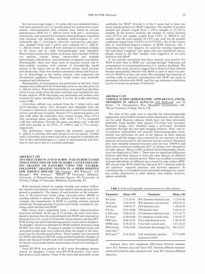
Six veal calves (age range: 1–18 weeks-old) were submitted from aveal farm operation over a 6 month period for postmortem exam-ination. Gastrointestinal (GI) contents were tested by enzymeimmunoassay (EIA) for C. difficile toxins A/B and C. perfringensenterotoxin, and screened for common enteropathogens associatedwith neonatal calf diarrhea, including enterotoxigenic E. coli,Salmonella spp. C. perfringens, rotavirus, coronavirus, and para-sites. Samples from only 2 calves were cultured for C. difficile.C. difficile toxins A and/or B were detected in intestinal contentsof all calves and no other enteropathogens were identified.Macroscopic lesions were similar in all cases and consisted of fibrin-ous enteritis, colonic edema, entero-colitis and multiplehemorrhages, dehydration, and pulmonary congestion and edema.Histologically, there were focal areas of mucosal erosion and fi-brino-cellular exudates, with colonies of clostridia-like bacillipresent in the lumen and on the mucosal surface small intestineand abomasum. There was marked transmural edema and focal ar-eas of hemorrhage in the lamina propria, with congested andthrombosed capillaries. Mesenteric lymph nodes were markedlycongested and edematous.
Subsequently, an investigation was performed on the farm. Fecalsamples were collected from 24 diarrheic calves at one time point forC. difficile culture. Three historical isolates recovered from diarrheiccalves 4 years earlier from the same veal farm were included for mo-lecular analysis. PCR-ribotyping was performed and isolates werescreened for genes encoding toxins A (tcdA), B (tcdB) and binarytoxin (cdtB).
Clostridium difficile was isolated from the 2 initial calves and22/24 diarrheic calves. Five ribotypes were identified from the27 isolates (3 historical, 2 postmortem calves and 22 from diarrheiccalves), and all were toxigenic. Sixteen (57%) possessed genes tcdAand tcdB, while the remainder were variant strains. Nine (32%)only possessed genes encoding tcdB, while 2 (7.1%) possessedtcdB and cdtB genes. Overall, the genes encoding tcdA, tcdB andcdtB were present in 16 (59%), 27 (100%) and 2 (7%) strains,respectively.
This preliminary report supports the potential capacity ofC. difficile to colonize and cause disease in several species. Furtherstudy of diarrheic and normal veal calves is required to elucidate therole of this enteropathogen as a cause of gastroenteritis and diar-rhea in veal calves and as a zoonotic pathogen.
ABSTRACT #52MYCOBACTERIUM AVIUM SUBSP. PARATUBERCULOSISTISSUE INFECTION OF YOUNG DAIRY CATTLE FOLLOW-ING GRAZING OF PASTURES USING THE ‘‘LEADER-FOLLOWER’’ GRAZING SYSTEM IN A HERD POSITIVEFOR JOHNE’S DISEASE. ME Fecteau1, RH Whitlock1, CDBuergelt2, RW Sweeney1. 1School of Veterinary Medicine,University of Pennsylvania, Kennett Square, PA.2University ofFlorida, College of Veterinary Medicine, Gainesville, FL.
With increased interest in organic farming and animal welfare,less intensive production systems that employ pasture grazing havegained in popularity. The impact of such practice on the transmis-sion of Mycobacterium avium subsp. paratuberculosis (MAP) toyearling animals is unknown. The purpose of the study was to in-vestigate the transmission of MAP to yearling animals exposedexclusively through grazing of pasture previously occupied by lac-tating cattle infected with MAP.
Nine Jersey steers originating from a Johne’s disease-positiveherd were included. At the age of 15 months, the steers were intro-duced to pastures heavily contaminated withMAP and remained onthose pastures for a total of 10 months. Fecal cultures and fecal RT-PCR testing were performed during pasture season and at slaughter.At slaughter, blood was obtained forMAP antibody detection usingELISA. For each steer, 28 separate samples of intestinal tissue andassociated lymph node were collected along the length of the intes-tinal tract for mycobacterial culture. These samples were processedfor MAP culturing on HEYM, using standard methods. Samples ofileum and ileocecal lymph node were examined histopathologicallyfor lesions of paratuberculosis and presence of acid-fast staining or-ganisms.
Fecal RT-PCR was positive in all 9 steers throughout pastureseason. At slaughter, 4 steers were RT-PCR positive. Two steershad positive fecal cultures. None of the steers had detectable serum
antibodies for MAP. Overall, 6 of the 9 steers had at least onetissue sample positive for MAP organisms. The number of positivesamples per animal ranged from 5 (18%) samples to 23 (82%)samples. In the positive animals, the number of colony formingunits (CFUs) per sample ranged from 1CFU to 300CFUs persample, and the total number of CFUs per steer for all samplescombined ranged from 25CFUs to 9,337 CFUs. Six of the 9 steershad no histolopathological evidence of MAP infection. The 3remaining steers were negative for acid-fast staining organismsbut individual Langhans’ type giant cells and epithelioid macro-phages found in the ileal samples were suggestive of an earlyinfection with MAP.
It was initially postulated that these animals were positive forMAP in their feces as MAP was ‘‘passing through’’ following oralconsumption on contaminated pasture (i.e., passive shedding). Theresults of this study, however, suggest that at least 6 of these animalsbecame permanently infected with MAP as each of them was pos-itive for MAP in at least one tissue. We concluded that exposure ofyearling cattle to pastures contaminated with MAP can result inpermanent infection with MAP, and that age resistance to infectioncan be overcome by pressure of infection.
ABSTRACT #53NORMAL ECHOCARDIOGRAPHIC APPEARANCE AND DI-MENSIONS IN ADULT ALPACAS. GD Hallowell1 and TJPotter2. 114, Threadcutters Way, Shepshed, Leicestershire, UK.2Royal Veterinary College, Herts, UK.
The aims of this study were to describe the echocardiographicappearance and establish normal cardiac dimensions and time indi-ces for adult Huacaya alpacas, which have not been previouslypublished. Eight healthy male alpacas (72�6 kg) were recruited.Standard images were obtained using a previously describedtechnique through left and right imaging windows. Inter- and in-tra-operator repeatability was assessed. Echocardiographic exam-ination was performed on each of the alpacas on three separateoccasions and the images were evaluated by one operator on threeseparate occasions and by one operator on one occasion. Statisticaltests used included repeated measures and one-way ANOVA andintra-class correlation coefficients (ICC’s). Images were obtained inall eight alpacas. Mean (�SD) standard echocardiographic dimen-sions are displayed in Table 1. There were no significant differencesbetween individual alpacas or from the same alpacas on differentdays except for pre-ejection period. There was excellent correlationbetween individuals on different days except for time indices (PEP,MV closure time, RTI) which also had poor ICC’s between individ-ual operators’ repeated measures and between operators. Inconclusion this is a straightforward repeatable technique for acquir-ing cardiac dimensions in adult alpacas; time indices, however,appear unreliable.
Table 1. Echocardiographic measurements in adult alpacas
Parameter Mean�SD Parameter Mean�SD
PA (cm) 2.31�0.41 RV diameter-diastole (cm) 1.12�0.36Ao (cm) 2.56�0.43 RV diameter-systole (cm) 0.83�0.32AoS (cm) 3.04�0.37 IVS-diastole (cm) 1.24�0.28PA:Ao 0.91�0.04 IVS-systole (cm) 1.68�0.25LAD (cm) 4.94�0.42 LV diameter-diastole (cm) 5.17�0.75ET (ms) 0.20�0.06 LV diameter-systole (cm) 3.10�0.72PEP (ms)�1 0.08�0.03 Free wall-diastole (cm) 1.26�0.39EPSS (cm) 0.29�0.18 Free wall- systole (cm) 1.67�0.32MVclosure
time (ms)1#0.10�0.06 Fractional shortening (%) 40.5�8.31
RTI (ms)1#� 0.14�0.05 Left ventricular ejection
fraction (LVEF)
0.77�0.09
�indicates there were significant differences between animals,1poor ICC between days and #poor ICC between different measure-
ments performed by same operators and �poor ICC between different
operators.
292008 ACVIM Abstracts

ABSTRACT #54THROMBOELASTOGRAPHY (TEG) IN CLINICALLYHEALTHY NEONATAL AND ADULT ALPACAS. D Bedenice1,EA Rozanski E1, B Wiinberg B2. 1Cummings School of VeterinaryMedicine at Tufts University, Grafton, MA.2The Royal Veterinaryand Agricultural University, Denmark.
Thromboelastography (TEG) is considered a rapid, reproducibletest of hemostatic function in whole blood. This analytical methodhas been previously evaluated in humans, dogs, and select species toidentify and quantify alterations of both hyper- and hypo-coagulative disease states. TEG may have similar diagnostic poten-tial in New World Camelids with hemostatic disorders, augmentingtraditional coagulation parameters. The objective of this study wasto establish a clinically applicable reference interval for reactiontime (R), clotting time (K), angle (a), and maximum amplitude(MA) of TEG on citrated whole blood from 21 clinically healthyalpacas.
Ten milliliters citrated whole blood from 15 clinically healthyadult alpacas (1–7 yrs) and 6 neonatal crias (1–14 days) was collect-ed by direct venipuncture. TEG analysis (using recombinant humantissue factor) was performed in duplicate at room temperature, 30minutes after collection. Prothrombin time (PT) and partial throm-boplastin time (PTT) were obtained concurrently.
Mean values of TEG parameters (1/� SD) for adult alpacas andneonatal crias were, respectively, R5 7.7 (2.9), 5.6 (2.2); K5 3.53(1.43), 2.55 (1.18); a5 48.11 (11.2), 56.21 (10.4); MA5 55.5 (8.35)and 59 (3.21) mm.Mean PT and PTT were 8.6 s (0.6) and 18.9 s (1.9)in adults vs. 9.4 s (0.6) and 18.1 s (1.96) in neonates. A significantdifference was not observed between age groups.
Compared to reported TEG results from dogs and horses,alpacas show relatively shorter clotting times (K). Thus, species-specific normal values are necessary for interpretation of TEGparameters.
ABSTRACT #55IVERMECTIN CONCENTRATIONS IN BLOOD ANDCEREBROSPINAL FLUID FOLLOWING INTRAVENOUSADMINISTRATION TO HEALTHY LLAMAS. Sarel vanAmstel, Ashley Portmann, Sherry Cox, Thomas Doherty, ShelleyNewman. University of Tennessee, College of Veterinary Medicine,Knoxville, TN.
The response to anthelmintic treatment in cases of meningealworm (Parelaphostrongylus tenuis) remains unpredictable. In a pre-vious study, ivermectin (IVM) was not detected in cerebrospinalfluid (CSF) following subcutaneous administration to healthy lla-mas. The aim of this study was to determine if a single IVadministration of the same dose (2.5ml/100 pounds bodyweight)of IVM [Ivomecs Injection Sterile 1% Solution (Merial)] results inmeasurable IVM concentrations in CSF. The IVM dose was addedto 1 L 0.9% NaCl and given over 30 minutes to 5 healthy llamas.CSF and blood were collected from indwelling spinal (lumbo-sa-cral) and jugular catheters respectively, at baseline and 2, 4, 6, 12,24, 48 and 60 hr after IVM administration. Serum and CSF werestored at �40 1C and IVM concentrations were determined usingHPLC.
Immediately after administration of IVM, 3 of the llamas showedtransient lethargy and decreased appetite. One llama developedacute neurological signs (paralysis and seizures) 7 days after IVMadministration and was euthanased 3 days later, having failed to re-spond to supportive treatment. Histopathologic examinationrevealed diffuse myelinic edema in the brain and spinal cord. Al-though it is possible that these histological changes resulted fromIVM administration, IVM was only detected in the CSF at the 4 hrsampling (0.11 ng/ml). Two other llamas developed laboratorychanges consistent with septic inflammation in the CSF (increase innucleated cells predominantly neutrophils) and in one of these casesfree and phagocytosed bacteria were present. The CSF returned tonormal in both llamas after antibiotic treatment and neither llamahad clinical signs at any stage. Results are reported as mean andrange.
The results indicate that CSF concentrations of IVM can beachieved following IV administration at the dose used in this study;however, it is not known whether these concentrations are thera-
peutic in cases of meningeal worm infestation. In addition, becauseof the possibility of IVM-induced neurological changes, it is recom-mended that IVM not be administered at this dose IV.
Serum IVM (ng/ml) CSF IVM (ng/ml)
2 hr 4751 (2291–7742) [n 5 5] 0.258 (0–0.57) [n 5 5]
4 hr 491 (102–615) [n 5 5] 0.282 (0.1–0.68) [n 5 5]
6 hr 298 (86–555) [n 5 5] 0.78 (0–2.49) [n 5 5]
12 hr 124 (58–222) [n 5 5] 0.145 (0–0.44) [n 5 4]
24 hr 67 (23–208) [n 5 5] 0.115 (0–0.22) [n 5 4]
48 hr 38 (10–118) [n 5 5] 0.09 (0–0.15) [n 5 5]
60 hr 22 (11–48) [n 5 5] 0 0 [n 5 4]
ABSTRACT #56CARDIOVASCULAR EFFECTS OF DOBUTAMINE AND NO-REPINEPRHINE INFUSION IN HEALTHY, ANESTHETIZEDALPACAS. CJ Vincent, AT Hawley, EA Rozanski, KM Lascola,D Bedenice. Cummings School of Veterinary Medicine at TuftsUniversity, North Grafton, MA.
The objective of this study was to characterize the cardiovasculareffects of dobutamine and norepinephrine infusion in isoflurane-anesthetized, healthy alpacas.
Eight adult alpacas (3 females, 5 intact males, 4.1 � 2.7 years)were evaluated. Initial baseline cardiovascular, respiratory, andmetabolic variables were obtained 30 minutes after induction of iso-flurane anesthesia. Four treatments (dobutamine at 4 and 8mg/kg/min; norepinephrine at 0.3 and 1 mg/kg/min) were administered inrandom order via constant rate infusion over 15 minutes, followedby repeat measurements of values and a 20 minute washout period.Subsequent baseline and post-treatment measurements were simi-larly repeated until both drugs and dosages were administered toeach animal. Baseline data in awake alpacas was obtained 18–24hours following recovery from anesthesia.
Both norepinephrine and dobutamine significantly elevated car-diac index and arterial blood pressure from baseline. Similarincreases in hemoglobin, oxygen content, and oxygen delivery wereobserved following administration of each drug at either dosage.Only dobutamine, however, reduced relative oxygen consumptionwhile improving overall oxygen balance. Furthermore, heart ratewas selectively enhanced by dobutamine and systemic vascular re-sistance by norepinephrine. Norepinephrine infusion resulted indose dependent changes in cardiovascular variables.
This study shows that both dobutamine and norepinephrine areappropriate choices to improve cardiac index, mean arterial pres-sure, and thus overall oxygen delivery in alpacas experiencingisoflurane induced hypotension. The lower infusion rates of bothdobutamine (4mg/kg/min) and norepineprhine (0.3mg/kg/min) arerecommended to avoid potential arrhythmogenic effects and exces-sive vasoconstriction, respectively.
ABSTRACT #57EXPRESSION OF SEROTONIN, TRANSFORMING GROWTHFACTOR-BETA1, AND EXTRACELLUAR MATRIX SIGNAL-ING MOLECULES IN MYOXOMATOUS MITRAL VALVETISSUE. MA Oyama1, SV Chittur2, JE Rush3, BW Keene4.1Department of Clinical Sciences, University of Pennsylvania,Philadelphia, PA, 2Center for Functional Genomics, State Universityof New York at Albany, Albany, NY, 3Department of ClinicalSciences, Tufts University, North Grafton, MA, 4Department ofClinical Sciences, North Carolina State University, Raleigh, NC.
The etiology and pathogenesis of canine myxomatous mitralvalve disease (MVD) are poorly understood. Using oligonucleotidemicroarrays, we have previously reported increased expression ofvarious serotonin (5HT) and transforming growth factor-beta1(TGF-B1) signaling molecules in myxomatous canine valve tissue.We have also shown that canine interstitial valve cells demonstrateincreased downstream mitogen activated protein-kinase (MAP-K)activity after exposure to in vitro 5HT, suggesting that serotoninand TGF-B1 may have the capacity to mediate both the prolifera-tive and degenerative changes seen in MVD. Based on these results,
30 2008 ACVIM Abstracts

we sought to further characterize the expression of key signalingcomponents of the 5HT and TGF-B1 pathways and extracellularmatrix in affected canine mitral valves. Mitral valve leaflets from7 dogs that were euthanized or died due to severeMVDwere excised,snap frozen in liquid nitrogen, and stored at �70C until processing.Mitral valve tissues from 5 healthy dogs that were euthanized for anunrelated study were collected as controls. Total RNA was isolatedand analyzed for integrity and quality. Transcription was evaluatedby real-time reverse transcriptase quantitative PCR (rt-qPCR) usinga commercial system (SybrGreen RT-qPCR, Applied Biosystems).Fifteen different transcripts were analyzed, including various 5HT-related molecules (5HT-receptor subtypes 1A, 1B, 2A, and 2B, and5HT transporter), TGF-B1-related molecules (TGF-B1, TGF-B1receptor, SMAD-2, and TAK-1), and extracelluar matrix compo-nents (MMP-1, 2, and 9, cathepsin K, collagen A, and fibronectin).Results were expressed in units of relative expression. For the 55HT-related molecules, transcriptional activity was detected in 33 of35 MVD samples (94%) vs 9 of 25 control samples (26%)(Po0.0001). Affected valves had significantly increased 5HT-recep-tor-1B as compared to control (MVD 29.8 vs control 0.0; Po0.05).For TGF-B1 components, no significant difference was found in thepresence or absence of expression or relative expression levels be-tween MVD and control. For extracellular matrix components,transcriptional activity was detected in 40 of 42 MVD samples(95%) vs 19 of 30 control samples (63%) (P50.0005). Affected tis-sue had significantly increased MMP-9 transcription as comparedto control (MVD 98.5 vs control 0.0; Po0.01). Expression of 5HTand extracellular matrix components is more prevalent in affectedmitral valves, and 5HT signaling may play a role in the pathology ofcanine MVD.
ABSTRACT #58SERUM SEROTONIN CONCENTRATION IS ELEVATED INDOGS WITH DEGENERATIVE MITRAL VALVE DIS-EASE. JW Arndt1, MA Oyama1, JM Connolly2, CA Reynolds1,RJ Levy2. 1Department of Clinical Sciences, School of VeterinaryMedicine, University of Pennsylvania, Philadelphia, PA.2Department of Pediatric Cardiology Research, The Children’sHospital of Philadelphia, Philadelphia, PA.
Little is known concerning the molecular mechanisms involved indegenerative mitral valve disease (DMVD). In humans, elevatedserotonin (5-HT) is associated with development of valvular lesions.Canine mitral valve cells demonstrate dose-dependent 5-HT-medi-ated ERK1/2 signaling, suggesting a possible link with canineDMVD. We sought to measure serum 5-HT concentration in dogswith DMVD, dogs predisposed to DMVD (small breed dogs weigh-ingo10 kg and without a murmur), and healthy large breed controldogs. Measurement of 5-HT was performed using a competitiveELISA (IB89527, Immuno Biological Laboratories, Inc., Minneap-olis, MN), which was validated for canine use. Seventy-nine dogswere enrolled (27 affected, 24 predisposed, and 28 controls), with17/27 affected and 15/24 predisposed dogs being Cavalier KingCharles Spaniels (CKCS). The assay demonstrated acceptable lin-earity (r50.96), parallelism (P40.05), and recovery(mean5100.9%). Analysis revealed significantly higher mean serum5-HT in affected dogs vs. control dogs (affected, 650.6 [SD5232.9]ng/ml vs. control, 474.3 [210.4]; Po0.05), and in predisposed vs.control dogs (predisposed, 765.9 [322.8] vs. control, 474.3;Po0.001). Subgroup analysis revealed that predisposed CKCS hadgreater mean serum 5-HT than predisposed non-CKCS (CKCS,903.9 [321.5] ng/ml vs. non-CKCS, 536.0 [153.7]; P50.004). Weconclude that dogs with clinically apparent DMVD as well asCKCS that are predisposed to DMVD have elevated serum 5-HT.Our results suggest that 5-HTmay play a role in the development ofDMVD in small breed dogs, and in particular in the CKCS. Furtherstudies involving the relationship between 5-HT, DMVD, breed,and platelet number, morphology, and function are warranted.
ABSTRACT #59GENETIC ASSOCIATION OF THE A31P AND A74T POLY-MORPHISM IN THEMYBPC3 GENE AND HYPERTROPHICCARDIOMYOPATHY IN MAINE COON CATS. G Wess,
C Schinner, K Weber, K Hartmann. Clinic for Small AnimalInternal Medicine, LMUUniversity of Munich, Germany.
Hypertrophic cardiomyopathy (HCM) is the most common felinecardiac disease and is inherited as an autosomal dominant trait. TheA31P and A74T single nucleotide polymorphisms (SNPs) in theMYBPC3 gene are thought to be causative mutations in MaineCoon cats. However, in many investigators’ experience phenotypesoften differ from genotypes. Echocardiographically confirmedHCM positive Maine Coon cats may have negative genetic test re-sults, and vice versa. Veterinarians are therefore often unsure whichrecommendations they should give breeders with various test re-sults. The aim of this study was to evaluate the association of theA31P and A74T SNPs and HCM in Maine Coon cats and to vali-date the clinical use of both genetic tests. The presence of theseSNPs in other breeds was assessed as well.
83 Maine Coon cats (mean age 71.3 months) and 68 cats of otherbreeds (mean age 102.4 months) were prospectively phenotyped us-ing echocardiography as phenotype-healthy or phenotype-HCM.Inclusion criteria were a minimum age of 24 months for males and36 months for females. The HCM-phenotype was defined as a leftventricular wall thickness of more than 6mm in diastole measuredin a 2D image using a VIVID 7 (General Electrics). Cats with hyper-thyroidism or hypertension were excluded. Taqmans genotypingassays were used for genotyping. The potential impact of the SNPswas assessed with PolyPhen software.
Of the 83 Maine Coon cats, 21.7% were positive and 78.3% neg-ative for A31P. None of the other breeds had a positive A31P SNP.From the genotype-positive cats, 83.3% were phenotype-healthyand 16.7% were phenotype-HCM. From the genotype-negativecats, 86.1% were considered phenotype-healthy and 13.9% pheno-type-HCM. 79 Maine coons were tested for the A74T SNP and35.4% were genotype-positive. The A74T SNP was present in otherbreeds as well. 78.6% of the Maine Coon genotype A74T SNP pos-itive cats had a normal ultrasound examination. The genotype-negative cats were considered as phenotype healthy in 88.2% and asHCM in 11.8%.
There was no significant allele frequency difference between HCMand healthy cats for both SNPs. Echocardiographic HCM positivecats were genotype-negative in 75% concerning the A31P and in50% concerning the A74T SNP. None of the genetic tests could pro-vide a useful predictive value of disease outcome. Computer basedprotein analysis of the evaluated SNPs indicated the potential im-pact of both SNPs to be ‘‘benign’’. The use of presently availablegenetic tests for selective breeding appears to be questionable ac-cording to these study results. The gold standard for the diagnosis ofHCM remains the annually echocardiographic examination.
ABSTRACT #60EFFECTOFRENALDYSFUNCTIONONN-TERMINALPRO-B-TYPE NATRIURETIC PEPTIDE; A CANINE BIOMARKERFOR HEART DISEASE. MK Schmidt1, CA Reynolds2, AHEstrada1, R Prosek1, HW Maisenbacher1, MM Sleeper2, MAOyama2. 1University of Florida College of Veterinary Medicine,Gainesville, FL.2University of Pennsylvania College of VeterinaryMedicine, Philadelphia, PA.
N-terminal B-type natriuretic peptide (NT-proBNP) is secretedfrom the cardiac ventricles in response to volume expansion or pres-sure overload. This peptide has previously been shown todistinguish cardiac and noncardiac causes of dyspnea in humansand dogs. In humans with kidney disease, BNP concentration canbe elevated due to decreased renal clearance, and clinical interpre-tation of these markers must take renal function into account. Theaim of this study is to determine the effects of kidney dysfunction onserum NT-proBNP concentrations in dogs. In total, 28 dogs wereexamined by physical examination, echocardiography, renal panel,non-invasive blood pressure measurement, urinalysis, and serumNT-proBNP assay (Canine CardioCare, Veterinary Diagnostics In-stitute, Irvine CA). Twenty-two healthy control dogs and 6 dogswith renal disease but without a heart murmur, valvular, or primarymyocardial disease were recruited. Mean NT-proBNP in controldogs was 282 pmol/L, range of 179 to 578pmol/L. Compared withcontrols, NT-proBNP was significantly increased in dogs with kid-ney dysfunction (mean51069 pmol/L; range 179–2071pmol/L;P50.0004). These results suggest that renal insufficiency may false-
312008 ACVIM Abstracts

ly increase serumNT-proBNP; however, a high degree of variabilitywas present within the kidney dysfunction group. Optimal NT-pro-BNP cut-off values for the diagnosis of heart disease or heart failurein dogs with concurrent renal dysfunction should be investigated.
ABSTRACT #61EFFECTS OF ATORVASTATIN ON ENDOTHELIAL FUNC-TION, LIPID PROFILES AND INFLAMMATORY MARKERSIN HEALTHY DOGS AND DOGS WITH CONGESTIVEHEART FAILURE. SM Cunningham, JE Rush, LM Freeman.Tufts Cummings School of Veterinary Medicine, North Grafton,MA.
HMG-CoA reductase inhibitors (statins) improve heart failureclass and survival in people with congestive heart failure (CHF), in-dependent of cholesterol reduction. Pleiotropic statin effects includeantioxidant and anti-inflammatory properties, antiarrhythmic ac-tions, and improvement of endothelial function. Impairedendothelium-dependent flow-mediated vasodilation (FMD) occursin people with CHF and is a strong predictor of adverse outcome.The goals of this study were to evaluate the tolerability of atorvas-tatin in healthy dogs and in dogs with CHF, to determine whetherdogs with CHF have endothelial dysfunction, and to evaluate theeffects of atorvastatin on markers of endothelial function, systemicinflammation, and oxidative stress in dogs with CHF.
In part I of the study, employee-owned dogs that were healthybased on history, physical examination, CBC, biochemistry profile,and urinalysis (n511) were treated with atorvastatin and reevaluat-ed on days 14 and 30. Testing on each day included an examinationand fasting CBC, biochemistry profile, and C-reactive protein(CRP) concentrations. FMD of the brachial artery was assessed in6 of 11 dogs on days 0, 14, and 30. Ultrasonographic changes inarterial diameter and blood flow velocities were measured at 10, 15,30, 45, and 60 sec after a 3-minute period of vessel occlusion. In partII of the study, client-owned dogs with ISACHC Class II or IIIaCHF (n513) were administered atorvastatin at 2mg/kg q 24 h for 8weeks. All dogs were assessed on days 0 and 56 of the study viaechocardiography, ECG, blood pressure (BP), and owner-complet-ed FETCH questionnaires. CBC, biochemistry profiles, lipidfractionation, concentrations of 8-F2a-isoprostanes, NT-proBNPlevels, and CRPwere measured at each visit. FMDalso was assessedin 5 of 13 dogs on days 0 and 56 of the study. Baseline parameterswere compared between control and CHF dogs using Mann-Whit-ney-U tests, while pre- and post-atorvastatin data were analyzedusing Wilcoxon signed ranks tests. At baseline, FMD assessmentrevealed a trend (p50.06) towards a blunted change in the brachialartery velocity profile at 15 seconds in CHF dogs compared tohealthy dogs. Atorvastatin was well-tolerated in control and CHFdogs and did not result in side effects or significant increases in he-patic transaminases or creatine kinase. Reductions in systolic BP(p50.04), total serum cholesterol (p50.01), non-HDL cholesterol(p50.002), and total leukocytes (p50.03) were noted in the CHFgroup after receiving atorvastatin. There were no significant chang-es in echocardiographic or FMD parameters or in FETCH scores.Healthy dogs also had a reduction in total cholesterol (p50.01) afteratorvastatin administration. Analysis of CRP, isoprostanes, andNT-proBNP concentrations is pending. In conclusion, atorvastatinwas well-tolerated at this dose in both groups of dogs and furtherinvestigation into the effects of statin therapy in dogs with CHF iswarranted.
ABSTRACT #62TROPONIN CONCENTRATIONS IN PATIENTS WITHMASSES OR TUMORS. G Farace1, A Beardow1, C Carpenter1,K Yeung1, M Zieba1, SJ Ettinger2, SD Forney2. 1IDEXXLaboratories, Inc., Westbrook, ME, 2California Animal Hospital,Los Angeles, CA.
During the course of the IDEXX/California Animal Hospitalcardiac study we have discovered a number of interesting associa-tions. One such association is the fact that enrolled patients withmasses or tumors have higher levels of troponin than similar pa-tients with cardiac disease alone.
Taking all 658 patients currently in the study, normal dogs(n5115) have a mean troponin of 0.34 ng/ml (95% CI 0.22–0.46);those patients with at least some minor echocardiographic changes(n5445) have a mean troponin concentration of 1.29 ng/ml (95%CI0.97–1.61), while patients with a mass or tumor (n598) have a meantroponin of 11.71 ng/ml (95% CI 4.99–18.46). Wilcoxon tests showthat the group with masses is significantly higher than both the nor-mal dogs and the cardiac disease population (po0.0001). Dividingthe cardiac patients into those with minor echocardiographic chang-es or with asymptomatic disease and those with clinical signs ofheart disease or heart failure gives a mean troponin of 0.79 ng/ml(95% CI 0.44–1.13) for the first group and a mean of 1.82 ng/ml(95% CI 1.29–2.36) for the second. Treating the patients with mass-es in the same manner, we have a group with masses and minorechocardiographic changes or asymptomatic disease and a groupwith masses and clinical signs of heart disease or heart failure. Themeans for the two groups are 21.46 ng/ml (95% CI 6.69–36.22) and5.12 ng/ml (95%CI 1.31–8.92), respectively. In both cases the groupof patients with masses is still significantly different from the com-parable cardiac group (po0.0001 and po0.0026); therefore severityof cardiac disease does not explain the observed difference.The percentage of patients with a troponin over 2.00 ng/ml in theentire cardiac disease only group is about one-third of that in thegroup with masses. This gives a 5.4-fold relative risk that elevatedtroponin is associated with a mass or tumor. Dropping to a 1.00 ng/ml cut-off still gives a 3.5-fold relative risk.
The entire cardiac and masses/tumor groups are similar in termsof N-terminal prohormone atrial natriuretic peptide concentration(1943 vs. 1766 fmol/ml, respectively) and N-terminal prohormonebrain natriuretic peptide concentration (1482 vs. 1190 pmol/L re-spectively). Again this indicates that the underlying cardiac diseaseis not the cause of the differences in troponin.
Taking into account the location of the mass, it is apparent thattroponin is not consistently elevated in the presence of a mass. Onlymasses located in the heart, spleen, or multiple locations show meantroponin levels over 1.00 ng/ml.Masses in organs such as the lung orliver do not result in a mean troponin greater than 1.00ng/ml.
Troponin is not consistently elevated in all cases where a mass ispresent so it cannot be used as a tumor screen; however, troponinlevels above 1.00 ng/ml may indicate that the patient has a mass ortumor, and that the mass is likely located in the heart, spleen ormultiple sites.
ABSTRACT #63GREAT DANE DILATED CARDIOMYOPATHY IS ASSOCI-ATED WITH ALTERED TRIADIN AND CALSTABIN2TRANSCRIPTION. MA Oyama1, CR Reynolds1, M Kuentzel2,SV Chittur2. 1Department of Clinical Sciences, School of VeterinaryMedicine, University of Pennsylvania, Philadelphia, PA.2Center forFunctional Genomics, State University of New York, Albany, NY.
Little is known regarding the molecular mechanisms underlyingdilated cardiomyopathy (DCM) in the Great Dane. Thus, we per-formed a widescale evaluation of transcriptional activity frommyocardial tissue of affected dogs. cRNA was generated from leftventricular tissues from 3 Great Danes that were euthanized due torefractory DCM and from 3 large breed dogs euthanized for non-cardiac reasons and hybridized to a second generation canine-specificmicroarray (Canine GeneChip 2.0, Affymetrix). Comparison of tran-scriptional activity revealed significant downregulation of 24transcripts and upregulation of 298 transcripts in affected dogs vscontrol. The two transcripts with the greatest differential expressionwere triadin (9.07-fold down-regulated) and calstabin2 (FKBP12.6)(61.3-fold up-regulated). Expression of these two transcripts was fur-ther investigated using RT-qPCR. Both triadin and calstabin2 areregulators of cardiac ryanodine receptor (RYR2) activity and hencemovement of Ca21 within myocardial cells. Triadin is a transmem-brane protein that links RYR2 to calsequestrin and is a criticaldeterminant of systolic Ca21 release from the sarcoplasmic reticulum.Triadin deficiency would putatively lead to reduced calsequestrin-RYR2 binding, ‘‘leaky’’ RYR2, and decreased efficiency of excita-tion-contraction coupling. Calstabin2 binds to the cytoplasmicdomain of RYR2 and promotes a closed RYR2 state; thus increasedexpression of calstabin2 may represent a compensatory response.Further study involving calcium transients, RYR2 function, and therole of triadin and calstabin2 in Great Dane DCM is warranted.
32 2008 ACVIM Abstracts

ABSTRACT #64CARDIOMYOCYTE CALCIUM TRANSIENTS IN GERMANSHEPHERD DOGS WITH INHERITED VENTRICULAR AR-RHYTHMIAS. SA Jesty,1 BG Kornreich1, J Cordeiro,2 CAntzelevitch,2 NS Moıse1. 1College of Veterinary Medicine,Cornell University, Ithaca, NY. 2Masonic Medical ResearchLaboratory, Utica, NY.
Inherited ventricular arrhythmias and abnormalities of repolar-ization have been documented in German Shepherd Dogs (GSDs).The purpose of this study was to determine the temporospatial as-pects of calcium cycling in the ventricular cells of affected GSDs andnormal dogs. The hypotheses were that calcium transients in theventricular cells of affected GSDs would be different from those innormal dogs, and that calcium sparks and early/delayed afterdepo-larizations (EADs/DADs) would be more numerous in cells ofaffected dogs as a result of the calcium cycling abnormalities.
Ten affected GSDs and 10 unaffected dogs were studied. Mid-myocardial cells (M cells) were harvested from dogs aftereuthanasia. Myocytes were loaded with Fluo 3, a fluorophore whoseemission spectrum is shifted in the presence of calcium. Myocyteswere field stimulated and XT line scan images were recorded usingconfocal microscopy.
M cells from affected GSDs displayed a high variation in the de-cay phase of the calcium transient (Tau), compared with normaldogs (Figure ). Affected dogs had a mixture of abnormal and nor-mal cells, whereas normal dogs had more consistent calcium cyclingacross cells. Additionally, the number of calcium sparks, EADs, andDADs was greater in affected GSDs.
These results indicate that abnormalities of calcium cycling, andspecifically cell to cell electrical heterogeneity, may be a mechanismcontributing to the inherited ventricular arrhythmias in GSDs.
ABSTRACT #65COMPARISON OF NT-PRO-BNP CONCENTRATION INCATSWITH ACUTE DYSPNEA FROM CARDIAC OR RESPI-RATORYDISEASE. PR Fox1, MA Oyama2, KMacDonald,3 CAReynolds2. 1Animal Medical Center, NewYork, NY; 2University ofPennsylvania, Philadelphia PA; 3Animal Care Center, RohnertPark, CA.
Biomarkers are increasingly used to help differentiate causes ofacute dyspnea. We aimed to determine whether NTproBNP bloodconcentrations clinically differentiated respiratory vs cardiac causesof acute dyspnea in cats.
Recruitment required presentation for acute dyspnea. Each catwas evaluated by medical history, physical examination, thoracicradiography, and echocardiography by a board certified cardiolo-gist. Cause of dyspnea was designated cardiac (CHF) or non-cardiac (primary respiratory disease). A central laboratory blinded
to diagnosis performed serum NTproBNP assays. NTproBNP con-centrations were compared by Mann Whitney test. Spearmancorrelation assessed NTproBNP concentrations vs echocardio-graphic measurements in CHF cats. Receiver operatingcharacteristic analysis evaluated NTproBNP outcome.
Median NTproBNP was significantly different between cats withdyspnea associated with CHF (n534) vs respiratory disease (n522)(Po0.0001). Median NTproBNP [interquartile range] was846 pmol/L [567–1160pmol/L] for CHF and 52pmol/L [24–119pmol/L] for respiratory disease. Correlations (BNP vs echo) wereLVPWd (P50.039, r50.354), LVd (P50.113), LVs (P50.053),%FS(P5073), IVSd (P50.336), LA (P50.099), LA/Ao (P50.12), andvertebral heart score (P50.995).
NTproBNP4180 pmol/L for dyspnea caused by CHF possessed94.1% sensitivity, 86.4% specificity, 91.4% positive predictive val-ue, and 90.5% negative predictive value vs respiratory (fitted ROCarea under curve, 0.976 [SE50.0183]). Thus, NTproBNP determi-nation may help differentiate cardiac vs. primary respiratory causeof dyspnea in cats.
ABSTRACT #66NT-PRO-BNP ASSAY DISTINGUISHES CARDIAC VS PRI-MARY RESPIRATORY CAUSES OF RESPIRATORY SIGNSIN DOGS. MA Oyama1, JE Rush2, EA Rozanski2, PR Fox3, CAReynolds1, S Gordon4, B Bulmer5, B Lefbom6, W Brown7, LLehmkuhl8, R Prosek9, M Lesser10, M Kraus11, M Bossbaly12, GRapoport13. 1University of Pennsylvania, Philadelphia, PA; 2TuftsUniversity, NorthGrafton,MA; 3The AnimalMedical Center, NewYork, NY; 4Texas A&M University, College Station TX; 5OregonState University, Corvallis, OR; 6Chesapeake Veterinary CardiologyAssociates, Vienna, VA; 7Veterinary Cardiology Consultants, Novi,MI; 8MedVet, Worthington, OH; 9University of Florida, Gainsville,FL, 10Advanced Veterinary Care Center, Lawndale, CA; 11CornellUniversity, Ithaca, NY; 12Heartsound Consultants, Langhorne, PA;13Angell Memorial Animal Hospital, Boston, MA.
In dogs with respiratory signs, differentiation of the underlyingetiology (i.e., congestive heart failure [CHF] vs. primary respiratorydisease such as pneumonia, chronic airway disease, fibrosis, etc.) isan important step towards accurate diagnosis and treatment. NT-pro-B-type natriuretic peptide (NT-proBNP) is elevated in dogswith CHF, and we sought to evaluate its ability to differentiate car-diac vs. non-cardiac etiology of respiratory signs in a prospectivemulticenter clinical study. Dogs with respiratory signs (i.e., tachyp-nea, cough, increased respiratory effort, etc.) that were judgedsevere enough by the owner to adversely affect the dog’s quality oflife were recruited from 13 sites. Dogs underwent physical exam,thoracic radiographs, and echocardiogram, after which a board-certified cardiologist categorized etiology of signs as follows: Group1, CHF; Group 2, primary respiratory disease; Group 3, primaryrespiratory disease with concurrent heart disease but without CHF;Group 4, equivocal. Serum NT-proBNP assay was performed at acentral laboratory blinded to the diagnosis. Complete data wasavailable for 116 dogs.Median NT-proBNP was significantly differ-ent between Groups 1 and 2 and between Groups 1 and 3(Po0.001). Group 1, n562, median NT-proBNP52445pmol/L,[interquartile range51499–3134]; Group 2, n521, NT-pro-BNP5413 pmol/L [245–852]; Group 3, n528, NT-proBNP5510 pmol/L [353–1212]). NT-proBNP 41200pmol/Lpossessed 85.5% sensitivity, 81.6% specificity, 85.5% positive pre-dictive value, and 81.6% negative predictive value for identifyingdogs with congestive heart failure (Group 1) vs. dogs with primaryrespiratory disease with or without concurrent heart disease(Groups 2 and 3). Area under the receiver-operating characteristiccurve was 0.908 (SE50.029).
ABSTRACT #67CIRCULATING NATRIURETIC PEPTIDES CONCENTRA-TIONS IN CATS WITH RESPIRATORY DISTRESS. D JConnolly, R J Soares Magalhaes, V Luis Fuentes, H M Syme, GCole, AM Boag. Royal Veterinary College, Herts, UK.
Cats in respiratory distress (RD) can represent a significant diag-nostic challenge. The ability to distinguish cardiac from non-cardiac
332008 ACVIM Abstracts
causes of RD is a vital initial step to achieving an accurate diagnosisand appropriate treatment. It is often not possible to do this on thebasis of history and physical examination, and the compromisedstate of cats with severe RD often limits diagnostic evaluation. Hu-man and canine studies have shown circulating B-type natriureticpeptide concentration to be an accurate predictor of congestiveheart failure (CHF), enabling patients with CHF to be differentiat-ed from those with non-cardiac causes of RD (RDNC). In contrastto dogs and humans, relatively little is known about the utility ofnatriuretic peptide testing in cats. It has been established that circu-lating NT-proANP and particularly NT-proBNP concentrationsmeasured by ELISA identified cats with heart disease and heart fail-ure. The study aim was to determine if circulating natriureticpeptide (NP) concentration could distinguish cats with RDNC fromthose with CHF.
The study recruited 91 cats from 1 university teaching hospitaland 2 private practices. Serum natriuretic peptide concentrationswere measured in 41 cats with RDNC (diseases included asthma,neoplasia, pyothorax, pneumonia, pleural effusion, bronchitis, rhi-nitis, nasopharyngeal stenosis, nasopharyngeal polyp) andcompared to cats with asymptomatic heart disease (AsymHDn517) and cats with RD due to CHF (RDCHF n533) usingsandwich enzyme immunoassays. The diagnoses in the cats withheart disease included hypertrophic cardiomyopathy, hypertrophicobstructive cardiomyopathy, or both (n536); restrictive card-iomyopathy (n510); dilated cardiomyopathy (n51); mitraldysplasia (n51); double-chambered right ventricle (n51); and idio-pathic third degree atrioventricular block (n51). The ability ofcirculating NP concentrations to distinguish cats with RDNC fromthose with RDCHF was explored using receiver operator curve(ROC) analysis. The RDCHF group had higher median NT-proANP and NT-proBNP concentrations (1690 and 523 fmol/mlrespectively) than the RDNC group (614 and 45 f/mol/ml, po 0.05and po0.05, respectively). The area under the curve was 0.88 and0.96 for the ROC analysis of the diagnostic accuracy of NT-proANP and NT-proBNP concentrations to discriminate RDCHFfrom RDNC cats. An optimum cut-off concentration of 986 fmol/ml for NT-proANP and 220 fmol/ml for NT-proBNP accuratelydiscriminated RDNC from RCCHF cats with a sensitivity andspecificity of 93.8% and 80.3% and 93.9% and 87.8% respectively.
Serum NP concentrations were different in cats with CHF com-pared with those with respiratory distress of non-cardiogenic origin.Evaluation of circulating NP concentrations may be helpful in theinitial approach to cats presenting with respiratory distress.
ABSTRACT #68CAN A ‘‘LOUD’’ RIGHT-APICAL SYSTOLICMURMUR PRE-DICT THE DIAGNOSIS OF CANINE PULMONARYHYPERTENSION? DG Ohad, I Lenchner, T Bdolah-Abram.The Koret School of Veterinary Medicine, The Hebrew Universityof Jerusalem, Rehovot, Israel.
The index of clinical suspicion of pulmonary hypertension (PHT)is typically low. We tested whether selected findings predict thediagnosis of PHT, as confirmed by a Doppler-derived systolic pres-sure gradient435mmHg between the right ventricle and atrium.
Data from 312 consecutive dogs were retrospectively analyzed tocalculate the correspondence ratio between pairs of non-quantita-tive (historical and/or physical) versus quantitative (Doppler)variables. Sensitivity, specificity, and predictive value indices werecomputed. The McNemar test was used to seek a trend among casesof disagreement.
The following (in a descending order) variables were more prev-alent in PHT (n596) than in non-PHT (n5216) patients: left apicalmurmur, cough, right apical murmur, syncope, ascites, dyspnea, astronger right than left apical murmur, hepatomegaly, and or-thopnea. Both left and right apical murmurs were louder in PHTthan in non-PHT patients.
A ‘‘loud’’ right apical murmur, whether isolated or combinedwith non-quantitative variables as ascites or syncope, had a lowsensitivity of o44% and a high specificity of 494% as a predictorof PHT. While an isolated stronger right than left apical murmurhad a positive predictive value (PPV) of 82% and was 98% specificfor PHT, when combined with syncope it had a PPV of 91%, andwas 96% specific for PHT. The combination of ascites with a Grade
�IV/VI right apical murmur had a PPV of 100% and a specificity of100% for PHT.
A louder-than-expected right apical systolic murmur combinedwith ascites and/or syncope should highly increase the index of sus-picion for PHT.
ABSTRACT #69TRANSVENOUS COIL EMBOLIZATION OF PATENT DUCT-US ARTERIOSUS IN SMALL DOGS. EHenrich, E Hassdenteufel,N Hildebrandt, C Fischer, M Schneider. Department of ClinicalStudies, Small Animal Clinic (Internal Medicine and Surgery), Justus-Liebig-University Giessen, Giessen, Germany.
The occlusion of patent ductus arteriosus (PDA) in small caninepatients is challenging for both surgical and interventional therapy.This prospective study examines the feasibility and success of trans-venous coil embolization of patent ductus arteriosus in small dogs.
Inclusion criteria were a left-to-right shunting PDA and a bodyweight equal to or less than 3.0 kg.The presence of congestive heartfailure was not considered an exclusion criterion. Patients with ad-ditional congenital cardiac diseases were excluded. Under generalanaesthesia the right femoral vein was accessed percutaneously us-ing a 4 French introducer sheet. The PDA was catheterizedretrogradely with a 4 French wedge catheter, a 4 French multipur-pose catheter, and a 0.018 inch guide wire. After switching to anangiographic catheter, the contrast medium was injected into thedescending aorta. PDA morphology was classified and the minimaland ampulla diameter of the ductus were measured. A commercial0.038 inch coil (Detachable Coil for PDA closure, Cook Deutsch-land GmbH) measuring at least twice the minimal diameter of thePDA was chosen. The coil was placed with approximately 1/2 loopof the coil anchored on the pulmonary side. An echocardiographicfollow-up was performed within 24 hours post intervention.
Twenty-one dogs underwent transvenous coil occlusion, withChihuahua and Yorkshire Terrier being the most common breeds(n56 and n55, respectively). The distribution of sex was 14 femaleand 7 male dogs. The ages of the patients ranged from 1.9 to 83.5months (median 7.7 months), and the body weight from 1.0 and2.9 kg (median 1.9 kg). The minimal diameter of the PDA measured1.2 to 2.4mm (median 1.8mm) and the PDA ampulla 2.4 to 5.9mm(median 4.6mm). All but one PDA had an aortic ampulla and apulmonary constriction (Type E, n516, Type A, n54). The remain-ing patient had two constrictions (one on the aortic and one on thepulmonary side, Type D). The coil implantation was successful in allpatients. 7 dogs received a coil with a 3mm loop-diameter while inthe other 14 dogs a coil with a 5mm loop-diameter was implanted.After the coil had been detached, repositioning of the pulmonaryloop of the coil with a 4 French wedge catheter became necessary inone dog. Two dogs developed temporary bradycardia after coil im-plantation requiring medical therapy and 16/21 (76%) dogs had noresidual shunting in the follow-up echocardiography after 24 hours.
In conclusion: For an experienced cardiologist, transvenous em-bolization of patent ductus arteriosus in small dogs is possible usinga 4 French catheter and a commercial detachable coil. Arterial ac-cess is not essential and the procedure is safe and successful.
ABSTRACT #70EMBOLIZATION OF LARGE PATENT DUCTUS AR-TERIOSUS (4 4.0mm) IN DOGS WITH A SINGLEPOLYESTER FIBERED DOUBLE-HELIX COIL. M Schneider,N Bierent, N Hildebrandt, C Fischer. Department of ClinicalStudies, Small Animal Clinic (Internal Medicine and Surgery),Justus-Liebig-University Giessen, Giessen, Germany.
Coil embolization of large patent ductus arteriosus (PDA) is diffi-cult, because coil stiffness is getting lower with increasing loopdiameter. To increase coilstiffness a Double-Helix configuration ofthe coil can be used. The aim of this prospective study was to provethe feasibility of embolization of large PDAs (4 4.0mm) with asingle polyesther fibered Double-Helix coil.
Ten consecutive dogs with an angiographically determinded min-imal PDA diameter of 4 4.0mm were included independent onclinical status of congestive heart failure (n57 NYHA III or IV) or
34 2008 ACVIM Abstracts
atrial fibrillation (n51). Detachable Double-Helix coils designed oftwo identical strains of stainless steel wire (0.052 inches) with poly-ester anchored were produced in various diameters (10, 11, 12,14, 16mm). Prior to the coil embolization an angiography of de-scending aorta was performed to classify the PDA morphologic(classification of Krichenko), to measure the PDAminimal and am-pulla diameter, and to define the shunt-grade. The shunt-ratio wascalculated using the Fick method. The PDA was catheterized in aretrograde manner and a 7 F Mullins sheet was placed through thePDA into the descending aorta. A Double-Helix coil with loop di-ameter greater than two times the PDA minimal diameter wasimplanted. Ten minutes after coil embolization calculation of theshunt-ratio and the angiography was repeated. In all dogs a singlecoil was employed regardless of residual shunting. Clinical andechocardiographic reexaminations were performed within threedays after the intervention. M-mode measurements were indexedto body weight by allometric scaling. Data prior and after interven-tion were compared by student t-test.
German Shepherd dogs (n54) were overrepresented. The medianage was 9.2 month (range 4.3–82.0). The median body weight was24.0 kg (range 8.1–36.0). The PDA was long and conical (Type E) inseven dogs and showed two constriction (Type D) in three dogs. Theminimal PDA diameter on the pulmonic site was 4.8–6.3mm (me-dian 5.9mm) and the width of the ampulla was 9.1–18.9mm(median 15.1mm). In 9/10 dogs the implantation of the primary se-lected coil was successful, in the remaining dog the next coil size hasto be used. After coil placement the shunt-ratio decreased signifi-cantly (mean� SD: 3.80� 0.97, range 2.4–5.3 to 1.38 � 0.46, range1.0–2.3, p o 0.0001, n510). Complete closure was documented byangiography in one and by echocardiography in three dogs. Despiteof incomplete closure in 7/10 dogs the volume overload was reducedin all dogs, leading to a significant reduction of the maximal aorticblood flow velocity (2.980 � 0.703 to 2.160 � 0.635, p 5 0.0016,n59) and of the index of left ventricular diastolic diameter (2.259 �0.398 to 2.025 � 0,370, p 5 0.0023, n510).
In conclusion, the polyester fibered Double-helix coil has thestiffness to be fixed in a large PDA up to 6.3mm and produce a re-duction of shunt flow.
ABSTRACT #71MYOTUBULAR MYOPATHY IN A FAMILY OF MANCHES-TER TERRIER DOGS. F.L. Robinson1, A.P. Misizin1, D.P.O’Brien2, G.S. Johnson2, J.E. Dixon1, G.D. Shelton1. 1School ofMedicine, University of California, San Diego, La Jolla, CA.2College of Veterinary Medicine, University of Missouri,Columbia, MO.
Three male Manchester Terrier littermates were presented at 2months of age for weakness and failure to thrive. The dam and sirewere clinically normal. The dam was previously bred to a differentmale with 2 male puppies showing similar clinical signs. Muscle bi-opsy specimens from all three puppies showed centrally placednuclei resembling fetal myotubes, or a central clear zone, withinmost type 1 fibers. Cryostat sections reacted for oxidative enzymesshowed attenuated activity at the periphery of the fibers, and denseoxidative staining at the center of the fibers, which lacked myosinATPase activity. Ultrastructural analysis correlated well with thehistochemical findings including myofilaments forming a compactperipheral rim around a central nucleus, or around a central zone,containing mitochondria, glycogen, or dilated membranous pro-files. In humans, recessive mutations in the myotubularin (MTM1)gene cause X-linked myotubular myopathy. MTM1 is a member ofa family of phosophoinositide 3-phosphatases that dephosphorylatephosphatidylinositol 3-phosphate and phosphatidylinositol 3,5-bis-phosphate. The canine MTM1 gene also resides on the Xchromosome. Accordingly, MTM1 was investigated as a possiblegene responsible for the described myotubular myopathy in Man-chester Terriers. MTM1 protein was analyzed in affected andnormal dog muscle. MTM1 protein in detergent extracts wasimmunoprecipitated with an anti-MTM1 antibody (Santa CruzBiotechnology) and protein-A agarose, then analyzed by SDS-PAGE and immunoblotting using an anti-MTM1 antibody.MTM1 protein was not detected in the muscle extracts from affect-ed dogs. MTM1 mRNA analysis of muscle was performed by RT-PCR. Exon 1 containing mRNA was not detected in affected dogs.In conclusion, myotubular myopathy in Manchester Terrier dogs is
clinically, histologically, ultrastructurally, and biochemically simi-lar to that of human X-linked myotubular myopathy. The absenceof MTM1 protein and exon 1 containing mRNA in muscle tissue ofaffected dogs suggests that loss of function mutations in this PI 3-phosphatase may be the causative genetic defect. Preliminary datasuggests a mutation in exon 1 of theMTM1 gene.
Originally presented at the World Muscle Society Meeting,Giardini-Naxos, Taormina, Sicily, October 2007.
ABSTRACT #72MYCOPHENOLATE MOFETIL THERAPY FOR ACQUIREDMYASTHENIA GRAVIS IN DOGS: A COMPARATIVE RET-ROSPECTIVE STUDY (1999–2007). CW Dewey1, MF Harb-Hauser2, S Cerda-Gonzalez1, JM Levine3, BL Badgley1, NJ Olby4,M Kent5, N Birnbaum6, GD Shelton7. 1Cornell University Collegeof Veterinary Medicine, Ithaca, NY; 2Pet Emergency and SpecialtyCenter of Marin, San Rafael, CA; 3Texas A&M University Collegeof VeterinaryMedicine, College Station, TX; 4North Carolina StateUniversity College of Veterinary Medicine, Raleigh, NC;5University of Georgia College of Veterinary Medicine, Athens,GA; 6Veterinary Internal Medicine Practice of N. Virginia,Manassas, VA; 7University of California-San Diego, LaJolla, CA.
Acquired myasthenia gravis (MG) is a common autoimmune ne-uromuscular disorder of dogs, the effective treatment of which canbe extremely challenging. Mycophenolate mofetil (MMF) is a lym-phocyte-specific immunosuppressive drug that has been usedsuccessfully in human autoimmune disorders and transplant recip-ients. There is anecdotal evidence of MMF efficacy in dogs withvarious autoimmune diseases. The purpose of this investigation wasto retrospectively evaluate the outcome of dogs with serologicallyconfirmedMG that were treated withMMF and compare theseMGpatients to MG dogs treated with pyridostigmine (PYR) alone.
Myasthenic dogs, 14 treated with MMF (mean dose-18.6mg/kgBID) and 8 treated with PYR alone (mean dose-1.16mg/kg BID-TID), were identified. All but 2 MMF dogs also received PYR.Dogs in both groups were of several breeds of both sexes, and allclinical forms (focal, generalized, acute fulminating and paraneo-plastic) of MGwere represented. Megaesophagus was present in 12/14 (86%) dogs in the MMF group and 7/8 (88%) dogs in the PYRgroup. At initial diagnosis, acetylcholine receptor (ACh R) anti-body titers ranged from 0.96 to 14.4 nM/L (mean-3.30 nM/L) in theMMF group, and from 1.21 to 7.13 nM/L (mean-3.97 nM/L) in thePYR group. Thymoma or lymphoma was identified in 1 MMF dogand 2 PYR dogs. Eight of 14 dogs (57%) treated with MMFachieved clinical remission of MG; 7 of these dogs had megaesoph-agus, 5 had pneumonia. Three of these 8 dogs (38%) came out ofremission during MMF weaning. Two of 8 PYR dogs (25%)achieved clinical and immune remission; 1 of these 2 dogs hadmegaesophagus and pneumonia. The 1-yr mortality rate (due toMG) was 37.5% for PYR dogs and 28.6% forMMFdogs. Based ona Fisher’s exact test (po0.05), the differences between groups in re-mission rates and mortality rates were not significant.
Results of this study suggest that clinical remission ofMG in dogsis more likely to be achieved with MMF than with sole PYR ther-apy; however, this was not statistically significant. Also, dogsattaining clinical remission with MMF should be monitored closelyfor disease recurrence duringMMFweaning (i.e., sustained immuneremission less likely). Further investigation of MMF as a therapyfor canine acquired MG with a larger group of dogs is warranted.
ABSTRACT #73INBORN ERROR OF METABOLISM IN GORDON SETTERPUPPIES: ORGANIC ACID PROFILE AND CANDIDATEGENE SEQUENCING. Gorgi AA1, O’Brien DP1, Shelton GD2,Johnson GS3. 1University of Missouri, College of VeterinaryMedicine, Department of Small Animal Medicine and Surgery,Columbia, MO. 2Comparative Neuromuscular Laboratory,University of California, San Diego, CA. 3University of Missouri,College of Veterinary Medicine, Department of VeterinaryPathobiology, Columbia, MO.
A fatal, neurologic disease of Gordon Setters called DUNGd bybreeders was first reported in the veterinary literature in 2000 as an
352008 ACVIM Abstracts

autosomal recessive trait. Gait and postural abnormalities and pro-gressive weakness begin at 3–4 weeks of age, progressing to visualdeficits and recumbency by 5–6 weeks of age. Serum chemistries andCSF analysis are normal and only subtle and non-specific histo-pathologic findings are observed at necropsy. Urine samples fromthree affected puppies were assayed by gas chromatography/massspectroscopy for organic acids. A distinct pattern of elevated glycineconjugates (3-methylcrotonylglycine, 2methylbutyrlglycine, hex-anoylglycine) was observed in all three samples. Urine from oneobligate carrier parent and one clinically normal littermate showeda similar pattern, and urine samples from one obligate carrier parentand two clinically normal littermates were normal. Based on the hu-man literature the organic acid findings were most consistent with amutation in the biotinidase (BTD) or holocarboxylase synthase(HLCS) genes. Primary methylcrotonyl CoA carboxylase (MCC)deficiency has also been reported albeit with much milder clinicalsigns. Coding regions of the BTD and HLCS genes have been se-quenced and no significant mutations identified. Sequencing of theMCC gene is pending.
ABSTRACT #74BIOMECHANICAL EVALUATION OF TWO INTERNAL FIX-ATION IMPLANTS USED FOR CANINE CERVICAL SPINEARTHRODESIS. DG Hicks, MJ Pitts, RS Bagley, A Vasavada, JSimon, AV Chen, FA Wininger. Washington State University,Pullman, WA.
Two fixation devices used to surgically arthrodese the canine cer-vical spine were evaluated in an in vitro biomechanical study.Twelve cervical spine cadaver specimens (C3-C6) were harvestedfrom skeletally mature neurologically normal dogs. Dual energy X-ray absorptiometry of each spine was obtained to determine bonemineral density for each vertebra. Digital radiographs of the spec-imens ruled out skeletal abnormalities and confirmed physealclosure. Biomechanical testing of each spine was done as intactspecimens and after fixation of the C4-C5 intervertebral space.Spines were randomly distributed between 2 implant treatmentgroups. Group 1 (n 5 6) underwent traditional pin-PMMA fixationby use of positive-profile threaded pins implanted in the vertebralbodies and group 2 (n 5 6) underwent screw-bar-PMMA fixationusing a novel implant design. In group 2, the construct consisted ofcortical bone screws implanted bilaterally in the transverse pro-cesses and a reinforcing bar was wired to the screw heads prior tocovering with PMMA. Four-point bending was used to approxi-mate a pure bending moment throughout the length of the spine.Each spine was deformed at a constant rate to the same angular de-formation. Load deformation curves were recorded duringextension during the third through fifth cycles of bending. Stiffnessin dorsal bending of unaltered spines was compared to similar mea-surements in surgically altered spines.
Treated spine specimens were significantly stiffer than controls (Po 0.0001) in dorsal bending (estimated mean difference [treatment– control] 5 0.098Nm/degrees, CI: [0.074, 0.122]). There was nostatistical difference in stiffness between the traditional pin-PMMAfixation and the novel screw-bar-PMMA fixation (P4 0.05).
ABSTRACT #75SAFETY AND PHARMACOKINETICS OF 4-AMINOPYRI-DINE DERIVATIVES IN DOGS. NJ Olby1, JW Humphrey1, MPapich1, N Parke1, K Spinapolis1, PMMehta1, T Harris1, R Shi2, DSmith2. 1North Carolina State University, Raleigh, NC; 2PurdueUniversity, West Lafayette, IN.
The myelin sheath is critical for secure axonal conduction butdemyelination is a consequence of many different pathological pro-cesses within the nervous system. Loss of myelin can lead to leakageof potassium from exposed potassium channels in the axonal mem-brane, blocking axonal conduction and producing neurologicaldeficits. 4-Aminopyridine (4-AP) is a potassium channel blockerthat restores conduction to demyelinated axons and has been shownto improve neurological function following spinal cord injury.However, it produces unwanted side effects such as tremors and sei-zures at doses close to the effective dose. In order to develop saferand more effective alternatives to 4-AP, derivatives have been tested
in in vitro and in vivo models of spinal cord injury in guinea pigs.Three drugs showed promise as therapeutic agents: methyl-, ethyl-and n-(4-pyridyl)-t-butyl-carbamate derivatives (MC, EC andt-BC). The aims of this study were to determine the safe range ofdoses of these drugs in dogs and to evaluate their pharmacokineticproperties.
Normal Beagle dogs aged between six and 18months were used. Acomplete blood cell count and serum biochemistry panel were per-formed prior to and 24 hours after drug administration. Each doghad a jugular catheter placed to facilitate blood sampling at regularintervals for the pharmacokinetic studies. Blood samples were he-parinized at the time of sampling and stored for drug concentrationmeasurements using high performance liquid chromatography(HPLC). Following drug administration, vital parameters and be-havior were recorded hourly for 6 hours, then at lower frequenciesfor the subsequent 18 hours. Drugs were administered to two dogsat a time at doses extrapolated from the work in guinea pigs; on eachsubsequent testing session the dose was doubled until either adverseeffects were noted or the dose exceeded the effective dose.
None of the drugs produced adverse effects in any of the dogs atany of the doses evaluated. HPLC measurement of plasma levelsshowed that theMC and EC derivatives were very similar to 4-AP intheir absorption and elimination. The MC derivative was adminis-tered at doses of 0.5 to 6mg/kg and the EC derivative wasadministered at doses of 0.5 to 2mg/kg. Peak blood levels werereached within 1 to 3 hours and there was little detectable drug re-maining by 24 hours after administration. The t-BC derivative wasfar more potent than the other compounds when tested in vitro andso was administered at doses ranging from 0.015mg/kg to 0.15mg/kg. It was absorbed rapidly reaching peak levels within 30 to 60minutes of administration. In all three drugs, plasma concentrationshigher than those that restored conduction in vitro were achievedwithout causing adverse effects. We conclude that these derivativesare safe alternatives to 4-AP and phase 1 clinical trials are warrant-ed. The lack of adverse effects may reflect poor penetration of theblood brain barrier and so measurement of CSF levels is indicated.
ABSTRACT #76PHASE 1 CLINICAL TRIAL OF 4-AMINOPYRIDINE DERIV-ATIVES IN DOGS WITH CHRONIC MYELOPATHIES. NJOlby1, N Parke1, K Spinapolis1, JW Humphrey1 PM Mehta1, THarris1, M Papich1, R Shi2, D Smith2. 1North Carolina StateUniversity, Raleigh, NC; 2Purdue University, West Lafayette, IN.
4-Aminopyridine, a potassium channel blocker, has been shownto improve neurological function in chronic canine spinal cord in-jury and has recently received FDA approval for the treatment ofmultiple sclerosis. However, it also produces side effects such astremors, anxiety and seizures. Carbamate derivatives of 4-AP weredeveloped by a group at Purdue University and tested for their abil-ity to restore conduction to the injured spinal cord in vitro. Effectivedrugs were then tested in an in vivomodel of spinal cord injury in theguinea pig. Three derivatives compared favorably with 4-AP inthese models and were shown to be safe in dogs. The aim of thisstudy was to perform a phase 1 clinical trial of 4-AP derivatives indogs with chronic myelopathies.
Dogs were recruited from the patient population of the NCSUCollege of Veterinary Medicine. Study participants had to havechronic, stable paraplegia due to an acute spinal cord injury, or tosuffer from degenerative myelopathy. All cases underwent a full di-agnostic workup and appropriate surgical treatment if indicated. Inthe first phase of the study the dogs were treated with 4-AP. Thedose of 4-AP was titrated over a period of 4 days to determine themaximum, safe dose. Dogs were videotaped during a neurologicalexamination and when walking on a non-slip surface each day of thedose titration. Once the safe dose was established, the dog was senthome on a placebo controlled, blinded trial with the owner for aperiod of 2 weeks; they received placebo for one week and 4-AP forthe other week. The owners kept a daily log of neurological statusand noted all changes in their dogs. At the end of the trial the own-ers were asked to identify which week the dog was on 4-AP. The trialwas then repeated with one of the derivatives.
The methyl-carbamate (MC) and n-(4-pyridyl)-t-butyl-carba-mate (t-BC) derivatives were evaluated. The MC derivative did notproduce any discernable effect in two dogs in which it was tested.Both dogs showed a slight improvement with 4-AP but the owners
36 2008 ACVIM Abstracts

were unable to detect an effect of MC during the blinded phase ofthe trial. The t-BC derivative appeared to produce a clear improve-ment in pelvic limb function in dogs with chronic paraplegia due toacute disc herniations during dose titration and all owners correctlyidentified the week during which the drug was being administered.In contrast, 4-AP did not produce a discernable effect in these dogs.The t-BC derivative was administered to one dog with degenerativemyelopathy and no effect was noted. Several owners noted thattheir dogs had increased anxiety when receiving 4-AP but this com-plication was not reported with either of the derivatives. Weconclude that the t-BC derivative may show promise as a safe drugfor the therapy of chronic canine spinal cord injury. Recruitment ofdogs to the phase 1 clinical trial is ongoing and if the results contin-ue to show promise, larger phase 2 and 3 clinical trials should beundertaken.
ABSTRACT #77MAGNETIC RESONANCE IMAGING CONTRAST EN-HANCEMENT OF THE TRIGEMINAL NERVE IN 42 DOGSWITHOUT EVIDENCE OF TRIGEMINAL NEUROPATHY. RPettigrew, T Schwarz, H Rylander. University of WisconsinVeterinary Teaching Hospital, Madison, WI.
Medical records and brain magnetic resonance imaging (MRI)were reviewed retrospectively from 2002–2007 in order to establishthe incidence of trigeminal nerve contrast enhancement in dogs withan otherwise normal MRI and no clinical evidence of trigeminalneuropathy. Only cases without abnormalities detected on bloodwork, CSF analysis or MRI, and where the neurological examina-tion did not reveal any brainstem or trigeminal nerve abnormalities,were included in the study. The MRI of 42 dogs were evaluated by aboard certified radiologist and board certified neurologist. Thetrigeminal nerve was divided into 3 regions identified as A, B, andC. The total number of evaluations performed by the reviewers was126. Only in 3/126 (2.4%) of evaluations could the trigeminal nervenot be identified. Region A was the only region where the trigeminalnerve could not be visualized (n53). In 120/126 or 95% of theseevaluations, contrast enhancement was observed. In region A 34/42(81%) of the dogs showed contrast enhancement. In region B 42/42(100%) of the dogs had contrast enhancement of the trigeminalnerve. In region C 41/42 (98%) of dogs had contrast enhancementof the trigeminal nerve. The intensity of contrast enhancement wasconsidered less than what was seen in the pituitary gland in 105/126(83%) of the evaluations. MRI contrast enhancement of the trige-minal nerve was seen in a majority of dogs with no clinical evidenceof trigeminal nerve pathology.
ABSTRACT #78HEREDITARY CEREBELLAR CORTICAL DEGENERATIONIN SCOTTISH TERRIERS. G Urkasemsin1, NJ Olby1, PMMehta1, JS Bell2. 1College of Veterinary Medicine, North CarolinaState University, Raleigh, NC. 2Tufts Cummings School ofVeterinary Medicine, North Grafton, MA.
Hereditary cerebellar degenerative diseases have been describedin several breeds of dog. These progressive neurodegenerative dis-orders cause neurological deficits that reflect gradual loss ofcerebellar and, in particular, of Purkinje neurons. Details of a Scot-tish Terrier with a cerebellar cortical degenerative disease have beenreported and the problem appears to be becoming more prevalentwithin the breed worldwide. This paper reports the clinical andhistopathological features, and mode of inheritance of this diseasein 58 affected Scottish Terriers.
Pedigrees and medical records of affected dogs were obtained andresults of diagnostic workup were recorded. A full neurologicalworkup included neurological examination, routine blood work, di-agnostic imaging of the brain, and cerebrospinal fluid (CSF)analysis. Videotapes of the dogs’ gait when walking, running, andnegotiating steps were evaluated. Owners were interviewed by tele-phone to obtain details of the onset and progression of their dog’ssigns, and were subsequently contacted every 3 months to documentprogression. A definitive diagnosis was reached using histopatholo-gy. Following euthanasia, the brain was removed, placed in 10%buffered formalin, embedded in paraffin and sections cut and
stained for histopathologic evaluation. Sections from an agematched normal Scottish Terrier were used for comparison. A ped-igree analysis was performed to determine the mode of inheritance.
Fifty-eight dogs were identified from 5 different countries; diagno-sis was confirmed at necropsy in 9 of these dogs. Affected dogs showedonset of gait abnormalities from 2 months to 5 years of age, with themajority showing signs in the first year of life. Owners first notedclumsiness of the pelvic limbs, particularly when running. While rateof progression varied, in the majority of dogs, owners reported onlyslow changes or stabilization of signs and dogs were not euthanizedbecause of neurological deterioration. Evaluation of the videotapesrevealed dysmetria of all 4 legs causing difficulty in negotiating steps.When running, some dogs were unable to control the caudal half ofthe body, causing pronounced bouncing of the hindquarters. Subtleatrophy of the cerebellumwas evident onMRI.Microscopically, therewas a significant depletion of granular and Purkinje neurons, thinningof the molecular layer, and gliosis. Distribution of the degenerationwas not uniform; in general the dorsal half of the cerebellumwasmoreseverely affected. Pedigree analysis suggested autosomal recessive in-heritance of this disorder in Scottish Terriers.
In conclusion, hereditary cerebellar degeneration is emerging as aproblem in the Scottish Terrier breed. The clinical phenotype in themajority of dogs is relatively mild and this is reflected in the restrict-ed distribution of the histopathological changes. Genotyping andlinkage analysis of families of affected dogs is underway with theaim of identifying the causative mutation.
ABSTRACT #79GLOBOID CELL LEUKODYSTROPHY IN A LITTER OFKELPIES WITH EPISODIC CEREBELLAR DISEASE. EBeltran1, LA Matiasek1, L De Risio1, C Mellersh1, SR Platt2.1Animal Health Trust, Newmarket, UK, 2University of Georgia,Athens, GA.
Globoid cell leukodystrophy (GLD) is a rare, autosomal reces-sive lysosomal storage disease that results in progressivedegeneration of white matter of the CNS and PNS in several dogbreeds. The disease is caused by mutations in the gene encodingfor the lysosomal enzyme galactosylceramidase (GALC), whichresults in an accumulation of psychosine (galactosylsphingosine),which is highly toxic to oligodendrocytes and Schwann cells.The genetic muation for GLD has been identified in West High-land White Terriers, Cairn Terriers, and Irish Setters. The clinicalsigns associated with this disease are variable and may reflect amultifocal syndrome, often presenting as ascending pelvic limbparesis, lower motor neuron signs, cerebellar signs or (partial) sei-zure activity. Episodic cerebellar signs have not been reported withthis disorder in dogs. We report the presentation and diagnosis ofGLD in Kelpies.
Three affected dogs (one female and two males) from a litter ofeight were all presented at the age of 14months with a 9 to 11-monthhistory of episodic paroxysmal events. Video footage of the eventsdepicted cerebellar ataxia of the head, trunk and limbs, hypermetriaand hypertonia of the pelvic limbs, and intermittent decerebellaterigidity. They occurred about every 3 weeks and were of 5 minutesduration on average. In between the episodes the dogs were neuro-logically normal. The frequency and severity of these events werenon-progressive after 12 months from diagnosis. Hematology andserum biochemistry, MRI of the brain, CSF analysis (includingPCR for canine distemper virus, Toxoplasma gondii, and Neosporacaninum), electrodiagnostic testing and pelvic limb muscle biopsieswere unremarkable. A skin biopsy was taken for fibroblast culturingfrom all three dogs. In two patients there was no detectable activityof GALC, and GALC activity was reduced in the remaining dog bymore than 50% compared to healthy controls. The parents and sib-lings were reported to be clinically unaffected. Investigations toidentify the genetic mutation leading to this untypical presentationof GLD in Kelpies are currently underway.
ABSTRACT #80LINKAGE ANALYSIS IN AMERICAN STAFFORDSHIRETERRIERS WITH HEREDITARY CEREBELLAR CORTICALDEGENERATION. N.J. Olby1, T. Harris1, P.M. Mehta1, M.Breen1, R. Thomas1, R. Myers2, D. Nielsen2. 1College of
372008 ACVIM Abstracts

Veterinary Medicine, 2Department of Genetics, North CarolinaState University, Raleigh, NC.
A hereditary neurodegenerative disease affecting primarily thecerebellum has emerged within the American Staffordshire Terrierbreed. The late onset of clinical signs and recessive nature of thedisease have resulted in widespread dissemination of the causativemutation in the breeding population of dogs. The purpose of thisstudy was to genotype families of affected dogs and to perform link-age analysis to identify the disease locus.
DNA samples and pedigrees were obtained from affected dogsand their relatives by dissemination of information through theStaffordshire Terrier Club of America. Affected individuals andtheir parents and siblings were genotyped with a genome-wide panelof 315 canine fluorochrome labeled microsatellite markers (repre-senting �10cM resolution), organized into 69 multiplex PCRgroups (MSS-2). PCR fragments were analyzed on an ABI-3700automated Genetic Analyzer (Applied Biosystems), with amplifiedfragments visualized by incorporation of a fluorescently labeledPCR primer (Applied Biosystems). Results were analyzed withGeneMapper 3.7 software (Applied Biosystems). Linkage analyseswere performed using the ‘‘lm_bayes’’ program from the computerpackage Morgan. This method utilizes a Markov-chain MonteCarlo (MCMC) approach for estimating pedigree likelihoods usingthe genotype and phenotype information provided.
DNA samples were obtained from over 60 affected AmericanStaffordshire terriers and 120 of their normal relatives. Forty-eightdogs were genotyped with the MSS-2 panel of microsatellites; pos-itive LOD scores were obtained on seven different chromosomes butthe only significant linkage was found on CFA9. Additional micro-satellites located in the linked region on CFA9 at approximately1cM intervals (details obtained from the UC Davis Canine LinkageMap) were genotyped in 92 dogs. In these dogs, the disease locusmapped to CFA9 with a maximum LOD score of 9.4. The linkedregion extended over the first 23-Mb of the chromosome, but therewas a peak of linkage extending over a distance of approximately3-Mb from markers FH2263 to CAP09S. This region contains thegenes for two isoforms of a voltage dependent calcium channelgamma subunit (CACNG1 and 4). The gamma subunit is implicatedin both calcium channel and glutamate receptor function. Weconclude that cerebellar cortical degeneration in AmericanStaffordshire Terriers maps to CFA9, a region containing a viablecandidate gene for the disease. Sequencing of the gene for this cal-cium channel subunit is underway in affected and normal dogs.
ABSTRACT #81ELECTROENCEPHALOGRAPHY OF THE DEVELOPINGFELINE BRAIN. Melissa Lewis,1 Wenge Ding,1 ColetteWilliams,2 Charles Vite1. 1School of Veterinary Medicine,University of Pennsylvania, Philadelphia, PA. 2School ofVeterinary Medicine, University of California, Davis, CA.
Electroencephalography (EEG) measures ongoing electrical ac-tivity in the brain and offers a way to non-invasively characterizebrain function. EEG represents a useful tool for examining cerebro-cortical development in young animals; however, limited data areavailable among certain species, including felines. Previous studiesof EEGs in kittens are limited in number and vary widely withrespect to how recordings were performed making broad general-izations and comparisons difficult. Various types of sedation used indifferent studies further complicate the picture of the developing fe-line brain because pharmacologic effects on the electrical activityrecorded by EEG must be considered. We characterized the normalpost-natal maturational process of the feline brain with regard toEEG by describing a standardized sedation protocol and recordingEEGs with subcutaneous electrodes at discreet postnatal ages fromtwo to 24 weeks of age.
Twenty five minute EEG recordings were performed in 20 cats atintervals of 2, 4, 6, 8, 12, 16, 20, and 24 weeks of age. Surface elec-trodes were placed on the head in a standard arrangement. Thepositioning of electrodes over appropriate brain regions was verifiedby post-mortem exam of one euthanized cat. Cats were sedated withmedetomidine hydrochloride and butorphanol and reversed withatipamezole. Sedation protocol limited significant motion artifactsand was not associated with any adverse effects. Recordings werevisually reviewed for the character of background electrical activity.
Computer-aided analysis was used to perform frequency spectralanalysis and to determine absolute and relative power of the back-ground activity.
Analysis of background frequency, amplitude, and absolute pow-er showed strong age-related electrical changes. Quantitativecalculation of absolute power showed minimum values in 2 weekold kittens within each frequency range reached maximum values in6 to 8 week old cats, and declined steadily until reaching a plateau in20 to 24 week old cats. There was no correlation between age andrelative power. Our data show that age is an important consider-ation when interpreting EEG data in a young cat.
EEG recordings were also made in developing cats with the neuro-developmental disorder Niemann-Pick type C. Affected cats showedabnormal spike and spike/wake activity as well as decreased normaltransients compared to unaffected cats. Recordings also indicateage-related electrical changes with regard to amplitude and frequen-cy of background activity.
ABSTRACT #82MINIMALLY-INVASIVE EXCISIONAL BRAIN BIOPSY ANDINTRACRANIAL BRACHYTHERAPY CATHETER PLACE-MENT IN DOGS. RA Packer, LJ Freeman, AE Fauber, MAMiller, WB Morrison. Purdue University School of VeterinaryMedicine, West Lafayette, IN.
A novel technique for minimally-invasive excisional brain biopsyand intracranial placement of a balloon-tipped brachytherapy cath-eter was evaluated in 5 purpose-bred dogs. CT-guidance was used toplan biopsy trajectory to a predetermined target with reference to alocalizer grid affixed to a craniotomy stand. The procedure was per-formed through a 1 cm skin incision and 6mm burr hole using a9-gauge biopsy device. Five cylindrical samples measuring 3–4mmin diameter by 7–12mm in length were removed over 5 cycles of thevacuum-assisted ATECs breast biopsy and excision system [Ho-logic Inc., Indianapolis, IN], leaving approximately a 1 cm3
resection cavity. A GliaSites [Proxima Therapeutics, Inc., Alphar-etta, GA] balloon-tipped intracranial catheter was placed throughthe burr hole into the resection cavity to simulate 125-I liquid bra-chytherapy, and explanted after 7 days.
Four of 5 dogs had favorable outcomes, and were evaluated for 4weeks post-operatively. One dog died within hours due to misguidedbiopsy. Neurological deficits were unilateral, focal and mild/mod-erate. Three dogs developed proprioceptive deficits; 2 had menacedeficits, 2 developed circling, and 1 had horizontal nystagmus andreduced response to noxious nasal stimulation. Neurological statusimproved throughout the study period.
Histologic quality of the biopsy specimens was excellent. Histo-logic healing response of the brain at necropsy 4 weeks post-operatively was narrowly confined to the margins of the biopsy de-fect and catheter trajectory.
This simple technique can be used to target lesions and obtainhigh-quality tissue samples efficiently, with minimal morbidity.Clinical trials and adjunctive studies are underway.
ABSTRACT #83THE PHARMACOKINETICS OF LEVETIRACETAM INHEALTHY DOGS FOLLOWING SINGLE AND MULTIPLEORAL DOSES. SA Moore, KR Munana, MG Papich, J Nettifee-Osborne. North Carolina State University College of VeterinaryMedicine, Raleigh, NC.
The use of levetiracetam (LEV) in veterinary medicine is increas-ing for both refractory and newly diagnosed epileptic patients.Although the pharmacokinetics after a single dose in dogs has beenreported, the pharmacokinetics after multiple doses is unknown.The objective of this study was to measure the pharmacokinetics ofLEV after an oral dose in dogs, and to determine if the pharmaco-kinetics would change after repeated dosing. Six healthy dogs wereadministered a single oral dose of LEV (20.8–22.7mg/kg). Bloodsamples were collected at baseline and intermittently for 24 hoursafter a single oral dose, and again after 6 days of Q8h dosing. Plas-ma LEV concentrations were measured by HPLC. Pharmacokineticdata was analyzed using a compartmental model. Peak (CMAX)concentration occurred in 0.8 hours (TMAX) with absorption T1/2 of
38 2008 ACVIM Abstracts
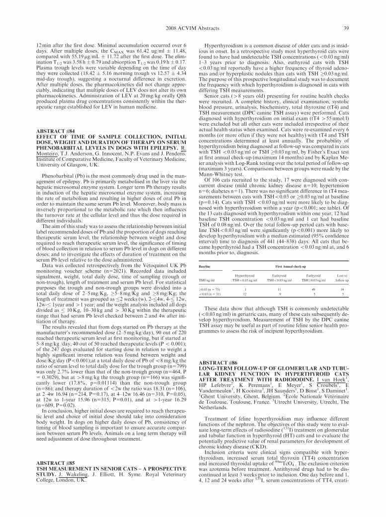
12min after the first dose. Minimal accumulation occurred over 6days. After multiple doses, the CMAX was 61.42 ug/ml � 11.48,compared with 55.19mg/mL � 11.72 after the first dose. The elim-ination T1/2 was 3.58 h� 0.79 and absorption T1/2 was 0.19 h� 0.17.Plasma trough levels were variable depending on the time of daythey were collected (18.42 � 5.16 morning trough vs 12.57 � 4.34mid-day trough), suggesting a nocturnal difference in excretion.After multiple doses, the pharmacokinetics did not change appre-ciably, indicating that multiple doses of LEV does not alter its ownpharmacokinetics. Administration of LEV at 20mg/kg orally Q8hproduced plasma drug concentrations consistently within the ther-apeutic range established for LEV in human medicine.
ABSTRACT #84EFFECT OF TIME OF SAMPLE COLLECTION, INITIALDOSE,WEIGHTANDDURATIONOF THERAPYON SERUMPHENOBARBITAL LEVELS IN DOGS WITH EPILEPSY. R.Monteiro, T.J. Anderson, G. Innocent, N.P. Evans and J. Penderis.Institute of ComparativeMedicine, Faculty of VeterinaryMedicine,University of Glasgow, UK.
Phenobarbital (Pb) is the most commonly drug used in the man-agement of epilepsy. Pb is primarily metabolised in the liver via thehepatic microsomal enzyme system. Longer term Pb therapy resultsin induction of the hepatic microsomal enzyme system, increasingthe rate of metabolism and resulting in higher doses of oral Pb inorder to maintain the same serum Pb level. Moreover, body mass isinversely proportional to the metabolic rate which then influencesthe turnover rate at the cellular level and thus the dose required indifferent individuals.
The aim of this study was to assess the relationship between initiallabel recommended doses of Pb and the proportion of dogs reachingtherapeutic serum level; the relationship between weight and doserequired to reach therapeutic serum level, the significance of timingof blood collection in relation to serum Pb level in dogs on differentdoses; and to investigate the effects of duration of treatment on theserum Pb level relative to the dose administered.
Data was collected retrospectively from the Vetoquinol UK Pbmonitoring voucher scheme (n52021). Recorded data includedsignalment, weight, total daily dose, time of sampling (trough ornon-trough), length of treatment and serum Pb level. For statisticalpurposes the trough and non-trough groups were divided into atotal daily dose of 2–5mg/Kg, �5–8mg/Kg and �8mg/Kg; thelength of treatment was grouped as �2 weeks (w), 2-�4w, 4-� 12w,12w-� 1year and41 year; and the weight analysis included all dogsdivided as � 10Kg, 10–30kg and 4 30Kg within the therapeuticrange that had serum Pb level checked between 2 and 4w after ini-tiation of therapy.
The results revealed that from dogs started on Pb therapy at themanufacturer’s recommended dose (2–5mg/kg/day), 90 out of 220reached therapeutic serum level at first monitoring, but if started at5–8mg/kg /day, 40 out of 50 reached therapeutic levels (Po 0.001);of the 247 dogs evaluated for starting dose in relation to weight ahighly significant inverse relation was found between weight anddose/Kg/day (Po0.001);at a total daily dose of Pb ofo8mg/kg theratio of serum level to total daily dose for the trough group (n5799)was only 2.7% lower than that of the non-trough group (n5464, P5 0.3029), but at 48mg/kg the trough group (n5166) was signifi-cantly lower (17.8%, p50.01114) than the non-trough group(n586); and therapy duration of o2w the ratio was 18.31 (n5106),at 2–4w 16.94 (n5214, P50.17), at 4–12w 16.46 (n5310, P50.05),at 12w to 1-year 15.96 (n5315, P50.01), and at 41-year 16.29(n5609, P50.02).
In conclusion, higher initial doses are required to reach therapeu-tic level and choice of initial dose should take into considerationbody weight. In dogs on higher daily doses of Pb, consistency oftiming of blood sampling is important to ensure accurate compar-ison between serum Pb levels. Animals on a long term therapy willneed adjustment of dose throughout treatment.
ABSTRACT #85TSH MEASUREMENT IN SENIOR CATS – A PROSPECTIVESTUDY. J. Wakeling, J. Elliott, H. Syme. Royal VeterinaryCollege, London, UK.
Hyperthyroidism is a common disease of older cats and is insid-ious in onset. In a retrospective study most hyperthyroid cats werefound to have had undetectable TSH concentrations (o0.03 ng/ml)1–3 years prior to diagnosis. Also, euthyroid cats with TSHo0.03 ng/ml reportedly have a higher frequency of thyroid adeno-mas and/or hyperplastic nodules than cats with TSH �0.03 ng/ml.The purpose of this prospective longitudinal study was to documentthe frequency with which hyperthyroidism is diagnosed in cats withdiffering TSH measurements.
Senior cats (48 years old) presenting for routine health checkswere recruited. A complete history, clinical examination, systolicblood pressure, urinalysis, biochemistry, total thyroxine (tT4) andTSH measurement (DPC canine TSH assay) were performed. Catsdiagnosed with hyperthyroidism on initial exam (tT4 455 nmol/l)were excluded but all other cats were included irrespective of theiractual health status when examined. Cats were re-examined every 6months (or more often if they were not healthy) with tT4 and TSHconcentrations determined at least annually. The probability ofhyperthyroidism being diagnosed at follow-up was compared in catswith TSH o0.03 ng/ml or TSH �0.03 ng/ml, by Fisher’s Exact testat first annual check-up (maximum 14 months) and by Kaplan Me-ier analysis with Log-Rank testing over the total period of follow-up(maximum 3 years). Comparisons between groups were made by theMann-Whitney test.
Of 106 cats recruited to the study, 17 were diagnosed with con-current disease (mild chronic kidney disease n510; hypertensionn56; diabetes n51). There was no significant difference in tT4 mea-surements between cats with TSHo0.03 or �0.03 ng/ml at baseline(p50.14). Cats with TSH o0.03 ng/ml were more likely to be diag-nosed with hyperthyroidism within a year (po0.001; see table). Ofthe 13 cats diagnosed with hyperthyroidism within one year, 12 hadbaseline TSH concentration o0.03 ng/ml and 1 cat had baselineTSH of 0.08 ng/ml. Over the total follow-up period cats with base-line TSHo0.03 ng/ml were significantly (po0.001) more likely todevelop hyperthyroidism with a median estimated (95% confidenceinterval) time to diagnosis of 441 (44–838) days. All cats that be-came hyperthyroid had a TSH concentrationo0.03 ng/ml at, and 6months prior to, diagnosis.
Baseline First Annual check-up
TSH ng/ml
Hyperthyroid
TSHo0.03 ng/ml
Euthyroid
TSHo0.03 ng/ml
Euthyroid
TSH�0.03 ng/ml
Lost to
follow up
�0.03 (n 5 75) 1 11 49 14
o0.03 (n 5 31) 12 9 5 5
These data show that although TSH is commonly undetectable(o0.03 ng/ml) in geriatric cats, many of these cats subsequently de-velop hyperthyroidism. Measurement of TSH by the DPC canineTSH assay may be useful as part of routine feline senior health pro-grammes to assess the risk of incipient hyperthyroidism.
ABSTRACT #86LONG-TERM FOLLOW-UP OF GLOMERULAR AND TUBU-LAR KIDNEY FUNCTION IN HYPERTHYROID CATSAFTER TREATMENT WITH RADIOIODINE. I van Hoek1,HP Lefebvre2, K Peremans1, E Meyer1, S Croubels1, EVandermeulen1, H Kooistra3, JH Saunders1, D Binst1, S Daminet1.1Ghent University, Ghent, Belgium. 2Ecole Nationale Veterinairede Toulouse, Toulouse, France. 3Utrecht University, Utrecht, TheNetherlands.
Treatment of feline hyperthyroidism may influence differentfunctions of the nephron. The objectives of this study were to eval-uate long-term effects of radioiodine (131I) treatment on glomerularand tubular function in hyperthyroid (HT) cats and to evaluate thepotentially predictive value of renal parameters for development ofchronic kidney disease (CKD).
Inclusion criteria were clinical signs compatible with hyper-thyroidism, increased serum total thyroxin (TT4) concentrationand increased thyroidal uptake of 99mTcO4
�. The exclusion criterionwas azotemia before treatment. Antithyroid drugs had to be dis-continued at least 3 weeks prior to inclusion. One day before and 1,4, 12 and 24 weeks after 131I, serum concentrations of TT4, creati-
392008 ACVIM Abstracts
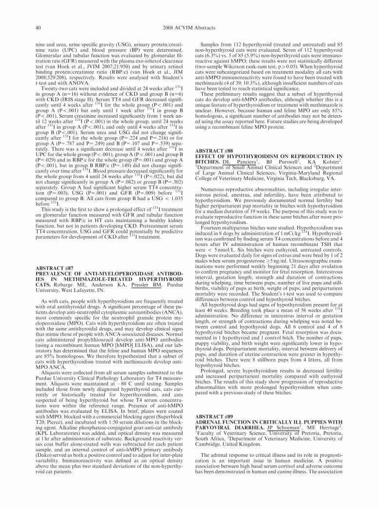
nine and urea, urine specific gravity (USG), urinary protein/creati-nine ratio (UPC) and blood pressure (BP) were determined.Glomerular and tubular function was evaluated by glomerular fil-tration rate (GFR) measured with the plasma exo-iohexol clearancetest (van Hoek et al., JVIM 2007;21:950) and by urinary retinolbinding protein/creatinine ratio (RBP/c) (van Hoek et al., JIM2008;329:208), respectively. Results were analysed with Student’st-test and with ANOVA.
Twenty-two cats were included and divided at 24 weeks after 131Iin group A (n516) without evidence of CKD and group B (n56)with CKD (IRIS stage II). Serum TT4 and GFR decreased signifi-cantly until 4 weeks after 131I for the whole group (Po.001) andgroup A (Po.001) but only until 1 week after 131I in group B(Po.001). Serum creatinine increased significantly from 1 week un-til 12 weeks after 131I (Po.001) in the whole group, until 24 weeksafter 131I in group A (Po.001), and only until 4 weeks after 131I ingroup B (Po.001). Serum urea and USG did not change signifi-cantly after 131I for the whole group (P5.224 and P5.216) or forgroup A (P5.787 and P5.249) and B (P5.107 and P5.539) sepa-rately. There was a significant decrease until 4 weeks after 131I inUPC for the whole group (Po.001), group A (Po.001) and group B(P5.029) and in RBP/c for the whole group (P5.001) and group A(Po.001), but in group B RBP/c (P5.149) did not change signifi-cantly over time after 131I. Blood pressure decreased significantly forthe whole group from 4 until 24 weeks after 131I (P5.022), but didnot change significantly in group A (P5.082) or group B (P5.302)separately. Group A had significant higher serum TT4 concentra-tion (P5.003), USG (P5.001) and GFR (P5.009) before 131Icompared to group B. All cats from group B had a USG o 1.035before 131I.
This study is the first to show a prolonged effect of 131I treatmenton glomerular function measured with GFR and tubular functionmeasured with RBP/c in HT cats maintaining a healthy kidneyfunction, but not in patients developing CKD. Pretreatment serumTT4 concentration, USG and GFR could potentially be predictiveparameters for development of CKD after 131I treatment.
ABSTRACT #87PREVALENCE OF ANTI-MYELOPEROXIDASE ANTIBOD-IES IN METHIMAZOLE-TREATED HYPERTHYROIDCATS. Robarge ME, Anderson KA, Pressler BM. PurdueUniversity, West Lafayette, IN.
As with cats, people with hyperthyroidism are frequently treatedwith oral antithyroidal drugs. A significant percentage of these pa-tients develop anti-neutrophil cytoplasmic autoantibodies (ANCA),most commonly specific for the neutrophil granule protein my-eloperoxidase (MPO). Cats with hyperthyroidism are often treatedwith the same antithyroidal drugs, and may develop clinical signsthat mimic those of people with ANCA-associated diseases. Normalcats administered propylthiouracil develop anti-MPO antibodies(using a recombinant human MPO [hMPO] ELISA), and our lab-oratory has determined that the feline and human MPO sequencesare 85% homologous. We therefore hypothesized that a subset ofcats with hyperthyroidism treated with methimazole develop anti-MPO ANCA.
Aliquots were collected from all serum samples submitted to thePurdue University Clinical Pathology Laboratory for T4 measure-ment. Aliquots were maintained at �80 C until testing. Samplesincluded those from newly diagnosed hyperthyroid cats, cats cur-rently or historically treated for hyperthyroidism, and catssuspected of being hyperthyroid but whose T4 serum concentra-tions were within the reference range. Presence of anti-hMPOantibodies was evaluated by ELISA. In brief, plates were coatedwith hMPO, blocked with a commercial blocking agent (SuperblockT20; Pierce), and incubated with 1:50 serum dilutions in the block-ing agent. Alkaline phosphatase-conjugated goat-anti-cat antibody(KPL Laboratories) was added, and optical density was measuredat 1 hr after administration of substrate. Background reactivity ver-sus coat buffer alone-coated wells was subtracted for each patientsample, and an internal control of anti-hMPO primary antibody(Dako) served as both a positive control and to adjust for inter-platevariability. Immunoreactivity was defined as an optical densityabove the mean plus two standard deviations of the non-hyperthy-roid cat patients.
Samples from 112 hyperthyroid (treated and untreated) and 85non-hyperthyroid cats were evaluated. Seven of 112 hyperthyroidcats (6.3%) vs. 5 of 85 (5.9%) non-hyperthyroid cats were immuno-reactive against hMPO; these results were not statistically different(two-sample Wilcoxon rank-sum test, p40.05). When hyperthyroidcats were subcategorized based on treatment modality all cats withanti-hMPO immunoreactivity were found to have been treated withmethimazole (4 of 39; 10.3%), although insufficient numbers of catshave been tested to reach statistical significance.
These preliminary results suggest that a subset of hyperthyroidcats do develop anti-hMPO antibodies, although whether this is aunique feature of hyperthyroidism or treatment with methimazole isunclear. However, because human and feline MPO are only 85%homologous, a significant number of antibodies may not be detect-ed using the assay reported here. Future studies are being developedusing a recombinant feline MPO protein.
ABSTRACT #88EFFECT OF HYPOTHYROIDISM ON REPRODUCTION INBITCHES. DL Panciera1, BJ Purswell2, KA Kolster2.1Department of Small Animal Clinical Sciences and 2Departmentof Large Animal Clinical Sciences, Virginia-Maryland RegionalCollege of Veterinary Medicine, Virginia Tech, Blacksburg, VA.
Numerous reproductive abnormalities, including irregular inter-estrous period, anestrus, and infertility, have been attributed tohypothyroidism. We previously documented normal fertility buthigher periparturient pup mortality in bitches with hypothyroidismfor a median duration of 19 weeks. The purpose of this study was toevaluate reproductive function in these same bitches after more pro-longed hypothyroidism.
Fourteen multiparous bitches were studied. Hypothyroidism wasinduced in 8 dogs by administration of 1mCi/kg 131I. Hypothyroid-ism was confirmed by finding serum T4 concentrations before and 4hours after IV administration of human recombinant TSH thatwere o 5 nmol/L. Six bitches were euthyroid, untreated controls.Dogs were evaluated daily for signs of estrus and were bred by 1 of 2males when serum progesterone �5 ng/ml. Ultrasonographic exam-inations were performed weekly beginning 21 days after ovulationto confirm pregnancy and monitor for fetal resorption. Interestrousinterval, gestation length, strength and duration of contractionsduring whelping, time between pups, number of live pups and still-births, viability of pups at birth, weight of pups, and periparturientmortality were recorded. The Student’s t-test was used to comparedifferences between control and hypothyroid bitches.
All hypothyroid dogs had signs of hypothyroidism present for atleast 40 weeks. Breeding took place a mean of 56 weeks after 131Iadministration. No difference in interestrus interval or gestationlength, or strength of contractions during whelping was noted be-tween control and hypothyroid dogs. All 6 control and 4 of 8hypothyroid bitches became pregnant. Fetal resorption was docu-mented in 1 hypothyroid and 1 control bitch. The number of pups,puppy viability, and birth weight were significantly lower in hypo-thyroid dogs. Periparturient mortality, interval between delivery ofpups, and duration of uterine contraction were greater in hypothy-roid bitches. There were 8 stillborn pups from 4 litters, all fromhypothyroid bitches.
Prolonged, severe hypothyroidism results in decreased fertilityand increased periparturient mortality compared with euthyroidbitches. The results of this study show progression of reproductiveabnormalities with more prolonged hypothyroidism when com-pared with a previous study of these bitches.
ABSTRACT #89ADRENAL FUNCTION IN CRITICALLY ILL PUPPIES WITHPARVOVIRAL DIARRHEA. JP Schoeman1, ME Herrtage2.1Faculty of Veterinary Science, University of Pretoria, Pretoria,South Africa, 2Department of Veterinary Medicine, University ofCambridge, United Kingdom.
The adrenal response to critical illness and its role in prognosti-cation is an important issue in human medicine. A positiveassociation between high basal serum cortisol and adverse outcomehas been demonstrated in human and canine illness. The association
40 2008 ACVIM Abstracts

of serum ACTH-stimulated cortisol and delta cortisol concentra-tions with outcome in critical illness is less clear.
This prospective, case controlled study was conducted on 63puppies with parvoviral diarrhea. The diagnosis was confirmed bydetection of viral particles on fecal electron microscopy. Seventeenhealthy puppies were used as controls. Blood samples were obtainedin each dog at admission prior to treatment and daily thereafter un-til death or discharge from the hospital. Immediately after the basalsamples were drawn, each dog was injected daily with 5 ug/kg ofACTH (tetracosactrin) intravenously. A second sample was taken 1hour later for serum ACTH-stimulated cortisol measurement andthe calculation of delta cortisol. Cortisol concentrations were deter-mined by a commercial canine radioimmunoassay kit (Coat-a-counts, DPC, CA). Dogs were retrospectively assigned to twogroups: survivors (n550) and non-survivors (n513). Hormone con-centrations between the survivors and non-survivors were comparedwith the Mann Whitney U test for non-parametric data. Signifi-cance was set at po 0.05.
Median day 1 (D1) basal cortisol and ACTH-stimulated cortisolwas significantly higher in patients than in controls (259 vs. 77 nmol/L) and (393 vs. 295 nmol/L); Po0.01 for both.Median delta cortisolwas lower in patients (68 vs. 203 nmol/L). In nonsurvivors vs. sur-vivors, D1 basal cortisol was significantly higher (539 vs 234 nmol/L), Po 0.05. ACTH-stimulated cortisol did not differ significantly(459 vs. 380 nmol/L); P 5 0.2, therefore delta cortisol was signifi-cantly lower in nonsurvivors (�60 vs 128 nmol/L); Po 0.05. On day3 (D3), basal cortisol was still higher in non-survivors, but not sig-nificantly so (189 vs. 68 nmol/L); P 5 0.18, yet ACTH-stimulatedcortisol was significantly higher in nonsurvivors (417 vs. 345 nmol/L); P o 0.05. In contrast to D1, delta cortisol was not significantlylower in nonsurvivors on D3 (179 vs. 256 nmol/L); P 5 0.2.
This study confirmed the previously described association be-tween high basal serum cortisol concentrations and mortality inparvoviral diarrhea dogs. Low delta cortisol was also associatedwith mortality on D1. However, the D3 delta cortisol was not sig-nificantly lower in nonsurvivors compared to survivors. This studyhighlights the important limitation of designating a patient as adre-nal insufficient on the basis of a one-off ACTH stimulation test inthe early stages of acute illness. During this stage basal cortisol pro-duction is close to maximum and delta cortisol would be low,without necessarily indicating adrenal insufficiency. The serial sam-pling in this study provided novel insights on the adrenal responseand re-enforces the need to consider basal-, delta- and ACTH-stim-ulated cortisol in the evaluation of adrenal reserve in canine criticalillness.
ABSTRACT #90URINARY CATECHOLAMINE AND METANEPHRINE TOCREATININE RATIOS IN DOGS WITH PHEOCHROMOCY-TOMA. P.H. Kook1, S. Quante1, P. Grest2, C.E. Reusch1. 1Clinicfor Small Animal Internal Medicine, 2Institute of Pathology,Vetsuisse Faculty, University of Zurich, Switzerland.
Pheochromocytomas (PHEO) are neuroendocrine tumors arisingfrom chromaffin cells of the adrenal medulla or extra-adrenal para-ganglia. In people, diagnosis crucially depends on biochemicalevidence of production of the secretory product of the tumor. Wide-ly used biochemical tests include measurements of urinarycatecholamines and its metabolites. In veterinary medicine, bio-chemical testing is difficult to perform due to limited availability oftechniques and lack of established reference ranges, and PHEO ismost commonly identified as an incidental finding at necropsy.Measurement of urine catecholamine concentrations has been doc-umented only in one dog with PHEO; however results wereinconclusive. Recently, we reported on catecholamine- and meta-nephrine:creatinine (crea) ratios of spot urine samples in healthydogs. The purpose of this study is to evaluate the biochemical diag-nosis of PHEO in 6 dogs with histologically confirmed disease.
Between October 2004 and July 2007, all dogs with ultrasono-graphic evidence of adrenal gland enlargement and clinicalsuspicion of PHEO were enrolled into the study. The final criterionfor inclusion was histopathological confirmation. Ten healthy cli-ent-owned dogs served as controls. During the study period 11client-owned dogs were evaluated and histopathology confirmingPHEO was available in 6/11 dog. Urine sampling procedures variedin dogs with PHEO. In one dog, urine was collected by the owner on
day 7 (d7) after discharge from the hospital. In five dogs urine wascollected during the initial work-up. Additional urine specimenswere sampled in two dogs on day 2 (d2), resp. on days 6 (d6) and 7(d7) after discharge by their owners. Results were compared totimely corresponding values from 10 healthy dogs collected in a vet-erinary hospital (t0), and 1 resp. 7 days later at home (t1, t7).Urinary free catecholamines comprising epinephrine, norepineph-rine, and dopamine and urinary fractionated metanephrinescomprising free and conjugatedmetanephrine and normetanephrinewere separated and quantitatively determined by high pressure liq-uid chromatography with electrochemical detection usingcommercial reagents (BIO-RAD, Munich, Germany). Values wereexpressed as ratios to crea (nM:mM).
Normetanephrine:crea ratios were consistently increased com-pared to controls, ranging from 103 to 6430 (t0: 14–91; median 59.5,t1: 32–100; median 57.5, and t7: 21–79; median 55).
Four dogs had increases well above (414; resp. 243 on d2, 476,925, and 6430 (d7)) even the highest results of healthy dogs at t0,whereas 2 dogs showed only smaller increases: (161) and (157 resp.103 on d6, and 131 on d7). Highest normetanephrine : crea ratioswere found in bilateral PHEO (2/6). All other ratios showed moreoverlap.
In conclusion, urinary normetanephrine:crea ratios are useful indiagnosing canine PHEO. In view of our first results these patientsshould ideally have their urine samples collected in their home en-vironment. Results in one dog suggest that documentation ofsmaller but persistently increased normetanephrine:crea ratios onrepeated measurements can further substantiate the diagnosis.
ABSTRACT #91PREDICTIVE VALUE OF POSTOPERATIVE PLASMA CON-CENTRATIONS OF ACTH, a-MSH AND CORTISOL FORRECURRENCE OF CUSHING’S DISEASE AFTER TRANS-SPHENOIDAL HYPOPHYSECTOMY IN DOGS. JM Hanson,JA Mol, BP Meij. Faculty of Veterinary Medicine, UtrechtUniversity, Utrecht, The Netherlands.
Transsphenoidal hypophysectomy is an effective treatment forCushing’s disease or pituitary-dependent hypercortisolism (PDH) inthe dog. However, recurrences occur after initial remission. The aimof this study was to analyze the predictive value of immediate post-operative plasma concentrations of ACTH, a–MSH, and cortisolfor recurrence after transsphenoidal hypophysectomy in dogs withPDH.
Transsphenoidal hypophysectomy was performed in 55 dogs withPDH. Plasma concentrations of ACTH, a–MSH, and cortisol weremeasured before surgery and 1, 2, 3, 4, 5 and 24 to 48 hours afterremoval of the pituitary gland. The prognostic value of the hormoneconcentrations for recurrences (disease-free period) was analyzedwith univariate Cox proportional-hazard analysis followed by step-wise multivariate analysis.
Forty-eight of the 55 dogs went into remission. Median disease-free period was 569 days. Hypercortisolism recurred in 12 dogs aftermedian 255 days. Postoperative plasma ACTH, cortisol and a–MSH concentrations at 4 and 5 hours after removal of the pituitarygland were significantly associated with recurrence. In multivariateanalysis the plasma ACTH concentration remained significant.
It is concluded that immediate postoperative measurement ofplasma concentrations of ACTH, a–MSH and cortisol is a valuabletool for evaluation of long-term outcome after transsphenoidal hy-pophysectomy in dogs with Cushing’s disease.
Originally presented at the European College of Veterinary In-ternal Medicine - Companion Animals Congress, September , 2007.
ABSTRACT #92SUSTAINED HYPERGLYCEMIA AND HYPERLIPIDEMIAIN CATS: CONTRIBUTION OF INFLAMMATION AND BE-TA-CELL LOSS TO THE PATHOPHYSIOLOGY OFDIABETES. E Zini1, M Osto2, M Franchini3, F Guscetti4, MDonath5, A Perren6, P Linscheid1, M Bouwman7, M Ackermann3,TA Lutz2, CE Reusch1. 1Clinic for Small Animal Internal Medicine;2Inst. of Vet. Physiol.; 3Inst. of Virol.; 4Inst. of Vet. Pathol.,Vetsuisse Faculty, University of Zurich, Switzerland.5Clinic forEndocrinol. and Diabetes, University Hospital Zurich,
412008 ACVIM Abstracts

Switzerland.6Dept. of Pathol., Technical University of Munchen,Germany.7Dept. of Clinical Sciences of Companion Animals,Utrecht University, The Netherlands.
Feline diabetes shares many similarities to human type 2 diabetesmellitus (T2DM), including islet amyloidosis and b-cell loss. Basedon cultured rodent and human islet cells, it has been demonstratedthat hyperglycemia and hyperlipidemia in T2DM have a negativeeffect on b-cell viability. Exposure to high glucose or lipid levels in-duces interleukin expression in cultured pancreatic islets, followedby Fas receptor up-regulation and apoptosis of b-cells. In addition,high glucose levels upregulate islet chemokines and attract neutro-phils and macrophages. Inflammatory cells may contribute topancreatic-islet cell death in T2DM. The objective of this studywas to investigate the above findings in vivo in cats.
Eleven healthy cats were infused for 10 days with glucose (n55)or lipids (n56) to target their blood concentrations at the approx-imate level found in untreated feline diabetes (glucose: 450–540mg/dl; triglycerides: 265–620mg/dl). As control groups, 10 healthy catswere either infused with saline (n55) or did not receive infusion(n55). On day 10, blood samples and pancreatic biopsies were col-lected. Levels of a1-acid glycoprotein were measured in plasmasamples. Isolated pancreatic islets were used to quantify mRNAtranscripts of cytokines (IL-1b, IL-6, TNF-a) and chemokines(IL-8, MCP-1) by real-time PCR. In addition, mRNA transcriptsof Fas receptor were measured. To quantify islet neutrophils,pancreatic sections were immunostained with insulin and my-eloperoxidase. Pancreatic sections stained with amylin or insulinwere used to quantify b-cells in pancreatic islets by morphometricanalysis. Statistical differences between groups were determinedwith nonparametric tests.
Compared to controls, hyperglycemic cats had a 50% reducednumber of b-cells per islet surface and an increased number of ne-utrophils relative to b-cells. More apoptotic cells were noted in thepancreatic islets by light microscopy. In hyperlipidemic cats thenumber of b-cells and neutrophils did not differ from control cats.Plasma levels of a1-acid glycoprotein were increased in cats on glu-cose and lipid infusion. Islet quantities of cytokine, chemokines andFas receptor transcripts were not different between groups.
Sustained hyperglycemia causes b-cell loss in pancreatic islets ofhealthy cats, possibly through increased apoptosis. Even thoughhyperglycemia and hyperlipidemia are accompanied by a systemicinflammatory response, inflammation does not seem to occur inpancreatic islets under the present experimental conditions. The in-creased number of neutrophils observed in the pancreatic islets ofhyperglycemic cats needs to be further explored.
ABSTRACT #93EFFECT OF HYPERGLYCEMIA AND HYPERLIPIDEMIAON THE EXPRESSION OF 11b-HYDROXYSTEROID-DEHY-DROGENASE AND GLUCOCORTICOID RECEPTOR ININSULIN SENSITIVE TISSUES. N. S. Sieber-Ruckstuhl1, E.Zini1, F. S. Boretti1, M. Meli2, M. Osto3, B. Sigrist1, M.Franchini4, P. Linscheid1, T.A. Lutz3, C. E. Reusch1. 1Clinic forSmall Animal Internal Medicine, 2Clinical Laboratory, 3Institute ofVeterinary Physiology and Center of Integrative Human Physiologyand 4Institute of Virology of the Vetsuisse Faculty University ofZurich, Zurich, Switzerland.
Type 2 diabetes mellitus is characterized by inadequate insulinsecretion and impaired insulin action. Once diabetes mellitus is es-tablished, hyperglycemia and hyperlipidemia further impair b-cellfunction and insulin sensitivity. Increasing evidence in humans andexperimental animals suggests that altered tissue cortisol metabo-lism may additionally promote insulin resistance. Of keyimportance for the tissue cortisol metabolism is the pre-receptor en-zyme 11b-hydroxysteroid dehydrogenase (11b-HSD) with its twoisoforms (11b-HSD1 and 11b-HSD2). 11b-HSD1 converts the in-active cortisone to cortisol and 11b-HSD2 converts cortisol tocortisone.
The aim of this study was to determine if exposure of cats to highglucose and lipid levels has an influence on the expression of 11b-HSD1, 11b-HSD2 and glucocorticoid receptor (GR) and by thismean on the tissue cortisol metabolism. Eleven cats were infused for10 days through the jugular vein with glucose (n55) or lipids (n56)
to clamp their blood concentrations at the level found in untreatedfeline diabetes (450–540mg/dl for glucose and 265–620mg/dl fortriglycerides). Ten control cats were infused with saline (n55) or re-ceived no infusion (n55). At the end of the 10 day period bloodsamples to measure cortisol and tissue biopsies from omental andsubcutaneous adipose tissue, liver and muscle to measure mRNAexpression for 11b-HSD1, 11b-HSD2 and GR by real-time PCRwere collected.
Lipid infused cats showed significantly higher 11b-HSD1 mRNAexpression in omental and subcutaneous adipose tissue than all oth-er cats, significantly lower 11b-HSD2 mRNA expression in omentaladipose tissue than all other cats, significantly lower 11b-HSD2mRNA expression in liver than control cats, significantly lower GRmRNA expression in omental adipose tissue than all other cats andsignificantly lower GR mRNA expression in subcutaneous adiposetissue than saline and glucose infused cats. Glucose infused catsshowed significantly lower 11b-HSD2 mRNA expression in liverthan control cats. Saline infused cats showed significantly higherGR mRNA expression in omental fat and significantly higher cor-tisol blood levels compared to untreated cats. In summary, two-week lipid infusion leads to higher 11b-HSD1 expression and lowerGR expression in adipose tissue and lower 11b-HSD2 expression inomental adipose tissue and liver.
Up-regulation of 11b-HSD1 and down-regulation of 11b-HSD2is expected to result in increased tissue cortisol concentrations.Down-regulation of GR possibly represents a self-protective mech-anism of the tissue against increased tissue cortisol levels. Inconclusion, these results indicate that hyperlipidemia has a pro-found effect on the 11b-HSD expression and support the connectionbetween hyperlipidemia, tissue cortisol metabolism and insulin sen-sitivity.
ABSTRACT #94THE EFFECT OF HYPOTHYROIDISM ON INSULIN SENSI-TIVITY IN DOGS. N Inteeworn, DL Panciera, WE Monroe, KESaker. Department of Small Animal Clinical Sciences, Virginia-Maryland Regional College of Veterinary Medicine, Virginia Tech,Blacksburg, VA.
Hypothyroidism causes abnormalities in glucose homeostasis in avariety of animal species. It has been documented as a cause of in-sulin resistance in dogs with concurrent diabetes mellitus. Whileseveral studies have been conducted in hypothyroid dogs to deter-mine insulin secretion and glucose concentrations during glucosetolerance tests, parameters of insulin sensitivity have not been de-termined to date. The purpose of the study was therefore toinvestigate the effect of hypothyroidism on glucose tolerance andinsulin sensitivity in dogs.
Sixteen female mixed breed dogs were studied. Dogs were ran-domly selected and allocated into two groups. In 8 dogs,hypothyroidism was induced by administration of 1mCi/kg 131Iwhile the remaining 8 dogs were euthyroid controls. Hypothyroid-ism was confirmed by finding serum T4 o 5 nmol/L before and 4hours after IV administration of human recombinant TSH. Exper-iments were performed on non-anesthetized, fasted dogs inanestrous approximately 12 months after hypothyroidism was in-duced. The insulin-modified frequently sampled intravenousglucose tolerance test (FSIGT) was used to determine insulin andglucose concentrations over a 3-hr time period. Data was analyzedby the Minimal Model Analysis (MINMOD) to calculate basal in-sulin and glucose concentrations, acute insulin response to glucose(AIRg), insulin sensitivity (Si), glucose effectiveness (the effect ofglucose itself in its own disappearance, Sg) and the disposition index(DI). Student’s t-test for unpaired data was used for comparisonsbetween groups. P-values o 0.05 were considered statistically sig-nificant.
One dog in the control group was excluded from the analysis dueto an exaggerated response to the exogenously administered insulinat 20 minutes. Basal glucose concentrations were mildly, but signifi-cantly higher in the control group, whereas basal insulinconcentrations were significantly lower. Minimal Model Analysisrevealed a lower insulin sensitivity in the hypothyroid group(Po0.001), whereas AIRg was higher (P50.01). Glucose effective-ness and disposition index were not statistically different betweengroups.
42 2008 ACVIM Abstracts
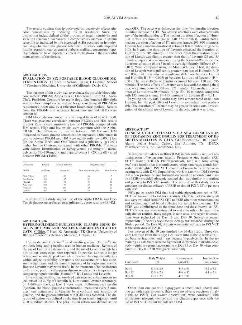
The results confirm that hypothyroidism negatively affects glu-cose homeostasis by inducing insulin resistance. Since thedisposition index, defined as the product of insulin sensitivity andsecretion remained unchanged, a compensatory increase in insulinsecretion as indicated by the increased AIRg occurred in hypothy-roid dogs to maintain glucose tolerance. In cases with impairedinsulin secretion, such as canine diabetes mellitus, concurrent hypo-thyroidism can have important clinical implications in the successfulmanagement of the disease.
ABSTRACT #95EVALUATION OF SIX PORTABLE BLOOD GLUCOSE ME-TERS IN DOGS. T Cohen, R Nelson, P Kass, E Feldman. Schoolof Veterinary Medicine, University of California, Davis, CA.
The purpose of this study was to evaluate six portable blood glu-cose meters (PBGM; AlphaTRAK, One-Touch, Elite XL, Accu-Chek, Precision, Contour) for use in dogs. One hundred fifty-eightvenous blood samples were assayed for glucose using all PBGMs inrandomized order and by a reference hexokinase method. Resultsfrom the PBGMs and reference hexokinase method (HM) werecompared.
HM blood glucose concentrations ranged from 41 to 639mg/dl.There was excellent correlation between PBGMs and HM results(Table). Results were consistently low for 4 PBGMs, compared withHM results. High and low results were common with the Alpha-TRAK. The difference in results between PBGMs and HMincreased as blood glucose concentrations increased. Differences inresults between PBGM and HM were significantly (po0.0001) lessfor the AlphaTRAK and One-Touch and significantly (po0.01)higher for the Contour, compared with other PBGMs. Problemswith correct identification of hypoglycemia (o70mg/dl), norm-oglycemia (70–120mg/dl), and hyperglycemia (4200mg/dl) variedbetween PBGMs (Table).
Correlation Percent Median difference Percent incorrect
PBGM coefficient low results from reference (mg/dl) identification
AlphaTRAK 0.92 55 18 2
One-Touch 0.96 87 21 4
Elite XL 0.97 98 45 16
Accu-Chek 0.96 99 45 16
Precision 0.93 100 49 20
Contour 0.94 99 73 39
Results of this study support use of the AlphaTRAK and One-Touch glucose meters based on significantly closer results with HM.
ABSTRACT #96HYPERINSULINEMIC-EUGLYCEMIC CLAMPS USING IN-SULIN DETEMIR AND INSULIN GLARGINE IN HEALTHYCATS. C Gilor, T Keel, KJ Attermeier, TK Graves. University ofIllinois College of Veterinary Medicine, Urbana, IL.
Insulin detemir (Levemirs) and insulin glargine (Lantuss) aresynthetic long-acting insulins used in human medicine. Reports ofthe use of Lantus in cats are rare, and the use of Levemir in cats hasnot, to our knowledge, been reported. In people, Lantus is longeracting and relatively peakless, while Levemir has significantly lesswithin-subject variability. Levemir is also associated with less unde-sired weight gain and decreased frequency of hypoglycemic events.Because Levemir may prove useful in the treatment of feline diabetesmellitus, we performed hyperinsulinemic-euglycemic clamps in cats,comparing regular insulin (Humulins R), Lantus and Levemir.
Five young, healthy, purpose-bred cats received subcutaneous in-jections of 0.5U/Kg of Humulin R, Lantus and Levemir separately,on 3 different days, at least 1 week apart. Following each insulininjection, the blood glucose concentration, measured every 5 min-utes, was maintained at baseline by a constant rate infusion ofglucose, and the glucose infusion rate (GIR) was recorded. The du-ration of action was defined as the time from insulin injection untilGIR stabilized at zero. The peak insulin action was defined as the
peak GIR. The onset was defined as the time from insulin injectionto initial increase in GIR. No adverse reactions were observed withany of the insulin products. The median duration of action of Hum-ulin R was 305 minutes (range: 140–370 minutes). Lantus had amedian duration of action of 470 minutes (range: 295–950 minutes).Levemir had a median duration if action of 800 minutes (range 525–915). In 3 cats, the duration of Levemir exceeded the duration ofLantus by 205–505 minutes. In the other 2 cats the duration of ac-tion of Lantus was slightly greater than that of Levemir (25 and 35minutes longer). When compared using the Kruskal-Wallis test thedurations of action of the 3 insulins were significantly different (P 50.012). When compared using the Mann-Whitney U test, the dura-tion of Levemir was significantly greater than that of Humulin R (P5 0.008), but there was no significant difference between Lantusand Humilin R (P 5 0.095) or between Lantus and Levemir (P 50.31). The peak effects of Lantus occurred between 120 and 585minutes. The peak effects of Levemir were less variable among the 5cats, occurring between 370 and 575 minutes. The median time ofonset of Lantus was 80 minutes (range: 50–110 minutes), comparedwith 135 minutes (range: 80–165 minutes) for Levemir (P 5 0.11).
In young healthy cats, Lantus may have a more rapid onset thanLevemir, but the peak effect of Levemir is somewhat more predict-able. The duration of Levemir may be greater in some cats. Investi-gation of the clinical use of Levemir in diabetic cats is warranted.
ABSTRACT #97CLINICAL STUDY TO EVALUATE A NEW FORMULATIONOF PROTAMINE ZINC INSULIN FOR TREATMENT OF DI-ABETES MELLITUS IN CATS. GD Norsworthy1, RC Lynn2.Alamo Feline Health Center, San Antonio, TX; IDEXXPharmaceuticals, Inc., Greensboro, NC.
Treatment of diabetes mellitus (DM) in cats usually requires ad-ministration of exogenous insulin. Protamine zinc insulin (PZIVETs Insulin, IDEXX Pharmaceuticals, Inc.) is a long actingbeef-pork insulin that is manufactured using pancreatic glands har-vested at slaughter. PZI VET insulin is known to be effective intreating cats with DM. Unpublished work in rats with DM showedthat a new protamine zinc formulation based on recombinant insu-lin (PZIR) provided glycemic control that was similar in durationand potency to PZI VET insulin. The purpose of this study was tocompare the clinical efficacy of PZIR to that of PZI VET in pet catswith DM.
Fifty pet cats with DM that had stable glycemic control on PZIVET insulin were selected for the study. On Day 0 of the study allcats were switched from PZI VET to PZIR after they were examinedand weighed and had blood collected for serum fructosamine. ThePZIR was administered at the same dose and dose interval as PZIVET. Cat owners were instructed to make no other changes in thedaily diet or routine. Body weight, insulin dose, and serum fructosa-mine were rechecked on Day 15 and Day 30. Subjective ownerimpression of the cat’s response to therapy was recorded during the30-day period. On Day 30, the cats were switched back to PZI VETat the same dose as PZIR.
Forty-seven of the 50 cats finished the 30-day study. Three catswere removed from the study; 1 cat went into diabetic remission, 1cat became fractious, and 1 cat became hypoglycemic. In the re-maining 47 cats there were no significant differences in insulin dose,body weight or serum fructosamine at Day 15 or Day 30 when com-pared to Day 0. PZIR was given twice daily.
Time point
Body Weight
(lb)
Fructosamine
(mmol/L)
Insulin Dose
(units/dose)
Day 0 13.8 � 2.8 445 � 91 4.2 � 3.5
Day 15 13.8 � 2.9 446 � 95 4.4 � 3.4
Day 30 13.7 � 2.8 432 � 91 –
Other than one cat with hypoglycemia (mentioned above) andone cat with hyperglycemia, there were no adverse reactions attrib-utable to PZIR. Cat owner observations were consistent withsatisfactory glycemic control and our clinical experience with theuse of PZI VET insulin for cats with DM.
432008 ACVIM Abstracts

The results of this study in pet cats are similar to the previousfindings in rats with DM. This study in diabetic cats also demon-strates that protamine zinc insulin based on recombinant insulin(PZIR) provides glycemic control that is comparable to that of PZIVET insulin at the same dose and dose interval.
ABSTRACT #98EFFICACY OF PROTAMINE ZINC RECOMBINANT INSU-LIN FOR TREATING DIABETES MELLITUS IN CATS. RWNelson1, K Henley2, C Cole2. 1School of Veterinary Medicine,University of California, Davis, CA. 2IDEXX Pharmaceuticals,Inc, Greensboro, NC.
The objective of this study was to evaluate the effects of a prota-mine zinc recombinant insulin (PZIR) on control of glycemia in catswith diabetes mellitus. One hundred twenty-six cats with newly di-agnosed diabetes and 13 cats with poorly controlled diabetes weretreated with PZIR twice daily for 45 days. Control of glycemia wasassessed on days 7, 14, 30, and 45 by evaluation of clinical response,body weight, serum fructosamine, and blood glucose concentrationsdetermined 1, 3, 5, 7, and 9 hours after administration of PZIR.Adjustments in dosage of PZIR were made as needed to attain con-trol of glycemia.
PZIR administration resulted in a significant (po0.001) decreasein 9 hr mean blood glucose, mean blood glucose nadir, and serumfructosamine concentrations and a significant (po0.05) increase inmean body weight in the 139 diabetic cats at day 45, compared withday 0. 84% of the diabetic cats, including 9 poorly controlled, at-tained good diabetic control by day 45, based on improvement inclinical signs, physical examination, and blood parameters used toassess control of glycemia. Hypoglycemia was identified in 160 of690 9-hr serial blood glucose curves involving 87 cats.
Results of this study demonstrate that PZIR is safe and effectivefor control of glycemia in cats with newly diagnosed and poorlycontrolled diabetes.
ABSTRACT #99COMPREHENSIVE PATHOLOGIC EVALUATION OF KID-NEY DISEASE IN DOGS AND CATS: 179 CASES (2005–2007). George E. Lees, Brian R. Berridge, and Fred J. Clubb, Jr.College of Veterinary Medicine, Texas A&M University, CollegeStation, TX.
Nephropathology in human medicine is a well-established sub-specialty in which thorough light microscopic (LM) examinationsare routinely combined with transmission electron microscopic(TEM) examinations and immunofluorescence (IF) evaluations toadequately characterize the pathologic features of diseases affectingkidneys. Pathologic findings are integrated with clinical findings torender specific morphologic and disease diagnoses and guide appro-priate therapy.We hypothesized that taking a similar approach to thepathologic evaluation of renal diseases in dogs and cats would im-prove diagnosis and treatment of kidney disorders in these animals.
Early in 2005, we established a diagnostic veterinary renal pa-thology service that (a) provides kits of materials and instructionsthat enable clinicians to properly obtain and submit specimens ofkidney suitable for TEM and IF, as well as histologic (LM) exam-inations, (b) routinely performs thorough LM, TEM, and IFevaluations, and (c) focuses on identifying pathologic implicationsfor clinical patient management (i.e., diagnosis, prognosis, treat-ment). Our initial experiences in this activity have yielded somenoteworthy observations.
Case submissions increased annually (2005, 19; 2006, 51; 2007,109) and totaled 179 by the end of 2007. Submissions mainly werefrom dogs (n, 165) and most were biopsies (154 biopsy and 11 nec-ropsy cases). Submissions from cats (n, 14) were too few tosummarize in a meaningful way. Glomerular diseases of varioustypes predominated in most (107/165; 64.8%) of the canine cases,and the remaining cases (58/165; 35.2%) were an eclectic mix ofother disorders, including juvenile-onset nephropathies, acute orchronic interstitial nephritis, and acute tubular injury. The biopsyspecimens varied in quality, but generally were sufficient for diag-nosis; only 9/154 (5.8%) were judged to be inadequate. Importantly,biopsy specimens, which typically are obtained at an earlier stage of
disease than necropsy specimens, exhibit a much more diverse andinformative range of pathologic changes than is evident in necropsyspecimens or is well characterized in the veterinary literature. Amembranoproliferative pattern of glomerular injury was the singlemost common histologic lesion observed, but seemingly had multi-ple causes, including some for which evidence of glomerularimmune-complex deposition could not be demonstrated.
We conclude that thorough nephropathologic evaluations, espe-cially of biopsy specimens that are properly obtained fromappropriate patients and correlated with relevant clinical informa-tion, often provide previously unappreciated insights about thecauses and pathogenesis of kidney diseases in dogs and cats. If theyare obtained, prepared, and submitted properly for LM, TEM, andIF evaluations, biopsy specimens are especially informative becausethey offer excellent tissue quality and often exhibit early stages ofdisease development, when distinctive diagnostic features are morereadily apparent and specific, well-targeted therapeutic interven-tions are more likely to improve clinical outcomes.
ABSTRACT #100URINARY N-ACETYL-b-D-GLUCOSAMINIDASE (NAG) IN-DEX IN CATS WITH VARIABLE AZOTAEMIA AND AS APREDICTOR OF KIDNEY DISEASE. R.E. Jepson, C. Vallance,H.M. Syme & J. Elliott. Royal Veterinary College, London, UK.
Diagnosis of feline chronic kidney disease is based on the devel-opment of azotaemia and inadequate urine concentrating ability bywhich time substantial damage to the kidney will already haveoccurred. NAG is a lysosomal enzyme expressed at high levels in theproximal tubular cells (PTC). Increased protein processing ordamage to the PTC may increase NAG release and activity in theurine. NAG activity may therefore be an early marker of kidneydamage.
Geriatric (49 years) cats with variable plasma creatinine concen-tration were recruited from 2 first opinion practices in centralLondon. Cats received a physical examination, plasma biochemist-ry, measurement of systolic blood pressure, urinalysis andevaluation of urine protein to creatinine ratio (UP/C). Total T4concentration was evaluated in all non-azotaemic cats. A colori-metric enzymatic assay involving the conversion of 3-Cresolsulfonphthaleinyl-N-acetyl-b-glucosaminide to 3-cresol-pur-ple was used to assess NAG activity. Non-azotaemic cats wereoffered re-examinations at 12 months and were classified accordingto their renal status at this time (non-azotaemic or azotaemic (plas-ma creatinine4 2.0mg/dL)).
NAG activity was standardised to urine creatinine concentra-tion to give the NAG index. Results are reported as median [25th,75th percentile]. NAG index and UP/C were logarithmically trans-formed for parametric statistical analysis. Cats were groupedaccording to their plasma creatinine concentration; I o1.6mg/dL,IIa 1.6–2.0mg/dL, IIb 2.0–2.8mg/dL, III 2.8–5.0mg/dL, IV45.0mg/dL and according to their magnitude of proteinuria; UP/Co0.2 non-proteinuric, UP/C 0.2–0.4 borderline proteinuric, UP/C40.4 proteinuric. A one-way ANOVA was used to comparegroups and where necessary a Bonferoni post-test was applied.Pearson’s correlation was used to assess the relationship betweenplasma creatinine and log UP/C with log NAG index. A Student t-test was used to compare log NAG index between non-azotaemiccats that remained non-azotaemic at 12 months and those whichdeveloped azotaemia.
NAG index was evaluated in 197 cats of which 93 were non-azotaemic. No significant difference was found in log NAG indexwhen cats were grouped according to plasma creatinine concentra-tion (p50.06). The correlation between plasma creatinine and logNAG index was poor (r50.154, p50.031). A significant differencewas found in log NAG index when cats were grouped by magnitudeof proteinuria (po0.001) and a significant positive correlation wasfound between log UP/C and log NAG index (r50.499, po0.001).In non-azotaemic cats NAG index was significantly higher in thosecats that developed azotaemia (n519, 1.52 [0.82, 5.32] U/g) than inthose that remained non-azotaemic at 12 months (n556, 0.64 [0.21,1.30] U/g; p50.0014).
NAG index may indicate early tubular damage in the cat beforethe onset of azotaemia and correlates with the magnitude of pro-teinuria and potentially tubular processing of protein in cats withvariable azotaemia.
44 2008 ACVIM Abstracts

ABSTRACT #101N-ACETYL-b-D-GLUCOSAMINIDASE INDEX AS AN EARLYBIOMARKER FOR CHRONIC RENAL INSUFFICIENCY INCATSWITHHYPERTHYROIDISM. C Lapointe, MC Belanger,M Dunn, C Bedard, M Moreau. Faculte de medecine veterinaire.Universite de Montreal, St-Hyacinthe, Quebec, Canada.
Hyperthyroid cats are at risk of developing chronic renal insuffi-ciency (CRI) and diagnostic tools currently used to screen for renalinsufficiency in hyperthyroid cats are either not reliable or imprac-tical. The aim of this study was to determine whether n-acetyl-b-D-glucosaminidase index (NAG) was a good biomarker for chronicrenal insufficiency (CRI) in these cats.
Twenty-four newly diagnosed hyperthyroid cats with a normalrenal biochemical profile and ten healthy (H) cats were enrolled andevaluated for HT4 at baseline. Hyperthyroid cats were started onmethimazole and they were reevaluated once euthyroid. At the endof the study, three groups were discemed: H, normal euthyroid (NE)and euthyroid cats with CRI (ECRI). Baseline group characteristicswere scrutinized to predict CRI. NAGi over time was also evaluated.
Baseline NAGi was significantly different between groups(P5.004). ECRI had a higher median value (13.12U/g) when com-pared to H (1.38U/g). NAGi values decreased significantly overtime in NE. Using a NAGi 4 2.76U/g, negative and positive pre-dictive values for CRI were 77.7% and 50%, while the combinationof a urinary specific gravity (USG) � 1.035 and a T4 4 7.80mg/dlenhance prediction level to 88.9% and 83.3%, respectively.
NAGi did not differentiate NE from ECRI at baseline. However,it appears to be a biomarker for active renal damage during HT4.The combination USG-T4 could optimize the identification of ap-propriate candidates for definitive treatment of HT4. NAGi couldalso be used to adjustmethimazole dosage duringmedical treatment.
ABSTRACT #102RETINOL BINDING PROTEIN IN SERUM AND URINE OFHYPERTHYROIDCATS BEFOREANDAFTER TREATMENTWITH RADIOIODINE. I van Hoek, E Meyer, L Duchateau, KPeremans, S Daminet. Ghent University, Ghent, Belgium.
Retinol binding protein (RBP) is a marker of renal tubular dam-age that is variably detected in urine of untreated hyperthyroid (HT)cats (van Hoek et al., JIM 2008;329:208–213). No data are availableon serum RBP in cats or on the influence of treatment on serum orurinary RBP. In humans, serum RBP concentrations can be signifi-cantly lower in hyperthyroidism compared to eu- andhypothyroidism. The objectives of this study were to evaluate se-rum and urinary RBP levels in HT cats compared to healthy cats(H), and the influence hereon of radioiodine (131I) treatment.
Inclusion criteria were clinical signs compatible with hyper-thyroidism, increased total thyroxin (TT4) serum concentration(nmol/L) and increased thyroidal uptake of 99mTcO4
�. Blood wastaken by jugular venipuncture and urine by cystocenthesis 1 day be-fore and 4, 12 and 24 weeks after 131I treatment. Aftercentrifugation, samples were aliquoted and stored frozen until as-sayed. A polyclonal rabbit anti-human RBP antibody was used in acommercial sandwich ELISA validated for RBP assessment in felineurine (van Hoek et al., JIM 2008;329:208–213). Parallelism of serialdilution curves of feline serum samples with trend lines from humanRBP standards, indicated adequate recovery of feline RBP in serum.RBP concentrations were expressed as mg/L in serum and as RBP/urinary creatinine (RBP/c, 10�2mg/mg creatinine) ratio in urine.
Ten HT cats and 8H cats were included. Mean � SD of serumRBP, urinary RBP/c ratio and serum TT4 concentration in HT catsbefore and after treatment and H cats are described below. A linearmixed model was used for statistical analysis.
HT cats 0 14 weeks 112 weeks 124 weeks H cats
Serum RBP 199 � 87 246 � 196 224 � 145 184 � 123 174 � 60
Urinary RBP/c 1.4 � 1.5 0.2 � 0.4 0.3 � 0.5 0.6 � 1.0 below Q. L.
Serum TT4 150 � 52 27 � 33 26 � 31 24 � 24 26 � 4
There was a significant difference between H and HT cats beforetreatment in urinary RBP/c ratio (P50.015) and in serum TT4 con-centration (Po0.001). Serum RBP did not differ significantly
between H cats and HT cats before (P50.494) or 4 weeks(P50.335), 12 weeks (P50.383) or 24 weeks (P50.835) after treat-ment. Serum TT4 concentration decreased significantly at all timepoints after treatment (Po0.001) and urinary RBP/c decreased sig-nificantly 4 and 12 weeks after treatment (P50.003). There was nosignificant change in serum RBP concentration (P50.796) beforeand after treatment. There was a significant correlation in HT catsbetween serum TT4 concentration and urinary RBP/c at all timepoints (0.42, P50.007), but no significant correlation between serumRBP and TT4 (0.03, P50.858) or urinary RBP/c (�0.16, P50.333).
SerumRBP fromHT cats does not differ statistically significantlyfrom those of H cats and does not change after treatment, supportingthe hypothesis that urinary RBP in HT cats is caused by a decreasedrenal function which is reversible upon treatment with 131I.
ABSTRACT #103RELATIONSHIP BETWEEN PROSTATOMEGALYWITH ORWITHOUTMINERALIZATION AND CYTOLOGIC DIAGNO-SIS IN 55 DOGS. CA Bradbury1, J Westropp2, R Pollard3.1School of Veterinary Medicine, Colorado State University, FortCollins, CO. 2Department of Medicine and Epidemiology, and3Department of Surgical and Radiological Sciences, School ofVeterinary Medicine, University of California at Davis, Davis, CA.
Prostatic neoplasia must be distinguished from other types of pros-tatic disease such as prostatitis, paraprostatic cysts, prostatic abscessand benign prostatic hypertrophy so as to direct therapy and allowprognostication. Mineralization has previously been associated withprostatic neoplasia in dogs. This studies purpose was to determine theconnection between prostatic mineralization seen with imaging andcytologic assessment of the prostate in a large number of dogs.
Medical records of 55 dogs (22 neutered, 33 intact) with radiographicor ultrasonographic evidence of prostatomegaly and a cytologic diag-nosis were retrospectively evaluated. Images were reviewed for mineral-ization and compared to results of fine needle aspiration (n521), trau-matic catheterization (n52), biopsy (n519) or necropsy (n513).
22/55 (40%) of dogs had prostatic neoplasia. Mineralization inneutered dogs had a positive predictive value (PPV) of 100%, neg-ative predictive value (NPV) of 50%, sensitivity and specificity of84% and 100% respectively. Mineralization in intact dogs had aPPV of 22%, NPV of 96%, and sensitivity and specificity of 67%and 77% respectively. All neutered dogs with prostatomegaly butnot prostatic neoplasia had bacterial prostatitis and were castratedwithin the previous 3 months due to signs of prostatic disease. Intactdogs with prostatomegaly and mineralization not diagnosed withneoplasia had paraprostatic cysts (n53), cystic hyperplasia (n52),benign prostatic hyperplasia (n51) or E.coli prostatitis (n51).
Prostatomegaly with mineralization detected by abdominal radi-ography or ultrasound is highly indicative of neoplasia in neutereddogs but less reliable in sexually intact dogs.
ABSTRACT #104TREATMENT OF URETHRAL AND BLADDER STONES IN28 DOGS WITH ELECTROHYDRAULIC LITHOTRIPSY. A.Defarges, M. Dunn. Faculte de Medecine Veterinaire, Universite deMontreal, St-Hyacinthe, QC, Canada.
Electrohydraulic lithotripsy (EHL) has been used as an alterna-tive to cystotomy in humanmedicine to remove urinary tract stones.This prospective clinical study was performed to evaluate the efficacyand safety of EHL to fragment bladder and urethral stones in dogs.
Dogs presenting between January 1 2005 and June 1 2007 diag-nosed with lower urinary stones by radiographs or ultrasoundwere included in the study. Physical exam, hematology, biochemist-ry, urinalysis and urine culture were performed at presentation.Under anesthesia, EHL and voiding urohydropulsion were per-formed. After 12 hours of fluids, patients were rechecked by ultra-sound and discharged with antibiotics and anti-inflammatory drugsfor 5 days. All patients were reevaluated 1 month, 3 and 6 monthsafter presentation by physical exam, urinalysis and ultrasound.
Twenty-eight dogs (19 males, 9 females) presenting bladder and/or urethral stones were included in the study and 32 procedures wereperformed. Median weight was 8.3 kg. Only 2 dogs necessitated a
452008 ACVIM Abstracts

cystotomy because of risk of obstruction. 79% of patients presentedcalcium oxalate stones, 14% struvite and 7%mixed. Stone-free ratewas 100% for urethral stones, 50% for bladder stones in femalesand 20% for bladder stones in males; 80% of cases presented noclinical signs 6 days after the lithotripsy. The recurrence rate at 6months was 28%.
Results of this study support the use of lithotripsy as a minimally in-vasive treatment for urethral stones and bladder stones in female dogs.
ABSTRACT #105EFFICACY AND SAFETY OF LASER LITHOTRIPSY TOMANAGE UROCYSTOLITHS AND URETHROLITHS INDOGS: 100 CONSECUTIVE CASES. J Lulich, C Osborne, HAlbasan, MMonga. University of Minnesota, Saint Paul, MN.
We hypothesized that laser lithotripsy was an effective and safemethod for removal of canine uroliths. To test this hypothesis weprospectively studied 100 dogs with naturally occurring urocysto-liths and/or urethroliths.
Lithotripsy was performed during anesthesia with a holmium-YAG laser via cystoscopy. Basket retrieval and voiding urohydro-propulsion were used to remove urolith fragments. Post-proceduralcontrast cystography was used to assess efficacy and safety. Urinesamples for analysis and culture were collected before and threetimes after urolith removal from the first 40 dogs.
Complete urolith removal was achieved in 82% of dogs; 52/66with urocystoliths, 17/17 with urethroliths, and 13/17 with urocys-toliths and urethroliths. Incomplete urolith removal was detected in18 cases; 9 dogs had uroliths�3mm in diameter; 9 dogs had urolithso3mm. Multivariate analysis revealed that gender (female) was themost significant predictor for success.
Two dogs developed post-procedural urinary tract obstruction.Hematuria (45RBC’s/hpf) was detected in 53% on day 0, 84% onday 1, 13% on day 4, and 3% on day 11. Leukocyturia (45WBC’s/hpf) was detected in 13% on day 0, 47% on day 1, 0% on day 4, and3% on day 11. Clients perceived that their dog exhibited urinarydiscomfort (410 on a 0 to 100 scale) in 62% on day 0, 60% on day1, 38% on day 4, and 3% on day 11.
Although associated with immediate and reversible hematuriaand noninfectious inflammation, lithotripsy is a safe and effectivemethod of managing urocystoliths and urethroliths in dogs.
ABSTRACT #106LASER LITHOTRIPSY AND CYSTOTOMY ARE EQUALLYEFFECTIVE FOR MANAGEMENT OF CANINE UROCYSTO-LITHS AND URETHROLITHS. J Bevan, J Lulich, C Osborne, EPluhar. University of Minnesota, Saint Paul, MN.
We performed a case-control study to compare effectiveness, re-sources and complications between lithotripsy and surgery. BetweenOctober 2004 andOctober 2006, urocystoliths and urethroliths affect-ing 39 male and 27 female dogs were managed via laser lithotripsy.Surgical records from 66 dogs of similar gender, weight, and similarurolith type, volume, and location were selected for comparison.
Incomplete urolith removal occurred in 15 dogs managed withlithotripsy. Seven had uroliths �3mm in diameter; 5 had uroliths 1to o3mm, and 3 had uroliths o1mm. Incomplete urolith removaloccurred in 9 dogs managed with surgery. Seven had uroliths �3mm in diameter; 1 had uroliths 1 to o 3mm, and 1 had urolithso1mm. Efficacy between lithotripsy and surgery was not different(p value 5 0.18). In the lithotripsy group, seven ancillary procedureswere performed to completely remove urolith fragments43mm. Inthe surgery group, 11 ancillary procedures were performed to com-pletely remove uroliths43mm.
Laser lithotripsy required more (23minutes) time (p value 50.0024). Procedure time was not different when unsuccessful caseswere culled. Neither procedure cost nor anesthetic time for litho-tripsy was different from surgical urolith removal. Duration ofhospitalization for dogs undergoing lithotripsy was significantlyshorter than dogs undergoing surgery.
There were no major complications (i.e. death or dehiscence) inany dog in either group. One dog developed acute urinary obstruc-tion 1 day post-lithotripsy.
Laser lithotripsy is a minimally invasive, comparable alternativeto surgery for managing canine urocystoliths and urethroliths.
ABSTRACT #107STRUVITE RELATIVE SUPERSATURATION: A GOOD PRE-DICTOR OF STRUVITE STONES DISSOLUTION INVITRO. C Tournier, E Malandain, S Abouhafs, S Aladenise, CVenet, C Ecochard, R Sergheraert, V Biourge. Royal CanineResearch Centre, Aimargues, France.
Relative supersaturation (RSS) is a method that allows to mea-sure the potential for a urine to dissolve or form crystals and thathas been validated in cats. The aim of this study was to assess ifstruvite RSS is a good predictor of in vitro struvite dissolution ki-netic in cat urine.
Two different commercial complete dry expanded diets with 2levels of Na content, designed to dissolve struvite uroliths, were se-lected: diet A (1.3% Na as fed) and diet B (0.4% Na). Those dietswere fed successively to 7 Chartreux cats for 2 weeks. Urinary vol-ume, pH, specific gravity and concentrations of 10 solutes (Ca, Mg,Na, K, NH4
1, phosphate, citrate, sulfate, oxalate, uric acid) weremeasured on the pooled urine collected during the last 7 days ofeach study period. Based on those data, the urinary relative super-saturation (RSS) for struvite (MAP) was calculated usingSupersatTM. Each pooled urine was divided in aliquots based onmean urinary volume produced daily per cats. The aliquots wereplaced in bottles and stored at �20 1C pending the test. Two groupsof 2 feline struvite stones (219 � 1mg) homogeneous in source,shape and weight were selected. On day 0, a bottle of each urine wasdefrosted, and struvite stones were added. The bottles were thenplaced in a water bath at 38 1C for 24 hours. After that, the urineswere filtered to collect the stones. The stones were lightly dried on anabsorbent paper and weighed, and the process restarted until com-plete dissolution of the stones.
During the study, cats remained healthy, maintained their bodyweight and consumed their diet adequately. Table 1 summarizes theresults.
When RSS iso 1 (undersaturation zone), urine dissolves struvitestones efficiently and the lower the RSS, the quicker the dissolutionkinetic. This observation shows that RSS is thus a good predictor ofurine potential to dissolve stuvite stones. Diet B has also beenshown to induce struvite dissolution in vivo. This in vitro modelmight thus be a good way to assess efficacy in vivo.
ABSTRACT #108QUANTIFICATION OF END-PRODUCTS OF PURINE CA-TABOLISM IN DOGS FED DIETS VARYING IN PROTEINAND PURINE CONTENT. E Malandain1, E Causse2, CTournier1, S Aladenise1 A Rigolet2, C Ecochard1, V Biourge1.1Royal Canin, Aimargues, France. 2Rangueil Hospital, Toulouse,France.
Urate and xanthine stones result from urine saturation of end-products of purine catabolism. In dogs urinary excretion of urateand xanthine is affected by breeds (i.e. Dalmatians) or drug treat-ment (allopurinol). To date, a purine and severely protein restricteddiet has been recommended to reduce urinary xanthine and urateexcretion in affected dogs. The purpose of this study was thus tostudy the influence of 3 dry-expanded diets with different purine andprotein content on urinary purine end-product excretion in dogs.
Six healthy neutered-female medium Schnauzers (8.8 � 0.44 y)were included in the study. They were successively fed 3 commercialdiets with various purine and protein content: A: a poultry andcorn-based diet (25% protein), B: an egg and rice diet (18% protein)and C: an egg and rice diet (9.8% protein as fed). Dogs were fedbased on the equation 132 kcal/kg0,75/d. Water was available ad lib-itum. After five days of adaptation, dogs were moved in metaboliccages and all urines were collected over 5 days. Purines (adenine,
Mean Urine
Volume
(mL/cat/day)
Urinary
pH
Specific
Gravity
MAP
RSS
Number of
days for
complete
dissolution
Dissolution
kinetic
(mg/day)
Diet A 107.9 6.1 1.052 0.2 16 14,6
Diet B 55.7 6.1 1.068 0.4 34 6,8
46 2008 ACVIM Abstracts

guanine, hypoxanthine, xanthine) and uric acid were assayed in thediet by HPLC. Urine samples were pooled and conserved at �20 1Cpending assays. End-products of purine catabolism (hypoxanthine,xanthine, uric acid and allantoin) were quantitatively assayed bycapillary electrophoresis. Data were analyzed by repeated measuredANOVA. Results are expressed as the mean � SEM.
Dogs maintained their weights over the duration of the study.Daily mean consumptions of purines were for diet A: 2727 �63, fordiet B: 686�19 and for diet C: 465�18mmol/d. Mean daily urinaryexcretion of total purine end-products were significantly higher fordiet A 6063 �497 than both diet B: 3962 �212 and diet C: 3309�353mmol/24h. The ratio of uric acid/(uric acid1allantoin) repre-senting the proportion of uric acid not converted into allantoin andexcreted in urine did not vary depending on the diet (A: 11.8 �3.9,B: 11.2 �3.3, C: 9.8 �2.5%) but significant individual variationswere noted between dogs (mean 11%, range 4.1–21%).
This study confirms the great influence of purine intake on purineend-products excretion. It shows that severe protein restriction isnot mandatory to decrease purine end-product urinary excretion.Measurements of total purine end-products excretion might be amore relevant method to assess a benefit of the diet on purine me-tabolite excretion in normal dogs than the excretion of uric acid.Evaluation of the ratio: uric acid/(uric acid1allantoin) in urinecould be helpful in identifying dogs with increased risk of formingpurine uroliths.
ABSTRACT #109INFLUENCE OF A LOW CARBOHYDRATE, DRY FORMU-LATED DIET ON URINARY SATURATION FOR CALCIUMOXALATE AND STRUVITE IN HEALTHY ADULT FEMALECATS. MMustillo, J Bartges, C Kirk, S Cox, B Young, T Moyers,H Byrd. The University of Tennessee, Knoxville, TN.
Urolithiasis is common in cats and calcium oxalate is the secondmost common mineral occurring in uroliths. Obesity is associatedwith increased risk for feline calcium oxalate urolith formation, andis oftenmanaged by feeding a low carbohydrate diet. In some humanbeings, consumption of a low carbohydrate diet is associated withincreased calcium oxalate urolith formation. We hypothesized thatfeeding a commercially available low carbohydrate diet to healthyadult female cats will increase urinary excretion of calcium and ox-alate, decrease urinary excretion of magnesium and citrate, induceaciduria, and increase urinary saturation for calcium oxalate whencompared with a commercially available adult maintenance diet.
Six healthy, spayed female cats, aged 4–7 years, and weighing3.5–6.5 kg, were evaluated. Cats were randomly assigned to be fed acommercially available maintenance diet (Purine ONE adult chick-en and rice dry formulation, Nestle Purina) or a low carbohydratediet (CNM DM dry formulation, Nestle Purina) in a cross-overdesign. Diets were fed for approximately 6 weeks at which time24-hour urine samples were collected using a modified litter box.Twenty-four hour urine samples were mixed, the volume recorded,and pH, sodium, potassium, chloride, calcium, magnesium, phos-phorous, citrate, oxalate, and ammonia concentrations weredetermined. Molar concentrations of these analytes were enteredinto a computer program (EQUIL 89d, University of Florida) fordetermination of relative supersaturation for calcium oxalate mono-hydrate and dihydrate, and struvite. Data were analyzed using1-tailed, paired t-tests; p � 0.05 was significant.
Body weight did not change between diet periods. Consumptionof the low carbohydrate diet was associated with significantly higherurine pH (6.96 � 0.26 vs 6.11 � 0.15), ammonia excretion (0.50 �0.22 vs 0.11� 0.08mM/kg/24hr), citrate excretion (0.036� 0.032 vs0.021 � 0.016mM/kg/24 hr), and sodium excretion (3.72 � 1.56 vs1.85 � 0.67mEq/kg/24hr), and significantly lower magnesium ex-cretion (0.33� 0.15 vs 0.98� 0.68mg/kg/24 hr) and urinary relativesupersaturation for calcium oxalate dihydrate (0.52� 0.29 vs 0.99�0.49) when compared with the adult maintenance diet. No signifi-cant difference was found for urinary volume, urinary excretion ofcalcium, chloride, creatinine, oxalate, phosphorous, or potassium,or urinary relative supersaturation for calcium oxalate monohy-drate or struvite.
Consumption of a low carbohydrate diet by healthy adult femalecats did not increase risk for formation of calcium oxalate orstruvite urolith formation, and was associated with lowering riskfor calcium oxalate dihydrate formation.
ABSTRACT #110THE EFFECT OF HETASTARCH ON SPECIFIC GRAVITYAND OSMOLALITY OF URINE IN THE DOG. L Smart, KHopper, J Aldrich, J George, PH Kass. William R. PritchardVeterinary Medical Teaching Hospital, University of California,Davis, CA.
Urine osmolality (UOSM) is a measure of the number of soluteswithin a given weight of urine, and signifies renal concentrating abil-ity. Urine specific gravity (USG) is used clinically to estimate UOSM.Although USG has been shown to have a linear correlation withUOSM in dogs, the relationship is altered when there are significantnumbers of high molecular weight (MW) molecules in the urine. Thepurpose of this investigation was to evaluate the effect of hetastarch(HES), with an average MW 600 and degree of substitution 0.7, onUSG as compared to UOSM. Our hypothesis was that USG wouldno longer predict UOSM in dogs given intravenous HES.
Eight healthy, employee-owned dogs were included in the treat-ment group. Four of these dogs also served as the control group.USG and UOSM were measured every 30 minutes from t50 min-utes to t5360 minutes. Dogs were administered 20mL/kg of eitherNaCl 0.9% (control group, n54) or HES (treatment group, n58)IV over one hour starting at t590 minutes.Washout period betweenexperimental protocols for the control group was a minimum of 4weeks. Two-way repeated measures analysis of variance was used toassess changes over time and differences between control and treat-ment groups. Time-specific group differences were evaluated usingStudent’s t-test with a sequentially rejective method of multiplecomparison adjustment.
There was a decrease in UOSM in both groups starting att5120minutes and continuing for the study duration, and therewas no significant difference in UOSM between treatment and con-trol groups across all time points. There was an appropriate decreasein USG from t5120 minutes for the control group. In the treatmentgroup, USG increased significantly at t5120 minutes (p50.0006),t5150 minutes (p50.0002) and t5180 minutes (p50.0044). Themost remarkable increase in USG occurred at t5150 minutes with amean USG of 1.070 � 0.021 (range 1.038–1.104).
Given the results of this study, USG does not correlate withUOSM, and therefore does not reflect renal concentrating ability,after a one-hour infusion of 20mL/kg of HES. Interpretation of anincreased USG after this therapy as an indicator of concentratedurine, rather than as an effect of hetastarch molecules within theurine, may lead to unfavorable treatment decisions.
ABSTRACT #111RATIO OF PERIURETHRAL TISSUE COLLAGEN TYPE III/TYPE I CONTENT IN INTACT AND SPAYED FEMALEDOGS. JK Byron, TK Graves, JF Cosman, EM Long, M Becker.University of Illinois College of Veterinary Medicine, Urbana, IL.
Collagen types I (CI) and III (CIII) are responsible for tensilestrength of tissue. It has been postulated in humans that a change inthe ratio of CIII/CI in periurethral tissues may play a role in thedevelopment of stress urinary incontinence in post-menopausalwomen; however, it is not known whether the observed changes aremore strongly influenced by age or hormonal status. Evaluation ofthe CIII/CI ratio in intact and spayed female dogs may help to an-swer this question and further describe those factors involved incanine urethral sphincter incompetence.
. Frozen sections of periurethral tissues from 14 intact and6 spayed female dogs were evaluated. All dogs were 4 15 kg and
472008 ACVIM Abstracts

between 1 and 7 years old. One of the spayed females had urinaryincontinence. Immunofluorescence was performed using anti-colla-gen I-FITC and anti-collagen III-Texas Red conjugatedmonoclonal antibodies. Three to 6 regions of tissue sections fromeach dog were imaged using a Leica TCS SP2 confocal microscope.Images were converted to binary and analyzed using Image J soft-ware (v1.37, NIH, USA) for pixel total area and area fraction. TheCIII/CI ratio was calculated for each image and averaged withineach dog. Comparisons were made between intact and spayed fe-male dogs with a Welch’s t test for unequal variances with andwithout the incontinent female included. No significant difference inCIII/CI total area or area fraction was found based on reproductivestatus. The incontinent dog was an outlier with a higher total areaand area fraction CIII/CI ratio than all continent dogs. These re-sults suggest that estrogen may not significantly influence theperiurethral CIII/CI ratio in female dogs. Collagen content in uri-nary incontinent female dogs should be further evaluated todetermine if incontinence is related to a higher CIII/CI ratio.
ABSTRACT #112EFFECT OF THYROXINE SUPPLEMENTATION ON GLO-MERULAR FILTRATION RATE IN HYPOTHYROIDDOGS. K. Gommeren1, H.P. Lefebvre2, G. Benchekroun3, S.Daminet1. 1Department of Small Animal Medicine and ClinicalBiology, Ghent University, Merelbeke, Belgium; 2Department ofClinical Sciences, National Veterinary School of Toulouse,Toulouse, France; 3Internal Medicine Unit, National VeterinarySchool of Alfort, Maisons-Alfort, France.
Glomerular filtration rate (GFR) is decreased in human hypo-thyroid patients, but information about kidney function in caninehypothyroidism is lacking. The objective of this study was to assessGFR in hypothyroid dogs, prior to substitutional therapy and afterreestablishment of a euthyroid state.
Hypothyroid dogs (n514) without gross abnormalities on renalultrasonography and urinalysis were included. Blood pressure mea-surement and exogenous serum creatinine clearance (ECC) test wereperformed before treatment (t0, n514), one month (t1, n514) and 6months (t6, n511) after supplementing levothyroxine (20mg/kg/dayPO) therapy. At t1, response to therapy was monitored by measure-ment of serum total thyroxine and thyrotropin. If thyroid treatmentneeded to be adjusted, it was reassessed after one month.
Statistical analysis was performed using a general linear model,results were expressed as mean�SD.
Age at t0 was 6.25�1.4 years, body weight decreased (Po0.05)from 35�18kg at t0 to 27�14kg at t6. All dogs remained norm-otensive throughout the study. Basal serum creatinine also decreased(Po0.05) from 121�37 to 98�20 and 104�28mmol/L at t0, t1 and t6,respectively. ECC conversely increased (Po0.01), the correspondingvalues were 1.6�0.4, 2.1�0.4 and 2.0�0.4mL/min/kg, respectively.
Decreased GFR was observed in hypothyroid dogs. However, re-establishment of a euthyroid state increased GFR significantly.
Originally presented at the European College of VeterinaryInternalMedicine – CompanionAnimals Congress, September 2007.
ABSTRACT #113SEASONAL VARIATION IN DIAGNOSTIC TESTS FOR PI-TUITARY PARS INTERMEDIADYSFUNCTION INNORMALAGED GELDINGS. CM Schreiber, AJ Stewart, EN Behrend, JWright, R Kemppainen, KA Busch. Auburn University College ofVeterinary Medicine, Auburn, AL.
The purpose of the study was to determine if seasonal variationsexist in Alabama in the results of various tests used to diagnose pi-tuitary pars intermedia dysfunction (PPID) in normal horses. Inaddition, we aimed to provide reference ranges for endogenousplasma adrenocorticotrophin (ACTH), a-melanocyte-stimulatinghormone (a-MSH), and serum insulin and cortisol concentrations,and cortisol concentrations 15 and 19 hrs after a dexamethasonesuppression test (DST) for each season of the year.
Fifteen healthy mixed breed geldings aged 12–25 years were used.Baseline blood samples were collected at 4 p.m. for determination ofendogenous ACTH, a-MSH, cortisol and insulin concentrations.Dexamethasone (40mg/kg) was then injected intramuscularly
(T50). Blood was collected at T515 hrs (7am) and T519 hrs(11am) for determination of cortisol concentrations. Sample collec-tion was repeated monthly for 12 months. Radioimmunoassayswere used to measure all hormones. Differences in hormone con-centrations by season and month were assessed using repeatedmeasures ANOVA; P � 0.05 was considered significant.
There was a significant effect of season on a-MSH (P50.0019)concentrations and cortisol response to DST at T519 hrs (P50.02).There was a tendency toward a seasonal effect on ACTH (P50.07),but no effect on insulin or cortisol at T50 or T515 hr. There was nosignificant time effect in any variable when analyzed by month.
The seasonal effect on tests for PPID appears to be less marked inAlabama compared to the northeastern United States, and insuffi-cient to affect the validity of tests evaluating ACTH, insulin orcortisol concentrations in most horses. Seasonal reference rangesshould be used for a-MSH.
ABSTRACT #114PREVALENCE AND RISK FACTORS FOR HYPE-RINSULINEMIA IN PONIES. CM McGowan1, R Geor2, TWMcGowan3. 1The University of Helsinki, Finland, 2Virginia Tech,Blacksburg, Virginia, USA, 3The University of Queensland,Brisbane, Australia.
Insulin resistance (IR) is a major factor in the susceptibility of po-nies to pasture-associated laminitis yet epidemiological research todate is limited. The aim of this study was to determine the prevalenceand risk factors of hyperinsulinaemia as an indicator of IR in ponies.
Pony studs within a 100-km radius of Gatton, SE Queensland,were identified via internet listings and relevant publications, con-tacted and visited on consent. Blood samples were obtained andanalysed for serum insulin (DSL RIA), plasma ACTH (Immulite1000), serum triglyceride and plasma leptin concentrations. Ponieswere evaluated for body condition score (BCS, 1–5 scale), increasedfat deposition (cresty neck, increased supraorbital fat) and historyor evidence of laminitis. Hyperinsulinaemia was defined as seruminsulin � 20 mIU/ml. Equine Cushing’s Syndrome (ECS) was de-fined as ponies� 15 years of age with ACTH450 pg/ml and used asan exclusion criterion.
Of the 26 pony studs or traders identified, 23 were able to be con-tacted and 22 available for visit (response rate 96%). The studypopulation consisted of 208 ponies; 70 Australian Ponies, 67 WelshMountain Ponies or Cobs (WMP), 51 Connemaras and 20 Shet-lands. Mean BCS was 4.0 � 0.5; mean age 10� 7.1 years (median 9;range 1–34 years). The majority of ponies were currently used forbreeding (53%), followed by young and dry stock (36%) and show-ing or riding (11%). 81% (152/188) of ponies were kept entirely onpasture, the other 19% being supplementary fed.
Twenty ponies (9.6%) were excluded based on suspicion of ECS.Of the remaining 188 ponies, the prevalence of IR was 27.7% (52/188) (95% CI: 21.3–34.1%). Mean BCS was greater in ponies withIR (4.2 � 0.6) vs. those without (3.8 � 0.5) (Po0.001). There was amild correlation between serum triglyceride and both serum insulin(Spearman’s r 5 0.36; Po0.001) and plasma leptin concentrations(Spearman’s r 5 0.39; Po0.001). There was a moderate correlationbetween plasma leptin concentration and body condition score(Spearman’s r 5 0.56; Po0.001). Univariate analysis showed an in-creased risk of IR in ponies provided supplementary feed (OR 3.68;95% CI 1.6–8.5). There was a significantly increased risk of IR inWMP (OR 2.5; 95% CI 1.3–5.0) and decreased risk in Connemaras(OR 0.2; 95% CI 0.1–0.5). Neither gender nor current use affectedrisk of IR, but risk was increased if owners reported a history oflaminitis (OR 7.4, 95% CI 2.6–21.0), or if laminitic rings were de-tected on physical examination (OR 3.1, 95% CI 1.5–6.3). Therewas also an increased risk of IR in ponies with a cresty neck detectedon physical examination (OR 4.6; 95% CI 2.3–9.1).
In conclusion, hyperinsulinaemia is prevalent in ponies in thispopulation and associated with both indicators of obesity and his-torical or current laminitis. The WMP breed appears to be atincreased risk in this population.
ABSTRACT #115COMPARISON OF 4 ASSAYS FOR SERUM INSULIN ANAL-YSIS IN THE HORSE. TW McGowan1, R Geor2, H Evans3,
48 2008 ACVIM Abstracts

MSillence4, KMunn4, CMMcGowan5. 1University of Queensland,Brisbane, Australia.2Virginia Tech, Blacksburg, VA, USA.3Cambridge Specialist Laboratories, UK.4Charles Sturt University,Wagga, Australia.5University of Helsinki, Finland.
Many different assays have been used to determine insulin con-centration in horses, the results of which are important in screeningfor insulin resistance and determining the prognosis in equids withEquine Cushing’s Syndrome. While these assays have generallybeen validated for use in horses, there are minimal data on compar-isons between different assays currently in use around the world.The aim of this study was to compare 4 assays (3 radioimmunoassaykits and one ELISA) for measurement of equine serum insulin con-centration.
Blood samples were collected as part of an epidemiological studyof insulin resistance in 208 ponies. Samples were centrifuged within3 hours of collection, aliquots of serum taken and frozen at �80 1Cuntil analysis. All samples were analysed using the DSL-1600 Insu-lin RIA kit (n 5 208). Aliquots from the same sample were alsoanalysed using the DPC Insulin RIA (n 5 137), DiaSorin InsulinRIA (n 5 59) and/or the Mercodia ELISA (n 5 22) kits. Serum andplasma aliquots from the same horse (n 5 17) were also comparedusing the DPC insulin RIA. All kits had been validated for use inhorses for their performance on sensitivity, parallelism, and preci-sion (intra-assay variation). All kits were within the acceptableparameters for recovery (�90%). All kits showed excellent dilution-al parallelism except the DSL RIA which tended to overestimate theinsulin concentration above a 1:4 dilution. Intra-assay variation wasgood for the DiaSorin RIA (o5%), Mercodia ELISA (5.3%), andDPC RIA (7.3%), and acceptable for the DSL 1600 RIA (10.9%).Insulin values ranged from 0.3–645mIU/ml and were not normallydistributed. A Spearman rank correlation coefficient (r) and coeffi-cient of determination (R2) were determined between pairs of testsusing linear regression. The Wilcoxon signed rank test was used todetermine any differences between data sets.
All tests were strongly significantly correlated. Serum and plasmainsulin both measured using the DPC RIA had the highest correla-tion (r 5 1.00, R2 5 0.98) and were not significantly different. Thedifferent assay kits had a range of r from 0.92 to 0.96, and R2 from0.84 to 0.96. However, despite excellent linear correlation, seruminsulin concentrations were not directly comparable between differ-ent assays, with significant differences detected between datasets(Po0.01). For example, at an insulin of 30 mIU/ml measured by theDPCRIA, values for the DSL,Mercodia and DiaSorin assays were,respectively, 20, 54 and 72 mIU/ml. Furthermore, at high serum in-sulin concentrations (4200mIU/ml), there was a reduction in thelinear correlation, especially with the DSL RIA kit. In conclusion,the 4 commonly used, validated assays for measurement of seruminsulin concentration were highly correlated but data from differentassays should not be directly compared.
ABSTRACT #116INCREASED ADIPOSITY IN HORSES IS ASSOCIATEDWITH DECREASED INSULIN SENSITIVITY BUT UN-CHANGED INFLAMMATORY CYTOKINE EXPRESSION INSUBCUTANEOUS ADIPOSE TISSUE. R Carter1, J McCutcheon1, T Burns2, J Belknap2, N Frank3, R Geor1. 1VirginiaTech, Blacksburg, VA.2The Ohio State University, Columbus,OH.3The University of Tennessee, Knoxville, TN.
Obesity and insulin resistance (IR) in horses are of intense interestto the equine practitioner due to associated sequelae, including la-minitis. In humans and in animal models of obesity, increasedadiposity leads to a chronic inflammatory state which underlies theIR. This study investigated whether an increase in adiposity overtime in horses is also associated with changes in insulin sensitivityand expression of inflammatory (circulating and subcutaneous ad-ipose tissue) cytokines. To induce weight gain, 12 Arabian geldingswere fed 200% of their digestible energy requirements for 4 months.Before and after weight gain, adiposity was measured, subcutaneousadipose tissue biopsies were obtained from the nuchal crest, andfrequently sampled intravenous glucose tolerance tests were per-formed and assessed by the minimal model of glucose and insulindynamics. Real time quantitative PCR was performed on the adi-pose tissue biopsy samples to assess mRNA concentrations ofinflammatory genes (TNF-a, IL-1b, IL-6, IL-8, IL-10), and plasma
concentrations of TNF-a were measured by ELISA (Endogen Inc.).Data were analyzed by the Mann-Whitney U test (a 5 0.05) and arereported as median (interquartile range). At the end of the trial,body weight was increased (P 5 0.001) by 18%, percent body fatestimated from subcutaneous fat thickness (ultrasonic assessment)had increased (P 5 0.002) from 14 (12 – 18)% to 23 (19 – 26)%, andresting insulin concentration was increased (P o 0.001) from 4.3(3.5 – 6.3) to 28 (20 – 43) mU/L. Insulin sensitivity decreased (P o0.001) to 1/4 its original value, accompanied by an over fourfold in-crease (P o 0.001) in acute insulin response to glucose. Thesechanges resulted in a similar disposition index (P 5 1.0), indicatingthat decreased insulin sensitivity was effectively compensated for byan increased insulin secretory response. In contrast to the majorityof data in human obesity, the decrease in insulin sensitivity was notassociated with increases in indicators of inflammation. Plasmaconcentrations of TNF-a and adipose tissue mRNA expression ofTNF-a, IL-1b, IL-6, IL-8, and IL-10 did not differ (P 4 0.05) be-tween pre- and post-weight gain samples. Thus, the results of thisstudy demonstrate that in horses an acute increase in adiposity isassociated with decreased insulin sensitivity and a compensatory in-crease in insulin secretory response independent of changes in thecurrently measured inflammatory cytokine expression in a subcuta-neous fat depot.
ABSTRACT #117URINE GLUCOSE CONCENTRATIONS DURING INTRAVE-NOUS GLUCOSE TOLERANCE TESTS IN HORSES. F.Toth1, N Frank1, K. Perdue1, R Geor2, SB Elliott1, RC Boston3.1University of Tennessee College of Veterinary Medicine,Knoxville, TN.2Middleburg Agricultural Research and ExtensionCenter, Virginia Polytechnic and State University, Middleburg,VA.3University of Pennsylvania, Kennett Square, PA.
When intravenous glucose tolerance tests are used, area under theblood glucose curve (AUCg) values are calculated, and it is assumedthat blood glucose levels decline in response to insulin. However, ithas not been determined whether blood glucose concentrations alsodecrease as a result of glucose loss into the urine. We hypothesizedthat urine glucose concentrations would increase significantly dur-ing the combined glucose-insulin tolerance test (CGIT) andfrequently-sampled intravenous glucose tolerance (FSIGT) test inhealthy adult horses. A new FSIGT test was also designed with theaim of eliminating glucose loss into the urine. Six horses were in-cluded in a crossover study. Treatments included the CGIT, whichinvolves infusion of 150mg/kg dextrose and 100mU/kg insulin, andtwo different FSIGT tests: the established procedure using 300mg/kg dextrose and 30mU/kg insulin and a new test using 100mg/kgdextrose and 20mU/kg insulin dosages. Urine samples were collect-ed every 10 minutes via a catheter inserted into the urinary bladder.Mean urine AUCg curve values were calculated for each procedure.Treatment effects were evaluated using ANOVA.
Glucosuria was detected and urine glucose concentrations in-creased significantly over time during each of the tests. Mean urineAUCg values differed significantly (P o 0.001) between groups,with the lowest value registered for the new FSIGT test. Resultsdemonstrate that glucosuria develops during intravenous glucosetolerance tests in horses and this may affect AUCg. This problemwas not resolved by selecting a lower dextrose dosage of 100mg/kg.
ABSTRACT #118SEASONAL CHANGE IN ENERGY METABOLISM OF PO-NIES COINCIDES WITH CHANGES IN PASTURECARBOHYDRATES: IMPLICATIONS FOR LAMINITIS. KHTreiber1, RA Carter1, PA Harris2, RJ Geor1. 1Virginia Tech,Blacksburg, VA. 2Equine Studies Group WALTHAM Centre forPet Nutrition, Leics, UK.
Obesity and altered insulin dynamics are associated with laminitisin grazing ponies. These metabolic predispositions may interactwith seasonal changes in pasture carbohydrates, increasing the riskfor laminitis. To evaluate potential seasonal interactions, we sam-pled �30 ponies and their pasture bimonthly on one farm innorthern Virginia in 2007. Pasture was sampled at 1400 h on 4 con-secutive days each month for measurement of water-soluble (WSC)
492008 ACVIM Abstracts

and ethanol-soluble (ESC) carbohydrates and starch. Blood wascollected between 0800 and 1100 h the following week to measureplasma glucose, non-esterified fatty acids (NEFA), triglycerides(TG), insulin and ACTH. Nine ponies developed clinical laminitisin late April/May and were categorized as CL. Data are shown inthe table below as means � SEM.
January March May July September
Pasture WSC, % 1.8 � 0.1c 0.85 � 0.1c 12.4 � 2.7a 6.7 � 0.2b 5.9 � 1.0b
Starch, % 0.4 � 0.1c 0.5 � 0.1bc 1.0 � 0.1a 0.9 � 0.2ab 1.0 � 0.1a
ESC, % 2.5 � 1.2b 1.2 � 0.1ab 8.6 � 1.6a 6.7 � 0.7ab 3.8 � 0.6ab
Glucose mg/dL 98 � 1bcd 100 � 2abcd 104 � 4ab 95 � 1bcde 91 � 1de
Ponies ACTH pg/mL 23 � 2b 23 � 2b 23 � 1b 21 � 1b 47 � 4a
NEFA mEq/L 0.22 � 0.0ab 0.23 � 0.0ab 0.13 � 0.01bd 0.11 � 0.01cd 0.1 7 � 0.01ac
Insulin NoCL 13.4 � 1.1b 16.1 � 1.2b 37.7 � 3.2a 15.2 � 1.3b 16.8 � 0.7b
mIU/L CL 43.1 � 2.7ab 52.6 � 4.3ab 140.6 � 22a 61.7 � 4.2ab 23.6 � 2.1b
TG NoCL 42.9 � 1.1ab 31.3 � 0.8cd 30.9 � 1.4bcd 40.5 � 1.0abc 42.2 � 1.2ab
mg/dL CL 53.8 � 1.0a 40.8 � 1.0a 56.8 � 2.4a 59.4 � 1.1a 50.5 � 0.6a
Means in the same row without the same subscript differ (P o0.05) according to within-subject ANOVA with Fisher-Hayter pair-wise comparisons. Over all months, ponies which developedlaminitis in May (CL) had higher insulin (P 5 0.002) and TG (P 50.027) than ponies which did not display clinical laminitis in 2007(NoCL), so data are reported separately.
In all ponies, plasma glucose and insulin were highest in spring(May) in association with the highest values for pasture WSC andESC. Conversely, plasma TG and NEFA declined around May.Therefore carbohydrate metabolism appears to be up-regulated andfat metabolism down-regulated during spring when pasture starchesand sugars are highest. These changes superimposed on already el-evated insulin concentrations or insulin resistance in high-riskponies may contribute to the development of laminitis in the spring.
ABSTRACT #119
ECHOCARDIOGRAPHIC REPEATABILITY OF EQUINEAORTIC VALVE PROLAPSE. GD Hallowell1 and I.M. Bowen2.114, Threadcutters Way, Shepshed, Leicestershire, UK. 2School ofVeterinary Medicine and Science, University of Nottingham, UK.
The aims of this study were to describe the most suitable views foridentification of aortic valve prolapse (AVP) and to report how re-peatable this finding was in the horse. Eight healthy Thoroughbredand Irish Draught cross horses (mean�SEM:600�26.7Kg) were re-cruited. Echocardiography was performed daily for five days oneach horse by two operators. Standard images of the aortic valvewere obtained and valves were interrogated using continuous,pulsed wave and color-flow Doppler. Images were evaluated byone operator for the five separate examinations and by one operatorfor a single examination. Inter- and intra-operator repeatabilitywere assessed. Only appropriate long-axis standard views of theaortic valve (AoV) were used for evaluation of the prevalence andrepeatability of AVP as non-standard views can artificially createthe appearance of valve prolapse. Data were compared using re-peated measures ANOVA, Student’s T, Friedman, and McNemar’stests as appropriate. Intra-class correlation coefficients (ICC’s) werecalculated. AVP was observed in 88% and physiological aortic re-gurgitation (PAR) in all horses on at least two days. Prolapse wasobserved in more than one cusp in 50% of horses and all three cuspsin one horse. Prolapse was observed to affect the right coronarycusp most commonly (100% of horses with AVP), the non-coronarycusp the next most common (50%) and the left coronary cusp leastcommonly (17%). This was most reliably identified on a right para-sternal long axis view of the left ventricular outflow tract and couldonly be seen on a right parasternal short axis view of the aortic valveif AVP was severe. There were no differences in any of the measure-ments, time indices, prevalence of AVP or PAR or dimensions of theregurgitant jet identified using color-flow Doppler over five days inthe same horse except for free wall diameter in systole (p50.018)and diastole (p50.019). There were no differences between mea-surements performed on images obtained by the two differentoperators and agreement regarding AVP and PAR between echo-cardiograms performed by different operators was between 79 and
100%. There were no differences between measurements performedon different occasions or between operators. Intra-class correlationcoefficients showed good (40.85) agreement between echocardio-grams obtained by different operators on the same day except forpre-ejection period (0.504) and inter-ventricular septum dimensions(0.304). There was extremely good (4 0.85) correlation betweenmeasures taken from the same horses on different days except forfree wall diameter in systole and diastole (0.537), AoV thickness(0.666) and PAR jet width (0.390). In conclusion, this is a repeatablereliable technique for identification of AVP and PAR in the horse.
ABSTRACT #120CARDIAC TROPONIN I CONCENTRATIONS IN PONIESCHALLENGED WITH INFLUENZA VIRUS. MM Durando1,EK Birks1, VB Reef1, SB Hussey2, DP Lunn2. 1University ofPennsylvania School of Veterinary Medicine, Kennett Square, PA.2Colorado State University College of Veterinary Medicine, FortCollins, CO.
This study was performed to determine the effect of influenza viruschallenge on circulating cardiac troponin I (cTnI) concentrations inponies. In people, viral infections are a common cause of myocardi-tis. Similarly, myocardial damage/myocarditis is thought to occursecondary to viral respiratory infections in horses. However there is alack of published information in the equine literature confirming acausal relationship between viral infections and myocarditis. The hy-pothesis was that influenza virus infection in ponies would causemyocardial damage, detectable by elevations in plasma cTnI. cTnI isa sensitive and specific biochemical marker for myocardial damagewith utility in horses, and has been used to monitor viral-associatedmyocarditis in people and in experimental models.
The study included 29 influenza-naıve yearling ponies: 23 werepart of an influenza vaccine study, with 11 unvaccinated ponies(UN) and 12 that received a primary vaccination series 6 monthspreviously with RecbombitekTM (Merial) (VAC). These 23 ponieswere challenged with 108 EID50 influenza virus A/eq/Kentucky/91using a nebulizer. An additional 6 ponies, housed in a similar man-ner but not exposed to influenza, were used as controls (CON).Physical examinations and body weights were recorded, and he-parinized blood collected from all groups 1 day prior to challenge,and on days 1, 3, 5, 7, 10, 14, 21, 24 and 28 after challenge. Repeatedmeasures ANOVA, chi–square, and/or clustered regression analyseswere used to evaluate possible relationships among the varioustreatment groups and cTnI. Significance was set at Po0.05.
All virus challenged ponies developed typical clinical signs of in-fection. All CON ponies had normal cTnI concentrations at all timepoints (mean�SD, 0.00�0.01 ng/ml). One VAC pony (0.32 ng/mlon day 5) and two UN ponies (0.46 ng/ml on day 28, and 0.13 ng/mlon day 14) had cTnI concentrations greater than our upper normallimit of 0.08 ng/ml. At all other sample times, cTnI concentrationswere o 0.05 ng/ml. There were no significant associations betweenbody weight, temperature or clinical event score (an indicator of se-verity of clinical disease) and cTnI (P 5 0.34, 0.93, and 0.13,respectively). There were no significant differences in cTnI betweengroups (mean�SD; CON, 0.00�0.01, VAC, 0.01�0.03 and UN,0.01�0.05 ng/ml; P 5 0.16). When separated into abnormal vs nor-mal cTnI, there were 0/6 abnormal in CON, 1/12 in the VAC, and 2/11 in UN, with no significant differences between groups (p 5 0.49).
This study shows that acute myocardial damage does not com-monly occur secondary to influenza virus infection in sedentaryponies; however, transient elevations in cTnI suggesting mild myo-cardial damage do occur. Possible relationships in activeperformance horses, as opposed to sedentary ponies, as well as timepoints beyond 28 days should be examined.
ABSTRACT #121TEMPORAL DETECTION OF LAWSONIA INTRACELLUL-ARIS USING SEROLOGY AND REAL-TIME PCR INTHOROUGHBRED HORSES RESIDING ON A FARM EN-DEMIC FOR EQUINE PROLIFERATIVE ENTEROPATHY. N.Pusterla1, R. Jackson2, R. Wilson2, J. Collier1, S. Mapes1, C.Gebhart3. 1School of Veterinary Medicine, University of California,Davis, CA. 2California Polytechnic State University, San LuisObispo, CA. 3College of Veterinary Medicine, University ofMinnesota, St. Paul, MN.
50 2008 ACVIM Abstracts

Equine poliferative enteropathy (EPE) caused by Lawsonia intra-cellularis has recently been recognized as an emerging disease infoals. The goal of this study was to document exposure to L. intra-cellularis in a population of Thoroughbred horses residing on a farmendemic for EPE.
The study population included 68 resident mare and foal pairswhich were sampled over a period of 11 months (January-November). Serum samples from mares at delivery and foals pre-and post-colostrum ingestion and monthly thereafter were collectedand tested for the presence of L. intracellularis antibodies byimmunoperoxidase monolayer assay (IPMA). Serum collected fromfoals was also used to determine the concentration of total solids.Additionally, feces from mares at delivery and foals post-partumand monthly thereafter were collected, processed for nucleic acidpurification and assayed for L. intracellularis using real-time PCR.
Thirty-seven mares (54.4%) had positive titers (�60) againstL. intracellularis by IPMA at the time of foaling with titers rangingfrom 60 to 240 (mean titer 133). The seroprevalence in mares in-creased with each foaling month (33% January, 47% February,55% March, 71% April, 100% May). All tested foals had negativeantibody titers (o60) against L. intracellularis prior to colostrumingestion. Passive transfer of colostral antibodies against L. intra-cellularis was documented in 36 foals (53.7%) with titers rangingfrom 60 to 240 (mean titer 123). Colostral antibodies remained de-tectable in the serum of foals for 1 (31 foals), 2 (4) and 3 (1) months.Exposure rates in foals by month ranged from 0 to 10% and theantibodies remained detectable for 1 to 2 months. Overall, 22 foals(33.3%) showed evidence of natural exposure to L. intracellularisthroughout the study period. None of the study foals developedsigns compatible with EPE and the concentration of total solids inthe serum of all foals remained within normal limits. Only one singlefecal sample collected from one foal in May tested PCR positive forL. intracellularis.
In conclusion, the results showed that colostral antibodies againstL. intracellularis were passively transferred to foals born on a farmendemic for EPE. Further, the exposure rates in mares and foalsvaried with the month of collection. The measurable immunologicalresponse following passive transfer of colostral antibodies or fol-lowing primary exposure appeared to be short-lived in foals.Despite high exposure to L. intracellularis in foals, no clinical EPEcases were recorded. It is possible that the high rate of foals withevidence of passive transfer of colostral antibodies against L. intra-cellularis may have reduced the incidence of clinical cases. Healthyresident mares and foals did not appear to shed L. intracellularis infeces.
ABSTRACT #122PREVALENCE OF RHODOCOCCUS EQUI ISOLATES RE-SISTANT TO MACROLIDES OR RIFAMPIN ANDOUTCOME OF INFECTED FOALS. S Giguere1, E Lee1, NDCohen2, MK Chaffin2, N Halbert2, RJ Martens2, RP Franklin3, CCClark4. 1University of Florida, Gainesville, FL. 2Texas A&MUniversity, College Station, TX. 3Equine Medical Center of Ocala,Ocala, FL. 4Peterson & Smith Equine Hospital, Ocala, FL.
The objectives of this study were to establish the prevalence ofR. equi isolates resistant to macrolides or rifampin and to determinethe outcome of foals infected with resistant isolates. Thirty-four iso-lates classified as resistant to erythromycin, clarithromycin,azithromycin, or rifampin, were obtained from 6 laboratories be-tween 1997 and 2007. For each isolate, minimum inhibitory concen-tration of the 4 antimicrobial agents was determined using both the Etest and broth macrodilution. Each isolate confirmed to be resistantto at least one antimicrobial agent was also evaluated for presence ofthe virulence plasmid using PCR amplification of the vapA gene.
Only 19 of the 34 isolates (55.9%) submitted as resistant wereR. equi isolates resistant to at least one drug. Two isolates wereresistant to rifampin only whereas 17 isolates were resistant to all3 macrolides and rifampin. Two of the 19 resistant isolates wereavirulent. The prevalence of resistant isolates at the University ofFlorida was not significantly different from that of Texas A&MUniversity. The overall prevalence of R. equi isolates resistant tomacrolides or rifampin was 3.7%. The survival rate of foals infectedwith resistant isolates (25%) was significantly lower (P o 0.004)than that of foals from which susceptible isolates were cultured(69.6%).
In conclusion, approximately half theR. equi isolates identified asresistant to a macrolide or rifampin by diagnostic laboratories areeither not R. equi, not resistant, or avirulent. Foals infected with re-sistant isolates are less likely to survive than foals infected withsusceptible isolates.
ABSTRACT #123EQUINE HERPES VIRUS-1 (EHV-1) RECRUDESCENCE ANDVIREMIA IN HOSPITALIZED CRITICALLY ILL HORS-ES. EA Carr1, N Pusterla2, H Schott1, J Dechant2, S Holcombe1.1Michigan State University College of Veterinary Medicine, EastLansing, MI. 2University of California, Davis, Davis, CA.
Outbreaks of EHV-1 appear to be occurring with increased fre-quency and reports of spread within a hospital population exist.Equine herpes virus-1 infection occurs through exposure to viralparticles shed in nasal secretions or placenta and tissues of an abort-ed fetus. Once infected, latency is a hallmark of herpes viruses.Recrudescence and nasal shedding can occur in animals with orwithout clinical evidence of disease making identification of poten-tially infectious horses challenging. Risk factors for recrudescenceand shedding are poorly documented but many types of stressors(e.g., weaning, castration, long-distance transport, movement to anew facility and immunosuppression) have been suggested. Horsespresenting to referral hospitals with severe illnesses are typically un-der significant stress and elevated serum cortisol and catecholamineconcentrations have been documented in such horses. Little infor-mation exists on the effect of acute critical illness on EHV-1recrudescence and shedding. Knowledge about this potential riskwould be important for development of effective biosecurity policiesto limit the risk of EHV-1 transmission in a hospital setting.
Horses greater than 6 months of age presenting with acute abdom-inal disorders (surgical colic or colitis) were included in the study. Forinclusion, horses with colic had to have had an exploratory celiotomyas part of their treatment course. Horses with colitis had to have ev-idence of systemic inflammatory response syndrome including atleast 3 of the following criteria; heart rate� 60bpm, plasma lactate�3.0mmol/liter, rectal temperature� 102.5F, or absolute neutropeniaor neutrophilia. Whole blood and nasal secretions were collected onday 1, day 4–7 and day 10–12. All samples (uncoagulated blood, na-sal secretions) were processed for nucleic acid purification and testedfor EHV-1 using real-time PCR assays targeting the glycoprotein B(gB) gene and the polymerase (ORF 30) gene.
One hundred and twenty-two horses met the inclusion criteria.There were 89 surgical colics and 33 colitis cases. Age ranged from 6months to 28 years with a median age of 12 years. There were 56mares (46%) 61 geldings (50%) and 5 stallions (4%). Samples werecollected from all horses on day 1, 75 horses (61%) on days 4–7 and 19horses (16%) on days 10–12. One hundred and seven horses survivedto discharge. None of the samples was positive for EHV-1 DNA.
These results suggest that nasal shedding and viremia of EHV-1in hospitalized critically ill horses with acute abdominal disorder isextremely rare. These results may be useful in the implementation ofbiosecurity protocols for decreasing the risk of EHV-1 infection inthe hospital setting.
ABSTRACT #124CONTROL OF EHV-1 VIREMIA & NASAL SHEDDING BYCURRENT COMMERICAL VACCINES. L. Goehring1, S.B.Hussey1, S. Rao1, R. Bigbie2, D.P. Lunn1. 1Colorado StateUniversity, Fort Collins, CO. 2Fort Dodge Animal Health,Overland Park, KS.
Equine herpesvirus-1 is a cause of significant morbidity and mor-tality in horses, and its most important disease manifestations areabortion and myeloencephalopathy. The pathogenesis of both thesediseases depends on establishment of viremia, and control of vi-remia is currently regarded as a likely critical goal forimmunoprophylaxis. An experiment was performed to determinewhether two current commercially available vaccines were capableof reducing EHV-1 viremia and nasal shedding.
The study design was a blinded, randomized challenge trial.Three groups of 8 yearling colt ponies, with no history of EHV-1infection, were established. Each pony group received one of three
512008 ACVIM Abstracts

treatments: vaccination with a modified live vaccine (MLV) againstEHV-1 (RhinomuneTM, Pfizer); vaccination with an inactivatedEHV-1 vaccine (Pneumabort-KTM, Fort Dodge); or injection witha saline placebo. Each treatment was administered 3 times, at inter-vals of 1month between the first two treatments, and 3monthsbetween the second and third treatments. All ponies were challengedby nasal instillation of 5� 107 pfu of EHV-1 (Findlay, Ohio 2003) 1month after the third treatment. Clinical signs of disease, includingrectal temperature, nasal discharge, anorexia, coughing, and de-pression, were recorded daily for 2 days prior to challenge infection,and 21 days post-challenge. Nasal shedding of virus and viremiawere measured on the same days, using a real-time PCR test proce-dure. Differences in viremia and viral shedding between groups wereanalyzed using a generalized linear model with ANOVA (SASTM),and differences were declared significant when P � 0.05.
All ponies demonstrated clinical signs of disease consistent withEHV-1 infection post-challenge infection, including pyrexia, nasaldischarge, inappetance and partial anorexia. The duration and se-verity of these signs appeared reduced in the vaccinated ponies butstatistical analysis has not been completed. In the control group, 6/8ponies were viremic on one or more days (total of 13 pony viremicdays), in the Rhinomune group there were 4/8 viremic ponies (totalof 5 pony viremic days), and in the Pneumabort-K group there were2/8 viremic ponies (total of 3 viremic days). Statistical analysis dem-onstrated a significant effect of vaccination on viremia on Day 5(P 5 0.0015). For nasal shedding, there was a reduction in sheddingin both groups, which was significant on Days 1–Day 11 post-chal-lenge.
This study demonstrated that both a commercial MLV and aninactivated vaccine containing a high antigen titer were capable ofsignificantly suppressing EHV-1 shedding after a highly pathogenicviral challenge. This is the first report of any suppression of viremiaby commercial vaccines, which may be important in controlling themost important clinical manifestations of EHV-1 infection.
ABSTRACT #125TREATMENT OF EHV-1 INFECTION USING RNA INTER-FERENCE. A Fulton, GA Perkins, S Peters, N Osterrieder, GRVan de Walle. Cornell University, College of Veterinary Medicine,Ithaca, NY.
RNA interference mediated by small interfering RNAs (siRNAs)is an important defense mechanism against viral infections in plants,and synthetic siRNAs have been shown to inhibit viral replicationof human immunodeficiency virus, herpes simplex virus and others.siRNAs bind to complementary target mRNA resulting in degra-dation and inhibition of protein expression. Equine herpesvirus type1 (EHV-1) causes myeloencephalitis, abortions and respiratory dis-ease and spreads rapidly by aerosolization of the virus andinhalation. Vaccines against the disease are marginally efficaciousand metaphylactic and therapeutic agents are not available. siR-NAs, delivered nasally, could prove to be a novel and efficaciousmethod of EHV-1 control. Effective siRNAs could be given (i) priorto co-mingling of horses at competitions, sales, and show events and(ii) during an EHV-1 outbreak to decrease viral load in the herd andprevent new infections. We investigated whether siRNAs againstEHV-1 genes responsible for viral replication and cell entry woulddecrease EHV-1 infection, which was studied first in vitro and thenin vivo.
In vitro: siRNAs against two essential EHV-1 genes were synthe-sized and transfected at various concentrations into rabbit kidney-13 cells, followed by infection with the neurovirulent EHV-1 strainAb4. Cell culture supernatant was collected at 24-h post infection(p.i.) and the number of infectious units determined by plaque as-says. In addition, the monolayers were examined to measure plaquesizes and morphology by fluorescent microscopy. Both siRNAs sig-nificantly reduced viral replication as measured by the magnitude ofvirus production and plaque size (po0.05, Student’s t-test), and amaximal effect was noted at 75 nM with an 80-fold reduction. In-terestingly, combining both siRNAs proved to be as effective as useof a single siRNA at 75 nM, but at a much lower concentration,namely 12.5 nM of each siRNA.
In vivo: Four-week old BALB/c mice were given various concen-trations of the siRNAs, alone or in combination, intranasally andwere subsequently infected with 105 PFU Ab4 at 0.5, 6, 12 and 24-hlater. Treatment with siRNAs not directed against EHV-1 genes,
the carrier (lipofectamine) and media were control groups. All micewere weighed daily for two weeks and the lungs were harvested (3mice per group) on days 2 and 4 p.i. for viral titers. Mice treatedwith siRNAs 30-min and 6-h prior to EHV-1 infection lost lessweight compared to the controls, and viral titers in the lungs weresignificantly lower (po0.05, Student’s t-test).
From these results, we conclude that siRNAs directed againsttwo genes vital to EHV-1 replication are highly effective at downregulating viral replication in vitro and also in vivo in a mouse modelof EHV-1 infection. Therefore, siRNAs show potential for prevent-ing EHV-1 infection in horses. The efficacy of siRNAs in thedefinitive host, the horse, will be tested in the near future.
ABSTRACT #126NITAZOXANIDE AND TIZOXANIDE INHIBIT EHV-1 ANDINFLUENZA TYPE A VIRUS REPLICATION IN VITRO. RJCallan, LV Ashton, and LS Goehring. Colorado State UniversityCollege of Veterinary Medicine and Biological Sciences, FortCollins, CO.
There is a need for development of specific antiviral treatment ofequine viral diseases. Nitazoxanide (NTZ, Navigator) and its me-tabolite, tizoxanide (TIZ), are thiazolide compounds thatdemonstrate antimicrobial activity against a variety of protozoal,bacterial, and viral organisms. The hypothesis for this study is thatNTZ and TIZ will decrease replication of equine herpesvirus type 1(EHV-1) and influenza type A viruses in cell culture. The purpose ofthe study was to determine the antiviral effective concentrations ofNTZ and TIZ that result in 50% (EC50) and 90% (EC90) reductionof equine herpesvirus type 1 (EHV-1) and equine influenza virus(EIV) production in culture supernatants. In addition, the cytotox-icity and selectivity index of the compounds on cell culture weredetermined.
Three EHV-1 and three EIV isolates were cultured on RK-13 orMadin-Darby Canine Kidney (MDCK) cells, respectively, in thepresence of varying concentrations of NTZ and TIZ. In vitro cyto-toxicity concentration (CC50) of NTZ and TIZ was determined asthe concentration resulting in a 50% decrease in viability of unin-fected cells at 96 hours incubation using an alamar blue reductionassay. Cells were grown in 96 well plates and infected with 7 plaqueforming units of virus in the presence of varying concentrations ofcompound. Viral titers of culture supernatants were determined at96 hours by plaque assay or TCID50. The EC50 and EC90 for thecompounds were determined by linear regression of supernatant vi-ral titers. The selectivity index was calculated as the EC50/CC50.
The CC50 of NTZ and TIZ for RK-13 cells was 15 mMand 25 mM,respectively. The CC50 of NTZ and TIZ for MDCK cells was 65 mMand 130mM, respectively. The mean EC50 and EC90 and the SI forEHV-1 and EIV are listed in the table below.
Viruses
Nitazoxanide Tizoxanide
EC50 (mM) EC90 (mM) SI50 EC50 (mM) EC90 (mM) SI
EH V-1 0.90 � 0.18 2.89 � 1.06 17.42 � 3.80 0.68 � 0.12 2.25 � 0.49 38.20 � 6.91
EIV 0.14 � 0.06 0.34 � 0.11 0.0021 � 0.001 0.14 � 0.08 0.34 � 0.14 0.0011 � 0.001
NTZ and its metabolite TIZ show antiviral activity against EHV-1 and EIV. Both compounds were more effective at inhibiting EIVreplication than EHV-1. The in vitro results indicate that NTZ maybe an effective drug for the prevention or treatment of EHV-1 andEIV infection in horses. Based on these results, further in vivo testingof NTZ for the control or treatment of EHV-1 or EIV in horses iswarranted.
ABSTRACT #127EFFECT OF PREDNISONE ADMINISTRATION ONTHROMBELASTOGRAPHY PARAMETERS IN HEALTHYBEAGLES. L Rose, C Bedard, M Dunn. University of MontrealFaculty of Veterinary Medicine, Saint-Hyacinthe, Quebec.
Long-term corticosteroid therapy has been associated with an in-creased risk of thromboembolic complications in dogs. The purpose
52 2008 ACVIM Abstracts
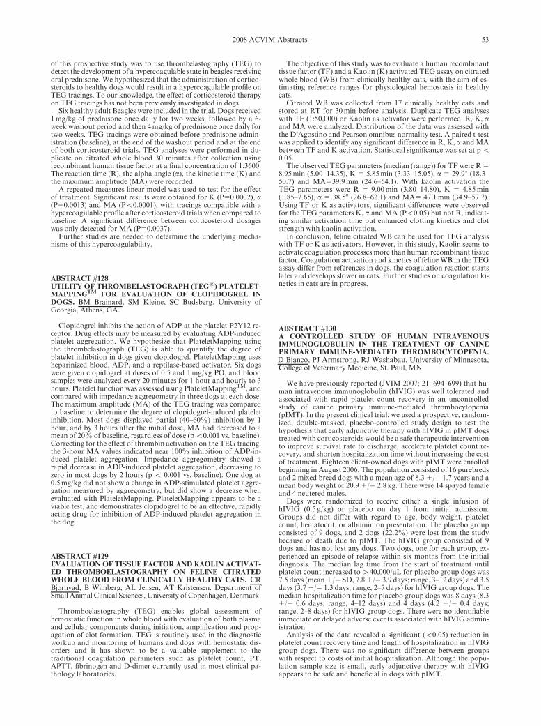
of this prospective study was to use thrombelastography (TEG) todetect the development of a hypercoagulable state in beagles receivingoral prednisone. We hypothesized that the administration of cortico-steroids to healthy dogs would result in a hypercoagulable profile onTEG tracings. To our knowledge, the effect of corticosteroid therapyon TEG tracings has not been previously investigated in dogs.
Six healthy adult Beagles were included in the trial. Dogs received1mg/kg of prednisone once daily for two weeks, followed by a 6-week washout period and then 4mg/kg of prednisone once daily fortwo weeks. TEG tracings were obtained before prednisone admin-istration (baseline), at the end of the washout period and at the endof both corticosteroid trials. TEG analyses were performed in du-plicate on citrated whole blood 30 minutes after collection usingrecombinant human tissue factor at a final concentration of 1:3600.The reaction time (R), the alpha angle (a), the kinetic time (K) andthe maximum amplitude (MA) were recorded.
A repeated-measures linear model was used to test for the effectof treatment. Significant results were obtained for K (P50.0002), a(P50.0013) and MA (Po0.0001), with tracings compatible with ahypercoagulable profile after corticosteroid trials when compared tobaseline. A significant difference between corticosteroid dosageswas only detected for MA (P50.0037).
Further studies are needed to determine the underlying mecha-nisms of this hypercoagulability.
ABSTRACT #128UTILITY OF THROMBELASTOGRAPH (TEGs) PLATELET-MAPPING
TMFOR EVALUATION OF CLOPIDOGREL IN
DOGS. BM Brainard, SM Kleine, SC Budsberg. University ofGeorgia, Athens, GA.
Clopidogrel inhibits the action of ADP at the platelet P2Y12 re-ceptor. Drug effects may be measured by evaluating ADP-inducedplatelet aggregation. We hypothesize that PlateletMapping usingthe thrombelastograph (TEG) is able to quantify the degree ofplatelet inhibition in dogs given clopidogrel. PlateletMapping usesheparinized blood, ADP, and a reptilase-based activator. Six dogswere given clopidogrel at doses of 0.5 and 1mg/kg PO, and bloodsamples were analyzed every 20 minutes for 1 hour and hourly to 3hours. Platelet function was assessed using PlateletMappingTM, andcompared with impedance aggregometry in three dogs at each dose.The maximum amplitude (MA) of the TEG tracing was comparedto baseline to determine the degree of clopidogrel-induced plateletinhibition. Most dogs displayed partial (40–60%) inhibition by 1hour, and by 3 hours after the initial dose, MA had decreased to amean of 20% of baseline, regardless of dose (po0.001 vs. baseline).Correcting for the effect of thrombin activation on the TEG tracing,the 3-hour MA values indicated near 100% inhibition of ADP-in-duced platelet aggregation. Impedance aggregometry showed arapid decrease in ADP-induced platelet aggregation, decreasing tozero in most dogs by 2 hours (p o 0.001 vs. baseline). One dog at0.5mg/kg did not show a change in ADP-stimulated platelet aggre-gation measured by aggregometry, but did show a decrease whenevaluated with PlateletMapping. PlateletMapping appears to be aviable test, and demonstrates clopidogrel to be an effective, rapidlyacting drug for inhibition of ADP-induced platelet aggregation inthe dog.
ABSTRACT #129EVALUATION OF TISSUE FACTOR AND KAOLIN ACTIVAT-ED THROMBOELASTOGRAPHY ON FELINE CITRATEDWHOLE BLOOD FROM CLINICALLY HEALTHY CATS. CRBjornvad, B Wiinberg, AL Jensen, AT Kristensen. Department ofSmall Animal Clinical Sciences, University of Copenhagen,Denmark.
Thromboelastography (TEG) enables global assessment ofhemostatic function in whole blood with evaluation of both plasmaand cellular components during initiation, amplification and prop-agation of clot formation. TEG is routinely used in the diagnosticworkup and monitoring of humans and dogs with hemostatic dis-orders and it has shown to be a valuable supplement to thetraditional coagulation parameters such as platelet count, PT,APTT, fibrinogen and D-dimer currently used in most clinical pa-thology laboratories.
The objective of this study was to evaluate a human recombinanttissue factor (TF) and a Kaolin (K) activated TEG assay on citratedwhole blood (WB) from clinically healthy cats, with the aim of es-timating reference ranges for physiological hemostasis in healthycats.
Citrated WB was collected from 17 clinically healthy cats andstored at RT for 30min before analysis. Duplicate TEG analyseswith TF (1:50,000) or Kaolin as activator were performed. R, K, aand MA were analyzed. Distribution of the data was assessed withthe D’Agostino and Pearson omnibus normality test. A paired t-testwas applied to identify any significant difference in R, K, a andMAbetween TF and K activation. Statistical significance was set at po0.05.
The observed TEG parameters (median (range)) for TF were R 58.95min (5.00–14.35), K 5 5.85min (3.33–15.05), a 5 29.91 (18.3–50.7) and MA539.9mm (24.6–54.1). With kaolin activation theTEG parameters were R 5 9.00min (3.80–14.80), K 5 4.85min(1.85–7.65), a 5 38.5o (26.8–62.1) and MA5 47.1mm (34.9–57.7).Using TF or K as activators, significant differences were observedfor the TEG parameters K, a and MA (Po0.05) but not R, indicat-ing similar activation time but enhanced clotting kinetics and clotstrength with kaolin activation.
In conclusion, feline citrated WB can be used for TEG analysiswith TF or K as activators. However, in this study, Kaolin seems toactivate coagulation processes more than human recombinant tissuefactor. Coagulation activation and kinetics of feline WB in the TEGassay differ from references in dogs, the coagulation reaction startslater and develops slower in cats. Further studies on coagulation ki-netics in cats are in progress.
ABSTRACT #130A CONTROLLED STUDY OF HUMAN INTRAVENOUSIMMUNOGLOBULIN IN THE TREATMENT OF CANINEPRIMARY IMMUNE-MEDIATED THROMBOCYTOPENIA.D Bianco, PJ Armstrong, RJ Washabau. University of Minnesota,College of Veterinary Medicine, St. Paul, MN.
We have previously reported (JVIM 2007; 21: 694–699) that hu-man intravenous immunoglobulin (hIVIG) was well tolerated andassociated with rapid platelet count recovery in an uncontrolledstudy of canine primary immune-mediated thrombocytopenia(pIMT). In the present clinical trial, we used a prospective, random-ized, double-masked, placebo-controlled study design to test thehypothesis that early adjunctive therapy with hIVIG in pIMT dogstreated with corticosteroids would be a safe therapeutic interventionto improve survival rate to discharge, accelerate platelet count re-covery, and shorten hospitalization time without increasing the costof treatment. Eighteen client-owned dogs with pIMT were enrolledbeginning in August 2006. The population consisted of 16 purebredsand 2 mixed breed dogs with a mean age of 8.3 1/� 1.7 years and amean body weight of 20.9 1/� 2.8 kg. There were 14 spayed femaleand 4 neutered males.
Dogs were randomized to receive either a single infusion ofhIVIG (0.5 g/kg) or placebo on day 1 from initial admission.Groups did not differ with regard to age, body weight, plateletcount, hematocrit, or albumin on presentation. The placebo groupconsisted of 9 dogs, and 2 dogs (22.2%) were lost from the studybecause of death due to pIMT. The hIVIG group consisted of 9dogs and has not lost any dogs. Two dogs, one for each group, ex-perienced an episode of relapse within six months from the initialdiagnosis. The median lag time from the start of treatment untilplatelet count increased to440,000/mL for placebo group dogs was7.5 days (mean 1/� SD, 7.8 1/� 3.9 days; range, 3–12 days) and 3.5days (3.7 1/� 1.3 days; range, 2–7 days) for hIVIG group dogs. Themedian hospitalization time for placebo group dogs was 8 days (8.31/� 0.6 days; range, 4–12 days) and 4 days (4.2 1/� 0.4 days;range, 2–8 days) for hIVIG group dogs. There were no identifiableimmediate or delayed adverse events associated with hIVIG admin-istration.
Analysis of the data revealed a significant (o0.05) reduction inplatelet count recovery time and length of hospitalization in hIVIGgroup dogs. There was no significant difference between groupswith respect to costs of initial hospitalization. Although the popu-lation sample size is small, early adjunctive therapy with hIVIGappears to be safe and beneficial in dogs with pIMT.
532008 ACVIM Abstracts

ABSTRACT #131THE EFFECT OF HETASTARCH IN VIVO ON PLATELETFUNCTION IN THE DOG. L Smart, KE Jandrey, JR Wierenga,F Tablin. William R. Pritchard Veterinary Medical TeachingHospital, University of California, Davis, CA.
Hydroxyethyl starch, with an average molecular weight 600 kdand degree of substitution 0.7 (HES), is an artificial colloid solutioncommonly used in veterinary medicine. HES has been shown to de-crease platelet function in humans in vivo, indicated by prolongedclosure times (CT) as measured by the Platelet Function Analyzer-100s (PFA-100s). HES has also been shown to decrease canineplatelet function in vitro by prolonging CT. Our hypothesis was thatintravenous HES in vivo prolongs CT in dogs.
Eight healthy, employee-owned dogs were included in the treat-ment group. Four of these dogs also served as the control group.Washout period between experimental protocols for the controlgroup was a minimum of 4 weeks. Baseline platelet count was great-er than 100,000/ml in all dogs. CTs were measured using collagenand adenosine diphosphate platelet agonists. Dogs were given20mL/kg of either NaCl 0.9% (control group, n54) or HES (treat-ment group, n58) IV over one hour. Blood was drawn for CTbefore the infusion, and at 1, 3, 5, and 24 hours after the start of theinfusion. Two-way repeated measures analysis of variance was usedto assess changes over time, and differences between control andtreatment groups. Time-specific group differences were evaluatedusing Student’s t-test with a sequentially rejective method of multi-ple comparison adjustment.
There was a significant change over time from 0 to 24 hours(p50.0001), a significant difference between groups across time(p50.0004), and evidence of a significant group-by-time interaction(p50.0069). At 3 hours, mean CT for the treatment group was 122.3� 18.1 seconds, which was significantly different (p50.0002) fromthe control group (71.0� 3.5 seconds). At 5 hours, mean CT for thetreatment group was 142.7 � 33.9 seconds, which was significantlydifferent (p50.0014) from the control group (75.0 � 8.6 seconds).Mean CT at 24 hours was within the reference interval for both thecontrol and treatment group (66.0 � 2.9 seconds and 81.8 � 11.9seconds respectively), however CT in three individual dogs in thetreatment group at this time point remained prolonged.
Given these results, a clinically relevant dose of HES adverselyaffects platelet function, as assessed by closure time. Individual dogsmay still have decreased platelet function 24 hours after a single20mL/kg dose of HES, and therefore, an increased risk of bleeding.
ABSTRACT #132AN EVALUATION OF 9570 DOGS BY BREED AND DOGERYTHROCYTE ANTIGEN TYPING. AS Hale1, JWerfelmann1, M Lemmons2, B Smiler2 and J Gerlach2. 1MidwestAnimal Blood Services, Inc., Stockbridge, MI. 2Michigan StateUniversity, E. Lansing, MI.
Nine thousand five hundred and seventy dogs were evaluated bybreed and dog erythrocyte antigen assay (DEA) for the purpose ofpredicting compatibility during routine transfusion service.
Retrospectively, data from these dogs was evaluated for DEAfrequency in relation to breed type and total incidence within thesubmitted population. Sample testing occurred between 1995 and2006. All dogs were evaluated for DEA by tube agglutination uti-lizing polyclonal antisera derived through canine alloimmunizationrecognizing DEA 1.1, 1.2, 3, 4, 5 and 7. Ninety breeds were repre-sented. All recognized AKC breed groups were represented in thesampling. Data frommixed breed dogs was also correlated. Popula-tion incidence for the total sampling is represented in the table below.
DEA Antigen Population Frequency
1.1 42%
1.2 12%
3 7%
4 98%
5 11%
7 20%
Twenty nine percent of all dogs tested positive for DEA 4 only(‘‘Universal’’ type). Breeds demonstrating an incidence of ‘‘univer-
sal’’ type greater than the average population frequency includedAiredale, American Bulldog, Boxer, Bull Mastiff, English Bulldog,English Mastiff, German Shepherd, Greyhound, Irish Wolfhound,Old English Sheepdog, Pitbull, Saluki and Scottish Deerhound.Twenty-six percent of all dogs tested positive for DEA 1.1 and 4only. Breeds demonstrating an incidence of DEA 1.1,4 greater thanthe average population frequency included Afghan, Bassett Hound,Bernese Mountain Dog, Border Collie, Bouvier des Flandres, Bull-mastiff, Bull Terrier, Chesapeake Bay Retriever, Cocker Spaniel,Dalmatian, English Mastiff, English Setter, English Springer Span-iel, Great Dane, Golden Retriever, German Shorthair Pointer,Great Pyrenees, Irish Setter, Labrador Retriever, Malamute, Mali-nois, Newfoundland, Poodle, Rottweiler, Samoyed, St Bernard,Standard Poodle and Vizsla. Using the currently recommended fieldtyping scheme, 55% of all dogs submitted for typing could be usedas a red blood cell donor without DEA mismatch if the recipientsare type matched for DEA 1.1.
ABSTRACT #133A GEL COLUMN BASED ANTIGLOBULIN TEST TO DE-TECT ERYTHROCYTIC AUTO- AND ALLOANTIBODIES INCATS. Mayank Seth, Karen V Jackson, Urs Giger. Section ofMedical Genetics, University of Pennsylvania, Philadelphia, PA.
The antiglobulin (Coombs’) test detects auto- and alloantibodiesdirected against erythrocytes and has been used to diagnoseimmune-mediated hemolytic anemia (IMHA) and to identifyblood type incompatibilities. Many techniques, including tubeand microplate agglutination, as well as flow cytometry and gel-col-umn tests, using varied species-specific reagents, have beendeveloped.
We report here on the use of a novel, commercially available,simple, feline antiglobulin containing gel column assay (DiaMed,Switzerland). Briefly, for the direct antiglobulin test (DAT) patient’s�2% red cell suspension was placed on top of one gel column withpre-loaded antiglobulin reagent and one without (saline control),and then the columns were centrifuged. In the indirect antiglobulintest (IAT) patient’s serum or plasma was incubated with a red cellsuspension on top of gel columns at 37 1C prior to centrifugation.With increasing red cell retention in the gel column the results weregraded from 0 to 41.
Of 64 anemic Coombs’ tested cats, 13 were DAT positive (�11;12 �21). Eight of these 13 DAT positive cats were clinically diag-nosed with either primary or secondary IMHA. The other 5 DATpositive cats did not show clinical signs of hemolysis and were diag-nosed with various underlying illnesses. Among the 51 DATnegative cats 8 were suspected to have a hemolytic anemia: 4 Abys-sinians and 2 domestic shorthair cats had increased osmotic fragilityof erythrocytes, 1 Somali had a pyruvate kinase deficiency and in 1case the cause of hemolysis remained unknown. When screeningsamples from 62 healthy blood donors, all cats were found to beDAT negative.
Antiglobulin enhanced gel column crossmatches were performedon select samples from 42 anemic cats to 264 donor blood units. Inaddition, samples from 8 healthy non-anemic donor cats with sus-pected compatibility issues were screened against 65 additionalunits. The major crossmatch (patient plasma and donor red cells)test revealed 90 incompatibilities, including 2 A-B mismatches and10 reactions involving the Mik-antigen. The minor crossmatch (do-nor plasma and patient/other donor red cells) test revealed 53incompatibilities, of which 6 were related to the presence of Mik-alloantibodies. Among the 143 incompatibilities observed overall,81 were appreciated only in antiglobulin-containing gel columnsand not saline gel columns.
In addition, we used the gel column technique with anti-Mik se-rum for the Mik typing of 132 cats, of which 4 samples (3%) werefound to be Mik negative. All Mik reactions were stronger in theantiglobulin-containing columns than in the saline columns.
In conclusion, the feline antiglobulin gel column test seems simpleand standardized to use and helpful in identifying IMHA. More-over, the use of the antiglobulin column in the major and minorcrossmatch test facilitates the identification of transfusion incom-patibilities (e.g. A, B, Mik and other antigens and theiralloantibodies), although the clinical relevance of some of these in-compatibilities is not yet determined.
54 2008 ACVIM Abstracts

ABSTRACT #134COMPARISON OF GEL COLUMN, CARD, CARTRIDGE,SLIDE AND TUBE TECHNIQUES FOR AB BLOOD TYPINGOF CATS. Mayank Seth, Karen V Jackson, Urs Giger. Section ofMedical Genetics, University of Pennsylvania, Philadelphia, PA.
The clinical importance of the feline AB blood group system withits type A, type B and rare type AB has been well recognized. In-deed, A-B blood incompatibilities, responsible for life-threateningacute hemolytic transfusion reactions, as well as neonatal is-oerythrolysis, can be avoided by prior blood typing, which may beperformed by several methods.
This study directly compares a gel column diffusion assay (GEL,DiaMed, Switzerland with lectin as anti-B), a card based agglutina-tion assay (CARD, DMS Laboratories, NJ), an immunochro-matographic cartridge (CHROM, Alvedia, France) and laboratoryslide (SLIDE) and tube (TUBE) agglutination techniques on sam-ples fromRyan’s Veterinary Hospital, the PennAnimal Blood Bankand external samples sent to the Transfusion Laboratory. A total of38 EDTA blood samples from healthy cats and 20 from sick catswith an emphasis on the less common type B and rare type AB weretyped using all of the above methods. In addition, samples from 432cats were tested only with the GEL and TUBE methods; the latterbeing historically the gold standard. All plasma samples from cats,which express the type B antigen on the erythrocyte surface, wereevaluated for the presence of anti-A alloantibodies (back-typing).
In the complete comparative typing study of 58 cats, 35, 14 and 9samples were determined to have type A, type B and type AB, re-spectively. Fifty-two (90%) samples gave the same typing resultswith all techniques. The CHROMmethodmisidentified 1 type A catas type AB and 2 AB cats; 1 as type A and 1 as type B. The CARDtest misidentified 1 type A cat as type AB and 4 AB cats; 1 as type Aand 3 as type B. The SLIDE and GEL methods both mistyped 1type A as an AB cat. Interestingly, samples from 2 FeLV positivedomestic cats were the source of many A-B blood type discrepan-cies. All B cats had strong anti-A alloantibodies, while AB cats hadno anti-A alloantibodies in their plasma. The reactions of the GELand CHROMmethods were generally clearest to assess and archive.
In the comparison study of GEL and TUBE typingmethods, 372,42 and 18 samples were determined to be type A, B and AB, respec-tively. GEL and TUBE tests gave the same results in 430 (99.5%) ofcases. GEL test results were inconclusive for 2 FeLV positive type Acats.
We conclude that currently available commercial laboratory andin-clinic techniques generally provide accurate typing results. TheGEL and CHROM methods made interpretation of results simple.The rare type AB posed the most discordant results and FeLV pos-itive cats potentially represent another unique subset in terms ofserological typing.
ABSTRACT #135THE EFFECTS OF WEIGHT LOSS ON GENE EXPRESSIONIN DOGS. Yamka RM, KG Friesen, X Gao, S Malladi, S Al-Murrani and L Bernal. Hill’s Pet Nutrition Center, Topeka, KS.
Seven neutered/spayed obese Beagles (4 35% body fat; averageage 5 5.86 � 3.34 years; average weight 5 17.0 � 3.7 kg) were iden-tified for a weight loss study to test the efficacy of a dry low-fat,fiber-enhanced therapeutic food formulated for weight loss (33.2%crude protein, 8.7% crude fat and 26.7% total dietary fiber on drymatter basis). The dogs were fed for weight loss (1.0 � RER) for aperiod of 4 months. During the study all dogs underwent DEXAand chemistry screen analysis at 0, 1, 2, 3 and 4 months. In addition,blood samples were collected into PAXgene blood RNA tubes atthe beginning and end of the study to characterize changes in geneexpression differences resulting from weight loss on the food. RNAwas extracted according to the procedures provided in the PAXgeneBlood RNA Kit Handbook (Qiagen, Valencia, CA). RNA was hy-bridized to an Affymetrix Gene Chip Canine-2 Genome Array andsignal normalized using Robust Multi-Array Average. Data was fil-tered using following a false discovery rate of 0.1 and 4 1.3-foldchange were considered different for day 0 and end of study. On av-erage, dogs lost 2.8 � 0.8 kg body fat (41.2% of initial fat mass) in 4months. Gene expression profiles were modified in these dogs after 4months of weight loss on the food. The nutrigenomic effect of thefood can be seen in the shift from an obese to a lean profile. Of the
genes identified, there was a down-regulation of genes associatedwith fat accumulation, including long chain fatty acid CoA ligase 1,growth factor receptor-bound protein 2 and hypoxia-inducible fac-tor 1 alpha. In summary, obese dogs fed the weight loss food lost 2.8� 0.8 kg (41.2% of initial fat mass) body fat in 4 months, which wasaccompanied by a shift from an obese to lean genomic profile.
ABSTRACT #136LECITHIN:CHOLESTEROL ACYLTRANSFERASE ACTIVI-TY AND ITS FATTY ACYL SPECIFICITY IN CATS FEDDIETS OF VARYING FATTY ACID COMPOSITION. R.Angell1, K. Bigley1, M. McClure, Y. Mitsuhashi1, D. Nagaoka1, J.E. Bauer1,2. 1Companion Animal Nutrition Laboratory, 2Faculty ofNutrition, Texas A&MUniversity, College Station, TX.
Beyond the fact that cats express low delta-6 desaturase activities,lipid metabolism in this species remains to be fully explored undervarying dietary or other experimental conditions including otherimportant metabolic enzymes such as lecithin:cholesterol acyltr-ansferase (LCAT), which is involved in reverse cholesteroltransport. To characterize LCAT activity and demonstrate its fattyacyl specificity in cats fed varying types of dietary fat, intact youngfemales were fed diets enriched with high-oleic sunflower (n 5 9),menhaden fish (n 5 10), or safflower (n 5 10) oil (8 g oil/100 g kib-ble) for 4 wk. Fasted blood samples were drawn at d0, d14, and d28for determination of plasma total (TC), unesterified (UC), and es-terified cholesterol (EC) concentrations, LCAT activity, and fattyacid (FA) composition of the EC fraction. UC decreased at d28compared to d0 and d14, while EC increased at d28 compared to d0and d14 (all p o 0.05). LCAT and TC showed no time or dieteffects. Plasma EC FA profiles reflected the specificity of LCAT forlinoleic acid (LA) and numerous diet and time effects were ob-served. Even though similar amounts of eicosapentaenoic acid(EPA) and docosahexaenoic acid (DHA) were supplied by the fishoil diet and significant amounts of EPA were detected in the plasmaEC fraction, no DHA was identified in this fraction, indicating verylittle or no affinity of feline LCAT for DHA. The fish oil diet sup-plied docosapentaenoic acid (DPA) as well, but this FA was alsoabsent in the plasma EC fraction, suggesting little or no affinity offeline LCAT for DPA. We conclude that feline LCAT has no mea-surable affinity for DHA, but that feline LCAT demonstratedspecificity for LA regardless of diet fed. Finally, it was noteworthythat some changes in feline plasma EC FA composition were statis-tically significant after only 14 days of feeding the experimentaldiets. This observation indicates rapid modification of feline FAmetabolism by dietary fatty acid composition. By d28, further sig-nificant differences due to time were seen, indicating that whileinitial differences can be seen in as little as 14 days, a longer studyperiod may be necessary to establish a metabolic steady state.Whether 28 days is enough time to achieve this effect cannot be de-termined by the present study. However, our studies in dogs andother species indicate that 28 days is suitable to achieve steady stateeffects of plasma fatty acids. Nonetheless, we did demonstrate that28 days feeding is a long enough study period to observe alterationsin feline FA metabolism. Finally, because no diet or time effectswere seen on LCAT activity, the values for all cats at all time pointswere averaged to obtain a basal LCAT activity for adolescent catsof 92.4� 4.5 nmol of UC esterified/ml of plasma/hr (mean� SEM).This is lower than an earlier reported value for LCAT activity inadolescent cats, and is likely due to differences in methodology. Thepresent work utilized an endogenous substrate method while theearlier study employed an artificial proteoliposome substrate tech-nique.
ABSTRACT #137THE IMPACT OF SIGNALMENT AND BODY CONFORMA-TION ON ACTIVITY MONITORING IN COMPANIONDOGS. DC Brown, KE Michel, M Love, C Dow. University ofPennsylvania, Philadelphia, PA.
An objective method for recording activity could be useful forassessing the behavior of companion dogs in their everyday envi-ronment as many of these behaviors have to do with the dogs’ levelof activity. The Actical Activity Monitor (AAM) is a sensitive,
552008 ACVIM Abstracts

watch-sized, accelerometer that continuously measures the intensi-ty, frequency and duration of movement for extended periods. Ourobjective was to describe the impact of signalment and body confor-mation on activity monitoring using the AAM in companion dogs.
At least 20 dogs were recruited in each of 5 weight ranges. Sig-nalment and 7 body conformation measurements were recorded.While wearing an AAM, each dog was led through a series of stan-dardized activities: lying down, walking laps and trotting laps. Inaddition, our hypothesis was that a dog’s age and body conforma-tion could influence the outcome of activity monitoring during anactivity that is less controlled; therefore all dogs were trotted up anddown stairs, permitting each dog to adjust its stride between steps.Linear regression analysis was used to determine which signalmentor body conformation factors were associated with the AAM activ-ity counts.
Data was collected on 104 dogs. There was no significant impactof signalment or body conformation on activity counts recordedduring lying down, walking laps, and trotting laps. However, whentrotting up and down stairs, there was a significant impact of age andbodyweight such that, for every 1 kg increase in bodyweight, there isa 1.9% decrease in average activity counts and for every 1 year in-crease in age, there is a 4.1% decrease in average activity counts.
When the activity is well controlled, there is no significant impactof signalment or body conformation on the average activity countsrecorded by the AAM. However, when dogs are allowed leeway inthe extent of effort exerted within an activity, older dogs and largerdogs will deliver lower average activity counts than younger andsmaller dogs and so these factors will need to be considered in stud-ies using these monitors as an outcome.
ABSTRACT #138ACTIVITY MONITORING CAN DIFFERENTIATE INTENSI-TY OF EXERCISE IN COMPANION DOGS. KE Michel, MLove, C Dow, DC Brown. University of Pennsylvania,Philadelphia, PA.
Extent of activity directly affects an individual dog’s energy bal-ance and factors into that dog’s caloric requirement. An objectivemethod that captures the activity level of dogs in their everyday en-vironment could aid in tailoring feeding recommendations to betterreflect an individual companion dog’s opportunity and inclinationto exercise. The Actical Activity Monitor (AAM) is a sensitive,watch-sized, accelerometer that continuously measures the intensi-ty, frequency and duration of movement for extended periods. Ourobjective was to investigate whether the AAM could be used todifferentiate the intensity of activity in companion dogs.
While wearing an AAM, each dog was led through a series ofstandardized activities: lying down, walking laps, and trotting laps.At least 20 dogs were recruited in each of 5 weight ranges. The Wi-lcoxon signed-rank test was used to compare sedentary activitycounts to walking activity counts and walking activity counts totrotting activity counts. Receiver operating characteristic curveswere generated to determine the optimal activity counts for predict-ing whether an animal was sedentary, walking, or trotting.
Data was collected on 104 dogs. The median counts (range) forsedentary activity was 10 (0–64), for the walking activity was 290(91–622), and for the trotting activity was 785 (359–1517). Bothsedentary and walking activity counts and walking and trotting ac-tivity counts were significantly different from one another(po0.001). At an activity count of 65 there is 100% specificity and100% sensitivity in distinguishing the sedentary activity from thewalking activity. At an activity count of 441 there is 92% specificityand 92% sensitivity in distinguishing the trotting activity from thewalking activity.
In conclusion, activity counts recorded by the AAM were highlyaccurate in differentiating among standardized activities in com-panion dogs.
ABSTRACT #139ANTIOXIDANT STATUS AND BIOMARKERS OF OXIDA-TIVE STRESS IN DOGS WITH DIABETES MELLITUS. JRSmith1, SK Cox1, SD Lauten1, RC Hill2, JW Bartges1, CA Kirk1.1College of Veterinary Medicine, The University of Tennessee,
Knoxville, TN. 2College of Veterinary Medicine, University ofFlorida, Gainesville, FL.
Increasing evidence implicates oxidative damage in the progres-sion and pathologic complications of human diabetics. This studyassessed antioxidant status and oxidative stress in dogs with diabe-tes mellitus (DM). Antioxidant status was measured in diabetic(n510) and control (n510) dogs by HPLC of vitamin E isomers,reduced (GSH) and oxidized glutathione (GSSG), and calculationof the GSH:GSSG ratio. Biomarkers of protein, lipid and DNAperoxidation (fructosamine, isoprostanes, and Comet assay, respec-tively), and neutrophil function were used to evaluate oxidativestress. Correlation between glycemic control and antioxidant status/oxidative stress was also investigated. A diabetic index was generat-ed using clinical signs, body condition score, insulin dose,fructosamine, fasted blood glucose and urinary glucose andketones. Diabetic dogs were separated into those with good (n54)or poor (n56) control.
Serum fructosamine concentration was significantly higher (DM5 472.9mmol/l, controls 281.3mmol/l; po0.0001), while specificvitamin E isomers, serum GSH (DM 5 885.8 ng/ml, controls 51188.1 ng/ml, p50.01) and 8-F2a-isoprostanes (DM 5 0.31 ng/ml,controls 5 3.74 ng/ml; po0.0001) were significantly lower in dia-betic dogs versus controls. Antioxidant status/ oxidative stress wasnot associated with glycemic control in diabetic dogs.
Despite strong association of DM with oxidative stress in hu-mans, this simple relationship is not found in diabetic dogs. Theyhave both increased and decreased parameters of systemic oxidativestress compared with control dogs. This may be due to higher levelsof antioxidants in canine therapeutic diets, the relatively short du-ration of disease in dogs compared to humans, or other factors.
ABSTRACT #140TRIGLYCERIDEREFERENCEVALUES FORAMEALCHAL-LENGE TEST TO ASSIST DIAGNOSIS AND MANAGEMENTOF CANINE HYPERLIPIDEMIA. KF Elliott, JS Rand, LMFleeman, JM Morton. Centre for Companion Animal Health, Uni.of Queensland, Australia.
Hyperlipidemia in dogs is associated with clinical disorders, in-cluding recurrent seizures and acute pancreatitis. Dietary therapy istypically recommended for management of hyperlipidemia and astandardized method for evaluating efficacy of dietary managementin controlling postprandial triglyceride concentrations is needed.This study aimed to determine reference values using 95% toleranceintervals for 24 hour fasting and postprandial peak triglyceride con-centrations for healthy dogs after a standard meal, to assist indiagnosis and management of hyperlipidemia.
Twelve (6 male, 6 female) lean, healthy, neutered, mixed-breeddogs were studied. Dogs were fed a dry commercial maintenancediet (fat 31%ME, CHO 45%ME, protein 24%ME, fiber 2 g/100 kcal) for 3 weeks. After a 24 hour fast, plasma triglyceride con-centrations were measured at �1, �0.08, 1, 2, 3, 4, 5, 6, 9, and 12hours after a meal (median amount eaten: 127 kcal/kg0.75). Raw andlog transformed data were tested for normal distribution using Ko-lmogorov-Smirnov test and for outliers using Grubb’s test. Todetermine the most appropriate time for blood sampling (the timemost closely associated with highest postprandial triglyceride con-centration), Spearman’s correlation coefficients, associated 95%confidence intervals (CI) and p values were calculated to assess cor-relations in raw data between postprandial peak triglyceride andtriglyceride concentrations at each time point. Tolerance intervalswere calculated with log-transformed data using a parametric method.
One dog had a postprandial peak triglyceride concentration 44%higher than the next highest dog; however its fasting value was to-ward the low end of the tolerance interval. It was found to be anoutlier using Grubb’s test and was not included in calculation oftolerance intervals. Triglyceride concentration at 6 hours after in-gestion of the standard meal had the closest association withpostprandial peak triglyceride concentration with and without thisdog (r50.93 and 0.91, respectively; po0.01; 95% CI: 0.77 to 0.98and 0.68 to 0.98, respectively). The tolerance interval for postpran-dial peak triglyceride concentration was 49 to 191mg/dL (0.54 to2.11mmol/L). The tolerance interval for triglyceride concentration6 hours after eating was 35 to 176mg/dL (0.38 to 1.94mmol/L). Thetolerance interval for 24 hour fasted triglyceride concentrations
56 2008 ACVIM Abstracts

sampled 5minutes prior to a meal was 14 to 91mg/dL (0.15 to1.00mmol/L). In 50% of the dogs, triglyceride concentrations didnot return to baseline by 12 hours after eating.
In conclusion, measurement of both fasting and postprandialpeak triglyceride concentrations after a standard meal are importantfor detecting hyperlipidemia and for evaluating responses to thera-peutic diets, because fasting concentrations alone are not likely to bepredictive of postprandial peak concentrations. The reference valuesdetermined in this study can be applied in a meal challenge test todogs which have been fasted 24 hours, fed the same standard meal,and sampled 5minutes prior to the meal and 6 hours after eating.
ABSTRACT #141LIPID METABOLISM EFFECTS IN CATS FED DIETS VARY-ING IN MEDIUM CHAIN FATTY ACIDS AND LINOLEICACID CONCENTRATIONS. L. Trevizan1,2, K. Bigley2, W.Anderson3, M.K.Waldron3, J.E. Bauer2,4. 1LEZO, Faculdade deAgronomia, Universidade Federal do Rio Grande do Sul, RS,Brazil, 2Comp. An. Nutr. Lab., 3Nestle Purina Pet Care, St. Louis,MO, USA, 4Faculty of Nutrition, Texas A&MUniv., Col Stn, TX.
Medium chain triglycerides (MCT) containing fatty acids of 6 to12 carbons are found in ingredients such as coconut oil and milkbyproducts. They are readily absorbed and efficiently metabolizedin mitochondria. However, in dogs and cats, some studies show thatca. 22% Metabolic Energy (ME) caused diet aversion, especiallywhen enriched in caprylic acid (C8:0). In dogs, 11%ME MCTshowed no effects on food intake, but cats have not been evaluatedat this concentration. Thus, we hypothesized feeding cats with11%ME MCT does not cause diet aversion. The objective of thisstudy was to evaluate a diet containing increased MCT by substi-tuting a portion of the safflower oil containing dietary linoleic acid(LA) with coconut oil. The high MCT diet (HMCT) contained11.4%ME MCT and 4.3%ME LA (29.9%ME protein, 32.3%MENFE, 37.8%ME fat) while the low MCT diet (LMCT) contained4.3%ME MCT and 14.5%ME LA (29.8%ME protein, 29.4%MENFE, 40.7%ME fat). Nineteen adult female cats were separated in-to two groups (LMCT, n59; HMCT, n510). The cats had been feda pre-experimental diet (PED) containing no MCT, 5.3%ME LA ,and 27% ME total fat for 30 days. Test diets were fed for 9 weeksusing week 1 as a transition period. Cats were initially fed accordingto their metabolic body weights (100�W0.67) and water offered adlibitum. Daily consumption records, weekly body weights (BW) andbody condition scores (BCS, 1 to 9) were used to adjust amounts fedand to calculate the daily metabolic energy factors for each cat sothat an ideal BCS of 5 was maintained. Blood samples (7ml inEDTA) were taken after overnight fasting via saphenous vein at d 0,14, 28 and 56 to measure plasma triglycerides (TG), total cholesterol(TC), lipoprotein distribution (LP) and lecithin cholesterol acyltransferase (LCAT). Repeated measures analyses (SAS, MixedModels Procedure) and Tukey (a50.05) multiple comparisons wereperformed. A significant time effect was observed on food con-sumption, BW, BCS and metabolic factor reflecting the previousPED diet. A diet effect on plasma TG revealed a 28% increase(p50.0234) with the HMCT diet. However, no diet effects were seenin TC, LP or LCAT activity. Time effects were observed in whichTC increased on d 14 and again d 28 but returned back to the d 14level at d 56 (p50.0001), LCAT increased at d 14, only. All LP (b,pre-b and a) in both diets reached peak values at d 28 (p50,0001)consistent with elevation of TC. No diet differences were seen infood intake, metabolic factor, and BW, demonstrating that11%ME MCT does not produce aversion in cats. The TC increasewas likely the result of increased dietary fat content of the test diets.A modest increase of plasma TG occurred with HMCT but it waswithin normal limits. Thus, it is feasible to formulate feline dietscontaining MCT without diet refusal. Such diets may be useful inboth normal cats and those with fat malabsorption syndromes.
ABSTRACT #142POST-FEEDING SATIETY AND WEIGHT LOSS EFFECT OFA VEGETABLE-BASED FIBER SUPPLEMENT IN BEA-GLES. Y. Mitsuhashi1,2, K. Bigley1, J.E. Bauer1,2. 1CompanionAnimal Nutrition Laboratory, 2Intercollegiate Faculty ofNutrition, Texas A&MUniversity, College Station, TX.
Dietary insoluble fiber is believed to support weight loss by in-creasing satiety and allowing increased amounts of foodconsumption without adding calories, whereas dietary soluble fiberhas potential to reduce cholesterol. Therefore, in this study, weevaluated a vegetable-based fiber supplement containing both solu-ble (SF) and insoluble fiber (IF) types on plasma lipid profiles,satiety, and weight loss in Beagles.
Two diets were fed differing in fiber content. Purina ONEs
healthy weight formula (ONE) was compared to a diet consistingof ONE plus a vegetable-based fiber supplement (FIB). Total di-etary fiber contents were 3.83 g/100 kcal (ONE) vs 6.89 g/100 kcal(FIB). The SF content of the FIB diet was 83.3% more than that ofONE (1.10 g/100 kcal vs 0.60 g/100 kcal in ONE) and IF content ofthe FIB diet was 79.6% more than that of ONE (5.80 g/100 kcal vs3.23 g/100 kcal in ONE). For the satiety studies, female adult Bea-gles with average body fat (BF) of 37.5�1.4 (SEM) %, and bodyweight (BW) of 13.4�0.6 (SEM) kg were randomly divided into 2diet groups. Diets were fed at 8am and 3pm (7h interval, n514) or,during a second trial, at 8am and 11am (3h interval, n512) for15min each over a 3 or 2 day period, respectively. Amounts offeredat each feeding were 1.2 timesMER using a cross-over design with 3days washout. Blood samples were collected after the 8am feeding at45min and 120min and food intakes were recorded. For the weightloss study, 7 obese Beagles were selected (average BF 45.1�1.6%,BW 15.2�1.0 kg, BCS 8.0�0.3 [9 point scale]) and divided into 2groups. The diets were fed once daily (ca. 60% of obese MER) for42 days. Postprandial blood samples were collected at 60min (days1, 28 and 42) for lipid and lipoprotein analysis. Food intakes andBW were recorded daily and weekly, respectively. Repeated mea-sures ANOVA was performed under a50.05. Where there wassignificance, paired t or one-way AOV / Kruskal-Wallis tests withTukey multiple comparisons were performed after checking nor-mality.
In the satiety trials, both the 3 and 7h interval food intakes werenot different from the control group indicating similar satiety ofboth diets. However, significantly fewer total calories were con-sumed with the FIB diet during the 3 h interval study (p50.017).Plasma triacylglycerol (TG) concentration was significantly in-creased at 120min post feeding independent of diet compared to45min (po0.01). In the weight loss trial, the FIB group significantlyreduced % BW (p50.002) and % BF (p50.008) over time. PlasmaTG, total cholesterol (TC) and lipoprotein (LP) did not change.Thus, the increased percentage of IF in the FIB diet provided fewercalories with the same degree of satiety as the higher calorie intakeof the control diet. In addition, this increase supported more effi-cient weight loss. However, the modestly increased amount of SFwith the FIB diet did not affect plasma lipid profiles. In conclusion,the FIB diet appeared to provide similar satiety at lower calorie in-take thereby promoting weight loss without modifying TG, TC andLP responses.
ABSTRACT #143ALTERATIONS OF LECITHIN:CHOLESTEROL ACYL-TRANSFERASE ACTIVITY AND CHOLESTEROL METABO-LISM IN OBESE BEAGLES DURING WEIGHT LOSS. R.Angell1, D. Bandy1, K. Bigley1, Y. Mitsuhashi1, D. Nagaoka1, T.Umeda2, K. Otsuji2, J. E. Bauer1,3. 1Companion Animal NutritionLaboratory, 3Faculty of Nutrition, Texas A&MUniversity, CollegeStation, TX, USA, 2Kao Corp., Tokyo, Japan.
Little research has focused on the relationship between leci-thin:cholesterol acyltransferase (LCAT) activity and cholesterolmetabolism in dogs even though it is the major lipid metabolic en-zyme involved in reverse cholesterol transport. To study alterationsin LCAT activity and lipid metabolism during weight loss in thisspecies, four experimental weight-loss diets were fed to 12 obese fe-male Beagles for 8wk in a partial crossover design (n 5 6). High-(HGI) or low-glycemic index (LGI) starch (waxy corn or high amy-lose corn, respectively) and diacylglycerol (DAG) or triacylglycerol(TAG) oil were combined to compose diets with similar fatty acid(FA) profiles. Both starches were gelatinized prior to formulation.Experimental diets were fed as a water-based gruel after acclimatingthe dogs to a similar gruel type basal diet. Dogs were fed the amountof kcal required to maintain their obese body weights. Consumptionrecords were kept daily and body weights were measured weekly.Fasted blood samples were obtained at baseline, wk4, and wk8 to
572008 ACVIM Abstracts

measure plasma LCAT activity using a radiolabeled endogenoussubstrate technique. Total (TC), unesterified (UC), and esterifiedcholesterol (EC) concentrations were determined via enzymaticmethods. Fatty acid composition of the plasma phospholipid (PL)and EC fractions were determined after total lipid extraction, frac-tionation via thin-layer chromatography, and capillary gaschromatography. All groups lost weight as a result of voluntary re-duction of food intake of the gruel-based type of diet. Plasma UCconcentrations increased in all groups from baseline to wk4 (p o0.05). LCAT activities increased from baseline to wk4 in all groupsand remained elevated at wk8 (p o 0.05). Plasma PL FA profilesreflected the diets fed with few diet or time effects. Plasma EC FAprofiles reflected the previously observed specificity of canine LCATfor linoleic acid (LA) again with minimal diet or time effects. Weconclude that an increase in LCAT activity and plasma UC concen-tration is observed in conjunction with weight loss in dogs. Dieteffects on these two parameters were not observed, indicating thatthe changes in LCAT activity and UC concentration were indeeddue to weight reduction because all diet groups experienced weightloss. Dietary oil type (TAG vs. DAG) did not affect the plasma ECor PL fatty acid composition. These findings are likely due to thesimilar fatty acid composition of the experimental oils and diets andindicates that positional isomers of oil do not affect resultant FAprofiles under the conditions employed.
ABSTRACT #144INFLUENCEOFGENDERANDSEXUALALTERATION STA-TUS ON FELINE ADIPONECTIN. AL Lusby, CA Kirk, JWBartges. The University of Tennessee College of VeterinaryMedicine, Knoxville, TN.
Adiponectin is a hormone secreted almost exclusively from ad-ipocytes that correlates closely with insulin sensitivity in humanbeings and rodents. In contrast to other adipokines, adiponectinlevels decrease as fat mass increases. This hormone holds potentialdiagnostic and therapeutic applications for feline obesity and dia-betes. Although previous studies have identified strong associationsbetween obesity and gender and/or reproductive status in cats, theinfluence of adiponectin in these associations has not been evaluat-ed. The purpose of this study was to identify the impact of genderand reproductive status on adiponectin and insulin resistance. Thesefindings may help to better understand the role of adiponectin inobesity and insulin resistance in the cat.
This study compared four groups of lean (BCS 4–6/9), healthy,young adult cats (1–6 years; n540) to determine the influence ofgender and gonadectomy on total serum adiponectin levels. Eachgroup consisted of ten cats in each of the following categories: intactfemale (IF), intact male (IM), neutered female (NF), neutered male(NM). General health status was assessed through physical exam,complete blood count, and chemistry panel with electrolytes. Bodyweight and body condition scores (BCS) were also recorded. Serumsamples for adiponectin were collected from fasted cats at one timepoint and stored at �80 1C. A commercial ELISA kit was used tomeasure adiponectin (B-Bridge International, Mountain View,CA). A linear ANOVA model was used for statistical analysis(SAS v.9.1) and compared gender, reproductive status, body weightwith total adiponectin.
Body weight differed between all groups except for IM and NF(Po.05) while BCS did not differ. There was no significant differ-ence between adiponectin levels among groups (Po.05). However,NF tended to have greater adiponectin levels compared to IM(P 5 0.07) and neutered animals tended to have higher adiponectinlevels compared to intact animals (P50.06). While this studyindicates total adiponectin levels are not influenced by gender orgonadectomy in domestic cats, there are strong trends that may beinfluenced by sample size.
ABSTRACT #145INFLUENCE OF DIETARY PROTEIN CONTENT ANDSOURCE ON FECAL QUALITY AND PROTEIN-DERIVEDFERMENTATION PRODUCTS IN DOGS DIFFERING INBODY SIZE. J Nery1, C Tournier2, V Biourge2, L. Martin1, HDumon1 and P Nguyen1. 1Ecole Nationale Veterinaire de Nantes,France. 2Royal Canin, Aimargues, France.
When given the same diet, large breed dogs produce feces ofpoorer quality than smaller ones due partially to higher ferment-ative activity in the hindgut. Undigested proteins are importantsubstrates for fermentation by the colonic microflora. They can bemetabolized to several bioactive products, which could have delete-rious effects on the colonic mucosa. The aim of this study was toevaluate the effect of dietary protein source and amount on fecalquality and concentration of some key protein fermentation prod-ucts in dogs differing in body size.
Twenty-seven female dogs of 6 different breeds (from 3.35 to34.65 kg BW) were divided in 4 groups according to BW (miniature:MI, medium: ME and maxi: MA) and propensity to have softer fe-ces (maxi tolerant: MT and maxi sensitive: MS). Five diets varyingin protein source and level were tested in a 2-phase protocol con-sisting of a crossover and Latin square designs respectively. Dietswere formulated to be isoenergetic and to have similar content offat, TDF and ash. Main dietary protein sources and levels were asfollows: wheat gluten meal in diets WGLP (CP519.9%) andWGHP (CP534.3%), mix of poultry and wheat gluten meal in dietWPMP (CP526.2%), and poultry meal in diets PMLP(CP519.3%) and PMHP (CP535.2%). Feces were scored dailyand fecal moisture was determined from a 7-day collection pool af-ter a 7-day adaptation period. Fresh stools were collected (1–2samples/dog and diet) and analyzed for ammonia and branched-chain fatty acids (BCFA). Data were statistically analyzed usingANOVA followed by Fisher’s PLSD post hoc test.
Fecal quality was lower both with poultry meal diets (po0.0001)and with high CP levels (po0.01) whereas fecal moisture varied on-ly with protein source (po0.0001). Compared to MI dogs, MS dogshad a higher fecal moisture and a lower fecal quality on all diets(po0.0001). Ammonia fecal concentration varied with dietary pro-tein source (po0.0001) and level (po0.0001). Specially diet PMHPinduced a significantly higher ammonia fecal concentration. MSdogs were particularly prone to have high ammonia concentrations(po0.0001). BCFA varied both with protein source (po0.001) andlevel (po0.0001). An effect of dog size (po0.01) was only observedon diets WGHP and WPMP.
Decreasing CP levels and introducing a source of highly digestibleprotein such as wheat gluten improves fecal score. Moreover it re-duces protein fermentation in the hindgut. This is of interest to theoverall hindgut health as ammonia production induces a faster cellturnover and modified intestinal cell’s morphology and metabolism,affecting colonic absorption functions. According to our resultsthese deleterious effects seem to be of higher importance inMS dogs.
ABSTRACT #146BODY CONDITION SCORING AND DEXA MEASURE-MENTS IN PHYSICALLY INACTIVE DOMESTICSHORTHAIRED CATS. CR Bjornvad1, PJ Armstrong2, DHNielsen1, E Svalastoga1, AT Kristensen1. 1Department of SmallAnimal Clinical Sciences, The Royal Veterinary and AgriculturalUniversity, Copenhagen, Denmark. 2College of VeterinaryMedicine, University of Minnesota, Twin Cities, MN.
In cats and humans, obesity is predisposing to development oftype 2 diabetes. Further, physical inactivity and indoor confinementpredispose for development of type 2 diabetes mellitus in cats. Thepurpose of this study was to determine the percentage of body fat inindoor confined physically inactive normal and overweight domes-tic shorthaired cats.
Twenty-five physically inactive and indoor confined domesticshorthaired cats were recruited through the Companion AnimalVeterinary Teaching Hospital, University of Copenhagen, Den-mark. Informed consents were obtained from all owners. Atadmission, the cats underwent a thorough health examination in-cluding physical exam, body condition scoring (9-point scale), urineanalysis, haematological and biochemical blood profile includingT4-measurement and FIV/FeLV-testing. Two cats were excludedfollowing the health examination. For the remaining 23 cats, bodycomposition and whole body bone mineral density was assessed un-der anaesthesia using dual-energy X-ray absorptiometry (DEXA).
All 23 cats had been neutered (nine females and 14 males). Me-dian age was 6 years (range 3–11). Following body conditionscoring, the cats were divided into 4 groups BCS5 (n55), BCS6(n56), BCS7 (n57) and BCS8 (n55). No cats had a BCS less than 5or above 8. Females and males were equally distributed in all groups
58 2008 ACVIM Abstracts

except BCS8 that only included male cats. The cats in BCS5 wereyounger (3 years (range 3–5)) than cats in the other groups (7 years(range 3–11)). Percent body fat for the groups were BCS5: 31.3%(range 27.4–33.7), BCS6: 37.4% (range 32.5–40.7), BCS7: 43.6%(range 34.4–49.0) and BCS8 47.9% (range 44.1–52.2). Percent bodyfat was significantly different between all groups. There was nodifference in Bone Mineral Density (g/cm2) between groups or be-tween males and females.
According to the 9 integer scale BCS system a BCS of five is con-sidered normal weight. On the other hand, normal weight catsshould have a body fat content of less than 30%. The relatively highbody fat content in the normal weight cats in this study could beattributable to the sedentary lifestyle following indoor confinement.The physical inactivity may result in less lean body mass comparedwith kennel cats measured in earlier studies. Concurring with otherstudies, cats start to gain weight at three to five years of age andmale neutered cats tend to become fatter than female neutered cats.
ABSTRACT #147THE DISPOSITION AND PHARMACOKINETICS OF THEOXIDATIVE METABOLISM SUBSTRATES MIDAZOLAM,PHENYTOIN, AND THEOPHYLLINE IN GREYHOUNDDOGS. Butch KuKanich, Michelle Hubin, and Jon Nauss.Kansas State University, Department of Anatomy and Physiology,Manhattan, KS.
The purpose of the study was to assess the disposition andpharmacokinetics of midazolam, phenytoin, and theophylline inhealthy Greyhound dogs. Midazolam is a CYP3A12 substrate indogs, whereas the definitive mechanisms of metabolism for pheny-toin and theophylline have not been identified in dogs. Phenytoin isa CYP2C9 and CYP2C19 substrate in humans and theophylline isprimarily a CYP1A2 substrate in humans. There is evidence sup-porting the role of CYP1A in the metabolism of theophylline indogs.
Six healthy Greyhound dogs, 3 male and 3 female, were used inthe study which was approved by the Kansas State University Insti-tutional Animal Care and Use Committee. On separate occasions,with at least a 7 day washout period, midazolam (0.25mg/kg),phenytoin (11mg/kg), and theophylline (7.88mg/kg) administeredas aminophylline, were administered IV. Blood samples were ob-tained at predetermined intervals for the determination of plasmadrug concentrations with mass spectrometry (midazolam) or fluo-rescence polarization immunoassay (phenytoin, theophylline). Thepharmacokinetics were estimated with noncompartmental analyses.
The half-life (t 1/2), clearance (Cl), and volume of distribution(Vd) for phenytoin, and theophylline were similar to values reportedin dogs (see Table I). The Cl and Vd for midazolam appear to bedifferent in Greyhounds. The results are suggestive that the dispo-sition and pharmacokinetics phenytoin and theophylline are similarin Greyhound and non-Greyhound, non-Beagle dogs, but midazo-lam may be different. Further studies are indicated.
ABSTRACT #148RONIDAZOLE PHARMACOKINETICS IN CATS AFTER IVADMINISTRATION AND ORAL ADMINISTRATION OF ANIMMEDIATE RELEASE CAPSULE AND A COLON-TARGET-ED DELAYED RELEASE TABLET. DN LeVine1, MG Papich1,JL Gookin1, GS Davidson1, JL Davis1, WC Stagner2, R Goldman1,L Williamson2. 1College of Veterinary Medicine, North CarolinaState University, Raleigh, NC. 2Campbell UniversityPharmaceutical Sciences Institute, Buies Creek, NC.
Tritrichomonas foetus, a parasite of feline colonic mucosa, causesunrelenting diarrhea for which ronidazole (RDZ) has been identi-fied as the only effective treatment. RDZ kills T.foetus in vitro (RDZ� 0.1 mg/ml) and has eliminated T.foetus in cats (30–50mg/kg POq12h for 14 days). RDZ produces neurotoxicity in some cats anddisposition of the drug in felines is unknown. The objective of thisstudy was to characterize the pharmacokinetics of RDZ in cats afterintravenous (IV) and oral administration and to assess a novel for-mulation for colon-targeted delivery of RDZ.
RDZ was compounded into immediate-release capsules (95mg),with immediate-release confirmed by in vitro dissolution studies. AnIV solution was prepared by dissolving RDZ in 5% dextrose in wa-ter (3.2mg/ml). RDZ was administered orally (mean dose 28.2mg/kg) and IV (�9.2mg/kg) to 6 healthy adult cats in a crossover designwith at least one-week washout between crossovers. Plasma wascollected from catheters over 48 hours and analyzed for RDZ usinghigh pressure liquid chromatography. RDZ was well-tolerated bythe cats after each administration.
Following IV administration of RDZ, the terminal half-life (t1/2)was 9.72�0.38 hrs and initial concentration (C0) was 14.94�2.77mg/ml. Volume of distribution at steady state was 0.66�0.06L/kg and the systemic clearance was 0.80�0.07mL/kg/min.Following oral administration, RDZ was rapidly and completelyabsorbed with detection in plasma of all 6 cats by 10min after dos-ing and systemic availability of 99.3�17.2%. The maximum plasmaconcentration (CMAX) of RDZ was 36.20�2.63mg/mL, time to max-imum plasma concentration (TMAX) was 1.39�1.29 hr, and terminalt1/2 was 10.44�0.83 hr. After recognizing that RDZ was rapidly ab-sorbed from immediate-release capsules we formulated delayedrelease RDZ by coating tablets with guar gum. Bacterial digestionof guar gum and subsequent drug release are theoretically restrictedto the colon where T.foetus resides. In vitro dissolution studies con-firmed that RDZ was not released from guar gum coated tablets ateither gastric or intestinal pH. These tablets were administered tofour cats orally (�32.9mg/kg) and absorption studies repeated. Thedelayed release formulation extended the TMAX to 16 hrs and pro-duced a CMAX of 28.90�9.77mg/ml. Drug absorption was complete(bioavailability 117.0 � 31.8%) and corresponded to time of pro-jected arrival of RDZ in the colon.
In conclusion, RDZ is rapidly and completely absorbed from im-mediate-release capsules in the proximal GI tract and persists inplasma for over 48 hrs. These attributes may predispose cats to ne-urotoxicity with twice-daily administration. By targeting release ofRDZ to the colon, the site of T. foetus infection, delayed releasetablets may provide improved efficacy at lower doses or less fre-quent intervals that reduce risk of systemic toxicity.
ABSTRACT #149RELATIONSHIP BETWEENMUTATIONSOF THEPANCRE-ATIC SECRETORY TRYPSIN INHIBITOR GENE ANDPANCREATITIS IN MINIATURE SCHNAUZERS AND DOGSOF OTHER BREEDS. MA Bishop, PG Xenoulis, MD Levinski,JS Suchodolski, and JM Steiner. Gastrointestinal Laboratory,Texas A&MUniversity, College Station, TX.
Anecdotal evidence suggests that Miniature Schnauzers (MS) arepredisposed to pancreatitis. We have previously reported the iden-tification of 3 mutations (2 exonic mutations: N20K, N25T and 1intronic mutation: IVS3126-27ins(T)33-39,15_61dup11) of thepancreatic secretory trypsin inhibitor gene (PSTI) in MS. In hu-mans, multiple mutations of the PSTI gene have been linked tohereditary pancreatitis. Therefore, the aim of this study was to eval-uate the prevalence of mutations in the PSTI gene in a population ofMS and dogs of other breeds, and to determine whether the pres-ence of these mutations is associated with pancreatitis.
A total of 4 groups of dogs were enrolled in this study: 55 MSwith pancreatitis, 13 dogs of other breeds with pancreatitis, 40healthy MS, and 17 healthy dogs of other breeds. Diagnosis of pan-creatitis was based on a combination of clinical signs suggestive ofpancreatitis and a serum cPLI concentration above the cut-off valuefor pancreatitis. A complete history and clinical findings were re-corded for each dog. Serum and whole blood were obtained fromeach dog. DNA was extracted from whole blood and PCR was per-formed to amplify the 3 regions of the gene with the previouslyreported mutations, using specific primers. PCR products were pu-rified and sequenced directly by automated cycle sequencing.
Table 1. Pharmacokinetic parameters of midazolam, phenytoin, and theophylline in Grey-
hound dogs. Pharmacokinetic parameters from previous studies in non-Greyhound dogs
are included for comparison.
Parameter Units Breed Midazolam Phenytoin Theophylline
Half-life hr Greyhound 1.2 5 9.3
non-Greyhound 1.31 4.52 8.43
Clearance mL/min/kg Greyhound 10.2 2.4 1
non-Greyhound 271 2.82 0.83
Volume of
Distribution
L/kg Greyhound 1.0 1 0.75
non-Greyhound 3.01 12 0.553
592008 ACVIM Abstracts

Sequences were compared with the published canine genome projectsequence for this gene and also among the 4 groups of dogs. Pro-portions of dogs with mutations of the PSTI gene were comparedamong groups using a Fisher’s exact test and odds ratios (OR) withtheir 95% confidence intervals (CI) were calculated.
The 2 exonic mutations were always linked and were commonlypresent in all 4 groups of dogs studied. The intronic mutation wasfound only in MS (46 of 55 MS with pancreatitis and 25 of 40healthy MS). Additionally, this mutation was always linked to thefirst 2 mutations. Of the MS with pancreatitis, 35/55 (63.6%) werehomozygous for the intronic mutation compared to 16/40 (40.0%)of healthy MS (OR 5 2.625; p 5 0.0366; 95% CI: 1.135–6.069).However, being heterozygous for the intronic mutation was not sig-nificantly associated with pancreatitis (p 5 0.4765).
Results of this study suggest that this intronic mutation is uniqueto MS. The 2 exonic mutations were found in both MS and dogs ofother breeds and probably represent single nucleotide polymor-phisms (SNPs). MS that were homozygous for the intronic muta-tion were significantly more likely to have pancreatitis thanMS thatwere heterozygous or did not have the mutation. Due to the factthat the intronic mutation was present in both healthy and diseasedMS, we hypothesize that pancreatitis in MS is likely multifactorial.Further studies are in progress to determine the role of this muta-tion and other risk factors in the development of pancreatitis inMS.
ABSTRACT #150SPECIFICITY OF CANINE PANCREAS-SPECIFIC LIPASE(SPEC CPL
TM) IN DOGS WITH A HISTOLOGICALLY NOR-
MAL PANCREAS. S Carley1, JE Robertson2, SJ Newman3, JMSteiner4, D Kutchmarik2, RL Relford2. 1Santa Barbara, CA.2IDEXX Laboratories, Westbrook, ME. 3University of Tennessee,Knoxville, TN. 4Gastrointestinal Laboratory, Texas A&MUniversity, College Station, TX.
Recently, an enzyme-linked immunosorbent assay (ELISA) forthe measurement of canine pancreas-specific lipase (Spec cPL) hasbecome commercially available. The purpose of this study was toevaluate the specificity of the Spec cPL in dogs with a histologicallynormal pancreas.
Forty-four dogs euthanized for reasons unrelated to this studywere enrolled. CBC, biochemistry panel, and Spec cPLwere analyzedprior to euthanasia and the entire pancreas removed post euthanasia.The pancreata were sectioned every 1 cm and reviewed by the samepathologist (SJN) using a previously published classification system(Newman, et al. Veterinary Pathology 18:115–118, 2006). A meancumulative score (MCS) for neutrophilic inflammation, lymphocyticinflammation, pancreatic necrosis, peripancreatic necrosis, edema,atrophy, fibrosis and nodules and disease activity index (AI) and dis-ease chronicity index (CI) were determined for each pancreas.
Seventeen client-owned dogs from a private veterinary practicewere entered into the study. Dogs were euthanized for medical rea-sons: trauma (6), dystocia (2), nonpancreatic neoplasia (3),osteoarthritis (1), anemia (1), diabetic ketoacidosis (1), pancreaticneoplasia (1), and pancreatitis (2). Twenty-seven dogs from an an-imal shelter were entered in the study. Each dog was examined andbloodwork was reviewed. One dog was diagnosed with diabetesmellitus, and one dog was underweight and anemic. The remaining25 dogs appeared clinically healthy with 6 dogs having mild changeson bloodwork.
Dogs were classified into 2 groups for statistical comparison. Onegroup contained dogs with a disease involving the pancreas (pancr-eatitis, diabetes mellitus and pancreatic neoplasia). The other groupcontained healthy dogs and dogs with a disease not involvingthe pancreas. A Student’s t-test comparing the MCS for eachlesion, AI and CI of the 2 groups revealed a statistical differencebetween the pancreata for all parameters. Population distribu-tion data was established for the dogs that were healthy or had dis-eases not involving the pancreas. A pancreas with a MCS for eachhistologic category, AI and CI which included 90% of this popula-tion was considered normal. Based on these criteria, 31 dogs had ahistologically normal pancreas. The Spec cPL was below the cutoff for pancreatitis in 30 of these dogs resulting in a specificity of96.8%.
In conclusion, the Spec cPL test is highly specific and is not ele-vated in dogs with a histologically normal pancreas.
ABSTRACT #151FELINE EXOCRINE PANCREATIC INSUFFICIENCY: 15CASES (1992–2007). K.A. Thompson1, N.K. Parnell1, A.E.Hohenhaus2, G.E. Moore1, M.P. Rondeau3. 1Purdue University,Lafayette, IN. 2The Animal Medical Center, New York, NY.3School of Veterinary Medicine, University of Pennsylvania,Philadelphia, PA.
Exocrine pancreatic insufficiency occurs uncommonly in cats andis poorly described in the veterinary literature. The purpose of thisretrospective study is to describe a population of cats with feline ex-ocrine pancreatic insufficiency (EPI). Databases from 1992–2007 offour referral practices were searched for cases of feline exocrinepancreatic insufficiency. Inclusion criteria included at least one clin-ical sign compatible with exocrine pancreatic insufficiency and afeline trypsin-like immunoreactivity (fTLI) of o 12 g/L or threeconsecutive day fecal proteolytic activity resultso 6mm.
Fifteen cats met the enrollment criteria and their medical recordswere reviewed. The median age of cases was 7 years (range: 0.3–15years). The majority of cats were castrated males (9). The mostcommon breed was the domestic short hair (9).
Clinical signs and physical examination findings were recordedfor all 15 cats. Weight loss was the most common clinical manifes-tation and found in 14/15 cats (93%), followed by diarrhea/loosestools (11), vomiting (5), polyphagia (4), anorexia (4), lethargy (4),and fecal incontinence (3). Stool was described as voluminous (7),loose (6), hematochezia (4), increased frequency (4), malodorous(4), discolored (4), steathorrea (3) and greasy (3). The most commonphysical examination findings included thin/emaciated body condi-tion in 9/15 cats (60%), muscle wasting (6), unthrifty hair coat (5),and thickened intestinal loops (5). Concurrent disease was present in11 of 15 cats (73%). The most common concurrent diseases wereurinary tract disease (5), enteritis (3), gastritis (2), Helicobacter in-fection (2), and hepatic disease (2).
The most common complete blood cell count abnormalities in-cluded normocytic, normochromic anemia 7/14 cats (50%), lymph-openia (6/13) and neutrophilic leukocytosis (4/13). The most com-mon serum chemistry abnormalities included hyperglycemia (5/13),increased ALT (5/13) and total bilirubin (4/13). Serum cobalaminconcentration was measured in 10 cats and was abnormal in all 10.The median serum cobalamin concentration was 99 ng/L (range:o27–176 ng/L [reference range 290–1499ng/L]). Serum folate con-centrations were increased in 4/10 cats with a median value of21.1mg/L (range 12.2–42.5mg/L [reference range 9.7–21.6mg/L]).
Fourteen of 15 cats received treatment including pancreatic en-zyme supplementation (13/14), parenteral cobalamin supplement-ation (7/14) and metronidazole (5/14). Data regarding response totreatment was available in 12/15 cats. Of these 12 cats, 11 responded(92%).
This study shows cats with EPI exhibit many signs typical of EPIin the dog; however the majority of cats with EPI will have a con-current disease and are likely to be cobalamin deficient.
ABSTRACT #152RELATIONSHIP BETWEEN SERUM fPLI AND TRIGLYCER-IDE CONCENTRATIONS IN CATS. JM Steiner, KM Aicher, JSSuchodolski, PG Xenoulis. Gastrointestinal Laboratory, TexasA&MUniversity, College Station, TX.
Hypertriglyceridemia has been reported as a risk factor for pan-creatitis in both humans and dogs. Serum feline pancreatic lipaseimmunoreactivity (fPLI) concentration has been reported to be asensitive and specific marker for pancreatitis in cats. The objectiveof this study was to investigate a possible relationship between se-rum fPLI and triglyceride concentrations in cats.
Serum samples from 279 cats were used for this study. SerumfPLI concentration (reference range: 2.9–6.0mg/L) was measured inall serum samples by an in-house radioimmunoassay. Serum tri-glyceride concentration (reference range: 25–133mg/dL) wasmeasured using an automated serum chemistry analyzer. Possiblecorrelation between serum fPLI and triglyceride concentrations wasevaluated using a Spearman test. The median serum fPLI concen-tration was compared between cats with a normal serum triglycerideconcentration, those with an increased serum triglyceride concen-tration, and also those with severe hypertriglyceridemia (4 500mg/dL). The proportion of cats with a serum fPLI concentration above
60 2008 ACVIM Abstracts

the reference range or the cut-off value for pancreatitis (12 mg/L)was compared between cats with normal serum triglyceride concen-trations and those with increased serum triglyceride concentrations.
Forty-six cats (16.5%) had an increased serum triglyceride con-centration (median: 209mg/dL; range: 134–2,147mg/dL), but only6 cats had severe hypertriglyceridemia. There was no significantcorrelation between serum fPLI and triglyceride concentrations(Spearman r 5 0.1085; p-value 5 0.0703). The median serum fPLIconcentration was not significantly different between cats with anincreased serum triglyceride concentration (68.3mg/L) and cats witha normal serum triglyceride concentration (56.8mg/L; p-value 50.0557). Median serum fPLI concentration in the 6 cats with severehypertriglyceridemia (164.6mg/L) was not significantly higher thanin cats with a normal serum triglyceride concentration (56.8 mg/L;p-value 5 0.090). Also, the proportion of cats with a serum fPLIconcentration above the upper limit of the reference range or thecut-off value for pancreatitis was not significantly higher in hype-rtriglyceridemic cats than in cats with normal serum triglycerideconcentrations (p-values 5 0.4368 and 0.1078, respectively).
Several cats with hypertriglyceridemia were identified in thisstudy, but only 6 cats had a severely increased serum triglycerideconcentration. In this study none of the parameters assessing therelationship of serum fPLI and triglyceride concentrations reachedstatistical significance. However, the median serum fPLI concentra-tion was higher, albeit not significantly so, in cats withhypertriglyceridemia or severe hypertriglyceridemia than in catswith normal serum triglyceride concentrations. While this studydid not show hypertriglyceridemia to be a risk factor for pancreatitisin cats, further studies are needed to confirm these results.
ABSTRACT #153ESOPHAGEAL AND GASTRIC ENDOSCOPIC FOREIGNBODY REMOVAL: COMPLICATIONS AND LONG-TERMFOLLOW-UP OF 102 DOGS. P Gianella, NS Pfammatter, IABurgener. Division of Small Animal Internal Medicine from theVetsuisse Faculty of the University of Bern, Switzerland.
Esophageal and gastrointestinal foreign bodies (FB) are a rela-tively common problem in dogs. The purpose of this study was toinvestigate complications and long term follow up of endoscopic FBremoval in a large case number.
A total of 114 FB were endoscopically removed between 03/2001and 11/2006. Six dogs were excluded due to FB in the nose (N54) orlung (N52). In the remaining 108 dogs, the FB was located in theesophagus (N560), the stomach (N538), or both (N510). Thoracicradiographs were diagnostic for esophageal FB in all cases, whereasabdominal radiographs were diagnostic for gastric FB in 46/48cases. The duration of clinical signs before presentation ranged from2 hours to 40 days (51 dogso 1 day, 20 1–3 days, 314 3 days, 6 notknown). Only 18 dogs were known to have ingested foreign materialat the time of presentation.
West Highland White Terrier (11/108), Yorkshire Terrier (9/108)and Bernese Mountain Dogs (9/108) were overrepresented com-pared to the hospital population. The most frequent clinical signsreported were vomiting (38%), retching/gagging (29%), cough anddyspnea (18%), anorexia (14%), and regurgitation (10%). Bonymaterial accounted for 47% of the FB, followed by plastic (15%),metal (10%) and greenies (7%). Six dogs with esophageal FB un-derwent fluoroscopy due to contrast medium in the esophagus andwere excluded. In the remaining 102 cases, endoscopy alone wassuccessful in 92 dogs (90%) and a gastrotomy, but no esophago-tomy, was required in 10 dogs.
Eight dogs (7.8%) had perforations (5 esophagus, 3 stomach),most commonly associated with bony FB (6/8). Four dogs died(3.9%) due to pneumothorax and cardiac arrest (2), respiratory ar-rest (1), or esophageal diverticle with sudden death (1), whereas 2dogs were euthanized with pleural effusion and pneumothorax (1)or mediastinitis (1). Two dogs had aspiration pneumonia, whereofone required thoracotomy due to a periesophageal abscess. Only 1dog developed an esophageal stricture and underwent successfulendoscopic balloon dilations. Long-term outcome was available for75/96 patients, whereof only 4 (5.3%) showed some coughing andretching a few days after discharge. Eight dogs died due to unrelatedproblems and 2 had another FB later on.
In conclusion, this study further supports the use of endoscopicFB removal. Even though the overall complication rate is low, po-
tentially life-threatening complications may develop. Clinical signsfor more than 1 day prior to endoscopy is associated with increasedmorbidity.
ABSTRACT #154THE EFFECTS OF BODY POSITIONING ON QUANTITA-TIVE ASSESSMENT OF SWALLOWING USING CONTRASTVIDEOFLUOROSCOPY IN NORMAL DOGS. CecilyBonadio1, Rachel Pollard2, Paul Dayton4, Caroline Leonard1,Stanley Marks3. 1Veterinary Medical Teaching Hospital, School ofVeterinary Medicine, 2Department of Surgical and RadiologicalSciences, School of Veterinary Medicine, 3Department of Medicineand Epidemiology, School of Veterinary Medicine, University ofCalifornia at Davis, Davis, CA. 4Department of BiomedicalEngineering, UNC-NCSU, Chapel Hill, NC.
Contrast videofluoroscopy is the imaging technique of choice forevaluating dysphagic patients because it allows for assessment ofanatomic and functional components of swallowing. In people,body position significantly alters the outcome of videofluoroscopicassessment of swallowing. The purpose of this study was to deter-mine if quantitative videofluoroscopic measures of swallowing weresignificantly affected by body position in dogs.
Healthy dogs (n515) with no history of dysphagia were recruitedfor this study. Dogs had normal physical examinations, CBC’s, andserum biochemistry panels. A restraint device was built to facilitateimaging of dogs in sternal recumbancy. Each dog underwent video-fluoroscopy of swallowing of liquid barium and barium soakedkibble in sternal and lateral recumbancy. Quantitative measures ofliquid and kibble swallowing (timing of swallowing, pharyngealconstriction ratio, esophageal transit time, and number of esopha-geal peristaltic waves) were compared between body positions. Thenumber of radiation safety violations per second was determined.
The time to opening of the upper esophageal sphincter was sig-nificantly longer for lateral versus sternal kibble swallows(p50.043). In lateral recumbency, 54% of liquid and 71% of kibbleswallows stimulated esophageal peristalsis. In sternal recumbency,81% of liquid and 90% of kibble swallows stimulated esophagealperistalsis. Significantly fewer radiation safety violations occurredin sternal versus lateral esophagrams (p50.003). Other variableswere not significantly different.
Body positioning affects the number of swallows that result inesophageal peristalsis. Radiation exposure to technical staff is sig-nificantly reduced for esophagrams performed in sternalrecumbency.
ABSTRACT #155SERUM MAGNESIM AND ZINC CONCENTRATIONS INDOGS WITH INFLAMMATORY BOWEL DISEASE. KKGingerich, NK Parnell, GE Moore. Purdue University College ofVeterinary Medicine, West Lafayette, IN.
Magnesium and zinc are essential trace elements absorbed in theproximal small intestine. Reduced magnesium and zinc concentra-tions have been documented in human malabsorptive conditionssuch as Crohn’s disease. Initial clinical signs of magnesium deficien-cy include vomiting and diarrhea, with progression to cardiac andneuromuscular abnormalities. Clinically relevant signs of zinc defi-ciency include acrodermatitis, poor wound healing and diarrhea.Currently, the effect of malabsorptive disease on magnesium andzinc concentrations in dogs is unknown.
The purpose of the study was to compare serum magnesium andzinc concentrations in normal dogs and dogs with inflammatorybowel disease. We hypothesized that, compared to healthy dogs,dogs with inflammatory bowel disease would have decreased serummagnesium and zinc concentrations. In a prospective clinical study,16 client-owned dogs were evaluated for chronic gastrointestinalsigns. Inflammatory bowel disease was diagnosed through histolog-ical evaluation of endoscopically obtained gastrointestinal biopsies.At the time of endoscopy, serum was collected for commercial mag-nesium and zinc analysis. Serum was also obtained from 16 age-(within six months) and sex-matched clinically healthy dogs. Themedian age of cases was 6.3 years (range:1.5–15.4 years), and themedian age of controls was 6.2 years (range: 1.5–14.9 years).
612008 ACVIM Abstracts

In dogs with inflammatory bowel disease, median serum albuminconcentrations [1.3 gm/dl (range: 0.9–3.8 gm/dl)] were significantlylower than for control dogs [3.4 gm/dl (range: 2.6–4.2 gm/dl)](p50.003). Albumin concentrations were significantly correlatedwith total magnesium (r50.636; p50.008) and zinc (r50.702;p50.003) concentrations in dogs with inflammatory bowel disease.There was no correlation between albumin concentrations and totalmagnesium (r5-0.195; p50.469) or zinc (r50.139; p50.608) con-centrations in control dogs. Median total magnesiumconcentrations for dogs with inflammatory bowel disease [1.6mg/dl (range: 0.8–2.4mg/dl)] were significantly lower than for controldogs [2.1mg/dl (range: 1.8–2.5mg/dl)] (po0.001). Median zinc con-centrations for dogs with inflammatory bowel disease [0.66 ppm(range: 0.25–2.12 ppm)] were significantly lower compared to con-trol dogs [1.00 ppm (range: 0.58–1.65 ppm)] (p50.044).
This study suggests that magnesium and zinc deficiency should beconsidered in dogs with inflammatory bowel disease and secondaryhypoalbuminemia. The clinical significance of these deficiencies indogs with inflammatory bowel disease is currently unknown andshould be further evaluated.
ABSTRACT #156FECAL N-METHYLHISTAMINE CONCENTRATIONS INNORWEGIAN LUNDEHUNDS WITH GASTROINTESTINALDISEASE. N Berghoff, JS Suchodolski, and JM Steiner.Gastrointestinal Laboratory, Texas A&M University, CollegeStation, TX.
It has previously been suggested that mast cells play a role in thepathogenesis of inflammatory bowel disease (IBD) and other gas-troenteropathies. Mast cells release inflammatory mediators,including histamine. Histamine is metabolized by two major enzy-matic pathways, one of which yields N-methylhistamine (NMH).The purpose of this study was to compare fecal NMH concentra-tions in a group of Norwegian Lundehunds with chronicgastroenteropathy to those in a group of healthy control dogs. Nor-wegian Lundehunds are known to have a high prevalence of chronicgastroenteropathies, and were thus chosen as an initial study group.
Three fecal samples each were collected from 10 healthy controldogs and 21 Norwegian Lundehunds with chronic gastrointestinal(GI) disease. Histopathology was not available, but all Lundehundshad clinical signs of GI disease and/or showed abnormal results ofGI function tests, including decreased serum cobalamin, folate, to-tal protein, and/or albumin concentrations and/or increased fecala1-proteinase inhibitor concentrations. Samples for NMH analysiswere extracted and analyzed as previously described (Tredget, EE.Journal of Chromatography B, 1997), using stable isotope dilutiongas chromatography/mass spectrometry. Data were analyzed fornormality and the medians of the maximum NMH concentrationfor the 3-day collection period and of the 3-day average NMH con-centration were compared between the two groups (Mann-WhitneyU test). Statistical significance was set at po0.05. Maximum and 3-day average NMH concentrations in the group of Lundehunds werealso evaluated for correlation with fecal a1-proteinase inhibitor con-centrations and serum cobalamin, folate, total protein, albumin,and C-reactive protein concentrations.
The triplicate samples from control dogs had maximum NMHconcentrations between 13.8 and 321.7 ng/g feces (median: 57.3 ng/g), whereas those for the Lundehund samples ranged from 85.9 to14,300ng/g (median: 374.4 ng/g; p50.0004). Median 3-day averageNMH concentrations in the Lundehunds were significantly higher(median: 284.7 ng/g; range: 66.9–9,319ng/g) than in the controldogs (median: 44.5 ng/g; range: 8.9–196.9 ng/g; p50.0005). FecalNMH concentrations did not correlate with fecal a1-proteinase in-hibitor concentrations or serum cobalamin, folate, total protein,albumin, or C-reactive protein concentrations.
These data show that fecal NMH was significantly increased in agroup of Lundehunds with chronic GI disease. A potential short-coming of this study is the lack of histopathology in the affectedLundehunds. However, all dogs examined had clinical signs and/orabnormal GI function tests. Intestinal mast cell degranulation andhistamine release may also play a role in other dogs with gastroen-teropathies. Thus, further studies are needed and ongoing in orderto assess fecal NMH concentrations in dogs of other breeds withgastroenteropathies.
ABSTRACT #157CHARACTERIZATIONOF THE BACTERIALMICROFLORAAND INNATE IMMUNITY RESPONSE IN GERMANSHEPHERDDOGSWITH ANTIBIOTIC-RESPONSIVE DIAR-RHEA. K Allenspach1, JS Suchodolski2, FM McNeill1, A House1,A Hendricks1, PG Xenoulis2, JM Steiner2, D Werling3.1Department of Veterinary Clinical Sciences and 3Department ofPathology and Infectious Diseases, 2Royal Veterinary College,University of London, UK, and the Gastrointestinal Laboratory,Texas A&MUniversity, College Station, TX.
The interaction of receptors mediating innate immunity and theintestinal bacterial microflora is believed to play an important rolein the etiology of antibiotic-responsive diarrhea (ARD) in GermanShepherd Dogs (GSDs). The aim of this study was to compare Toll-like receptor (TLR) 2 and TLR4 mRNA expression in duodenal bi-opsies from GSDs with ARD and healthy controls and tosimultaneously assess the mucosa-adherent bacterial microflora inthe same dogs.
Seven GSDs with ARD and 11 healthy retired racing Grey-hounds were evaluated for this study. TLR2 and TLR4 mRNAexpression was assessed using quantitative real-time PCR (qRT-PCR). The microflora were evaluated by obtaining cytology brushsamples during gastroduodenoscopy followed by DNA extractionand PCR amplification using universal 16S rDNA primers. Con-structed clone libraries were compared between groups using theUniFrac distance metric and the RDP Classifier. Indices for bacte-rial diversity and species richness were calculated and comparedusing t-tests.
TLR2 and TLR4 mRNA expression was significantly higher inthe GSDs compared to healthy controls (TLR2 in GSDs vs con-trols: p50.03, TLR4 in GSDs vs controls: p50.01). In addition,dogs with ARD showed a 2–4 times higher expression of TLR2mRNA compared to TLR4 (p50.02). Principal component analysisof the bacterial intestinal clone libraries revealed clustering of indi-vidual dogs within each group, indicating that the small intestinalmicroflora of GSDs and control dogs are composed of distinct mi-crobial communities. The abundance of several bacterial groupswas altered in GSDs compared to control dogs. There was a trendfor GSDs to be enriched in sequences belonging to Proteobacteria(p50.08). Healthy dogs were more likely to harbor members of Ba-cteroidetes (including Bacteroides spp. and Prevotella spp.) andClostridiaceae (po0.001).
In conclusion, TLR2 and TLR4 mRNA expression is upregulat-ed in the duodenum of GSDs with ARD, which is accompanied byan imbalance of the intestinal microflora compared to healthy con-trol dogs. The increased TLR mRNA expression could contributeto an abnormal immune response, resulting in the development ofARD in GSDs.
ABSTRACT #158MAST CELL STABILIZATION WITH BETA-ADRENERGICRECEPTOR BLOCKADE ATTENUATES DYSFUNCTIONALCARDIAC REMODELING IN CANINE MITRAL VALVE RE-GURGITATION. AR Dillon1, DM Tillson1, J Hathcock1, TDenney1, C Killingsworth2, P Betty2, LJ Dell’Italia2. 1AuburnUniversity, Auburn, AL. 2University of Alabama-Birmingham,Birmingham, AL.
Dogs with induced mitral valve regurgitation (MR) have beenshown to lose myocardial collagen, develop mast cell infiltrates inthe LV wall, undergo cardiomyocyte lengthening and use an inher-ent renin-angiotensin system (RAS) unaffected by systemic ACEinhibitors. The mechanism of this dysfunctional cardiac remodelingsecondary to volume overload appears distinct from changes result-ing from cardiogenic or pressure overload remodeling. The goal wasto characterize this cardiac remodeling (structurally and molecular-ly) in order to compare these findings with hemodynamic studies,and the geometric changes and LV wall stress obtained through car-diac magnetic resonance imaging (c-MRI). Study groups includednormal dogs (n514), dogs with MR (n59) without medication,dogs with MR (n56) receiving a b1-receptor blockade (b1-RB), anddogs with MR (n56) receiving b1-RB and a mast cell stabilizer(MCS).
Mild mitral regurgitation (MR) was induced by chordae tendon-ae rupture. Sufficient regurgitation was achieved when dogs had 1) a
62 2008 ACVIM Abstracts

6–12% decrease in forward stroke volume (SV), 2) increased LVEDP or decreased LV ESP, 3) increased pulmonary artery wedge(Paw), and an auscultable mitral murmur. Hemodynamic parame-ters were collected before and immediately after MR induction andagain at 4 months. C-MRI images (Picker Vista 1.0T magnet;DICOM images; 8mm slices) were collected during the cardiac cy-cle before MR induction and again at 4 months.
Treatment with b1-RB improved isolated cardiomyocyte frac-tional shortening and B-receptor responsiveness, but did notattenuate increases in cardiomyocyte length or LV-EDV or affecteccentric LV remodeling. Improved cardiomyocyte function did nottranslate to benefits in LV wall stress, load dependent LV fractionalshortening, or peak LV dP/dt. Treatment with b1-RB (with or with-out MCS) attenuated epicardial extracellular collagen loss but notendocardial collagen loss, by picric acid-Sirius red assay (TypeI&III). Treatment with b1-RB1MCS normalized cardiomyocytelength and restored Ca11 transients. LV peak 1dP/dt was de-creased in b1-RB treated dogs compared to baseline but wasnormalized in dogs receiving b1-RB1MCS.
All groups had increased LV mass and total SV compared tobaseline. Forward SV compared to baseline was decreased in MRand b1-RB dogs, but was preserved in b1-RB1MSC dogs. 3-DMRIimages showed an increase in LV-EDV and LV 3-D radius/wallthickness in MR and b1-RB dogs, but these and LV-ED wall stresswere decreased in b1-RB1MCS dogs compared to MR.
These data suggest use of b1-RB and MCS in dogs with MR in-duced volume overload does attenuate dysfunctional LV andcardiomyocyte remodeling and improves LV function and geome-try, without attenuating the previously described interstitialcollagen loss. The effect of MCS may be directly on cardiomyocytesor through preservation of cardiomyocyte-collagen scaffolding pro-teins affecting outside-in signaling and eccentric remodeling.
ABSTRACT #159AUTOLOGOUSMESENCHYMAL STEM CELLS IN EXPERI-MENTAL MITRAL VALVE REGURGITATION OF DOGSATTENUATED COLLAGEN LOSS AND DYSFUNCTIONALMYOCARDIAL REMODELING. RH Presley1, AR Dillon1 DMTillson1 Niemeyer1, J Hathcock1, TS Denney2, LJ Dell’Italia3.1Auburn University College of Veterinary Medicine, Auburn, AL.2Auburn University College of Engineering, Auburn, AL.3University of Alabama at Birmingham Center for Heart FailureResearch, Birmingham, AL.
In early mitral valve regurgitation (MR) of dogs, increases in leftventricular (LV) diastolic wall stress induces neurohormonal andinterstitial activity, including mast cell recruitment, the dissolutionof collagen in the cardiac extracellular matrix (ECM), and elonga-tion of cardiomyocytes. In ischemic cardiomyopathy, mesenchymalstem cells (MSCs) were capable of transforming into myofibroblastsand attenuating additional myocyte loss. The purpose of this studywas to determine if implantatedMSCs in dogs withMR and volumeoverload would 1) remain at the site of the injection in the absenceof inflammation, 2) alter the collagenmatrix in the ECM, 3) vary theactivity of interstitial mast cells, and 4) alter the mRNA LV profile.Seven intact mongrel dogs without known heart disease had MSCsisolated from bone marrow samples using an accepted plastic ad-herence technique and each sample cultured. The MSCs werelabeled with 0.9 ım polystyrene spheres impregnated with contrastagents of iron oxide (62.4%) microparticles and a fluorescein-5 is-othiocyanate analog (Dragon Green). Viability of the cells (470%) was determined by Trypan Blue exclusion; and fluorescein la-bel was documented inside the MSCs.
On Day 1, MR was induced via chordal rupture using a flu-oroscopic-guided catheterization method. Immediately after MR, a1mL aliquot containing 2.18�0.82 � 107 (range 1.0–3.5 � 107) ofautologous labeled MSCs was injected into the LV free wall in asingle location via LV needle catheter. On Day -7, Day 1 post-op,and Day 38, cardiac MRIs were recorded, wall stress and geometrycalculated, and the MSC in LV located via the iron oxide. On Day 1Pre and Post MR, and Day 40 hemodynamics were collected. Afterhumane euthanasia, LV was collected from the MSC injection site,adjacent to the site, and distant from the site. Samples were evalu-ated for mRNA pathways, collagen density, mast cell counts, andhistopathology.
In the area ofMSC implantation, determined by Picric Acid SiriusRed stain, a significantly higher collagen volume percent was noted,suggesting fibroblast transformation by the MSCs. Hemodynamicdecreases in forward cardiac output from baseline were significantlylower( po0.05) in dogs treated with MSCs (19.7% �7.5) than MRplacebo dogs (n54) (39.4%�9.2). Increases in SVR were also higher(po0.05) in MR placebo dogs (84.6%�53.8) thanMSC treated dogs(32.9%�21.0). By cardiac MRI, on Day 1 post MR and Day 38,MSCs were noted with no loss in signal. These data suggest auto-logousMSCs injected into the LV of dogs with earlyMR remained inthe myocardium, promoted stabilization of the ECM and attenuatedthe initial dysfunctional cardiac remodeling. Global administrationof MSCs may have clinical application in MR of dogs.
ABSTRACT #160ECHOCARDIOGRAPHIC ASSESSMENT OF LEFT VEN-TRICULAR AND MITRAL VALVE GEOMETRY IN CATSWITH HYPERTROPHIC CARDIOMYOPATHY. KE Schober,A Todd. Department of Veterinary Clinical Sciences, College ofVeterinary Medicine, The Ohio State University, Columbus, OH.
Hypertrophic cardiomyopathy (HCM) is the most common car-diac disease in the domestic cat. Obstruction of the left ventricularoutflow tract (LVOT) is frequently observed in HCM (referred to asHOCM) and is usually due to systolic anterior motion and mid-to-late systolic contact of the mitral valve with the interventricular sep-tum (IVS). Altered LV geometry secondary to abnormal myocardialgrowth as well as intrinsic mitral valve abnormalities secondary tocongenital mitral valve malformations may both favor obstructionof the LVOT but may both be associated with a different outcomeand a different response to treatment. This study addresses the hy-pothesis that abnormalities of the LVOT and the mitral valve arecommon in cats with HOCM and can be detected by two-dimen-sional (2D) transthoracic echocardiography.
A total of 102 cats (30 normal control cats, 43 cats with HCMwithout obstruction, and 29 cats with HOCM) underwent a compre-hensive transthoracic 2D echocardiographic examination includingthe assessment of chamber size, wall thickness, and anatomy of themitral valve apparatus and the LVOT using an ultrasonographicunit (Vivid 7, GE, Milwaukee, WI) and a transducer array of 7 to10MHz nominal frequency. A total of 20 variables were assessed,including the size of the left atrium (2 variables), anatomy of the leftventricle (8 variables) and the LVOT (2 variables), and morphologyof the mitral valve (8 variables). All cats were imaged in lateral re-cumbency with the transducer from underneath. Normal controlcats, cats with HCM, and cats with HOCM were compared usinganalysis of variance on ranks for unpaired observations and theHolm-Sidak test or Dunn’s test for post-hoc analyses.
Cats with HOCM had increased (P�0.05) LV wall thickness, LVmyocardial area, number of papillary muscles and papillary musclearea, and increased length of the anterior mitral valve leaflet com-pared to cats without obstruction. There was no difference(P40.05) between groups with regard to age, body weight, sex,IVS thickness, chordae tendinae length, mitral annulus dimension,distance of the mitral valve leaflet coaptation point to the LV freewall, inter-papillary muscle distance, position of the papillary mus-cles in the LV cavity, and the IVS-to-aortic root angle.
Abnormalities of LV geometry, anatomy of the papillary mus-cles, and length of the anterior mitral valve leaflet are common incats with HOCM, suggesting a possible role of such abnormalities inthe genesis of the obstruction. Additional imaging and post-mortemstudies are needed to validate our echocardiographic findings.
ABSTRACT #161COMPARISON OF ECHOCARDIOGRAPHIC INDICES OFMYOCARDIAL STRAIN TO INVASIVE MEASUREMENTSOF LEFT VENTRICULAR SYSTOLIC FUNCTION. NMPonzio, JD Bonagura, KE Schober. The Ohio State UniversityCollege of Veterinary Medicine, Columbus, OH.
Strain and strain rate (SR) are recently developed echocardio-graphic indices of regional myocardial systolic function that arerelatively unaffected by passive motion, cardiac translation, and
632008 ACVIM Abstracts

tethering effects. Unlike Doppler-derived strain and SR, two-di-mensional (2D) techniques are angle-independent.
This prospective study tested the hypothesis that 2D strain anal-ysis of longitudinal and radial strain and SR can predict an invasive,gold-standard measure of global left ventricular (LV) systolic func-tion (1dP/dtmax) over a range of hemodynamic and inotropic states.Simultaneous cardiac catheterization and Doppler echocardiogra-phy were performed in seven healthy, anesthetized dogs.Measurements of 2D-derived strain variables, conventional echo-cardiographic indices of LV systolic function, and LV 1dP/dtmax
were compared over six treatment periods that altered loading andinotropic conditions. Statistical methods included linear regressionand calculation of Pearson’s or Spearman’s correlation coefficients.
All myocardial segments could be analyzed. Linear relationshipswere identified between 2D-derived strain variables and LV 1dP/dtmax with longitudinal SR and global strain demonstrating thestrongest correlations (r40.75, Po0.0001). Correlation of tradi-tional echocardiographic indices of LV systolic function with LV1dP/dtmax were lower (r o0.74, P o0.0001). Correlations betweenglobal longitudinal strain and traditional echocardiographic indicesof LV systolic function were relatively low (all r o0.70,Po0.0001) except for LV ejection fraction (r 5 �0.876).
These results indicate that 2D strain and SRmay represent usefulnoninvasive indices of LV systolic function in dogs. Further studiesare needed to develop reference ranges and assess repeatability inhealthy canine populations and to assess this methodology in dogswith cardiac disease.
ABSTRACT #162PHYSIOLOGIC PERIPHERAL PULMONARY ARTERYSTENOSIS IN NEONATAL CAMELIDS. BA Scansen,1 KESchober,1 JD Bonagura,1 A Varga,1 KA Scansen2. 1Department ofVeterinary Clinical Sciences and 2Nationwide Children’s Hospital,The Ohio State University, Columbus, Ohio.
Heart murmurs are common in neonatal camelids and are oftenrelated to cardiac malformation. In many crias, ejection murmursare detected in the setting of an echocardiographically-normalheart. We sought to characterize the origin for some of these inno-cent murmus and identified a condition that appears similar toperipheral pulmonic stenosis (PPS) of infancy. PPS is a recognizedcause of innocent murmurs in children and arises from flow turbu-lence due to physiologic narrowing and acute angulation of thebranch pulmonary arteries (PA). This narrowing resolves within thefirst 3–6 months of life as pulmonary vascular resistance falls.
We performed an observational study of 13 camelids (10 alpacas,3 llamas) with a mean age of 4.8 days (range 1–14 days). Each criawas evaluated for a systolic murmur (grade I to III/VI) by Dopplerechocardiography (DE). No cardiac malformations were evident.However, in each case flow acceleration and turbulence were iden-tified in the distal main and branch PAs. The maximal velocity(mean 1/� SD) at the pulmonic valve measured by pulsed-wave DEwas 0.86 m/s (1/� 0.14 m/s) and flow velocity increased significantlyto 1.93m/s (1/� 0.4m/s) at the branch PAs (po0.001; paired t-test).Mean aortic outflow velocities were 1.04 1/� 0.21m/s in these crias.
Three cases were available for follow-upDE at a mean age of 63 1/� 9 days and revealed amaximal velocity at the pulmonic valve of 0.881/� 0.05m/s and no significant difference in velocity at the branchPAs (mean 0.96 1/� 0.2 m/s; p50.443). One alpaca underwent cardi-ac catheterization revealing a systolic pressure gradient of 25mmHgbetween the main and branch PAs on the 8th day of life with no mea-surable gradient during repeated catheterization at 78 days of life.
Although observational in nature, these data suggest physiologicnarrowing of the branch PAs may result in a systolic murmur in im-mature camelids. Physiologic PPS may therefore be a clinicallyimportant differential in newborn camelids with systolic heart mur-murs. As in children, this narrowing may resolve during the firstmonths of life. Further studies are warranted to more completelycharacterize this flow disturbance and related morphology in came-lids as well as other domesticated species.
ABSTRACT #163THE EFFECTS OF DEXTROSE AND MANNITOL ON TNF-aPRODUCTION FROM LPS-STIMULATED FELINE
PBMC. CE Haak, AE DeClue. University of Missouri College ofVeterinary Medicine, Columbia, MO.
High glucose concentration and osmolality have been shown toalter tumor necrosis factor (TNF)-a production from human pe-ripheral blood mononuclear cells (PBMC). The objective of thisstudy was to evaluate the effect of dextrose and mannitol on lip-opolysaccharide (LPS)-induced TNF-a production from felinePBMC. Blood was collected from 4 adult cats and PBMC were har-vested immediately using a histopaque technique. Cellular viabilitywas assessed using trypan blue exclusion. Cell counts were normal-ized to 2 � 106 cells/well in RPMI, horse serum, penicillin andstreptomycin. The cells were incubated at 371 C in either culturemedia alone (control), dextrose 600mg/dl (HD), dextrose 400mg/dl(MD), mannitol 600mg/dl (HM) or mannitol 400mg/dl (MM) for24 hours. Then, LPS (25 ng/ml or 50 ng/ml) was added to the media.At 48 hours, cell culture supernatant was collected and TNF-a ac-tivity quantified using a cell killing bioassay. Data were analyzedusing an ANOVA and post-hoc Fisher LSD method with a p-valueofo0.05 considered significant. LPS induced significantly increasedcell culture supernatant mean�SD TNF-a activity (50 ng/ml,844�936; 25 ng/ml, 458�379; 0 ng/ml, 164�258 pg/ml; po0.001) ina dose dependent manner regardless of treatment. TNF-a activitywas significantly greater in the cell culture supernatant from thePBMC incubated with HM (717�62 pg/ml) than supernatant fromPBMC incubated with HD (396�62 pg/ml, p50.005), MD(382�73 pg/ml; p50.007) or control (331�214pg/ml, po0.001). Inthis study, mannitol had a greater pro-inflammatory effect on LPSstimulated feline PBMC than dextrose, ex vivo.
ABSTRACT #164MEASUREMENT OF FELINE CYTOKINE GENE EX-PRESSION IN A MODEL OF FELINE RENAL TRANS-PLANTATION. LR Aronson, J Stumhofer, C Hunter. Universityof Pennsylvania, School of Veterinary Medicine, Philadelphia, PA.
Renal transplantation is currently an accepted treatment optionfor cats with acute and chronic renal failure. Following successfultransplantation and immunosuppression consisting of cyclosporine(CyA) and corticosteroids (Dex), the incidence of acute rejection inthe cat can be as high as 26%. Recent studies in the human literaturehave found that cytokines produced by T cells play a critical rolein transplantation immunology particularly in the area of kidneyrejection.
The purpose of this study was to evaluate the effects of currentimmunosuppressive therapy including CyA and Dex as well as anovel immunosuppressive agent, (hu)CTLA4-Ig, on cytokine pro-duction in an in vitro feline model.
Peripheral blood (3–4ml) was collected from 16 healthy cats andthe peripheral blood mononuclear cells (PBMC) isolated by densitygradient centrifugation. Peripheral blood mononuclear cells wereplated in triplicate at a concentration of 2 � 106 cells/mL and stim-ulated with either the mitogen Con A (10mg/ml) alone or Con A inthe presence of CyA (0.05mg/ml), Dex (1x10⁻⁷M), a combinationof CyA and Dex or (hu)CTLA4-Ig (10mg/ml). An ELISA was per-formed from the supernatant for INFg, GM-CSF, IL-4 and IL-10.Pairwise comparisons were performed using the Wilcoxan signranktest with significance set at po0.05.
Compared to mitogen alone, CyA, Dex, the combination of CyAand Dex and (hu)CTLA4-Ig all caused a significant decrease inINF-g and GM-CSF production. CyA and the combination of CyAand Dex caused a significant decrease in IL10 production. No sig-nificant difference in IL4 production in the presence of any drugcompared to stimulus alone was identified.
The percutaneous needle biopsy remains the gold standard fordiagnosing acute rejection in both human and veterinary patients,but it is an invasive procedure and complications can occur. In hu-man transplantation, the evaluation of cytokine production fromblood or urine can be useful in detecting acute rejection at a stagesufficiently early to allow therapeutic intervention. Results from thisstudy may be useful in developing a similar noninvasive test thatwould be invaluable for the evaluation of the feline renal transplantpatient.
Originally presented at the ACVS Symposium, Chicago, IL, Oc-tober 2007.
64 2008 ACVIM Abstracts

ABSTRACT #165EVALUATION OF SMART PILL CAPSULE FOR ASSESS-MENT OF GASTRIC EMPTYING TIME, AND SMALLBOWEL, COLONIC, AND WHOLE GUT TRANSIT TIMES INDOGS. Christopher Mole, Frederic Gaschen, Lorrie Gaschen.School of Veterinary Medicine, Louisiana State University, BatonRouge, LA.
The SmartPill pH.p capsule is equipped with pH, pressure, andtemperature sensors, and transmits information to a receiver placedon the patient’s back. Gastric emptying time (GET), intestinal tran-sit time including small (SBTT) and large bowel transit times(LBTT), as well as total transit time are calculated from these data.The goal of this study was to evaluate the performance of the Smart-Pill pH.p capsule for assessment of changes in canine GI motility.
Six healthy dogs were used. After a baseline was determined,modification of the transit times was attempted with administrationof prokinetics (metoclopramide and cisapride), acepromazine, andchange to a high fat and a canned diet. Additionally, GET was as-sessed using ultrasound in parallel to SmartPill pH.p. Results wereanalyzed using the Wilcoxon matched pairs test for difference inmedians or linear regression as appropriate.
Baseline times are shown as median and range: GET 476min.(400–875), SBTT 203min. (171–374), and LBTT 1882min. (1396–2558). SmartPill pH.p was able to detect significant prolongation ofGET following administration of acepromazine (702min., 620–925)(po0.02), consumption of a high fat diet (900min., 440–1130)(po0.05) and consumption of a canned diet (795min., 615–1049)(po0.05). SBTT was significantly shortened in dogs receiving cisa-pride (138min., 83–263) (po0.05). Finally, GET as measured withSmartPill correlated (r50.70) with t50% as measured with ultra-sound.
SmartPill pH.p is able to detect changes in gastric and intestinaltransit times due to various medications, and the method correlateswith ultrasound evaluation of GET in dogs.
ABSTRACT #166THE EVALUATION OF THEWIRELESS CAPSULE (SMART-PILLTM) FOR MEASURING GASTRIC EMPTYING AND GITRANSIT IN NORMAL DOGS. F. Andrews1, R. Denovo1, R.Reese1, S. Elliott1, T.Moyers1, D. Barthel2, M. Lyman2, G. Daniel1.1University of Tennessee, College of Veterinary Medicine,Knoxville, TN and 2SmartPill Corporation, Buffalo, NY.
Gastric emptying scintigraphy (GES) is used to measure gastricemptying in dogs, but the technique is non-ambulatory and requiresingestion of a radioactive meal. Also, GES does not provide infor-mation regarding small and large bowel transit time (SLBTT) andtotal bowel transit time (TTT). Recently, wireless ambulatory cap-sule technology, the SmartPillTM GI Monitoring System (SP) wasintroduced to measure gastrointestinal (GI) pH, pressure and tem-perature, and provide GE and transit time in humans. The purposeof this study was to compare GES to GEmeasured by the SP and toevaluate the SP for measurement of SLBTT, and TTT in normalhealthy dogs.
Six healthy adult (age: 5 to 8 yrs) dogs (4 F and 2 MC), weighing26.4� 1.7kg were used in the study. Two weeks prior to starting thestudy, dogs were acclimated to a standard canned food diet (Beef andChicken Entree, Science Diet). To assess GE, SLBTT and TTT, foodwas withheld at 8:00 PM the evening prior to performing the study.On the morning of the study, each dog consumed the standard mealmixed with 99m TcDisofenn (6 mCi). Once the meal was consumed,the SP was administered orally and an additional small meal of thestandard diet (55 gm) was given to facilitate passage of the SP intothe stomach. Dogs were place in a cage and the SP wireless data re-ceiver mounted on the cage. Gastrointestinal pH was collected every5 seconds for 24 hours, along with luminal pressure and temperatureuntil the pill was passed in the feces. After administration of the SP,GES was performed in a standard manner for 4 hours. GEmeasuredby the SP was defined as the time from ingestion to a sudden rise inpH44 of at least 3 pH units from baseline. The TTT was measuredfrom the time of ingestion of the wireless capsule until there was adrop in temperature or abrupt loss in signal associated with a bowelmovement. By subtractingGE time fromTTTwe calculated SLBTT.Data was presented as mean (SEM). GE measured by SP and GESwere analyzed using PROC CORR in SAS.
Over 90% of the data packets were received from the SP duringthe study period. The pill was recovered from each dog in the study.Mean (SEM) for GE with the SP was 6.34h (0.94) and GES(T10%)was 4.0 h (0.4). There was a strong correlation (CC50.7587; P50.08).Mean SLBTT was 31.74h (7.40) and mean TTT was 38.35h (7.78).
The SP represents a novel non-scintigraphic method for assessingGE in dogs and correlates well with GES. The GE measured by theSP suggests that it empties near the end of emptying of a solid meal.Also, the SP provides information regarding SLBTT and TTT. TheSmartPillTM represents a novel, ambulatory, non-radioactive meth-od to measure GE, SLBTT, and TTT and may be helpful indetermining delayed GE and alterations in bowel motility.
ABSTRACT #167INITIAL EXPERIENCEWITH ENDOSCOPIC RETROGRADECHOLANGIOGRAPHY AND BILIARY STENT PLACEMENTIN NORMAL DOGS. A Berent1, C Weisse1, M Kochman2.1Matthew J. Ryan Veterinary Hospital of the University ofPennsylvania, Philadelphia, PA.2Hospital of the University ofPennsylvania, Philadelphia, PA.
Endoscopic retrograde cholangiography (ERC) is a minimallyinvasive imaging modality used in humans for the diagnosis andtreatment of extrahepatic bile duct obstruction (EBDO). Tradition-al therapy in veterinary patients has been surgical. The purpose ofthe present study was to describe a minimally invasive technique us-ing ERC and endoscopic biliary stenting of the common bile duct(CBD) in normal dogs.
Six purpose-bred dogs with normal biliary tracts had duodenoscopyperformed using a side-viewing duodenoscope. Under endoscopicguidance a sphincterotome was used to cannulate the CBD and pan-creatic duct (PD) and contrast was used to opacify the duct usingfluoroscopy. A guidewire was advanced through the catheter and upthe CBD. A 5 or 7 French polyurethane stent was advanced over theguidewire into the CBD and patency was documented.
ERC was successful in 5/6 dogs ranging in weight from 10.5 to26kg. Biliary stenting was possible in 4/6 dogs. No major complica-tions occurred during the procedure. Two minor complicationsincluded the submucosal injection of contrast at the major duodenalpapilla making canulation and stent placement impossible (1) or diffi-cult (1). No dog had evidence of GI or CBD perforation. All stentswere able to be removed endoscopically with traction after placement.
ERC and endoscopic biliary stenting is possible in normal dogsbut difficult for those not trained in these techniques. Further in-vestigation of this minimally invasive technique for the relief ofEBDO as a future alternative to surgery is recommended.
ABSTRACT #168EVIDENCE OF INSULIN RESISTANCE IN HEALTHYMINIATURE SCHNAUZERS WITH IDIOPATHIC HYPE-RTRIGLYCERIDEMIA. PG Xenoulis,1 Y Mitsuhashi,2
JE Bauer,2 MD Levinski,1 JS Suchodolski,1 and JM Steiner1.1Gastrointestinal Laboratory, and 2Companion Animal NutritionLaboratory, Texas A&MUniversity, College Station, TX.
Miniature Schnauzers have been reported to have a high preva-lence of idiopathic hypertriglyceridemia, which has been suspectedto be familial in this breed. In humans, familial forms of hype-rtriglyceridemia have been associated with insulin resistance. Theeffects of familial hypertriglyceridemia on insulin function have notyet been studied in dogs. Thus, the aim of this study was to deter-mine whether idiopathic hypertriglyceridemia in MiniatureSchnauzers is associated with insulin resistance as determined bythe homeostasis model assessment (HOMA).
Blood samples from 18 healthy Miniature Schnauzers with idio-pathic hypertriglyceridemia and 23 healthy Miniature Schnauzerswith normal serum triglyceride concentrations were used in thisstudy. Samples were collected after food had been withheld for atleast 12 hours. All dogs were free of clinical signs of any disease forat least 3 months prior to blood collection, had no history of achronic disease, and were not receiving any medications that areknown to affect lipid metabolism. The effect of lipemia was evalu-ated by comparison of each parameter before and aftercentrifugation of lipemic serum samples. Mean age and body weight
652008 ACVIM Abstracts

were calculated and compared between groups. Serum insulin andglucose concentrations were measured and the HOMA index(HOMA index 5 fasting insulin (mU/L) X fasting glucose (mmol/L)/22.5) was calculated, and medians were compared betweengroups using a Mann-Whitney U test. Proportions of dogs with se-rum insulin concentrations above the reference range werecompared between groups using a Fisher’s exact test, and odds ra-tios (OR) with 95% confidence intervals (CI) were calculated. Non-esterified fatty acid (NEFA) concentrations, which might be in-creased in patients with insulin resistance, were also measured in 14control and 12 hypertriglyceridemic dogs.
The effect of lipemia on insulin concentrations was not significant(p50.16). There was no significant age (p50.14) or body weight(p50.078) difference between the 2 groups. Median serum insulinconcentration was significantly higher in hypertriglyceridemic Min-iature Schnauzers (28.4mU/L) than in controls (12.2mU/L;po0.001). Also, the proportion of dogs with serum insulin concen-trations above the reference range was significantly higher inhypertriglyceridemic Miniature Schnauzers (44.4%) than in con-trols (13.0%; p50.036; OR55.3; 95% CI51.2–24.6). The medianHOMA index score was significantly higher in hypertriglyceridemicMiniature Schnauzers (5.9) than in controls (2.9; p50.003). Therewas no significant difference in mean serum concentrations ofNEFA between hypertriglyceridemic dogs and controls (p50.528).
The results of the present study suggest that idiopathic hype-rtriglyceridemia in Miniature Schnauzers is associated with insulinresistance. Further studies are in progress to determine the preva-lence and significance of insulin resistance in hypertriglyceridemicMiniature Schnauzers.
ABSTRACT #169APPLICATION OF THE
13C-GALACTOSE BREATH TEST
FOR ASSESSMENT OF CANINE LIVER FUNCTION. SilvaS,1 Wyse C,2 Goodfellow M,1 Yam P,3 Preston T,4 PapasouliotisK,1 and Hall E1. 1Department of Clinical Veterinary Science,University of Bristol, Bristol, UK. 2Department of Anatomy,University of Bristol, Bristol, UK. 3Division of CompanionAnimal Studies, Institute of Comparative Medicine, University ofGlasgow, Glasgow, UK. 4Scottish Universities EnvironmentalResearch Centre, Glasgow, UK.
The 13C-galactose breath test (13C-GBT) has been validated fornon-invasive, quantitative assessment of liver metabolic function inhuman medicine, but the use of this test in veterinary medicine hasnot been investigated.
The aim of this study was to evaluate the application of the 13C-GBT for assessment of canine liver function through evaluation of agroup of healthy dogs (n 5 23) and a group of dogs with liver dys-function (n516). All dogs in the liver disease group displayedclinical signs of, and results of clinical investigations consistentwith, hepatic dysfunction. Furthermore these individuals were sub-sequently diagnosed with parenchymal liver disease (n 5 8) orprimary hepatic vascular abnormalities (n 5 8) on the basis of he-patic biopsy histopathology and diagnostic imaging.
After withholding food for 12 hours, each dog ingested a testmeal consisting 13C-galactose (5mg/kg) and unlabelled galactose(25 g/m2) dissolved in skimmed milk. Exhaled breath samples werecollected using a face mask, 20 minutes before ingestion of the testmeal and then at regular intervals thereafter for 6 hours; sampleswere stored at room temperature. No adverse effects were observedeither during or after the collection period. The proportion of13CO2/
12CO2 in the exhaled breath samples was measured by iso-tope ratio mass spectrometry. Non-linear regression analysis wasused to calculate twomathematical indices (t1/2 and tmax) to describethe rate of recovery of 13CO2 in breath.
There was no significant difference in values of tmax and t1/2 in thediseased group compared to the healthy controls, but there wasconsiderable inter-subject variation in both groups. This variationmay be due to differences in the rate of gastric emptying among an-imals, which could preclude detection of alterations in hepaticmetabolism of galactose. The results of this study do not supportthe application of the 13C-GBT for assessment of canine liver func-tion, although the application of this test following intravenoussubstrate administration may warrant further investigation.
This study was funded by a Comparative Gastroenterology Soci-ety research grant.
ABSTRACT #170MUSCLE ENZYME CONCENTRATIONS IN ENDURANCEHORSES ELIMINATED DUE TO LAMENESS. JK Kingston1,A Barnes2, S Beetson2, C Kuiper3. 1School of Veterinary Science,University of Queensland, Brisbane, Australia. 2School of Veterinaryand Biomedical Sciences, Murdoch University, Perth Australia.3Kentucky Equine Research, Brighton, Victoria, Australia.
Lameness is the most common reason for elimination of horsesfrom endurance competitions. Due to the nature of these competi-tions, the causes of lameness are not quantified or determined.Exercise associated increases in serum muscle enzyme concentra-tions are commonly reported in endurance horses duringcompetition. It is unclear what role, if any, muscle damage mayplay in lameness in endurance horses. The aim of the present studywas to investigate changes in muscle enzyme concentrations in hors-es eliminated for lameness from a 160 km endurance ride.
Horses in the study were competitors in Tom Quilty Gold CupNational Championship 160 km ride held in Western Australia inSeptember 2007. Blood samples were collected before the ride andat the completion of the ride (either after the final veterinary exam-ination at 160 km or after the horse was eliminated at one of theduring-ride veterinary check points). Veterinary examinations wereperformed on horses before, during, and at the finish of the ride asper standard endurance ride protocol.
Of the 48 horses participating in the study, 18 (38%) completedthe ride, 16 (33%) were eliminated for lameness, 11 (23%) wereeliminated for metabolic concerns (insufficient recovery to contin-ue) and 3 (6%) were withdrawn at the riders’ discretion. Mean speedof the finishers was 8.7 � 0.6 km/hr. There was no clinical evidenceof muscle damage or cramping in any of the pre-ride or post-rideveterinary examinations. All horses except for one had creatine kin-ase (CK) and aspartate aminotransferase (AST) concentrationswithin normal reference range in pre-ride blood samples. Seven(15%) of the 48 horses had post-ride CK activities greater than10,000 iu/l, of these 4 were eliminated due to lameness, 1 was elim-inated for metabolic concerns and 2 successfully finished the ride.The CK and AST concentrations for finishers, lameness elimina-tions and metabolic eliminations were 3722 � 6886 and 1028 �1040 iu/l; 11926� 24788 and 1272� 1259 iu/l; and 2288� 2296 and735 � 796 iu/l, respectively. Although there was no significantdifference in CK and AST activity at the end of the ride betweenfinishers and non-finishers, 2 horses eliminated for lameness had thegreatest increases in CK and AST activity.
Similar to the results of previous studies, the majority of horseseliminated from endurance competition had clinical signs of lame-ness. While a number of the lame horses also showed significantincreases in muscle enzyme activities, a direct association betweenlameness and muscle enzyme activity could not be demonstrated.Despite being the major cause of elimination from endurance com-petition, there is limited information on causes of lameness duringendurance events. Further studies exploring reasons for lameness inendurance competitions are needed.
ABSTRACT #171LAMINAR OXIDANT STRESS AND ANTIOXIDANT GENEEXPRESSION IN THE BLACK WALNUT EXTRACT MODELOF EQUINE LAMINITIS. TL Westerman, A Pettigrew, JKBelknap. The Ohio State University College of VeterinaryMedicine, Columbus, OH.
Due to the central role of oxidant stress in organ injury in humansepsis, we investigated both the presence of laminar oxidative injury,and the protective ‘‘antioxidant response’’ of the laminar tissue in theblack walnut extract (BWE) model of equine laminitis. Laminar oxi-dative injury was assessed in the forms of lipid peroxidation andprotein carbonylation. Lipid peroxidation of tissue was evaluated via4-hydroxy-2-nonenal (4-HNE) immunochemistry (slot blot), whereasprotein carbonylation was assessed via ELISA. Laminar tissue antiox-idant response was assessed by analyzing mRNA concentrations ofcritical isoforms of antioxidant enzymes superoxide dismutase (SOD)and glutathione peroxidase (GPx). Archived snap frozen tissue sam-ples (laminae, skin, lung and liver) were used from the BWE model attwo time points post-BWE administration (3H [onset of leucopenia], n5 5, and 12H [onset of lameness], n 5 5); these samples were com-
66 2008 ACVIM Abstracts

pared to control group samples (3HC [3H post water administration],n 5 5, and 12HC [12H post water administration], n 5 5). Real timequantitative PCR (RT-qPCR) was performed to assess mRNA con-centrations of SOD isoforms (SOD 1-3), cytosolic glutathioneperoxidase (cGPx) and phospholipid hydroperoxide GPx (phGPx, im-portant in protection from membrane lipid peroxidation). Significantincreases in 4-HNE concentrations were evident in laminar tissue atboth 3H and 12H time points post-BWE (compared to controlgroups), whereas no increase in 4-HNE was present in the lung, liveror skin samples. No changes in laminar protein carbonylation werepresent between principal and control groups. Marked increases in theexpression of laminar SOD-2 (mitochondrial SOD) was present atboth the 3H and 12H time points (po0.05). Laminar cGPX mRNAconcentration did not change significantly, but laminar phGPxmRNA concentration increased at the 12H time point (po0.05). In-creased laminar 4-HNE concentration with no increase in proteincarbonylation possibly indicate acute oxidant stress in the form of lipidperoxidation to the laminar cells, but not the end stage oxidative injurycharacterized by protein carbonylation. phGPx, an enzyme whichblocks membrane lipid peroxidation, undergoes slow induction in thelaminae. This late induction in the presence of a rapid onset of oxidantstress may contribute to the marked lipid peroxidation in affectedlaminae. The increased laminar SOD-2 (mitochondrial SOD) mRNAconcentrations indicate a similar pattern of SOD-2 expression as pre-viously reported in organ injury in rodent models of sepsis and humansepsis patients. Increased SOD-2mRNA in light of a recently reportedlack of laminar SOD activity at the 3H time point in the BWE modelsuggests either a post-transcriptional inhibition, or possibly a delayedincrease in laminar SOD concentration and activity. These results in-dicate that oxidant stress may contribute to the development of acutelaminitis and should be further studied to determine the potential ofoxidant stress as a therapeutic target in equine laminitis.
ABSTRACT #172THE POLYSACCHARIDE STORAGE MYOPATHY PHENO-TYPE IN QUARTER HORSE-RELATED BREEDS IS MODI-FIED BY AN RYR1 MUTATION. ME McCue, SJ Valberg,M Jackson, M Lucio, L Borgia and JR Mickelson. University ofMinnesota, College of Veterinary Medicine, St. Paul, MN.
A dominant mutation in the GYS1 gene causes one form ofPolysaccharide Storage Myopathy (PSSM) in Quarter Horses.Horses with the GYS1 mutation have a variable clinical phenotyperanging from subclinical disease to exertional rhabdomyolysis, toacute recumbency and death. Variable phenotypic expression maybe attributed to environmental factors such as diet and exercise aswell as modifying genes. We have identified one family of GYS1PSSM horses with a severe and occasionally fatal PSSM phenotype.The purpose of this study was to determine if a modifying gene(s)contributes to phenotypic variability of severely affected PSSMQuarter Horses and related breeds.
A severely affected GYS1 PSSM family was selected for studyfrom 3 families used in a whole genome association study to identifythe GYS1 mutation. The genotypes from these families and addi-tional GYS1 PSSM horses were re-evaluated after identification ofthe GYS1 mutation. Our goal was to identify loci that were highlyassociated with the PSSM phenotype in the severely affected family,but not associated with the PSSM phenotype in the other 2 PSSMfamilies. A microsatellite marker was found on ECA10 that met thiscriteria (p 5 0.000072 severely affected family; p 5 0.3 horses out-side this family). The microsatellite marker was approximately 2Mbfrom the RYR1 gene leading to the hypothesis that a mutation inRYR1, previously reported to cause malignant hyperthermia (MH)in Quarter Horses, was modifying the GYS1 phenotype. Horses inthe severely affected family, 203 additional Quarter Horses hetero-zygous for the GYS1 mutation and hair root samples from 330randomly selected Quarter Horses were genotyped for the RYR1mutation by restriction fragment length polymorphism assay. Ofthe 44 horses with the GYS1 mutation within the severely affectedfamily, 26 had the MH mutation (59%). In contrast, the prevalenceof the MHmutation in horses heterozygous for the GYS1mutationoutside the family was 1.5% and in randomly selected horses 0.6%.From this data we hypothesized that the MHmutation in combina-tion with the GYS1 mutation was responsible for a more severephenotype. To test this hypothesis, serum creatine kinase (CK) datawere evaluated from 9 horses with the GYS1 mutation, 4 of which
also had the MH mutation. CK activity was determined 4 h afterexercise for 15 days while horses consumed a high starch diet. MeanCK activity was significantly higher (t test po0.0001) in horses withboth MH and GYS1 mutations (3591 � 720U/L compared to 1584� 220.5U/L).
In conclusion, although the phenotype of horses with the GYS1mutation is influenced by diet and exercise, it may also be modified bythe presence of theRYR1MHmutation. Therefore, in Quarter Horsesand related breeds it may be prudent to test for both these mutations.
ABSTRACT #173PRESENCEOFTHEGYS1MUTATION INDIVERSEBREEDSOFHORSESWITH POLYSACCHARIDE STORAGEMYOPA-THY. ME McCue, SJ Valberg, M Jackson, M Lucio and JRMickelson. University of Minnesota, College of VeterinaryMedicine, St. Paul, MN.
Polysaccharide Storage Myopathy (PSSM) is a common cause ofneuromuscular disease in Quarter Horse related breeds, Draft andWarmblood horses when diagnosed by the presence of abnormal,amylase-resistant polysaccharide in skeletal muscle specimens. Us-ing less stringent diagnostic criteria of increased amylase-sensitiveglycogen, PSSM has also been diagnosed in a variety of other lightbreeds. Recently, a genome wide association study was used toidentify a dominant mutation in the GYS1 gene of Quarter Horseswith PSSM. This GYS1 mutation was present in 80% of PSSMQuarter Horses that were evaluated in the genome wide associationstudy. In 20% of PSSM Quarter Horses in that study, PSSM wasnot linked to the GYS1 gene, suggesting that there may be a sepa-rate, non-GYS1 (type 2) form of PSSM.
The first objective of this study was to determine the prevalence ofthe GYS1mutation in PSSM horses (biopsy diagnosis) from diversebreeds. The second objective was to determine if the prevalence ofthe GYS1 mutation differed between horses diagnosed with PSSMbased on grade 1 (amylase-sensitive) or grade 2 (amylase-resistant)polysaccharide. Nine hundred and one PSSM horses from 36 differ-ent breeds, as well as horses of mixed breed origin, were identifiedfrom the Neuromuscular Disease Laboratory database: 831 caseshad whole blood or tissue that was available for DNA isolation andgenotyping for the GYS1 mutation. Cases were genotyped using arestriction fragment polymorphism assay.
The PSSM mutation was identified in horses from 17 differentbreeds including Quarter Horses, Paints, Appaloosas, 5 Draft horsebreeds, Haflingers, 3 Warmblood breeds, Morgans, Mustangs,Rocky Mountain Horses and Tennessee Walking Horses, as well asmixed breed horses. The prevalence of the GYS1mutation in PSSMhorses was high in Draft (87%) and Quarter Horse related breeds(72%) and lower inWarmbloods and other light horse breeds, basedon grade 2 diagnostic criteria. Overall, the PSSM mutation waspresent in 16% of grade 1 and 70% of grade 2 PSSM cases.
In conclusion, the GYS1 mutation causes PSSM in at least 17different horse breeds and is the predominant form of PSSM inDraft-related and Quarter Horse-related breeds. Muscle biopsiesfrom horses with the GYS1 mutation are most often characterizedby the presence of abnormal, amylase-resistant polysaccharide in-clusions. False positive diagnosis, as well as the possibility of asecond glycogenosis in horses with neuromuscular disease (type 2PSSM), may explain the absence of the GYS1 mutation in horsesdiagnosed with excessive glycogen accumulation in muscle.
ABSTRACT #174DOSE-DEPENDENT EFFECT OF CAFFEINE ON INTRACEL-LULAR CALCIUM CONCENTRATION OF CULTUREDSKELETAL MYOTUBES DERIVED FROM EQUINESKIN. Fernandez-Fuente M1, Terracciano CMN2, Piercy RJ1,3.1Comparative Neuromuscular Diseases Laboratory, Department ofVeterinary Clinical Sciences, Royal Veterinary College, UK.2Harefield Heart Science Centre, Imperial College London, UK.3Dubowitz Neuromuscular Centre, Imperial College London, UK.
Muscle from Thoroughbreds with the genetic disorder RecurrentExertional Rhabdomyolysis (RER) displays altered sensitivity tocaffeine in comparison with muscle from normal horses, suggesting
672008 ACVIM Abstracts

that affected animals may have an inherited defect in the regulation ofskeletal muscle intracellular calcium. At present, definitive diagnosisof the disorder requires an in vitro muscle contraction test that is notwidely available to practitioners. With a view to investigating alter-native and less invasive diagnostic methods, we hypothesised thatfibroblasts derived from equine skin biopsy samples, when convertedtomuscle cells through forced expression of an equine muscle-specifictranscription factor (MyoD), would display a dose-dependent in-crease in intracellular calcium concentration in response to caffeine.
Fibroblasts cultured from 4mm punch skin biopsy samples, asep-tically obtained from ponies, were subsequently transduced with alentivirus (pCMV-eqMyoD-IRES-EGFP) that would force the co-expression of equine MyoD and green fluorescent protein. Unlikeuntransduced cells, virus treated cells were transformed to myo-tubes as verified by the expression of muscle-specific proteins over7–15 days by immunocytochemistry and western blot. Myotubeswere subsequently loaded with Indo-1 (10 ug/ml) for 30 minutes andintracellular calcium responses to increasing doses of caffeine (0–10mM) in the bathing solution (37 1C) were measured by fluores-cence. Caffeine was washed out between each test dose.
A dose-dependent and reproducible effect of caffeine on intracellu-lar calcium concentration was detected. Specifically, a rise inintracellular calcium concentration was first detected with 1mMcaffeine and a maximum response was detected with 10mM caffeine.On washout, intracellular calcium concentration returned to baseline.
These preliminary findings suggest that observation of responsesof cultured skin cells that have been converted to muscle cellsthrough forced expression of MyoD, may provide a less invasivemethod than muscle biopsy for the study of muscle intracellular cal-cium regulation and the pathophysiology of RER. Further study isrequired to determine whether skin-derived myotubes can be used ina practical diagnostic assay for RER in Thoroughbreds.
ABSTRACT #175EFFECTS OF PRETREATMENT WITH DEXAMETHASONEOR LEVOTHYROXINE SODIUM ON ENDOTOXIN-IN-DUCED INSULIN RESISTANCE IN HORSES. F. Toth1, NFrank1, R Geor2, SB Elliott1, RC Boston3. 1University ofTennessee College of Veterinary Medicine, Knoxville, TN.2Middleburg Agricultural Research and Extension Center, VirginiaPolytechnic and State University, Middleburg, VA. 3University ofPennsylvania, Kennett Square, PA.
Endotoxemia has been associated with laminitis, and transientinsulin resistance (IR) develops after administration of exogenouslipopolysaccharide (LPS) to horses. We hypothesized that restinginsulin sensitivity would affect the magnitude of IR induced by LPS.Horses were pretreated with dexamethasone (20mg/day PO) to in-duce IR or levothyroxine sodium (LT4; 48mg/day PO) to increaseinsulin sensitivity. Twenty adult mares were randomly assigned tocontrol (no pretreatment; n 5 8), dexamethasone (n 5 4), and LT4(n 5 8) groups. After the 14-day pretreatment period, horses werechallenged by intravenous administration of 20 ng/kg body weightEscherichia coli O55:B5 LPS. Frequently-sampled intravenous glu-cose tolerance test procedures were performed at �14 days, �3 h,and 20 h relative to LPS administration. Areas under the plasmaglucose (AUCg) and serum insulin (AUCi) curves were calculated.
Significant treatment� time effects were detected for AUCg (P 50.018) and AUCi (P o 0.001) for the 14-day pretreatment period.Treatment with dexamethasone for 14 days significantly (P o0.001) increased pre-LPS mean AUCg and mean AUCi values by24% and 364%, respectively, suggesting a significant decrease in in-sulin sensitivity over time. Furthermore, pretreatment withdexamethasone exacerbated IR induced by LPS. Mean AUCg andAUCi values did not change significantly over 14 days in the LT4group and this drug prevented LPS-induced IR. Results suggest thathorses already suffering from IR are likely to show greater distur-bances in insulin sensitivity when endotoxemia develops, and LT4pretreatment ameliorates these responses to LPS.
ABSTRACT #176ADMISSION AND SEQUENTIALLY MEASURED PLASMALACTATE CONCENTRATIONS AS PROGNOSTIC INDICA-TORS IN ADULT EQUINE EMERGENCIES. BS Tennent-
Brown, RC Boston, PA Wilkins. University of Pennsylvania (NewBolton Center), Kennett Square, PA.
Blood lactate concentration [LAC] may be a useful indicator ofdisease severity and outcome in equine patients. We hypothesizedthat prognosis for survival decreases in the face of persistently in-creased [LAC].
Adult emergency admissions (excepting ophthalmologic, superficialwound, and septic synovitis) were included. Admission [LAC] wasmeasured on a commercial blood gas analyzer. Subsequent [LAC] weredetermined at 6, 12, 24, 48, and 72 hours after admission with a lactatemonitor validated for use in the horse. Animals euthanized for finan-cial reasons were not included. Logistic regression was used to evaluaterisk, as captured in the odds ratio of death with increased [LAC].
Two hundred forty-nine horses were enrolled. All 6 samples wereobtained in 108 cases. Overall survival was�82%. All colic patientstreated medically survived, survival in surgically-managed colic pa-tients was �83%. All gastrointestinal ruptures were non-survivors.Overall, risk of non-survival was significantly (P � 0.05) increased7.3-fold for each mmol/L increase in [LAC] at admission,�4-fold at12 hours, �5-fold at 24 hours, �4 fold at 48 hours and �50-fold at72 hours. A diagnosis of ‘Small Intestinal Strangulating’ colic hadan �37-fold increased risk of death for each 1mmol/l increase in[LAC] at admission, followed by ‘Colitis’ (�20-fold), ‘Other’’ (�19-fold), and ‘Reproductive’ (�11-fold).
Increased [LAC] appears to have utility as a prognostic indicatorat admission and subsequent time periods. Further evaluation of itsuse, particularly by specific diagnosis category, and utilizing surviv-al analysis techniques, is forthcoming.
ABSTRACT #177INVESTIGATION OF THE INNERVATION OF THE CANINEMITRAL VALVE BY IMMUNOHISTOCHEMICAL STAIN-ING. GJ Culshaw, AT French, GT Pearson, BM Corcoran. Royal(Dick) School for Veterinary Studies, University of Edinburgh,Edinburgh, UK.
Sympathetic, parasympathetic and sensory nerve fibres have beenidentified in mitral valves from many species but not from dogs.This innervation suggests valvular movement and tone are not en-tirely passive.
The aims of this study were to use immunohistochemistry to con-firm, map and characterise innervation within mitral valves fromdogs of different ages.
Anterior mitral valve leaflets were collected from 11 healthy dogs.Four dogs were less than 1 year of age, 4 dogs were 5 years of ageand 3 dogs were estimated as 9–10 years of age. Innervation wasassessed qualitatively and semi-quantitatively.
Innervation was confirmed in leaflets in all 3 groups but it wasmarkedly reduced in the aged group. Myxomatous valve disease(MVD) was present in the valves of dogs 5 years of age and older.Innervation was densest proximally and mainly associated with theepimysial, perimysial and endomysial layers of the muscle within thevalve and with blood vessels. Innervation was reduced within themiddle zone of the valve and absent distally. Chordal innervationwas not identified. Nerve fibres were mostly sympathetic, some wereparasympathetic and some sensory.
This study confirms that the canine mitral valve is innervated. Inner-vation is mainly sympathetic and within muscle and around bloodvessels proximally suggesting a role in valvular function and health.Zonal distribution of innervation is similar to that in other species.Density of innervation reduces with age orMVD. Further investigationof the role of innervation in the pathophysiology of MVD is required.
ABSTRACT #178RETROSPECTIVE DESCRIPTION OF CANINE VENTRICU-LAR SEPTALDEFECT. JA Abbott1, KHawkes1, MT Small2, CEAtkins2, TC DeFrancesco2, RL Pyle1, BW Keene2. 1Virginia-Maryland Regional College of Veterinary Medicine, Virginia Tech,Blacksburg, VA. 2College of Veterinary Medicine, North CarolinaState University, Raleigh, NC.
In order to describe the clinical characteristics of canine ventric-ular septal defect (VSD), we reviewed case records of patients that
68 2008 ACVIM Abstracts
brashearl
Rectangle